Translation of in vitro to in vivo pyridinium oxime potential
in tabun poisoning
Maja Katalinić
1, Nikolina Maček Hrvat
1, Jana Žďárová Karasová
2, Jan Misik
2, and Zrinka Kovarik
1Institute for Medical Research and Occupational Health, Zagreb, Croatia1, Faculty of Military Health Sciences, Hradec Kralove, Czech Republic2
[Received in November 2015; CrossChecked in November 2015; Accepted in November 2015]
Even if organophosphorus (OP) nerve agents were banned entirely, their presence would remain a problem as weapons of terror (like in Syria). Oxime antidotes currently used in medical practice still fall short of their therapeutic purpose, as they fail to fully restore the activity of cholinesterases, the main target for OPs. As orphan drugs, these antidotes are tested too seldom for anybody’s benefit. Over the last few decades, search for improved reactivators has reached new levels, but the translation of data obtained in vitro to in vivo application is still a problem that hinders efficient therapy. In this study, we tested the strengths and weaknesses of extrapolating pyridinium oxime antidotes reactivation efficiency from in vitro to in vivo application. Our results show that this extrapolation is possible with well-determined kinetic constants, but that it also largely depends on oxime circulation time and its tissue-specific distribution. This suggests that pharmacokinetic studies should be planned at the early stages of antidote development. Special attention should also be given to improving oxime distribution throughout the organism to overcome this major constraint in improving overall OP therapy.
KEY WORDS: acetylcholinesterase; antidotes; butyrylcholinesterase; K048; nerve agents; pharmacokinetics, tissue-specific activity
Exposure to organophosphorus compounds (OP) such as nerve agents and certain pesticides causes diverse health complications including nervous, endocrine, reproductive, and immune system changes (1, 2). Their acute toxicity is mainly the result of the inhibition of acetylcholinesterase (AChE; EC 3.1.1.7) activity in the central and peripheral nervous system. Current treatment relies on the use of anticholinergic drug atropine to reduce the effects of accumulating acetylcholine usually combined with oximes that reactivate phosphylated AChE (3-5). However, the potential of the oximes used in medical practice today, such as 2-PAM, HI-6, and obidoxime, is limited primarily due to the very specific properties of each of the enzyme-OP conjugates (6-9).
Just as any other drug, oximes have to comply with a great number of requirements to be accepted for human use. Antidotes of this kind fall into the category of orphan drugs, which means that trials are seldom performed. Even preclinical studies on animal models are limited due to ethical issues and high security regulations for work with OPs (1). Therefore, selecting the right compound for further studies based on promising in vitro results undoubtedly presents the greatest challenge. This is even more true when we consider that so many results available today still lack a critical angle on the oximes’ antidotal potencies and how
applicable they are in vivo. This kind of information would greatly help to develop more effective therapies against OP poisoning.
With this in mind, we tested the strengths and weaknesses of extrapolating pyridinium oxime antidote results from in vitro to in vivo application. For our study, we chose the oxime K048 and OP compound tabun, since previous antidotal research on tabun highlighted oxime K048 as the leading pyridinium reactivator of tabun-inhibited AChE (10-13). As there is strong in vitro and in vivo evidence favouring this oxime-OP model, we believed that it would serve well for translation of the oximes’ potential as reactivator of OP-inhibited AChE to its antidotal potential against OP poisoning.
We focused on oxime distribution, elimination, and tissue-specific AChE reactivation capacity in rats exposed subcutaneously to a single sub-lethal dose of tabun (3/4
LD50). This route and dose were chosen to ensure toxic manifestations at all levels without loading the animals with excess poison (that would mask the effect of the oxime). Oxime K048 was applied intraperitoneally in a single dose approximating the in vitro determined enzyme-oxime (Ki) or phosphorylated enzyme-oxime (KOX) dissociation constant (10, 12), calculated on the estimated whole body fluid content (15). This dose ensures oxime concentration that efficiently reactivates AChE in vitro (10).
The tissue in which we monitored reactivation determines the final outcome of OP poisoning: blood,
activity in blood is used as a marker of exposure to OPs and is considered to be the first line of defence before OPs reach vital sites (5, 16, 17). AChE inhibition in diaphragm leads to life-threatening difficulties in breathing, whereas inhibition in the brain has severe and long-term consequences, all of which lead to death.
We also followed the activity of butyrylcholinesterase (BChE, EC 3.1.1.8), a stoichiometric scavenger of OPs (5, 16, 18, 19). Much effort has been put so far into improving BChE’s endogenous scavenging capacity, mainly by trying to convert it into a pseudo-catalytic scavenger by adding a BChE reactivation-specific oxime. Certain progress has been made with K048 analogues, pyridinium oximes K127 and K117, against tabun (20) and with some non-pyridinium oximes against VX-, cyclosarin-, and paraoxon-BChE conjugates (21). Although our oxime model should be specific to AChE reactivation (11), data obtained on BChE will give additional information for the extrapolation of in vitro pyridinium oxime efficiency to its efficiency in vivo.
METHODS
Chemicals
Oxime K048 (1-(4-hydroxyiminomethylpyridinium)-4-(4-carbamoylpyridinium) butane dibromide) (Mr=460.16 g mol‒1) was a gift from Dr Kamil Kuča,
University of Hradec Kralove, Czech Republic. It was kept at room temperature and dissolved in water or atropine immediately before use. Tabun (ethyl N,N-dimethyl phosphoroamidocyanidate) was purchased from NC Laboratory, Spiez, Switzerland. A stock solution of 5,000 μg mL‒1 was prepared in isopropyl alcohol. Further
dilutions were made in saline immediately before use. Atropine sulphate (Kemika, Zagreb, Croatia) was dissolved in water. Non-ionic detergent 1 % Triton X-100 was purchased from SERVA Electrophoresis GmbH, Heidelberg, Germany. Acetylthiocholine iodide (ATCh), DTNB (5,5’-dithiobis(2-nitrobenzoic acid)), BW284C51 (1,5-bis(4-allyldimethyl-ammoniumphenyl)-pentane-3-one dibromide), ethopropazine ((10-[2-diethylaminopropyl] phenothiazine)hydrochloride), and chemicals for high performance liquid chromatography (HPLC) [acetonitrile gradient grade LiChrosolv, disodium hydrogen phosphate dihydrate, citric acid monohydrate, ethylenedinitrilo tetraacetic acid disodium salt dihydrate (Na2EDTA),
1-octane sulphonic acid sodium salt, perchloric acid (70 %), and phosphoric acid] were purchased from Sigma-Aldrich (St. Louis, MO, USA). The water used for HPLC was double-distilled and deionised HPLC-grade.
Animal treatment and tissue isolation
Adult male Wistar rats (240-280 g body weight) were kept in Macrolone cages under controlled conditions (room
12 h), received a standard diet, and had free access to water. This study was performed with the approval of the Ethics Committee of the Institute for Medical Research and Occupational Health, Zagreb, Croatia and the Faculty of Military Health Sciences in Hradec Kralove, Czech Republic.
The rats were divided into five groups (four animals in each) for every time point (0.5 h, 1 h, and 6 h): (a) saline (control group); (b) K048; (c) tabun; (d) tabun + atropine; (e) tabun + atropine + K048. The first two groups (saline and K048 group) received intraperitoneally (i.p.) 2 mL kg‒1
of saline or a 1/4 LD50 dose of K048 (LD50=238.3 mg kg‒1
body weight; 22), respectively. A sub-lethal dose (3/4 LD50) of tabun (LD50=317.5 μg kg‒1 body weight; 22) was
administrated subcutaneously (s.c.) to the three groups receiving tabun. One minute after tabun exposure, the tabun + atropine group received a 10 mg kg‒1 body weight i.p.
dose of atropine, while the tabun + atropine + K048 group received a combination of 1/4 LD50 K048 and atropine (10 mg kg‒1).
At time points 0.5 h, 1 h, and 6 h, blood samples were taken directly from the heart. Plasma was separated by centrifugation and stored at ‒20 °C until analysis. After isolation, the brain and diaphragm were rinsed with saline and stored at ‒20 °C until analysis. After thawing, homogenates were prepared as 100 mg mL‒1 diaphragm
homogenate and 40 mg mL‒1 brain homogenate, diluted in
a corresponding volume of 0.1 mol L‒1 sodium phosphate
buffer, pH 7.4 (supplemented with 1 % Triton X-100 for the diaphragm sample preparation). The tissue homogenates were prepared using an IKA-Werke ultra turrax homogeniser (LABSCO, Friedberg, Germany). The homogenates were centrifuged at 3000 g and 4 °C for 10 min. The supernatants were used for the cholinesterase enzyme activity measurement promptly after the preparation (23).
Pharmacokinetics
The pharmacokinetics of K048 was determined in a group of six male Wistar rats for each time point. This group received the same dose as the K048 group for cholinesterase activity measurement (1/4 LD50; i.p.). The animals were
narcotised with carbon dioxide, and blood taken from the heart through the thoracic cavity into heparin-coated tubes, gently mixed, and centrifuged at 3000 g and 10 °C for 15 min (Universal 320R, Hettich, Germany) to obtain plasma. The animals were perfused transcardially with saline (0.9 % NaCl) for 8 min (50 mL min‒1). After
with perchloroacetic acid (300:25). The samples were centrifuged at 11,000 g and 10 °C for 10 min. The obtained supernatants were used for analyses.
HPLC separation conditions for K048 in plasma and tissue samples
Analytical column LiChrospher® 60 RP-select B (5 µm)
LiChroCART® 125-4 (Merck, Damstadt, Germany) with
guard column LiChrospher® 60 RP-select B (5 µm) was
used for analysis. The mobile phase contained 56.2 mmol L‒1
Na2HPO4, 47.9 mmol L‒1 citric acid, 0.027 mmol L‒1 Na2EDTA, 0.925 mmol L‒1 octane sulphonic acid, and
75:950 acetonitrile:aqueous phase. The flow rate of the mobile phase was 1 mL min‒1. The pH was adjusted to 3.5
with H3PO4. Sample volume was 20 µL. All chromatograms were obtained at 40 °C. Samples were analysed with UV detection. The maximum wavelength of K048 was 275 nm.
All pharmacokinetic analyses were performed on an Agilent 1260 Series liquid chromatograph (Palo Alto, CA, USA) composed of degasser, quaternary pump, light-tight autosampler unit set, thermostated column compartment, and MS detector LCQ Fleet (Thermo Finnigan, San Jose, CA, USA). Agilent ChemStation software and statistical software Prism4 (GraphPad Software, San Diego, CA, USA) were used for analysis of results.
Enzyme activity assay
Plasma, diaphragm, and brain samples were analysed for AChE and BChE activity using the spectrophotometric Ellman method (24, 25). Final sample dilutions for the assay were: plasma - 50 times; brain - 625 times; and diaphragm - 66.7 times. AChE and BChE activities were distinguished using AChE- and BChE-selective inhibitors BW284C51
(26, 27). The activity was measured in a 0.1 mol L‒1 sodium
phosphate buffer, pH 7.4, at 25 °C using ATCh (1.0 mmol L‒1)
and DTNB (0.3 mmol L‒1). All of the measurements were
performed on a Cary 300 spectrophotometer (Varian Inc, Australia). For diaphragm samples increase in absorbance was monitored at 436 nm and for plasma and brain samples at 412 nm over 4 min. Enzyme activity was expressed as micromoles of ATCh hydrolysed per minute and millilitre of tissue homogenate (undiluted) or plasma sample from which ratio to control activity was calculated (% of control activity). For the statistical analysis we used one-way analysis of variance with Bonferroni correction (post hoc). Values p<0.05 were considered statistically significant.
RESULTS AND DISCUSSION
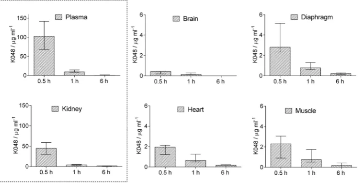
Figure 1 summarises K048 distribution and elimination across tissues. As a positively charged compound, K048 is quickly eliminated from the organism (28-30). In our study the entire dose (60 mg kg‒1 body weight) was eliminated
within a few hours from i.p. application. This could be seen as an advantage of pyridinium oxime-based antidotes, but at the same time as a disadvantage considering effective therapy and dosage regimes. Moreover, earlier studies by Thiermann et al. (17, 31) have argued in favour of repeated or continuous infusion with a pyridinium oxime antidote to counter the persistence of OPs in the organism and ensure full recovery and survival.
K048’s distribution profile shows that it reaches the highest concentration of ≈200 μmol L‒1 in the plasma
30 min after i.p. injection (Table 1). Considering AChE reactivation efficiency in vitro and the fact that pyridinium oximes are not metabolised in serum (32), this concentration
Figure 1 Rat plasma and tissue concentrations of K048 after a single i.p. dose of 238.3 mg kg‒1;(1/4 of the LD
50, see ref. 20). Data are
of K048 should suffice for AChE reactivation in vivo (10). The second highest concentration of K048 was observed in the kidneys (≈100 μmol L‒1), also half an hour after i.p.
injection. This is not unusual, since renal clearance is the main elimination pathway of pyridinium oximes (28, 32). If we look at the other organs such as the diaphragm, skeletal muscle, and heart, we can see that positively charged pyridinium compounds have difficulty crossing biological barriers and not just the blood-brain barrier (maximum brain concentration was less than 1 μmol L‒1; 33). The problem
of delivering oxime antidotes to the brain has been addressed by many approaches so far. Several groups of researchers have tried to synthesise uncharged oximes that would be able to cross the blood-brain barrier (34-38), but these new classes of oximes fall short of pyridinium oxime efficiency when it comes to tabun reactivation (37, 39). Much research has also been done to improve or modify
transport of oximes across the blood-brain barrier. Some used membrane transporters by linking an oxime to a sugar moiety and some employed nanoparticles to act as carriers (40, 41). Resolving distribution of an antidote across the organism perhaps requires different approach, not necessarily limited to modifying oxime structure.
To get an idea of what we can expect in the sense of in vivo reactivation efficiency, we made calculations (Table 1) based on reactivation constants obtained in vitro (10). These calculations indicate that with the obtained concentrations an efficient AChE reactivation of 80 % in the first 30 min could be expected only in the plasma/blood of tabun poisoned rats.
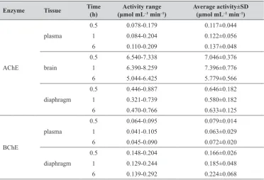
Table 2 shows cholinesterase activity in vivo in the
control groups (animals that received saline only), which serve as the baseline. Tissue-specific reactivation of AChE and BChE activity in treated animals over time is given in Table 1 Estimated AChE reactivation in the tissue calculated from the HPLC‒determined maximal oxime K048 concentration (Figure 1) and based on constants obtained for K048‒assisted reactivation in vitro and the equations describing reactivation
Tissue Mean oxime concentration (determined in 30 min) Recalculated kobs
(min‒1) reactivation in 30 minEstimated AChE
Plasma 102.6 µg mL‒1 223 µmol L‒1 0.0496 78 %
Brain 0.43 µg mL‒1 0.9 µmol L‒1 0.0006 2 %
Diaphragm 2.81 µg mL‒1 6.1 µmol L‒1 0.0039 11 %
Kidney 44.6 µg mL‒1 97 µmol L‒1 0.0347 64 %
Heart 1.95 µg mL‒1 4.2 µmol L‒1 0.0027 8 %
Muscle 2.31 µg mL‒1 5.0 µmol L‒1 0.0032 9 %
*Calculations were done using kinetic constants for hAChE: k+2=0.074 min‒1, KOX=110 μmol L‒1, and equations: ln([EP]0/[EP]t)=kobs∙t
and kobs=k+2∙[OX]/(KOX+[OX]); where [EP]0 and [EP]t are the concentrations of phosphylated enzyme at time zero and at time t,
respectively, kobs is the observed first‒order rate constant of reactivation at any given oxime concentration [OX], KOX is the dissociation
constant of the phosphylated enzyme‒oxime complex and k+2 is the maximum first‒order reactivation rate constant (10)
Table 2 AChE and BChE activities in control rats (n=4)
Enzyme Tissue Time (h) (μmol mLActivity range‒1 min‒1) Average activity±SD(μmol mL‒1 min‒1)
AChE
plasma
0.5 0.078-0.179 0.117±0.044
1 0.084-0.204 0.122±0.056
6 0.110-0.209 0.137±0.048
brain
0.5 6.540-7.338 7.046±0.376
1 6.390-8.259 7.396±0.776
6 5.044-6.425 5.779±0.566
diaphragm
0.5 0.446-0.887 0.646±0.182
1 0.321-0.739 0.580±0.182
6 0.470-0.766 0.633±0.125
BChE
plasma
0.5 0.064-0.095 0.079±0.014
1 0.041-0.105 0.063±0.029
6 0.045-0.090 0.072±0.020
diaphragm
0.5 0.148-0.204 0.166±0.026
1 0.129-0.244 0.185±0.048
Figures 2 and 3, respectively, and is expressed as percentage of the baseline. In rat plasma, AChE activity contributed to the total measured activity with 60 %, while the remaining 40 % was assigned to BChE. This ratio was probably due to the presence of free AChE in the plasma and partial haemolysis of rat erythrocytes before centrifugation of the whole blood. In the diaphragm, AChE activity accounted for almost 80 % of the total measured activity, while in the rat brain, AChE accounted for 95 % of the total measured activity, which is why we omitted BChE from evaluation in the brain.
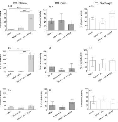
Therapy with K048 was effective in restoring AChE activity only in plasma; the highest reactivation of around 80 % was reached within the first 30 min (Figure 2). With time and elimination (cf. Figure 1) this effect diminished. At 6 h, AChE activity dropped to the level measured in rats treated with tabun alone. In other words, the remaining tabun in plasma continued to inhibit the reactivated AChE.
This finding confirms that it is vital to monitor AChE plasma activity during therapy and to adapt and repeat antidote dosing not only to keep AChE activity recovered but also to bioscavenge the remaining OP with the antidote before it affects the brain and other vital organs. In the diaphragm or the brain K048 did not significantly restore enzyme activity. These results confirm our extrapolation of the in vitro to in vivo data based on the oxime’s tissue-specific concentrations.
In addition, it was interesting to see the extent of AChE inhibition in each tissue (cf. Fig. 2). In plasma it was significant. In the brain, AChE inhibition was evident shortly after tabun injection, but it did not drop below 30 % of the control group. In the diaphragm, AChE inhibition was not as severe as would be expected and stayed within 40-50 % of the control value at all time points. A similar tissue-specific distribution was reported for other nerve agents like sarin, cyclosarin, VR, and VX (42).
Figure 2 The effects of oxime K048 on AChE activity in the plasma, brain, and diaphragm of tabun-poisoned rats after 0.5, 1, and 6 h of therapy. Activity is expressed as the percentage of mean control activity (Table 2). K048 alone did not affect AChE activity and is
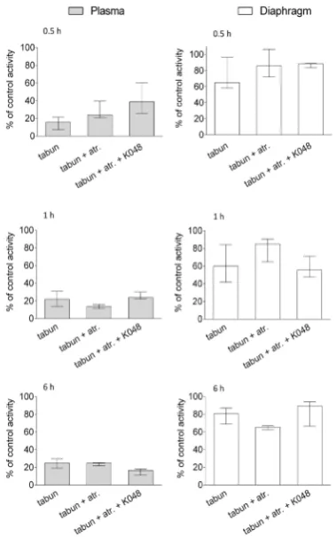
of tabun-inhibited BChE should be insignificant (11). In other words, kinetic constants of kmax=0.0022 min‒1 and KOX=630 μmol L‒1 predict a reactivation maximum of only
2 % with pharmacokinetically determined oxime plasma concentration (230 μmol L‒1 in 30 min, cf. Table 1). Indeed,
according to the in vivo results (Figure 3), K048 therapy did not preserve BChE activity in any of the tested samples, which confirms that BChE in combination with K048 can act only as a stoichiometric scavenger of tabun and not as a catalytic one.
In conclusion, our results show that it is possible to
correlate in vitro to in vivo AChE or BChE reactivation efficiency based only on well-determined kinetic constants. This approach should be applicable for future research of pyridinium oximes. However, this extrapolation would not be possible without us knowing the exact concentration of the oxime in a specific tissue after application. Therefore, pharmacokinetic studies should be planned for the early stages of antidote development. Pharmacokinetic profile can also be predicted with in silico models as a way to select a promising compound before performing in vivo experiments (43).
reactivation are also concentration-dependant (44, 45), such as muscle regeneration efficiency, interaction with cholinergic receptors, cell viability, and oxidative stress, all of which can contribute to or diminish the outcome of therapy. Furthermore, future oxime development should focus on improving distribution across the organism to overcome maybe the main hindrance in improving therapy. Current antidotal research mostly revolves around passive diffusion linked to lipophilicity (46, 47). This perspective requires changing the oxime’s structure to obtain more desirable properties, but such changes greatly affect the oxime’s potency to reactivate inhibited cholinesterases. A step in the right direction could be to consider active transport or involvement of transporters as a way of improving therapy (48). Maybe in doing so, and based on wide experimental background obtained so far, we can reach a milestone we set out to achieve decades ago.
Acknowledgements
We thank Dr Kamil Kuča for kindly providing us with oxime K048. We also wish to thank Ms Zvonka Frelih, Dr Tomaž Marš, Ms Jasna Mileković, and Dr Suzana Žunec for skilful help in conducting in vivo experiments. This study was supported by the NATO Reintegration Grant (EAP.RIG.981791), Ministry of Science, Education and Sports of the Republic of Croatia (Grant 022-0222148-2889), the Croatian-Slovenian bilateral grant (BI-HR/07-08-020) and by Croatian Science Foundation (Grant 4307).
REFERENCES
1. Gupta R. Toxicology of Organophosphate and Carbamate Compounds. 1st ed. London: Elsevier Academic Press; 2005. 2. Malekirad AA, Faghih M, Mirabdollahi M, Kiani M, Fathi
A, Abdollahi M. Neurocognitive, mental health, and glucose disorders in farmers exposed to organophosphorus pesticides. Arh Hig Rada Toksikol 2013;64:1-7. doi: 10.2478/10004-1254-64-2013-2296
3. Gray AP. Design and structure-activity relationships of antidote to organophosphorus anticholinesterase agents. Drug Metab Rev 1984;15:557-89. doi: 10.3109/03602538 409029973
4. Dawson MR. Review of oximes available for treatment of nerve agent poisoning. J Appl Toxicol 1994;14:317-31. PMID: 7822680
5. Doctor BP, Maxwell DM, Ashani Y, Saxena A, Gordon RK. New approaches to medical protection against chemical warfare nerve agents. In: Somani SM, Romano JA Jr, editors. Chemical warfare agents: toxicity at low levels. New York (NY): CRC Press; 2002. p. 191-214.
6. Kovarik Z, Radić Z, Berman HA, Simeon-Rudolf V, Reiner E, Taylor P. Mutant cholinesterases possessing enhanced capacity for reactivation of their phosphonylated conjugates. Biochemistry 2004;43:3222-9. PMID: 15023072
Figure 3 The effects of oxime K048 on BChE activity in the plasma and diaphragm of tabun-poisoned rats after 0.5, 1, and 6 h of therapy. Activity is expressed as the percentage of mean control activity (Table 2). K048 alone did not affect BChE activity and is not shown. Data are presented as medians with interquartile
ranges. Statistical significance was determined using one-way
analysis of variance and Bonferroni correction. Differences were
phosphorylated butyrylcholinesterase. Chem Biol Interact 2010;187:167-71. doi: 10.1016/j.cbi.2010.02.023
21. Radić Z, Dale T, Kovarik Z, Berend S, Garcia E, Zhang L, Amitai G, Green C, Radić B, Duggan BM, Ajami D, Rebek JJr, Taylor P. Catalytic detoxification of nerve agent and pesticide organophosphates by butyrylcholinesterase assisted with nonpyridinium oximes. Biochem J 2013;450:231-42. doi: 10.1042/BJ20121612.
22. Kovarik Z, Lucić Vrdoljak A, Berend S, Čalić M, Kuča K, Musilek K, Radić B. Evaluation of oxime K203 as antidote in tabun poisoning. Arh Hig Rada Toksikol 2009;60:19-26. doi: 10.2478/10004-1254-60-2009-1890
23. Camp S, Zhang L, Krejci E, Dobbertin A, Bernard V, Girard E, Duysen EG, Lockridge O, De Jaco A, Taylor P. Contributions of selective knockout studies to understanding cholinesterase disposition and function. Chem Biol Interact 2010;187:72–7. doi: 10.1016/j.cbi.2010.02.008
24. Ellman GL, Courtney KD, Andres VJr, Featherstone RM. A new and rapid colorimetric determination of acetylcholinesterase activity. Biochem Pharmacol 1961;7:88-90. doi: 10.1016/0006-2952(61)90145-9
25. Eyer P, Worek F, Kiderlen D, Sinko G, Stuglin A, Simeon-Rudolf V, Reiner E. Molar absorption coefficients for the reduced Ellman reagent: reassessment. Anal Biochem 2003;312:224-7. doi: 10.1016/S0003-2697(02)00506-7
26. Reiner E, Šinko G, Škrinjarić-Špoljar M, Simeon-Rudolf V. Comparison of protocols for measuring activities of human blood cholinesterases by the Ellman method. Arh Hig Rada Toksikol 2000;51:13-8. PMID: 11059068
27. Simeon-Rudolf V, Šinko G, Štuglin A, Reiner E. Inhibition of human blood acetylcholinesterase and butyrylcholinesterase by ethopropazine. Croat Chem Acta 2001;74:173-82. 28. Kušić R, Bošković B, Vojvodić V, Jovanović D. HI-6 in man:
blood levels, urinary excretion, and tolerance after intramuscular administration of the oxime to healthy volunteers. Fundam Appl Toxicol 1985;5(6part2):S89-S97. doi: 10.1093/toxsci/5.6part2.89
29. Kalász H, Hasan MY, Sheen R, Kuča K, Petroianu G, Ludányi K, Gergely A, Tekes K. HPLC analysis of K-48 concentration in plasma. Anal Bioanal Chem 2006;385:1062-7. PMID: 16763789
30. Zdarova Karasova J, Chladek J, Hroch M, Josef F, Hnidkova D, Kuca K. Pharmacokinetic study of two acetylcholinesterase reactivators, trimedoxime and newly synthesized oxime K027, in rat plasma. J Appl Toxicol 2011;33:18-23. doi:
10.1002/jat.1699
31. Worek F, Baecker M, Thiermann H, Szinicz L, Mast U, Klimmek R, Eyer P. Reappraisal of indications and limitations of oxime therapy in organophosphate poisoning. Hum Exp Toxicol 1997;16:466-72. PMID: 9292287 32. Benkö B, Kalász H, Ludányi K, Petroianu G, Kuca K, Darvas
F, Tekes K. In vitro and in vivo metabolisms of K-48. Anal Bioanal Chem 2007;389:1243-7. PMID: 17768608 33. Zdarova Karasova J, Zemek F, Kassa J, Kuca K. Entry of
oxime K027 into the different parts of rat brain: Comparison with obidoxime and oxime HI-6. J Appl Biomed 2014;12:25-9. doi: 10.1016/j.jab.2013.01.001
34. Mercey G, Verdelet T, Saint-André G, Gillon E, Wagner A, Baati R, Jean L, Nachon F, Renard PY. First efficient uncharged reactivators for the dephosphylation of poisoned approaches to nerve agent poisoning. Toxicol Lett
2011;206:5-13. doi: 10.1016/j.toxlet.2011.04.006
8. Wilhelm CM, Snider TH, Babin MC, Jett DA, Platoff GE Jr, Yeung DT. A comprehensive evaluation of the efficacy of leading oxime therapies in guinea pigs exposed to organophosphorus chemical warfare agents or pesticides. Toxicol Appl Pharmacol 2014;281:254-65. doi: 10.1016/j. taap.2014.10.009
9. Kovarik Z, Maček Hrvat N, Katalinić M, Sit RK, Paradyse A, Žunec S, Musilek K, Fokin VV, Taylor P, Radić Z. Catalytic soman scavenging by the Y337A/F338A acetylcholinesterase mutant assisted with novel site-directed aldoximes. Chem Res Toxicol 2015;28:1036-44. doi: 10.1021/acs.chemrestox.5b00060.
10. Čalić M, Lucić Vrdoljak A, Radić B, Jelić D, Jun D, Kuča K, Kovarik Z. In vitro and in vivo evaluation of pyridinium oximes: Mode of interaction with acetylcholinesterase, effect on tabun- and soman poisoned mice and their cytotoxicity. Toxicology 2006;219:85-96. doi: 10.1016/j.tox.2005.11.003
11. Lucić Vrdoljak A, Čalić M, Radić B, Berend S, Kuča K, Kovarik Z. Pretreatment with pyridinium oximes improves antidotal therapy against tabun poisoning. Toxicology 2006;228:41-50. doi: 10.1016/j.tox.2006.08.012
12. Kovarik Z, Čalić M, Šinko G, Bosak A. Structure-activity approach in the reactivation of tabun-phosphorylated human acetylcholinesterase with bispyridinium para-oximes. Arh Hig Rada Toksikol 2007;58:201-9. doi: 10.2478/v10004-007-0013-7
13. Kovarik Z, Čalić M, Bosak A, Šinko G, Jelić D. In vitro
evaluation of aldoxime interactions with human acetylcholinesterase. Croat Chem Acta 2008;81:47-57.
14. Katalinić M, Kovarik Z. Reactivation of tabun-inhibited acetylcholinesterase investigated by two oximes and mutagenesis. Croat Chem Acta 2012;85:209-12. doi: 10.5562/cca1815
15. Foy JM, Schnieden H. Estimation of total body water (virtual tritium space) in the rat, cat, rabbit, guinea-pig and man, and of the biological half-life of tritium in man. J Physiol 1960;154:169-76. doi: 10.1113/jphysiol.1960.sp006571
16. Saxena A, Sun W, Luo C, Myers TM, Koplovitz I, Lenz DE, Doctor BP. Bioscavenger for protection from toxicity of organophosphorus compounds. J Mol Neurosci 2006;30:145-7. doi: 10.1385/JMN:30:1:145
17. Thiermann H, Eyer F, Felgenhauer N, Pfab R, Zilker T, Eyer P, Worek F. Pharmacokinetics of obidoxime in patients poisoned with organophosphorus compounds. Toxicol Lett 2010;197:236-42. doi: 10.1016/j.toxlet.2010.06.005 18. Lenz DE, Maxwell DM, Koplovitz I, Clark CR, Capacio BR,
Cerasoli DM, Federko JM, Luo C, Saxena A, Doctor BP, Olson C. Protection against soman or VX poisoning by human butyrylcholinesterase in guinea pigs and cynomolgus monkeys. Chem Biol Interact 2005;157-158:205-10. PMID: 16289064
19. Rosenberg YJ, Laube B, Mao L, Jiang X, Hernandez-Abanto S, Lee KD, Adams R. Pulmonary delivery of an aerosolized recombinant human butyrylcholinesterase pretreatment protects against aerosolized paraoxon in macaques. Chem Biol Interact 2013;203:167-71. doi: 10.1016/j. cbi.2012.11.004
vivo reactivation by oximes of inhibited blood, brain and peripheral tissue cholinesterase activity following exposure to nerve agents in guinea pigs. Chem Biol Interact 2010;187:207-14. doi: 10.1016/j.cbi.2010.03.006
43. Voicu V, Rădulescu FŞ, Medvedovici A. Relationships between the antidotal efficacy and structure, PK/PD parameters and bio-relevant molecular descriptors of AChE reactivating oximes: inclusion and integration to biopharmaceutical classification systems. Expert Opin Drug Metab Toxicol 2015;11:95-109. doi: 10.1517/17425255. 2015.980813
44. Katalinić M, Miš K, Pirkmajer S, Grubič Z, Kovarik Z, Marš T. The cholinergic and non-cholinergic effects of organophosphates and oximes in cultured human myoblasts. Chem Biol Interact 2013;203:144-8. doi: 10.1016/j. cbi.2012.09.015.
45. Žunec S, Kopjar N, Želježić D, Kuča K, Musilek K, Lucić Vrdoljak A. In vivo evaluation of cholinesterase activity, oxidative stress markers, cyto- and genotoxicity of K048 oxime-a promising antidote against organophosphate poisoning. Basic Clin Pharmacol Toxicol 2014;114:344-51. doi: 10.1111/bcpt.12158
46. Voicu VA, Bajgar J, Medvedovici A, Radulescu FS, Miron DS. Pharmacokinetics and pharmacodynamics of some oximes and associated therapeutic consequences: a critical review. J Appl Toxicol 2010;30:719-29. doi: 10.1002/
jat.1561
47. Zdarova Karasova J, Pohanka M, Musilek K, Zemek F, Kuca K. Passive diffusion of acetylcholinesterase oxime reactivators through the blood-brain barrier: influence of molecular structure. Toxicol in Vitro 2010;24:1838-44. doi: 10.1016/j.tiv.2010.05.009
48. Sakurada K, Matsubara K, Shimizu K, Shiono H, Seto Y, Tsuge K, Yoshino M, Sakai I, Mukoyama H, Takatori T. Pralidoxime iodide (2-PAM) penetrates across the blood-brain barrier. Neurochem Res 2003;28:1401-7. doi: 10.1023/A:1024960819430
Translacija učinkovitosti piridinijevih oksima kod trovanja tabunom iz in vitro sustava u in vivo primjenu
Iako su organofosforni živčani bojni otrovi potpuno zabranjeni za upotrebu, njihova je prisutnost i dalje velik problem, posebice kao kemijsko oružje u terorističkim napadima (poput nedavnih u Siriji). Oksimi koji se danas koriste kao protuotrovi u tretmanu nemaju dostatno djelovanje na reaktivaciju aktivnosti kolinesteraza, glavnih meta djelovanja organofosfornih spojeva. Valja napomenuti kako se klinička testiranja ovih protuotrova rijetko provode zbog svoje iznimne specifičnosti. Tijekom zadnjih desetljeća učinjen je napredak u istraživanju novih učinkovitijih protuotrova, međutim još je uvijek veliki nedostatak u poboljšavanju terapije translacija in vitro dobivenih rezultata u in vivo primjenu. Ovom studijom ispitali smo mogućnosti ekstrapolacije reaktivacijske učinkovitosti određene za oksimske protuotrove iz in vitro u in vivo sustav. Naši rezultati pokazuju kako je ova translacija moguća uz detaljno određene kinetičke parametre in vitro i uz poznavanje distribucije oksima i vremena cirkulacije u organizmu. Takav rezultat ističe važnost planiranja i farmakokinetičkih istraživanja već u samom početku razvoja protuotrova. Također, poseban naglasak u istraživanju trebalo bi staviti i na poboljšanje tkivo-specifične distribucije oksima u organizmu čime bi se poboljšala cjelokupna terapijska učinkovitost.
KLJUČNE RIJEČI: acetilkolinesteraza; butirilkolinesteraza; farmakokinetika; K048, protuotrovi; specifična aktivnost u tkivima, živčani bojni otrovi
2011;47:5295-7. doi: 10.1039/c1cc10787a
35. Sit RK, Radić Z, Gerardi V, Zhang L, Garcia E, Katalinić M, Amitai G, Kovarik Z, Fokin VV, Sharpless KB, Taylor P. New structural scaffolds for centrally acting oxime reactivators of phosphylated cholinesterases. J Biol Chem 2011;286:19422-30. doi: 10.1074/jbc.M111.230656 36. Radić Z, Sit RK, Kovarik Z, Berend S, Garcia E, Zhang L,
Amitai G, Green C, Radić B, Fokin VV, Sharpless KB, Taylor P. Refinement of structural leads for centrally acting oxime reactivators of phosphylated cholinesterases. J Biol Chem 2012;287:11798-809. doi: 10.1074/jbc.M111.333732 37. Renou J, Mercey G, Verdelet T, Păunescu E, Gillon E,
Arboléas M, Loiodice M, Kliachyna M, Baati R, Nachon F, Jean L, Renard P-Y. Syntheses and in vitro evaluations of uncharged reactivators for human acetylcholinesterase inhibited by organophosphorus nerve agents. Chem Biol Interact 2013;203:81-4. doi: 10.1016/j.cbi.2012.09.023 38. Kliachyna M, Santoni G, Nussbaum V, Renou J, Sanson B,
Colletier JP, Arboléas M, Loiodice M, Weik M, Jean L, Renard P-Y, Nachon F, Baati R. Design, synthesis and biological evaluation of novel tetrahydroacridine pyridine- aldoxime and -amidoxime hybrids as efficient uncharged reactivators of nerve agent-inhibited human acetylcholinesterase. Eur J Med Chem 2014;78:455-67. doi: 10.1016/j.ejmech.2014.03.044
39. Kovarik Z, Maček N, Sit RK, Radić Z, Fokin VV, Sharpless KB. Taylor P. Centrally acting oximes in reactivation of tabun-phosphoramidated AChE. Chem Biol Interact 2013;203:77-80. doi: 10.1016/j.cbi.2012.08.019
40. Garcia EG, Campbell JA, Olson J, Moorad-Doctor D, Morthole IV. Novel oximes as blood-brain barrier penetrating cholinesterase reactivators. Chem Biol Interact 2010;187:199-206. doi: 10.1016/j.cbi.2010.02.033