Original Article
Comparison of some Molecular Markers for Tick Species Identification
*Eman M. Abouelhassan1; Hamdy M. ElGawady1; Ahmad Anwar AbdelAal1; Amal K.
El-Gayar1; Maria D Esteve-Gassent2
1Department of Veterinary Parasitology, Suez Canal University, Ismailia, Egypt
2Department of Veterinary Pathobiology, Texas A and M University, College Station, Texas, United
States of America
(Received 15 Mar 2018; accepted 22 Apr 2019)
Abstract
Background: Ticks are obligate blood-sucking ectoparasites of vertebrates. Since many tick identification studies are based on the analysis of 16S rDNA, 12S rDNAand ITS-1, 2 rDNA genes, we aimed to compare the performance of these molecular markers of common use for the identification of ticks, under a diagnostic laboratory environment.
Methods: Overall, 192 tick specimens were collected through the state of Texas from January 2014 to August 2015 and the species was determined by both morphology and molecular amplification using the 16S rDNA, 12S rDNA, ITS1 and ITS2.
Results: The species collected were identified by molecular techniques as Dermacentor albipictus, D. variabilis, Am-blyomma americanum, Ixodes scapularis, A. cajennense, Rhipicephalus sanguineus and Carios capensis. ITS1 and ITS2 were not able to prove consistent amplification and therefore have been considered as potential markers for tick iden-tification.
Conclusion: The use of mitochondrial genes in tick identification showed to provide more consistent results in the diagnostic environment.
Keywords:Ticks; Dermacentor; Amblyomma; Rhipicephalus; Ixodes
Introduction
Ticks are obligate blood-sucking ectopar-asites of vertebrates, causing great economic losses to livestock with its direct and indirect effects on hosts. Bloodsucking by large num-bers of ticks causes a reduction in live weight and anemia among domestic animals, while their bites also reduce the quality of hides (1). In addition, certain ticks will cause tick paral-ysis, which is an acute ascending flaccid mo-tor paralysis caused by the injection of a tox-in by the tick while feedtox-ing. However, the major effects caused by ticks are due to their ability to transmit protozoan, bacterial and viral diseases to livestock, companion animals and humans (16, 17). Ticks are currently consid-ered to be second only to mosquitoes as vec-tors of human infectious diseases in the world (9, 23). A number of bacterial zoonotic infec-
tious diseases (2) such as anaplasmosis, ehr-lichiosis, and lyme borreliosis are transmitted by ticks.
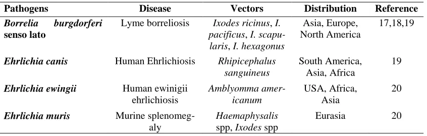
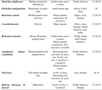
Table 1 shows the pathogens transmitted by different species of ticks as Ixodes species are the vectors of lyme borreliosis, Amblyomma americanum is the vector of Ehrlichia chaffeen-si, tick identification helping in the diagnosis of disease transmitted with, and misidentifi-cation of the may lead to difficult and even wrong disease diagnosis.
separate keys are normally used (2, 3). In cer-tain situation, damage to tick body parts essen-tial for their identification (such as capitulum and adjacent structures) may occur during re-moval of attached ticks to their hosts. In ad-dition, bad preservation of tick samples often occurs, leading to incorrect identifications (4). Identification of some tick species like Rhip-icephalus sanguineus and Amblyomma cajen-nense is difficult due to the fact that they have been classified as a species complex (11, 12). For instance, A. cajennense is a complex of 6 species, while R. sanguineus group compris-es a total of17 different species. In these com-plexes, the different species are geographically separated, due to the large geographical range of distribution, and the expected adaptation of tick populations to different environmen-tal conditions (12). Therefore, morphological identification of these species is not sufficient and the further molecular information is need-ed for a correct species determination (11, 12). These difficulties may be reduced when using molecular techniques for tick identifi-cation(3).Anotherbenefit from molecular tech-niques is that with those samples that tick DNA integrity has not been compromised, from the total DNA extraction, a collection of different tick-borne pathogens can be detected by molec-ular methodologies such as conventional PCR, and real-time PCR (4). Therefore, with one sin-gle extraction, both the agent and the vector species can be determined(4). Moreover, the availability of genetic sequence will provide the opportunity to study both the vector pop-ulation diversity, as well as the pathogen they carry, and potentially even their relationships (13). Currently, there is a lack of whole tick
genomes readily annotated
(https://www.vectorbase.org/), with Ixodes scapularis being the only one currently avail-able
(https://www.vectorbase.org/organisms/ixod es-scapularis) (13). This lack of information limits the advancement of the development of new molecular methods for the study of these
arthropods. One of the limitations is the large size of tick genomes (15). For instance, the average haploid genome of the tick I. scapu-laris has been calculated at 2262Mbp in length, while the A. americanum is around 3108Mbp. If we compare this to the human genome, tick genomes tend to be twice as bigger as the hu-man genome. Part of the difference in size is due to the presence of non-coding regions with extended tandem repeats, that difficulty signif-icantly the sequencing and annotation of those genomes (15). Consequently, different molec-ular markers have been traditionally used for the phylogeny of ticks (8). Those include the nuclear ribosomal genes 18S rDNA, 28S rDNA and ITS-1, 2 rDNA as well as mitochondrial genes such as 16S, 12S, COI, COIII) rDNA (8, 9).
El-Fiky and El Kammah (3) and Chitimia (4) successfully used the internal transcribed spacer (ITS) for the identification of Derma-centor marginatus, Ixodes ricinus, Haema-physalis, Boophilus, and Rhipicephalus san-guineus tick species. On the other hand, the 16S rDNA were able to construct the phy-logeny of both hard and soft ticks (6, 8, 9). 16SrDNAwereusedinmolecularclassification of metastriate ticks (Dermacentor, Amblyom-ma and Rhipicephalus respectively (10, 13).
Since many tick identification studies are based on the analysis of 16S rDNA, 12S rDNA and ITS-1, 2 rDNA genes, the objective of the present study was to compare the performance of these molecular markers of common use for the identification of ticks, under a diag-nostic laboratory environment (13).
Materials and Methods
Tick sample collection
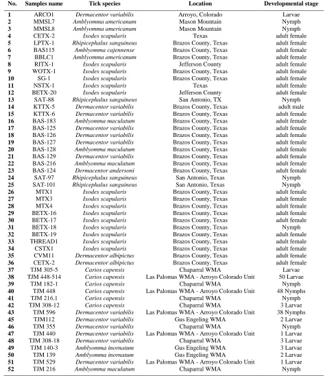
service provided by the Lyme Laboratory at Texas A and M University, from January 2014 to August 2015. On the other hand Group, B contains 133 tick pools collected from wild-life through an ecology project conducted in collaboration with Dr. Castro-Arellano at Tex-as State University (Table 2). For better DNA extractions, larvae and nymphs were analyzed in pools, sometimes the pools contain one lar-vae and other up to 50 larlar-vae of the same tick species collected from the same location in the same sampling effort. Nymphs, on the other hand, were pooled in groups of from one till 15 specimens, following the same strategy de-scribed for larvae for optimal DNA extraction. For the majority of these ticks, the morpho-logical identification was not enough to de-termine the species, mostly due to either bad storage of samples or loss of mouth-parts while removal the tick from the host. These ticks were immersed in 70% ethanol solution and then processed for DNA extraction and molecular identification using 16S rDNA PCR specific primers and sequencing the PCR product.
DNA Extraction
DNA wasextracted from the tick samples using Wizard® SV Genomic DNA Purifica-tion kit (Promega CorporaPurifica-tion, Madison, WI) following manufacturer’s recommendations with modifications. Briefly, ticks were incu-bated for 10min at 70 ºC in 200µl of Nuclei Lysis Solution, plus 50µl of 0.5M EDTA, 40 µl of a 20mg/ml Proteinase K solution, and 5µl RNase A Solution. After the initial diges-tion and for the optimal extracdiges-tion of DNA from the arthropods, adult individual ticks were homogenized utilizing the bead mill Bead Rup-tor 24 (Omni International, Inc., Kennesaw, GA), un-engorged ticks were homogenized with 1.4mm ceramic beads while 2.8mm ce-ramic beads were used with engorged ticks. Af-ter homogenization, tubes were centrifuged at 10,000×g to eliminate tick debris. Superna-tants were collected and 250µl of Wizard® SV Lysis Buffer was added to each sample and
the mixture. The mixture was run through fil-ter columns at 13,000×g for 3min. DNA bound to filter was washed and eluted following man-ufacturer recommendations. To extract DNA from the tick immature stages (nymphs and larvae) pools of a maximum of 15 nymphs or 50 larvae were made. Specimens received a code indicating the type of pool generated. All immature specimens were stored at -80 ºC with 100µl TE buffer for at least one hour. Specimens were homogenized utilizing pestles while the samples were frozen, fol-lowed by DNA extraction procedures using the prepGEM™ (ZyGem Ltd., New Zeland) Insect DNA Extraction kit following manu-facturer’s recommendations. Briefly, tick sam-ples were mixed with ultra-pure water, 10x buffer provided in the kit, and 1µl of the prepGEM™ enzyme (ZyGem Ltd., New Zeland). The mixture was incubated at 75 °C for 15min followed by incubation at 95 °C for 15min. The extracted DNA concentration and purity were measured using a NanoDrop, and stored at -20 until use.
Molecular identification of ticks based on 16S rDNA Gene, 12S rDNA and ITS-1 and 2 rDNA Genes
La-boratoies Inc., Hercules, CA). Positive bands were excised from the gel and purified using the Wizard® SV Gel and PCR clean-up sys-tem (Promega Corporation, Madison WI) fol-lowing manufacturer’s recommendations. The purified products were sent for sequencing (Eton Biosciences, San Diego, CA). Sequences were analyzed through BLAST® in MacVec-tor 14.0.0 software(MacVector Inc.,Cary, NC).
PCR was performed first using16S rDNA Gene primers. The same samples were also tested using specific primers for first and sec-ond internal transcribed spacers (ITS-1 and ITS-2rDNA) (Table 3) following methodol-ogies (4). The PCR reaction was done using the following cycling condition: initial dena-turation at 95 ºC for 5min followed by forty cycles of 95 ºC for 45sec, 55 ºC for 1min and 72 ºC for 90sec with a final extraction at 72 ºC for 1min. The amplification products were sep-arated on 1.6% agarose gel containing 0.4µg/ ml of ethidium bromide (Bio-Rad Laboratoies Inc., Hercules, CA) and the gel was run at 90 volts for 40–60min. Gels were visualized us-ing the ChemiDoc touch imagus-ing system (Bio-Rad Laboratoies Inc., Hercules, CA).
In 12S rDNA, PCR was done following the cycling condition: initial denaturation at 95 ºC for 5min followed by forty cycles of 95 ºC for 30sec, 40 ºC for 30sec and 72 ºC for 30sec, with a final extraction at 72 ºC for 5 min. The amplification products were sepa-rated and visualized as mentioned before on 1.6% agarose gel containing 0.4µg/ml of eth-idium bromide (Bio-Rad Laboratoies Inc., Her-cules, CA) and the gel was run at 90 volts for 40–60min. Gels were visualized using the ChemiDoc touch imaging system (Bio-Rad Laboratoies Inc., Hercules, CA).
Sequence analysis
Positive bands were excised from the gel and purified using the Wizard® SV Gel and PCR clean-up system (Promega Corporation, Madison WI) following manufacturer’s rec-ommendations. The purified products were
sent for sequencing (Eton Biosciences, San Diego, CA). Sequences were analyzed through BLAST® using MacVector 14.0 software (MacVector Inc., Cary, NC).
Results
Overall, 192 ticks were analyzed from dif-ferent developmental stages (larvae, nymph, males and engorged females) collected through the state of Texas. In this collection, the tick specimens were identified based on the16S rDNA PCR products as Dermacentor albipic-tus, D. variabilis, Amblyomma americanum,
Ixodes scapularis, A. cajennense Rhipiceph-alus sanguineus and Carios capensis (Table 4), the GenBank accession numbers from KX673167 to KX673180.
Comparison between 16S rDNA Gene, 12S rDNA Gene and (ITS-1, 2) rDNA Genes
In order to evaluate which genetic mark-er pmark-erform best undmark-er diagnostic conditions, comparison between the amplification of four genes 16S rDNA, 12S rDNA, ITS1 and ITS2 rDNA genes was done. Positive bands from 16S rDNA, 12S rDNA PCR were excised from the gel and cleaned and submitted for se-quencing. Sequences were analyzed through BLAST® in MacVector 14.0.0 software (MacVector Inc., Cary, NC).
in spite of the samples number are not rep-resentative, but changing in the tick PCR am-plification of the four genes was observed as good amplification for the samples were ob-served as both 16S rDNA, 12S rDNA Gene showing good PCR amplification in all the sample but (ITS-1, 2) rDNA Genes failed to amplify some samples species, (Fig. 1).
The samples utilized were Dermacentor albipictus (CVM11, CETX-2), Dermacentor variabilis (ARCO1), Amblyomma american-um (MMSL5, MMSL9), Ixodes scapularis
(SAT 88, SAT 97, and SAT 101).
In ITS-1PCR, one of the three samples of
Rhipicephalus sanguineus (SAT 101),two sam-ples of Amblyomma americanum (MMSL5, MMSL9), Ixodes scapularis samples CSTX-2, CSTX-3) and Dermacentor albipictus sam-ples(CVM11, CETX-2) were amplified, but there were no amplification in Dermacentor variablis (ARCO1), Amblyomma cajennense
(BAS 115) and the other two samples of R. sanguineus (SAT 88, SAT 97).
ITS-2 PCR, one of the three samples of
Rhipicephalus sanguineus (SAT 101),two sam-ples of Amblyomma americanum (MMSL5, MMSL9), Dermacentor variablis (ARCO1),
Amblyomma cajennense (BAS 115), Ixodes scapularis samples CSTX-2, CSTX-3) and
Dermacentor albipictus samples (CVM11, CETX-2) were amplified, but the two samples of R. sanguineus (SAT 88, SAT 97) failed to amplify.
Phylogenetic analysis
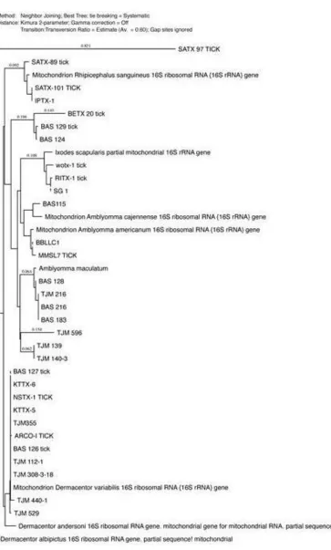
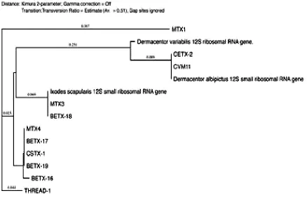
The phylogenetic analysis was performed using MacVector 14.0 software (MacVector Inc., Cary, NC) and the tree was constructed using neighbor-joining (NJ) methods (Figs. 2-4). The low degree of sequences variation observed within most of the species of the soft and hard tick trees based on the 16S rDNA since they all share the same ancestor. Nev-ertheless, there is one sequence in the hard tick population studied Rhipicephalus sanguineus
that showhigher variation.
Fig. 1.PCR amplification utilizing Tick samples of different species using: (A) the ITS-2PCR reaction (B): ITS-1PCR reaction and (C): 16S r DNA Gene, (D)12SrDNA DNA ladder is located on the left and right sides of the gel, fragment sizes are represented in base pairs (bp), 1: Rhipicephalus sanguineus sam-ple (SAT 97), 2: Rhipicephalus sanguineus sample (SAT88), 3: Rhipicephalus sanguineus sample (SAT 101), 4: Amblyomma americanum sample (MMSL5),
5: Amblyomma americanum sample (MMSL9), 6
Amblyomma cajennense (BAS 115 TICK): 7: Ixodes scapularis sample (CSTX-2), 8: Ixodes scapularis
sample (CSTX-3), 9: Dermacentor albipictus sam-ple(CVM11), 10: Dermacentor albipictus sam-ple(CETX-2), 11: Dermacentor variabilis
sam-ple(ARCO1) and12: negative control.
Table 1. Important tick-borne diseases of humans
Pathogens Disease Vectors Distribution Reference
Borrelia burgdorferi
senso lato
Lyme borreliosis Ixodes ricinus, I. pacificus, I.
scapu-laris, I. hexagonus
Asia, Europe, North America
17,18,19
Ehrlichia canis Human Ehrlichiosis Rhipicephalus
sanguineus
South America, Asia, Africa
19
Ehrlichia ewingii Human ewinigii
ehrlichiosis
Amblyomma amer-icanum
USA, Africa, Asia
20
Ehrlichia muris Murine
splenomeg-aly
Haemaphysalis
spp, Ixodes spp
Ehrlichia chaffeensis Human monocytic ehrlichiosis
Amblyomma amer-icanum
North America 17,18,19
Ehrlichia ruminantium Heartwater in
rumi-nant
Amblyomma spp Africa, Carib-bean
20
Rickettsia conorii Mediterranean
spot-ted fever
Rhipicephalus sanguineus, R.
turanicus
Africa, Asia, Europe
17,18,19
Coxiella burnetii Q fever Many species Africa, Asia,
Europe, North America,
Aus-tralia
17,18,19
Rickettsai rickettsii Rocky Mountain
spotted fever
Amblyomma amer-icanum,
Rhipicephalus sanguineus, Der-macentor variablis
North, South and Central
America
17,18,19
Anaplasma
phago-cytophilum
Human granulocytic anaplasmosis
Haemaphysalis concinna, H.
punc-tate, Ixodes rici-nus, I. pacificus, I.
scapularis Rhipicephalus
bursa
North America, Europe
17,18,19
Flavivirus Tick borne
encepha-litis
Ixodes ricinus, Haemaphysalis concinna, H.
punc-tate
Asia, Europe 18,19
Babesia divergen, B. microti
Babesiosis Ixodes ricinus, I. scapularis
North America, Europe
17,18,19
Fig. 2. Thephylogenetic analysis was constructed using neighbor joining method, to construct thetickphylogenetic tree of some of soft tick species sequences from the Genbank and our sequences samples are included based on 16S r
DNA sequences
Fig. 3. Thephylogenetic analysis was constructed using neighbor-joining method, to construct thetickphylogenetic tree of some of hard tick species sequences from the Genbank and our sequences samples are included based on 16S
Fig. 4. Thephylogenetic analysis was constructed using neighbor-joining method, to construct thetickphylogenetic tree of some of hard tick species sequences from the Genbank and our sequences samples are included based on 12S
r DNA sequences
Table 2. Ticks samples utilized in this study according to their distribution and stages
Distribution Adult female Adult male Nymph Larvae
Arroyo, Colorado 1
Mason Mountain 2
Brazos County, Texas 24 1 1
Jefferson County 2 2 1
Texas 2
San Antonio, TX 3
Gus Engeling WMA 2 5
Tejas Ranch 6
Chaparral WMA 4 10
Las Palomas WMA-Arroyo Colorado Unit 86 52
Total 28 3 99 74
Table 3.Primers using in PCR
Gene F-Primers R-Primers Ref
Tick 16S rDNA 5´-TTGGGCAAGAAGACCCTATGAA -3´ 5´- CCGGTCTGAACTCAGATCAAGT-3´ (5)
Tick 12S rDNA 5´-GAGGAATTTGCTCTGTAATGG -3´ 5´-AAGAGTGACGGGCGATATGT-3´ (21)
ITS-1rDNA 5´-TCATAAGCTCGCGTTGATT-3' 5´-AGCTGGCTGCGTTCTTCAT - 3' (3)
Table 4.Ticks samples utilized in this study according to their distribution and species
No. Samples name Tick species Location Developmental stage
1 ARCO1 Dermacentor variabilis Arroyo, Colorado Larvae
2 MMSL7 Amblyomma americanum Mason Mountain Nymph
3 MMSL8 Amblyomma americanum Mason Mountain Nymph
4 CETX-2 Ixodes scapularis Texas adult female
5 LPTX-1 Rhipicephalus sanguineus Brazos County, Texas adult female
6 BAS115 Amblyomma cajennense Brazos County, Texas adult female
7 BBLC1 Amblyomma americanum Brazos County, Texas adult female
8 RITX-1 Ixodes scapularis Jefferson County adult female
9 WOTX-1 Ixodes scapularis Brazos County, Texas adult female
10 SG-1 Ixodes scapularis Brazos County, Texas adult female
11 NSTX-1 Ixodes scapularis Texas adult female
12 BETX-20 Ixodes scapularis Jefferson County adult female
13 SAT-88 Rhipicephalus sanguineus San Antonio, TX Nymph
14 KTTX-5 Dermacentor variabilis Brazos County, Texas adult male
15 KTTX-6 Dermacentor variabilis Brazos County, Texas adult female
16 BAS-183 Amblyomma maculatum Brazos County, Texas adult female
17 BAS-125 Dermacentor variabilis Brazos County, Texas adult female
18 BAS-126 Dermacentor variabilis Brazos County, Texas adult female
19 BAS-127 Dermacentor variabilis Brazos County, Texas adult female
20 BAS-128 Amblyomma maculatum Brazos County, Texas adult female
21 BAS-129 Dermacentor variabilis Brazos County, Texas adult female
22 BAS-216 Amblyomma maculatum Brazos County, Texas adult female
23 BAS-124 Dermacentor andersoni Brazos County, Texas adult female
24 SAT-97 Rhipicephalus sanguineus San Antonio, Texas Nymph
25 SAT-101 Rhipicephalus sanguineus San Antonio, Texas Nymph
26 MTX1 Ixodes scapularis Brazos County, Texas adult female
27 MTX3 Ixodes scapularis Brazos County, Texas adult female
28 MTX4 Ixodes scapularis Brazos County, Texas adult female
29 BETX-16 Ixodes scapularis Brazos County, Texas adult female
30 BETX-17 Ixodes scapularis Brazos County, Texas adult female
31 BETX-18 Ixodes scapularis Brazos County, Texas Nymph
32 BETX-19 Ixodes scapularis Brazos County, Texas adult female
33 THREAD1 Ixodes scapularis Brazos County, Texas adult female
34 CSTX1 Ixodes scapularis Brazos County, Texas adult female
35 CVM11 Dermacentor albipictus Brazos County, Texas adult female
36 CETX-2 Dermacentor albipictus Brazos County, Texas adult female
37 TJM 305-5 Carios capensis Chaparral WMA Larvae
38 TJM 448-514 Carios capensis Las Palomas WMA - Arroyo Colorado Unit 50 Larvae
39 TJM 182-1 Carios capensis Chaparral WMA Nymph
40 TJM 448 Carios capensis Las Palomas WMA - Arroyo Colorado Unit 48 Nymphs
41 TJM 216.1 Carios capensis Chaparral WMA Nymph
42 TJM 308-12 Carios capensis Chaparral WMA 3 Larvae
43 TJM 596 Dermacentor variabilis Las Palomas WMA - Arroyo Colorado Unit 38 Nymphs
45 TJM112 Dermacentor variabilis Gus Engeling WMA 2 Larvae
46 TJM 355 Dermacentor variabilis Chaparral WMA Nymph
47 TJM 440 Dermacentor variabilis Las Palomas WMA - Arroyo Colorado Unit 1 Larvae
48 TJM 308-18 Dermacentor variabilis Chaparral WMA 3 Larvae
49 TJM 140-3 Amblyomma inornatum Gus Engeling WMA 3 Larvae
50 TJM 139 Amblyomma inornatum Gus Engeling WMA 2 Larvae
51 TJM 529 Dermacentor variabilis Las Palomas WMA - Arroyo Colorado Unit 1 Larvae
52 TJM 216 Amblyomma maculatum Chaparral WMA Nymph
Discussion
on DNA sequences to solve the problems with morphological tick identification, and some-times the morphological identification is not enough for detect the species so amplification of the16S rDNA using it as a methods for tick genetic identification, and comparison be-tween the amplification of the the16S rDNA, 12S rDNA, ITS-1 rDNA and ITS-2 rDNAfor the same samples species.
16S rDNA and 12S rDNAGenesare a mi-tochondrialribosomal DNA gene, mtDNA con-sidered one of the most commonly used genes for molecular identification of ticks due to the fact that it is relatively easy to work with them due to their higher copy number (8). In addi-tion, mtDNA sequences are a good phyloge-netic marker mostly for groups of organisms, diverged relatively, since mtDNA has a higher rate of base substitution than most nuclear markers (9). The problem for the mitochon-drial gene is it can transfer to the nucleus lead-ing to error in the phylogeny after the ampli-fication and sequencing (8). The difference between the 16S rDNA and 12S rDNA Genes that the evolution is faster in 12S rDNA Genes (8).
Regarding tick species, 16S rDNA was used and succeeded to construct phylogeny of both hard and soft ticks (6, 7, 8) and 16S rDNA is useful in constructing their tick phy-logenetic tree, but there is a problem asso-ciated with 16S rDNA is that using this gene alone is not sufficient for getting full resolu-tion for the tree so the best way to solve it accompanied it with another gene like 12S rDNA(6, 8). We utilized these genes for di-agnostic purpose only not for phylogeny, and the problems are usually associated with the phylogeny.
Overall, 192 tick samples (larvae, nymph, males and engorged females) were evaluated using16S rDNA PCR and 12S rDNA the PCR positive bands have to be sequencing. The se-quencing analysis determined that the tick spe-cies collected in the study were: Amblyomma americanum, Dermacentor albipictus, Ixodes
scapularis, D. variabilis, A. cajennense, Car-ios capensis and Rhipicephalus sanguineus
(Table 3). Therefore, using 16S rDNA and 12S rDNA are good in molecular tick identi-fication of all these species utilized in our study. The first and the second internal transcribed spacers region of the nuclear ribosomal gene cluster (ITS-1, ITS-2), consist of three genes 18SrDNA, 5.8SrDNA and 28SrDNA. These three rDNA genes are transcribed making a single transcript of RNA separated by the ITS-1 and ITS-2 regions (7). (ITS-ITS-1, ITS-2) con-sidered the fastest evolving DNA genes (9). Because of these facts, the ITS-1, 2 rDNA are not good in amplification of some of the tick species in our study, they failed to am-plify some tick species as mentioned before. Internal transcribed spacer was used success-fully for the identification of Dermacentor mar-ginatus, Ixodes ricinus, Haemaphysalis, Booph-ilus, and Rhipicephalus sanguineus tick spe-cies (3, 4).
Nevertheless, the problem with internal transcribed spacer is that genes are evolving rapidly so in some species they failed to am-plify as reported before in ticks (8). ITS-2 was utilized for identification Iranian hard tick and ITS-2 failed to amplify some of his sam-ples. Therefore, these markers were mostly useful to study close related species (10). Even some of our samples are closely related to each other as Rhipicephalus species and Derma-centor and it failed to amplify some of them, therefore, it is better to clone the PCR products and work with it as haplotypes, not individu-als make the studies more expensive (8).
Conclusion
are good for tick molecular identification for very closely related species but with our few samples even they are not representative sam-ples it did not work with the closely related tick species.
Acknowledgements
Thanks to the Lyme Laboratory at Texas A and M University and Dr Maria D Esteve-Gassent for her support and effort.
The authors declare that there is no con-flict of interests.
References
1. Zahid Iqbal Rajput, Song-hua Hu, Wan-Jun Chen, Abdullah G. Arijo, Chen-wen Xiao (2006) Importance of ticks and their chemical and immunological control in livestock. J Zhejiang Univ Sci B.7(11): 912–921.
2. Durden LA, Keirans JE (1996) Nymph of the genus Ixodes (Acari: Ixodidae) of the unuted states: taxonomy, identification key, distribution, Thomas Say Puplica-tions in Entomlogy. Entomolgical Soci-ety of America, Lanham, Maryland. 3. El-Fiky ZA, Sayed MA, El Kammah KM
(2005) Molecular markers of some tick genera in Egypt based on the internal transcribed spacer. Arab J Biotech. 8 (1): 61–66.
4. Chitimia L, Lin RQ, Cosoroaba I, Braila P, Song HQ, Zhu XQ (2009) Molecular characterization of hard ticks from Ro-mania by sequences of the internal tran-scribed spacers of ribosomal DNA. Par-asitol Res. 105: 1479–1482.
5. Patrı´cia RO, Gervasio HB, Sandra ED, Kelly CS, Erika TN, Matias PJS, Maria ICM (2005) Comparison of the exter-nal morphology of Rhipicephalus san-guineus (Latreille, 1806) (Acari: Ixodi-dae) Ticks from Brazil and Argentina.
Vet Parasitol. 129: 139–147.
6. Black WC, Piesman J (1994) Phylogeny of hard- and soft-tick taxa (Acari: Ixodida) based on mitochondrial 16S rDNA se-quences. Proc Natl Acad Sci USA. 91 (21): 10034–10038.
7. Difference Between Hard Ticks and Soft Ticks:
http://www.orkin.com/other/ticks/hard-soft-ticks-difference
8. Cruickshank RH (2002) Molecular mar-kers for the phylogenetics of the mites and ticks.Syst Appl Acarol. 7: 3–14. 9. Dabert M (2006) DNA markers in the
phylogenetics of the Acari. Biol Lett. 43(2): 97–107.
10.Abdigoudarzi M, Noureddine R, Seitzer U, Ahmed J (2011) rDNA-ITS2 Identi-fication of Hyalomma, Rhipicephalus Dermacentor and Boophilus spp. (Aca-ri: Ixodidae) collected from different ge-ographical region of Iran. Adv Stud Bi-ol. 3(5): 221–238.
11.Filipe Dantas, Maria Stefania Latrofa, Gi-ada Annoscia, Alessio Giannelli, Anto-nio Parisi, Domenico Otranto (2013) Morphological and genetic diversity of
Rhipicephalus sanguineus sensu lato from the New and Old Worlds. Para-sites and Vectors. 6: 213.
12.NavaS, Beati L, Labruna MB, Cáceres AG, Mangold AJ, Guglielmone AA (2014) Reassessment of the taxonomic status of Amblyomma cajennense with the de-scription of three new species, Amblyom-ma tonelliae n. sp., Amblyomma in-terandinum n. sp. and Amblyomma pati-noi n. sp., and reinstatement of Am-blyomma mixtum, and Amblyomma sculp-tum(Ixodida:Ixodidae).TicksTickBorne Dis. 5(3): 252–276.
13.Catherine AH (2010) Genome analysis of Major Tick and Mite Vectors of Hu-man Pathogens. Behalf of the Tick and
Mite Genomes:
/Genome-Analysis-of-Major-Tick-and-Mite-Vectors Consortium.
14.Anderson JM, Ammerman NC, Norris DE (2004) Molecular differentiation of metastriate tick immatures. Vector Borne Zoonotic Dis. 4(4): 334–342.
15.Geracia SN, Johnstonb JS, Robinsona JP, Wikelc SK, Hilla CA (2007) Variation in genome size of argasid and ixodid ticks. Insect Biochem Mol Biol. 37: 399–408. 16.Michael KF, Martin R, Jürgen H, Mareike
R, Andreas G, Alexander K (2014) Hu-man tick infestation pattern, tick-bite rate, and associated Borrelia burgdorferi s.l. infection risk during occupational tick exposure at the Seedorf military train-ing area, northwestern Germany. Ticks Tick Borne Dis. 5: 594–599.
17.Rajput ZI, Hu S, Chen W, Arijo AG, Xiao C (2006) Importance of ticks and their chemical and immunological control in livestock. J Zhejiang Univ Sci B. 7(11): 912–921.
18.Jongejan F, Uilenberg G (2014) The global importance of ticks. Parasitology. 129: S3–14.
19.Piesman J, Eisen L (2008) Prevention of tick-borne diseases. Annu Rev Entomol. 53: 323–343.
20.Dantas-Torres F1, Chome BB, Otranto D (2012) Ticks and tick-borne diseases: a One Health perspective. Trends Parasi-tol. 28(10): 437–446.
21.Maria DE, Nettles BR (2015) Molecular Detection of Pathogentic Ehrlichia spp. in Texas. Department of Veterinary Patho-biology, Texas and M University, Col-lege Station, Texas, United States of America.
22.Williamson PC, Billingsley PM, Teltow GJ, Seals JP, Turnbough MA, Atkinson SF (2010) Borrelia, Ehrlichia,and Rick-ettsia spp. in Ticks Removed from Per-sons, Texas, USA. Emerg Infect Dis. 16(3): 441–446.