T
T
h
h
e
e
r
r
a
a
n
n
o
o
s
s
t
t
i
i
c
c
s
s
2016; 6(3): 369-391. doi: 10.7150/thno.13438 ReviewDNA Methyltransferase Activity Assays: Advances and
Challenges
Wan Jun Poh, Cayden Pang Pee Wee and Zhiqiang Gao
Department of Chemistry, National University of Singapore, Singapore 117543
Corresponding author: Tel: 6516-3887, Fax: 6779-1691, e-mail: [email protected]
© Ivyspring International Publisher. Reproduction is permitted for personal, noncommercial use, provided that the article is in whole, unmodified, and properly cited. See http://ivyspring.com/terms for terms and conditions.
Received: 2015.08.03; Accepted: 2015.10.12; Published: 2016.01.06
Abstract
DNA methyltransferases (MTases), a family of enzymes that catalyse the methylation of DNA, have a profound effect on gene regulation. A large body of evidence has indicated that DNA MTase is potentially a predictive biomarker closely associated with genetic disorders and genetic diseases like cancer. Given the attention bestowed onto DNA MTases in molecular biology and medicine, highly sensitive detection of DNA MTase activity is essential in determining gene regulation, ep-igenetic modification, clinical diagnosis and therapeutics. Conventional techniques such as isotope labelling are effective, but they often require laborious sample preparation, isotope labelling, so-phisticated equipment and large amounts of DNA, rendering them unsuitable for uses at point-of-care. Simple, portable, highly sensitive and low-cost assays are urgently needed for DNA MTase activity screening. In most recent technological advances, many alternative DNA MTase activity assays such as fluorescent, electrochemical, colorimetric and chemiluminescent assays have been proposed. In addition, many of them are coupled with nanomaterials and/or enzymes to significantly enhance their sensitivity. Herein we review the progress in the development of DNA MTase activity assays with an emphasis on assay mechanism and performance with some discussion on challenges and perspectives. It is hoped that this article will provide a broad coverage of DNA MTase activity assays and their latest developments and open new perspectives toward the de-velopment of DNA MTase activity assays with much improved performance for uses in molecular biology and clinical practice.
Key words: DNA methyltransferase, DNA methylation, fluorometry, colourimetry, electrochemistry, chemi-luminescence, electrochemiluminescence.
1. Introduction
The methylation of DNA, referring to the cova-lent modification of nucleotides in DNA with methyl groups, is one of the most common epigenetic events taking place in mammal genome [1,2].Two of the four nucleotides, i.e. cytosine and adenine, can be methyl-ated, but adenine methylation is only found in pro-karyotes [3]. Consequently, methyltransferases (MTases) are categorised into three groups on the basis of the nucleotides and positions they methylate, namely, those methylate N6 of adenine (m6A EC 2.1.1.72), those methylate N4 of cytosine (m4C EC 2.1.1.113) and those methylate C5 of cytosine (m5C EC 2.1.1.37). All the known DNA methyltransferases
en-gage S-adenosyl methionine as the methyl donor in their methylation processes. It is known that the most common DNA methylation in occurs not only at the 5-carbon position of the pyrimidine ring of cytosine within CpG sites, but also at the 6-nitrogen position of the purine ring of adenine in the symmetric tetranu-cleotides 5’-G-A-T-C-3’ [4,5]. Three different DNA MTases, namely DNMT1, DNMT3A and DNMT3B, have been identified in mammals [6]. Among them, DNMT1 is the most abundant DNA and considered as the key methyltransferase in mammals. DNA meth-ylation mainly occurs at the 5-carbon position of the pyrimidine ring of cytosine at the CpG sites in
Ivyspring
mammal genome [7].The methyl moiety is grafted onto cytosine at the CpG sites by transferring a methyl group from S-adenosyl-L-methionine to cytosine cat-alysed by DNA MTases [8,9].Figure 1A details the reaction mechanism of cytosine methylation in DNA catalysed by DNA MTase. As illustrated in Figure 1A, the methylation cytosine employs a conjugate addi-tion reacaddi-tion between a nucleophilic thiolate (depro-tonated thiol) of the cysteine residue of MTase and 6-carbon of the pyrimidine ring of cytosine, forming a covalent intermediate between the thiolate and the 6-carbon to activate the 5-carbon for methyl addition. The thiolate in the cysteine residue acts a strong nu-cleophile, attacking the 6-carbon atom of the pyrimi-dine ring of cytosine to form a covalent bond between the thiolate atom and the 6-carbon atom. A glutamate residue in the vicinity of the reaction site stabilises the negative charge on cytosine. Nucleophilic attack then occurs on the methyl group
ofS-adenosyl-L-methionine, which is converted
toS-adenosyl-L-homocysteine. Finally, β-elimination takes place across the 5-carbon and 6-carbon bond, dislodging MTase form the methylated cytosine. On the other hand, the methylation of exocyclic amino nitrogen atoms of DNA takes rather different path-ways although the same methyl donor is involved in the process. Taking adenine as an example, instead of employing a conjugate addition reaction to activate the 5-carbon for methyl addition, methylation at the exocyclic amino position proceeds with the
deproto-nation of the nitrogen being methylated followed by a direct methyl transfer from S-adenosyl-L-methionine to the exocyclic amino group (Figure 1B) [10-13].The carbonyl of the peptide chain between two prolines interacts with the target amino group via hydrogen bonding and the deprotonation of the target amino group is facilitated by the aspartate(65) residue of MTase. Methyl transfer then takes place by the nu-cleophilic attack of the deprotonated amino group to
the methylsulfonium group of S-adenosyl-L-
methionine.
DNA methylation is one of the central regulating processes in the epigenetics of living organisms. In-creasing evidence has suggested that methylated DNA is an important regulator in many biological processes like X-chromosome inactivation, genomic imprinting and gene expression [14-16].For instance, genes with high levels of methylation in their pro-moter regions are transcriptionally silent [17]. Aber-rant DNA MTase activity, which often leads to aber-rant DNA methylation patterns, is closely associated with many types of genetic disorders and a large
number of human malignancies [18,19].Both
hypo-methylation (under-hypo-methylation) and hypermethyla-tion (over-methylahypermethyla-tion) have been identified in vari-ous types of cancer [20,21],such as breast cancer [22], ovarian cancer [23],lung cancer [24],colon cancer [25], urological cancer [26]and leukemia [27]. Hypermeth-ylation of CpG islands present in the respective pro-moter regions silence tumour suppressor genes [28]. Consequently, the ab-sence of tumour
sup-pressing proteins strongly encourages the onset of tumour for-mation. On the other hand, due to the ab-normal loss of methyla-tion status at the repeti-tive regions of genes that are not part of the CpG islands [28,29], hypomethylation affects the genomic stability and correlates with the progression and severity of the disease [30].The epigenetic effect of DNA methylation on onco-genesis is therefore be-lieved by either the ac-tivation of protoonco-genes due to hypo-methylation or the inac-tivation of tumour
pressor genes due to hypermethylation. Recent stud-ies have also indicated that DNA MTase is potentially a predictive biomarker and therapeutic target in the diagnosis and treatment of various types of cancer [31-34] since aberrant DNA MTase activity usually occurs far before other signs of malignancy [35].
As the first step toward improving human health, the activity of DNA MTase at cellular level has to be screened in order to gain an insight into the regulation of methylation and determine therapeutic
strategy [36-39]. Conventional techniques used in
screening the activity of DNA MTase include high-performance liquid chromatography (HPLC) [40],methylation-specific polymerase chain reaction (PCR) [41], gel electrophoresis [42], high performance capillary electrophoresis (HPCE) [43],as well as using isotopically labelled S-adenosyl-L-methionine for methylation [44,45]. Even though these techniques still prove to be useful and employed in laboratory practice, they are not without limitations. For exam-ple, most of the above-mentioned techniques not only employ bulky and expensive equipment, but also re-quire complicated sample preparation and an unde-sirable amount of time for data analysis. With these limitations, there have been continuous efforts to de-velop new DNA MTase activity screening assays hoping to overcome some of the limitations. In recent years, such new assays utilising other signal trans-duction strategies such as fluorometry, chemilumi-nescence, electrochemistry and colourimetry have been developed. This article reviews the progress in the research and development of DNA MTase activity assays and their applications. It starts off with a brief summary of conventional techniques and then moves onto introducing newly developed assays with great details and with typical examples illustrating their working principles and possible advantages they could bring to DNA MTase activity screening along with some discussion on challenges and perspectives. We hope that this article will provide a comprehen-sive overview of the current status of the field and open new perspectives toward the development of novel concepts and strategies for DNA MTase activity assays with much improved performance and relia-bility.
2. DNA MTase activity assays
2.1 Radio DNA MTase assays
As one of the earliest platforms and the standard assay for screening DNA MTase activity, the iso-tope-labelling was widely used in the detection of the activity of DNA MTase and methylated DNA in early days [46,47].During methylation catalysed by DNA MTase, isotope-labelled methyl moiety is grafted on
DNA and thus granting the methylated DNA radio-activity. This radioactivity can be conveniently de-tected either by a scintillation counter or by means of an autoradiograph. Unfortunately, the involvement of radioactive agents is the biggest drawback which has limited this assay in centralised laboratories. The search for radiation-free DNA MTase activity assays led to the development of alternatives like methyla-tion-specific PCR [48],HPCE [43] and HPLC [41,48], while methylation-specific PCR is considered as the
“gold standard’. In methylation-specific PCR, a sub-strate DNA is first treated by sodium bisulphite dur-ing which all unmethylated, but not methylated, cy-tosines are converted to uracils. Subsequently, PCR is carried out with primers specific for methylated ver-sus unmethylated DNA. Owing to the exceptional power of PCR amplification and the high accuracy of sequencing, methylation-specific PCR is sensitive to 0.1% methylated alleles of a given CpG island locus and can be performed on minute amounts of DNA samples extracted from various specimens [48].On the other hand, the excellent separation efficiency of HPCE and HPLC enables the detection of individual nucleotides including methylated nucleotides after the substrate DNA is enzymatically hydrolysed. Alt-hough non-radioactive, the above-mentioned DNA MTase activity assays require bulky and often expen-sive equipment, tedious and time-consuming sample preparation and data analysis, and cumbersome de-tection schemes that are undesirable in clinical prac-tice. Therefore, there is a great demand in the research and development of DNA MTase activity assays that possess the attractive features of conventional DNA MTase activity assays and yet overcome their draw-backs. Over last decade, several detection strategies like fluorometry, colourimetry, electrochemistry, chemiluminescence and electrochemiluminescence (ECL) have been engaged in the construction of DNA MTase activity assays. In the following sections, dif-ferent types of DNA MTase activity assays will be discussed with typical examples elaborating on their working principle and performance.
necessi-tate the engagement of more sensitive chromogens and/or amplification strategies. In the following sec-tion, both approaches will be discussed in detail with representative examples to illustrate their working principles. There have not been reported cases of col-orimetric assays apart from the usage of gold nano-particles or amplification strategies according to the best of our knowledge. For colorimetric methods to execute sensitively, there must be an absorption in the UV-visible range with a high extinction coefficient, which limits the choice of chromophores.
Owing to their ultrahigh extinction coefficient (108−109 M−1 cm−1) of the surface plasmon resonance absorption with a strong dependence on inter-particle distance, gold nanoparticles (AuNPs) have attracted much attention in the development of colourimetric assays for DNA MTase activity screening. Since the distance-dependent plasmonic absorption exhibits a distinct change in UV-vis absorption or solution col-our upon altering the distance among the AuNPs, strategies that can associate DNA MTase activity with the distance among the AuNPs have been proposed in DNA MTase activity assays [49]. One of such strate-gies is to coat the AuNPs with DNA MTase substrate and build a mechanism associated with DNA meth-ylation (Figure 2) [50].As seen in Figure 2, the AuNPs coated with doubles-stranded DNA (ds-DNA) that contains a DNA MTase recognition sequence and a terminal thiol at 5′-end in one of the two strands. The high negative charge density on the surface of the AuNPs ensures the AuNPs stably and homogene-ously dispersed in solution. When the coated AuNPs are incubated with exonucleases I and III, the DNA layer on the AuNP surface is enzymatically digested to mononucleotides and hence significantly diminish the negative charges on the AuNPs, thereby resulting in the aggregation of the AuNPs accompanying with a concomitant red-to-purple colour change of the solu-tion. On the other hand, in the presence of DNA MTase, the DNA MTase is covalently tagged at the 6-carbon position of the 5-aza-C base ring in the ds-DNA because the absence of a proton at the 5-nitrogen position prevented β elimination (Figure 1B). The methylated DNA strongly resists exonucle-ase digestion, and thus the AuNPs remain homoge-neously dispersed in solution. It was observed that the absorbance of the AuNP aggregates at 526 nm shows a linear correlation with the activity of the DNA MTase between 2 and 32 U/mL, achieving a detection limit of 0.5 U/mL. The ability of MTase to methylate specific DNA sequence has not yet been tested in real biological samples. In another report, AuNPs decorated with single-stranded DNA (ss-DNA) complementary to the two single-stranded ends of the substrate DNA were employed in the
construction of a colourimetric DNA MTase assay [51]. Because of the strong electrostatic repulsion among the AuNPs arising from the negative charges on the surface of the AuNPs, the DNA-coated AuNPs are homogeneously dispersed in solution in the ab-sence of the substrate DNA, thus showing a wine-red colour. On the other hand, in the presence of the sub-strate DNA, hybridisation between the DNA-coated AuNPs and the substrate DNA induces the aggrega-tion of the AuNPs, giving the soluaggrega-tion a blue colour. The DNA-coated AuNPs again are homogeneously dispersed in solution when both the substrate DNA and an endonuclease are added to the AuNP solution because of the instantaneous cleavage of the hybrid-ised substrate DNA by the endonuclease. However, if the hybridisation is followed by a period of incuba-tion with DNA MTase and then the addiincuba-tion of the endonuclease, the AuNPs firmly remain aggregated since the methylation of the hybridised substrate DNA inhibits its cleavage by the endonuclease. Dur-ing methylation, a covalent complex forms between the sulfhydryl group of cysteine in the Pro-Cys motif of MTase and the 6-carbon position of 5-aza-dC base ring followed by the transfer of a methyl group from S-adenosyl-L-methionine to the 5-nitrogen position, where the absence of a proton prevents β elimination and thus causes the trapping of DNA MTase.
Figure 2. (A) Schematic illustration of the AuNP-based colourimetric DNA MTase activity assay and (B) Trapping mechanism of MTase. ( Re-produced with permission from reference [50].)
cleaved by certain endonucleases like DpnI, a meth-ylation-responsive DNAzyme strategy for the detec-tion of DNA MTase activity was proposed by Li and co-workers (Figure 3) [53]. As illustrated in Figure 3, the key in the design of the hairpin probe is the inte-gration of both methylation-responsive sequence (segment III) and DNAzyme sequence (segment I) into one probe together with two spacers (segment II and IV) and a loop (segment V) with the pre-requite that no G-quadruplex is formed before methylation and DpnI cleavage. Therefore, in the hairpin probe the DNAzyme sequence (region I) is partially caged in the stem of the hairpin probe by spacer III, thus prevent-ing the formation of G-quadruplex. When both MTase and DpnI are introduced, successive enzymatic ac-tions on the hairpin probe (MTase catalytic methyla-tion and DpnI catalysed hairpin cleavage) release the complete DNAzyme sequence to solution. The freed DNAzyme sequence spontaneously folds into G-quadruplex and acquires its peroxidase-like activi-ty upon binding to hemin. Finally, catalysed oxidation of ABTS2- to its coloured product ABTS•- is realised in the presence of H2O2. The same authors went one step further to incorporate target-cycling amplification into the assay to enhance its sensitivity. In the pres-ence of DpnI, methylation-induced scission of the substrate DNA initiates the target-cycling amplifica-tion and consequently the producamplifica-tion of the DNAzyme. A detection limit of 0.25 U/mL was ob-tained. Biological sample matrix was not incorporated into this highly sensitive DNAzyme-based assay to monitor MTase activity.
Figure 3. Schematic illustration of the DNAzyme-amplified MTase activity assay. (Reproduced with permission from reference [53].)
Colourimetry has also attracted considerable at-tention of biotechnical industry in the development of MTase activity/inhibition kits [55-59]. To significantly enhance assay sensitivity and throughput, enzymatic amplifications such as horse reddish peroxidase
(HRP) in a microplate format are usually adopted in those kits. The specificity of the assays is safeguarded by the use of antibodies. For example, In the EpiQuik™ DNA methyltransferase activi-ty/inhibition assay kit, cytosine-rich DNA substrates are first immobilized on strip wells of a microplate [55]. After methylation, 5-methylcytosine-specific an-tibody is introduced to specifically bind to the meth-ylated DNA substrate. After which, an enzyme-linked immunosorbent assay-like reaction is performed to colourimetrically quantify the methylated DNA and hence the activity of MTase.
2.3 Fluorescent DNA MTase activity assays Fluorescent DNA MTase activity assays involve the absorption of light which excites fluorophores to promote electrons from ground state to excited states. The electrons rapidly relax to the lowest energy levels of the excited states and then emit light of longer wavelength(s) when the electrons eventually return to the ground state. Comparing to other DNA MTase activity assays, the advantages of fluorescent assays are their straightforwardness in the detection proce-dures and high sensitivity [60],but the complicated optics of fluorometer often limits their use in central-ised laboratory. Moreover, neither DNA nor methyl-ated DNA is intrinsically fluorescent. It is necessary to build a fluorescence generation mechanism in fluo-rescent MTase assays so that the methylation events can be detected by a fluorometer. Fluorescent DNA MTase activity assays can be broadly classified into direct and amplified assays. Generally, the perfor-mance of the amplified DNA MTase activity assays is better than their unamplified counterparts. On the other hand, the downsides of the amplified DNA MTase activity assays are their high cost and some-time some-time-consuming multistep procedures.
2.3.1 Direct fluorescent DNA MTase activity assays Fluorometry is one of the most frequently used forms of detection in DNA MTase activity assays. Fluorophores and quenchers are usually attached either to ss-DNA or a ds-DNA strands which manifest as hairpin probes or simply as double helix probes. These fluorescent probes have been extensively used in assays for drug development [61], in the detection of nucleic acids, proteins and enzymes [62,63]. In most fluorescent MTase activity assays, fluorescence signal generation is accomplished by fluorescence resonance energy transfer (FRET). In most cases, the fluoro-phores receive energy from light and when the quenchers are in close proximity, the quenchers ab-sorb the energy and emit as heat, thus preventing the fluorophores from fluorescing [64].
DNA MTase activity assays have been creatively de-veloped by various research groups around the world. It can be broadly sub-categorized into two groups, with and without incorporating nanomateri-als, which were discussed below. Though these in-novative assays have relatively low detection limits, there still remains a driving force to construct simple, easy to operate and highly sensitive MTase activity assays.
Figure 4. Schematic representation of the DNA MTase activity assay based on fluorophore (TAMRA) and quencher (DABCYL)-tagged DNA hairpin probes. (Reproduced with permission from reference [65].)
A) Assays without the usage of nanomaterials
The most basic form of such assays were shown by Li and co-workers [65]in 2007 and Wood et al.[66] in 2010 in which only a fluorophore and a quencher are attached to the substrate DNA. Both groups de-signed DNA hairpin probes with fluorophores and quenchers respectively attached at their ends (Figure 4). The hairpin probes emit no fluorescence since the fluorophores are completely quenched by the quenchers through FRET. In order to ensure mini-mum false negatives, there are only a few bases at the loop section of the hairpin probes. Otherwise, a com-plementary sequence can hybridise with the loop, it might cause the emission of fluorescence and thus, no means of detecting DNA MTase activity. Once the hairpin probes are catalytically methylated by DNA MTase, a restriction endonuclease DpnI is activated to cleave the probes at the methylated sites, producing ss-DNA strands and very short ds-DNA strands (<10 base pairs) with the fluorophores and quenchers at-tached. However, due to the instability of the short ds-DNA strands (Their melting temperature is lower than the reaction temperature), they readily dissociate
for the fluorescence to be restored. One major re-quirement for this kind of assays to be carried out successfully is the proper design of the DNA probes. The probes must have optimal number of base pairs in the double-stranded segment for them to dissociate to ss-DNA strands during enzymatic actions. Conse-quently, this assay enables real-time monitoring with a detection limit of 0.8 U/mL[65] and the study of the methylation kinetics with measurable rates of DNA methylation down to 0.34 ± 0.06 fmol/s [66]. It is also worth mentioning that the assay in Li’s group took 30 min to complete, in contrast with 3 hours of using gel electrophoresis. However when this assay was inves-tigated as a potential screening for inhibitory drugs, restriction endonuclease Dpn I was particularly sen-sitive to the antitumour drugs concentration at 100
μM, resulting in the inability of the enzyme to cleave the methylated sequence. This drawback may limit the versatility of such assays in clinical settings. The testing of MTase activity has not been done in real biological samples, which is crucial to assess the assay applicability.
Also without the assistance of nanomaterials, Chen and co-workers implemented an emis-sion-based approach using induced aggregation of polyphenyl systems [67]. They proposed a DNA MTase activity assay using a positively charged tetraphenylethene derivative as an aggrega-tion-induced emission (AIE) fluorogen [67]. Electro-static interactions occur between AIE molecules and negatively charged DNA strands which emit strong fluorescence. In this assay, a short strand of DNA du-plex containing a methylation sequence is tagged with quencher molecules at its two ends. This form of ag-gregation exhibits efficient super-quenching until DNA MTase and the restriction endonuclease DpnI perform their functions. Thus, the cleavage of the methylated DNA strands results in four separate sin-gle strands, two of which contain the quencher. Fluo-rescence is observed due to the induced aggregation of the tetraphenylethene derivative and the other two single strands without the quencher molecules which serves as a means to detect DNA MTase activity. Although this assay does not require additional en-zymes to amplify the signal, its performance is im-pressive with a detection limit of 0.25 U/mL while maintaining a simple and economical approach. In the screening application of enzyme MTase inhibitors, a
low drug concentration of 1 μM was employed, thus
mixtures.
Only in the work of Wang et al. [68],one can find an example of label-free assay without any fluorescent labels. There are particularly several crucial issues of such merging and combinations. For example, the inclusion of nanoparticles in the strategies necessi-tated cumbersome nanomaterial preparation and derivatisation procedures which will be discussed in the following section. Some required complex designs of labelled DNA probes which are readily impaired by external nonspecific events. These potential con-cerns are avoided by adopting perylene bisimide de-rivatives into this assay.
Perylene bisimide derivatives are currently of considerable interest in optical applications for their remarkable properties of high fluorescence quantum yield and resistance against photooxidation [69,70]. Furthermore, their ability to form self-assembled ag-gregates harnesses their potential in the construction of supramolecular functionalities [71].More interest-ingly, perylene excimers exhibit strong emission during their aggregation. At monomeric level, how-ever, they emit weak fluorescence at a distinctly dif-ferent wavelength. As such, switching between monomer molecules and aggregates (mono-mer-excimer transition) is an effective means of fluo-rescence signal generation. Perylene, being negatively charged, aggregates at the introduction of a cationic polymer via electrostatic interaction. The addition of ss-DNA strands of adequate length initiates competi-tion for the cacompeti-tionic polymer with perylene since it is highly anionic in nature. Wang and co-workers in 2014 thus proposed a DNA MTase activity assay to use excimer emission as a platform of detecting en-zymatic action [68]. In addition to perylene molecules and the cationic polymer, DNA duplexes without hydroxyl groups at their 3’ termini, a terminal deoxy-ribonucleotidyl transferase (TdT) and deoxyribonu-cleoside triphosphates (dNTPs) are deployed in a MTase activity assay. TdT acts as a DNA polymerase without specificity to any oligonucleotides and in-corporates mononucleotides into a growing primer at 3’-OH end [71]. In the absence of DNA MTase, there is excimer emission due to the 3’-OH-removed substrate DNA which is an inactivated primer for TdT. The cationic polymer thus favourably binds to perylene aggregates. At the enzymatic action of DNA MTase and restriction endonuclease DpnI, the substrate DNA formerly unable to be lengthened by TdT, now is cleaved to give two fragments with 3’-OH groups. Elongation of these two strands occurs with the as-sistance of TdT at 3’-OH ends, which eventually leads to cationic polymer binding, restoring the fluores-cence of perylene monomers. Thus, the excim-er-monomer transition mechanism is established and
consequently the fluorescence signal associated with DNA methylation is used to monitor the methylation event with the lowest detectable activity of 0.2 U/mL. To figure out MTase activity in real biological sample, it was tested in 5% calf serum and was concluded the complex sample matrix does not affect its activity significantly.
B) Assays with the usage of nanomaterials
In the increasing popularity of nanomaterials, some groups incorporated nanoparticles such as sil-ver nanoclusters [61],carbon nanotubes [73] and gold nanorods [74] into the assays based on the strong af-finity each type of nanomaterials has with either a particular base present in the substrate DNA, with ss-DNA or ds-DNA. Consequently, the review below seeks to give an overview yet detailed explanation of these novel assays.
valid concern. However, the authors have not as-sessed this aspect of MTase activity.
Besides integrating silver in the form of nano-particles, carbon nanotubes with their intricate nanostructure and unique optoelectronic properties, have gained much attention in recent years. Their interaction with biological materials has thus been investigated and found that multi-walled carbon nanotubes (MWCNTs) have a strong interaction with ss-DNA but not to ds-DNA [78].This type of interac-tion is due to π-π stacking since both the MWCNTs and nucleobases are made up of aromatic rings. In 2014, Huang et al. harnessed these aspects of the MWCNTs’ properties to DNA MTase activity assay
[73].To match the requirement of the MWCNTs, a
carefully-designed DNA has to be synthesised such as it comprises both ds-DNA and ss-DNA segments. The former is vital for the catalytic action of methyla-tion-sensitive endonucleases while the latter is used to
‘wrap around’ the MWCNTs. As for the mode of de-tection, fluorescence polarization (FP) as a signal generator is integrated into this assay. FP quantifies the fluorescence intensity of rotational motion of the fluorophore tagged on the DNA. When free in solu-tion, the fluorescent molecule rotates at a rapid rate which leads to a low FP value. Only at a slow motion when it binds with another material or molecule does the FP value goes up. As such, a fluorescein amidite (FAM) molecule is tagged to the double-stranded end while its single-stranded end is bound to a MWCNT. DNA MTase functions itself to methylate at the spe-cific recognition sequence and due to the inability of the restriction endonuclease DpnII to cleave the methylated site, the DNA-MWCNT conjugate con-tinues to rotate at a slow rate with a high FP value. By the same mechanism, in the absence of the DNA MTase, DpnII will then cleave at the unmethylated site, leaving a small fragment bound to the FAM which drastically decreases the FP value. On this ac-count, this design serves as an effective detection strategy for DNA MTase activity with a very low de-tection limit of 1.0 x 10⁻⁴ U/mL. The authors also ver-ified the application of this assay with another DNA MTase, EcoRI MTase. After replacing the duplex DNA substrate specifically for EcoRI MTase, the MWCNT-based amplified assay was also able to achieve the same detection limit of 1.0 x 10⁻⁴ U/mL. This proof gives rise to the generality of this assay with different DNA MTase, obtaining an edge over the other fluorescent assays. On the downside, the method of using FP is greatly dependent on its con-jugation chemistry. The size and amount of MWCNTs has to be optimised in the paper to effectuate signifi-cant and sensitive FP value. To evaluate this in real biological samples, MTase at three different
concen-trations were spiked into buffer solution and in di-luted human serum (both containing the required reagents) and comparable responses were obtained.
Figure 5. Schematic representation of the signal-on fluorescence DNA MTase activity assay. (Reproduced with permission from reference [80].)
In other cases, graphene oxide nanosheets, a type of carbon nanomaterials, also found applications in DNA MTase activity assays arising from their re-markable structural and electronic properties [79]. Likewise, inorganic graphene analogues such as two-dimensional MoS2 nanosheets have also been applied in the construction of DNA MTase activity assays. For example, Deng and colleagues proposed a signal-on fluorescent DNA MTase activity assay
em-ploying a MoS2 nanosheet-mediated fluorescence
quenching strategy (Figure 5) [80]. A
activ-ity assays [81.82].With the usage of such novel MoS2 nanosheets, it poses some difficulty in obtaining low background signal and additional step of centrifuga-tion is required to decrease the signal. This assay has not yet been utilised in real biological fluids, thus the implication of matrix effect on the MTase activity is still unknown.
In a recent report, FRET was employed between gold nanorods (AuNRs) and fluorescein FAM-tagged substrate ds-DNA by Wang and colleagues [74].Due to high affinity or aggregation formation between the AuNRs and the substrate ds-DNA, the substrate ds-DNA thus forms a hybrid with the FAM-tagged DNA which will have electrostatic interaction with the AuNRs. This causes the fluorescence of the FAM-tagged DNA to be quenched by the AuNRs, showing a much diminished fluorescence intensity. The restoration of the fluorescence of FAM occurs when the substrate DNA is methylated and digested by the endonuclease, resulting in weak electrostatic attraction between the AuNRs and the digested DNA fragments. This change in fluorescence intensity in-dicates the presence of the activity of DNA MTase down to the activity level of 0.25 U/mL. In their in-vestigation, the authors altered the DNA substrate sequence in order to prove the selectivity of Dam MTase. This could become a major concern as it was unlike in other papers, where those researchers tested their assays with a few different MTases with the ob-jective of ensuring the high selectivity of specific MTase with the same DNA substrate. In addition, in using this assay as a screening test for potential drug to inhibit MTase, there is no reported IC50 value for 5-fluorouracil, which is crucial for assessing drug ef-fectiveness. This assay has not yet been operated in real biological fluids, thus the resultant activity of enzyme MTase is still unknown.
2.3.2 Amplified fluorescent DNA MTase activity assays The inclusion of enzyme such as exonuclease III could be harnessed for signal amplification through target recycling. Signal amplification progressed fur-ther by tapping on the advances made in DNA re-search like isothermal DNA amplification techniques. Consequently, rolling circle amplification (RCA) [83] and processes involving nicking enzymes [84] have evolved into DNA MTase activity assays for im-provement purposes. On the other hand, the need for label-free DNA MTase activity assays has grown tremendously in recent years. In the work by Xue and colleagues [85], though there are no fluorescent tags attached to DNA, the assay utilises a few other exog-enous materials while amplifying response by iso-thermal exponential amplification reaction (EXPAR). With that comes a few downsides in this approach.
For example, the use of several exogenous reagents like DNA ligase, polymerase, exonuclease I and III adds on to the degree of complexity and non-specificity in RCA [86].
Figure 6. Schematic illustration of the DNA MTase activity assay based on Exo III-mediated target recycling. (Reproduced with permission from reference [87].)
target recycling mechanism. The fluorescence from FAM is therefore greatly amplified after a sufficient period of incubation with Exo III, thus allowing the sensitive detection of DNA MTase activity. Instead of the fluorophore-quencher pair, Ma and colleagues replaced the quencher with graphene oxide while keeping the exonuclease-mediated target recycling in their DNA MTase assay [88].Leveraging on the effi-cient digestion ability of T7 exonuclease and the super fluorescence quenching efficiency of graphene oxide, this assay produced a high signal-to-background ratio and satisfactory sensitivity without the requirement of a specific recognition sequence for enzymatic cyclic amplification, thus greatly simplifying the design of the assay with much reduced cost. This method has not yet been brought forth in biological sample mix-ture.
Since 2010, isothermal amplification techniques have been applied to DNA MTase activity assays, emerging much effort in the research field thereafter [83-85].Their main attraction is that they could in-crease the detection sensitivity to an extremely high level in an exponential manner while retaining the flexibility to vary the type of detection methods. Consequently, the incorporation of colorimetry, fluo-rometry, electrochemical techniques or chemilumi-nescence is thus made possible. One of the most common isothermal amplification techniques is RCA whose function is to generate long DNA strands of repeated sequences with DNA polymerase as the cat-alytic enzyme in the synthesis. Bst DNA polymerase was selected due to its high fidelity in specificity, simplicity in protocol design and good precision in isothermal amplification process [89]. For instance, Liu and co-workers in 2014 developed a procedure which combines RCA with molecular beacons, or commonly known as fluorescence-quenched hairpin probes [83].This procedure produced a detection limit of 0.18 U/mL and exhibited ease in operation and simplicity without having to include the usage of many enzymes. Nonetheless, it requires the specific methylation at the adenine in the 5’-G-A-T-C-3’ se-quence catalysed by DNA MTase and the specific cleavage at the methylated adenine catalysed by DpnI. The production of a shorter hairpin probes and ds-DNA strands then occurs, followed by the dissoci-ation of the unstable ds-DNA strands. The long ss-DNA strand plays the role of a signal primer to hybridise to a circular DNA template and conse-quently triggers the replication of a much longer strand by Bst DNA polymerase (large fragment) [90] together with dNTPs. Each strand produced com-prises of many repeated units which complements the sequence present in the molecular beacons. These fluorescence-quenched probes, upon short duplex
formation emit fluorescence. Accordingly, the enzy-matic actions of methylation and cleavage of the hairpin probes lead to an amplified signal during RCA and hence, much enhanced fluorescence inten-sity from the molecular beacons. This technique has not yet been tried out in real biological sample mix-ture.
In the realm of isothermal amplification tech-nology, DNA nicking enzymes have been viably uti-lised in strand displacement amplification (SDA), nicking enzyme amplification reaction (NEAR) and EXPAR for their ability of great signal amplification. Being found within the category of restriction enzyme endonucleases, they recognise ds-DNA and specifi-cally cleaves only one strand of the duplex [91,92]. Due to their high specificity and efficiency, they are frequently found operating in assays involving the detection of nucleic acids, protein and small biological molecules [93-96].Thereby, Zhao and co-workers in 2013 developed a DNA MTase activity assay based on recurring cycles of the action of a nicking enzyme Nt.BbvCl [84].In their assay, molecular beacons and Nt.BbvCl are critical participants besides the intri-cately designed hairpin-shaped DNA probes with 5’
overhangs, DNA MTase and methylation-sensitive restriction endonuclease DpnI. Selective methylation and cleavage at the methylated site which is situated in the stems of the hairpin probes produce shorter hairpins and destabilised DNA duplexes at the reac-tion temperature of 37°C. The presence of the 5’
overhangs pre-determines the length of the DNA du-plexes. Initially, no fluorescence was observed due to the quenching ability of the close locations fluoro-phore and quencher as part of the design of the hair-pin probes. The release of the longer ss-DNA permits the duplex formation to a signalling molecular beacon which consequently activates a turn-on fluorescence readout. Alongside with this is the mechanical action of the nicking enzyme Nt.BbvCl whose only active site is at the fluorescence-active molecular beacon. Thus, the long ss-DNA strands go through a series of recycling by triggering a series of hybridisation to the hairpin probes and nicking. With the effect of ampli-fying the signal as a direct consequence of DNA methylation, this assay produced in a detection limit of 0.06 U/mL within a period of 60 min of action. It is also worth mentioning that the proposed scheme has prospective in detecting MTase activity in complex biological fluids.
10-5 U/mL. In addition to the usual hairpin DNA probes, MTase and S-adenosyl methionine, this assay uses an amplification template (denoted as “XaXaY”
sequence), dNTPs, two kinds of enzymes (DNA polymerase and nicking enzyme) as well as ZnPPIX. The first step is the methylation and digestion of the palindromic sequence 5’-G-A-T-C-3’ in the stem por-tion of the hairpin DNA probes, generating shorter hairpins and highly unstable DNA duplexes. The DNA duplexes readily dissociate into two single strands, one of which functions as a signal primer to the ‘X’ portion of the amplification template, which starts off the second step. The second step involves the activation of DNA polymerase to perform the replica-tion on the template until it reaches the “a” portion (nicking site) when nicking enzyme Nt.BstNBI is ac-tivated. It cleaves at the nicking site on the newly formed strand, leaving the amplification template intact. However, only a short oligonucleotide se-quence “T” forms and it has inadequate stability to remain as a duplex. This transient duplex dissociate instantaneously after its formation which then initi-ates the cycle of elongation and sequence “T” release at the two “X” portions. The formation of transient duplex at “Y” portion results in the production of re-porter oligonucleotide R which serves as a scaffold for producing the G-quadruplex. In the third step, the G-quadruplex acts as the host to ZnPPIX and in so doing, emits a strong fluorescent signal upon inter-acting with ZnPPIX. It exhibits great potential in ap-plying this system in real biological matrix since the authors had tried using five different concentrations of Dam MTase and achieved good quantification. 2.4 Chemiluminescent/bioluminescent DNA MTase activity assays
Chemiluminescent/bioluminescent DNA MTase activity assays engage chemical/biochemical reac-tions that produce one or more reaction intermediates in their electronically excited states, which eventually return to their ground states through the emission of light. Contrary to fluorescent and colorimetric DNA MTase assays where a light source and sophisticated optics are required, the main advantages of chemilu-minescent DNA MTase assays are their simplicity of instrumentation and high sensitivity due to the ab-sence of background light [97].The main drawback of chemiluminescent platforms is that there is very lim-ited choice [97].Fortunately, for the development of chemiluminescent DNA MTase activity assays, a
well-known chemiluminescent platform –
lumi-nol-peroxidase-H2O2 system can be conveniently adopted. Therefore, a commonly employed approach is the oxidation of luminol to 3-aminophthalate using hydrogen peroxide as the oxidant and horseradish
peroxidase (HRP) as the catalyst [98,99]. Chemilumi-nescence is thus emitted and measured to quantify the level of DNA MTase activity with relatively good de-tection sensitivity. Isothermal signal amplification offers exponential signalling of the enzymatic action of DNA MTase and is therefore often operating to-gether with chemiluminescent detection of DNA MTase activity [100].Likewise, a well-known biolu-minescent platform – luciferase-luciferin-adenosine triphosphate system can be conveniently adopted in the development of DNA MTase activity assays alt-hough there are only a few bioluminescent platforms available [101. In addition, the three methods dis-cussed below did not test using real biological matrix, thus MTase activity cannot be established.
ampli-fication produced an extremely low detection limit of 1.29 x 10⁻⁴ U/mL.
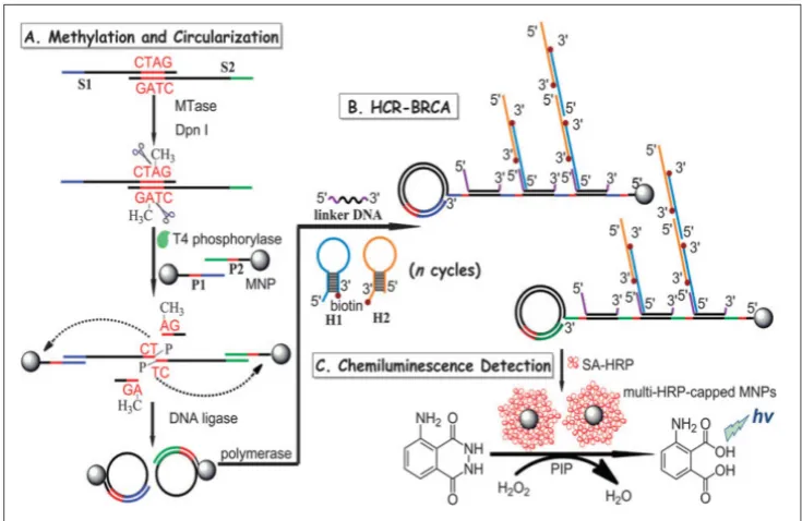
In another report, a combination of hybridisation chain reaction (HCR), branched rolling circle ampli-fication (BRCA) and the coupling of magnetic nano-particles with the aim of catalysing the chemilumi-nescence-generated reaction was reported by Bi and co-workers in 2013 (Figure 7) [103].As shown in Fig-ure 7, consecutive enzymatic actions of DNA MTase and restriction endonuclease DpnI efficiently resulted in four fragments after the hybridisation of DNA strands S1 and S2. At the 5’ ends of two newly-formed shorter fragments, T4 phosphorylase then plays its part to add a padlock-like probe so as to prepare for RCA after T4 DNA ligase joins the complementary ends of the longer DNA fragment to produce a circu-lar template. At the action of DNA polymerase, RCA commences to produce repeating sequences along the elongated strand which is designed to complement linker DNA to yield branched-like structures of overhanging ends of each linker DNA. HCR events can then be propagated between alternating hairpin probes H1 and H2. Since biotin molecules are tagged at 3’ termini of H1 and H2 probes, they recognise streptavidin-HRP (SA-HRP) conjugates and bind on to them. At this point, multitudes of HRP-labelled RCA products are the outcome of the HCR-BRCA process of a single DNA MTase methylation and DpnI cleavage. The final stage of this assay requires the
“fishing-out” of the HRP conjugates by the magnetic nanoparticles of which catalyse luminol oxidation enhanced by p-iodophenol, which manifests a
pow-erful amplification of chemiluminescence readout. Similarly, by adopting the lucifer-ase-luciferin-adenosine triphosphate bioluminescent platform coupled with a methylation-resistant cleav-age and in vitro protein expression, a highly sensitive bioluminescent assay was developed for the detection of DNA MTase activity [104].Using luciferase report-er DNA as substrate DNA for the DNA MTase and MboI as the methylation-resistant endonuclease, DNA MTase activity is quantified by measuring the bioluminescence of the expressed luciferase since methylated luciferase reporter DNA that resists Mbol cleavage could be expressed in cells to produce lucif-erase. The assay produced a wide dynamic range between 0.2–100 U/mL with a detection limit of 0.08 U/mL. Being isothermal in nature, the use of the methylation-resistant cleavage and protein expression approach offers the possibility of in vivo DNA MTase activity imaging and DNA MTase inhibitor screening. 2.5 Electrochemical DNA MTase activity as-says
Electrochemical DNA MTase assays involve the measurements of electrical quantities, such as current, voltage, charge and resistance, to reflect the activity of DNA MTase. They are advantageous over many other types of DNA MTase activity assays because of their low cost, high sensitivity, the ability to perform on-site monitoring and great amenability to minia-turisation and integration with microfabrication technology. The development of electrochemical techniques for bioanalysis has always been helmed as
one of the popular re-search areas in modern analytical chemistry [105,106]. Both direct and amplified electro-chemical DNA MTase activity assays have been proposed. The following section details the de-velopment of electro-chemical DNA MTase activity assays. General-ly, two approaches, namely DNA methyla-tion-initiated cleavage and the use of methyl-ated DNA binding pro-tein coupled with elec-trochemical reporters or electrochemical lumi-nescence generators, are employed in the con-struction of
chemical DNA MTase activity assays. Similar to fluo-rescent assays, to further enhance sensitivity, various enzymatic amplification strategies are incorporated in the electrochemical DNA MTase activity assays. However, comparing to fluorescent DNA MTase ac-tivity assays, the amplification strategies are rather limited because of the heterogeneous nature of elec-trochemical detection.
2.5.1 Direct electrochemical DNA MTase activity assays
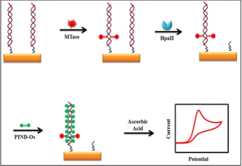
Some of the recently developed electrochemical platforms for screening and monitoring the activity of DNA MTase include electrochemical assays based on restriction endonucleases together with [Ru(NH3)6]3+[107], ferrocene and its derivatives [108,109],coomassie brilliant blue G250 [110],an elec-troactive and catalytic intercalator [111],methylene blue [112-116],carbon nanotubes [117],graphene [118] and graphene oxide [119], methylation sensitive cleavage utilising terminal transferase-mediated ex-tension [120]and the use of methyl binding domain protein (MBD) protein [121-123]and antibody [124]. For instance, a simple and highly sensitive electro-chemical DNA MTase activity assay was proposed by Deng and colleagues (Figure 8) [111].After a mono-layer of a substrate ds-DNA containing the endonu-clease recognition sequence of 5′-CCGG-3′ is immobi-lised on a gold electrode, successive incubations of the substrate DNA-coated electrode with DNA MTase and endonuclease HapII result in the methylation of the substrate DNA and subsequent cleavage of un-methylated DNA off the electrode [125]. Since the methylated substrate DNA resists HapII digestion, only the methylated DNA remains on the electrode surface after the incubations. A final incubation of the treated electrode in a solution containing a threading intercalator–(N,N′ -bis(3-propylimidazole)-1,4,5,8-na-phthalene diimide functionalised with two electro-catalytic redox Os(bpy)2Cl+ moieties introduces the intercalator to the methylated DNA through thread-ing intercalation. After a thorough rinsthread-ing, the elec-trode is tested in ascorbic acid solution where the in-tercalator on the methylated DNA catalyses the oxi-dation of ascorbic acid, producing a measurable cur-rent signal, thus enabling the assessment of the meth-ylation event and specifically the activity of DNA MTase. Much better sensitivity was obtained by working with the electrocatalytic oxidation of ascorbic acid instead of the intercalator itself although the amount of the intercalator on the electrode surface also directly correlates to the activity of DNA MTase. A linear relationship between the activity of DNA MTase and the catalytic oxidation current of ascorbic acid was obtained between 0.08 and 120 U/mL with a
current sensitivity of 0.046 μA mL/U. The concept behind this technique is based on the action of the restriction enzyme with specificity toward methyla-tion, resulting in the electrochemical signal being de-tected by the conversion of the methylation state of the target DNA. This highly sensitive method has not yet been experimented with real biological sample matrix for more appropriate assessment.
Figure 8. Schematic representation of the electroactive and catalytic intercalator-based DNA MTase activity assay. (Reproduced with permis-sion from reference [111].)
Moreover, a multiplexed signal-on electrochem-ical assay using DNA-modified electrodes has also
been proposed by Muren and Barton [121]. As for
ds-DNA strands along with their labels on the elec-trode surface, thereby producing a much-diminished current.
It is undeniable that researchers are now equipped with better understanding in cell biology. Coupling with extensive studies in molecular biology, researchers are now tapping on the vast potential of immunology in population screening and disease diagnosis. The high specificity arises from the se-quence rearrangement of the variable chains in the different types of immunoglobulins upon adherence to foreign or abnormal biological receptors and ex-tracellular matrix components [127].Such an ability of the immunoglobulins enables researchers to take the advantage of the high specificity and completely compatibility with DNA MTase to produce immuno-globulins (antibodies) for DNA MTase [128,129].One such technique in this context is the utilisation of electrochemical immunoassay to the molecular recognition of the epitopes of MBD protein. MBD protein is chosen as a good candidate in the detection of methylation because four out of the five polypep-tides belonging to the MBD protein family have high affinity and specificity toward CpG motifs that have been symmetrically methylated [130-132]. Conven-tional methods in the detection of methylation involve chemical treatment and labelling of 5-methylcytosine. Those processes are in principle undesirable because sample treatment often leads to skewed data due partly to the loss and chemical changes of the analytes [133,134].The high target specificity of MBD protein toward methylated CpG sequences offers an excellent opportunity for the construction of much simplified MBD protein-based assays for the detection of meth-ylated NDA and DNA MTase activity by eliminating most of the sample processing steps.
Recently, Hiraoka et al. demonstrated the use of MBD protein in isolation and precipitation of meth-ylated DNA and quantify the DNA methylation levels by introducing a substrate DNA to luciferase-fused
zinc finger assay [135]. The advantage of the
MBD-protein's specificity to methylation in conjunc-tion with the eliminaconjunc-tion of sample treatment with bisulphate and PCR amplification, makes MBD pro-tein a useful tool for probing DNA methylation and DNA MTase [135-138].For instance,Xu et al. utilised a recombinant MBD1 protein acting as the DNA meth-ylation recognition unit, coupling with a signal unit using a protein staining agent coomassie brilliant blue G250, to develop an electrochemical assay specifically targeting the detection of DNA MTase activity [121]. Another example can be found in the work of Yin et al [110]. After a substrate ds-DNA on an electrode is methylated at the symmetrical sequence of 5'-CCGG-3', a MBD protein MeCP2 is introduced
which selectively binds to the methylated 5'-CCGG-3'. This mode of operation is similar to the previous as-say. The methylated ds-DNA site is bound by the specific recognition unit of MeCP2 protein. Further, MeCP2 carries a His tag at its C-terminal end for se-lective binding by the anti-His tag antibody from HRP labelled and immunoglobulin G functionalised AuNPs (HRP-IgG-AuNPs). The HRP-IgG-AuNPs act as electrochemical signal amplifiers due to the loading of a large number of the IgG-HRP conjugates. Thus, HRP on the electrode efficiently catalyses the oxida-tion of hydroquinone to benzoquinone in the presence of H2O2. Current generated by the electrochemical reduction of benzoquinone is therefore utilised to analyse the activity of DNA MTase. Owing to the high specificity of MeCP2, excellent selectivity down a single base mismatch was observed. Upon further verification, it is believed that this assay could be ap-plied for screening DNA MTase inhibitors and for developing new anticancer drugs.
2.5.2 Amplified electrochemical DNA MTase activity assays
and Gao [150]. Hence, by introducing these two enti-ties into electrochemical immunoassay, a highly sen-sitive assay for DNA MTase activity is expected. To-ward to this goal, the recombinant MBD1 protein, ALP labelled IgG tagged AuNPs (ALP-IgG-AuNPs) and anti-his-tag antibody tagged AuNPs (His-tag-AuNPs) were integrated into an electro-chemical assay for the detection of DNA MTase activ-ity [121].A dual-amplification scheme is realised by the electrochemical oxidation of 1-naphthol produced from the hydrolysis process of enzyme ALP on its substrate. The mechanism of action involves the target recognition of methylated CpG sequences on ds-DNA by His-tagged recombinant MBD1 protein, followed by specific binding by His-tag-AuNPs. These His-tag-AuNPs then trigger the aggregation of the ALP-IgG-AuNPs through another specific binding with the antibody that is attached to the His-tag-AuNPs. The high loading capability of the antibody on the His-tag-AuNPs has resulted in a huge number of the ALP-IgG-AuNPs binding and tagging the methylated sequence of the substrate DNA. Hence, a much amplified electrochemical signal is produced due to the large production of 1-naphthol when ALP catalyses the hydrolysis of its substrate [121].Another feature of the assay is that another en-zyme, HpaII endonuclease, is used to increase the specificity of the electrochemical signal generation process. This is done through the digestion of un-methylated CpG DNA sequences by cleaving 5’-CCGG-3’. Meanwhile, this digestion is blocked by methylation, hence resulting in the inability in the formation of the ALP-IgG-AuNPs conjugates to in-terference or contribute to the detection level of actual amount of methylated CpG sequences [121].In an-other study, a similar approach to the detection of DNA MTase activity was reported by Jing and co-workers [115].In their assay, AuNPs are also used to enhance the electrochemical signals through am-plification. The mechanism of action involve the ac-cumulation of methylene blue onto the AuNPs con-jugated with ds-DNA sequences that have been methylated by DNA MTase and digested by MboI restriction endonuclease. The intercalation of meth-ylene blue onto the ds-DNA creates electron-transfer paths through methylene blue molecules through which a current signal is detected. Un-methylated ds-DNA strands are digested by MboI at the 5’-G-A-T-C-3’palindrome sequence site [151].
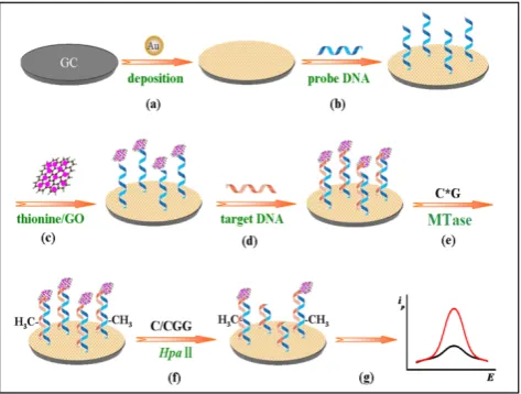
Other nanomaterials such as graphene oxide (GO) have all been tested as signal generators in elec-trochemical MTase activity assays. An example can be found in the work of Li and co-workers (Figure 9) [119]. In their assay, GO is utilised to amplify the signal and HpaII is engaged to improve the selectivity
of the assay. The principle of the assay is similar to the electrochemical signal amplification by the electro-catalytic intercalator as discussed earlier [111].In this assay, a significantly amount of the signal generator thionine is loaded onto GO. Consequently, as low as 0.05 U/mL MTase activity was detected. In addition, the excellent discrimination ability of HpaII between methylated and unmethylated substrate DNA strands produced single-base mismatch selectivity. No re-ported assessment was done in the presence of real biological matrix in this newly developed method.
Figure 9. Schematic illustration of the procedures of the gene-specific DNA methylation detection and DNA MTase activity assay based on the electrochemical signal amplification of GO and restriction endonuclease. (Reproduced with permission from reference [119].)
2.5.3 Photoelectrochemical DNA MTase activity as-says
QDs. As such, the presence of a strong EET effect when the AuNPs are in close proximity to the CdSe QDs effectively counters the photocurrent from ex-ternal light source, resulting in a low photocurrent generation. This only occurs in the presence of ds-DNA methylation by DNA MTase after the lysates or analytes are introduced to the electrode for hy-bridisation with the probe DNA on the AuNPs. This is because methylation prevents binding of the re-striction enzyme HpaII endonuclease onto the hy-bridised DNA for cleavage. Hence, strong EET is
preserved [153]. On the other hand, unmethylated
ds-DNA on the electrode is cleaved off by HpaII, re-sulting in the detachment of the AuNPs from the CdSe QDs, and consequently the reduction of EET. This, in turn, allows a high proportion of photocur-rent to be received by the electrode and a high curphotocur-rent signal registered [152]. This newly developed photo-electrochemical method has not been assessed using real biological fluids for quantification of MTase ac-tivity.
2.6 ECL DNA MTase activity assays
ECL, also known as electrogenerated chemilu-minescence, is a marriage of electrochemistry and luminescence. Like chemiluminescence, to achieve ECL, one or more intermediates in electronically ex-cited state must be produced in a highly exergonic reaction and upon relaxation to the ground state light is emitted. Instead of chemical reactions found in chemiluminescence, ECL generates the intermedi-ates(s) via an electrochemical reaction usually elec-trooxidation. Since the discovery of ECL in the 1960s through the use of aromatic hydrocarbons [154,155], great advances have been made especially after the discovery of the tris-2,2’-bipyridylruthenium(III) ECL
system in the 1970s [156]. Through introducing
chemically active moieties to tris-2,2’-bipyridyl ru-thenium(III), this ECL-generating species has been used extensively as an ECL label in the design of various bioanalytical platforms for highly sensitive and selective detections of a wide range of bioanalytes ranging from small biomolecules to proteins and nu-cleic acids [157,158].Apart from sharing similar at-tractive features with chemiluminescence like sim-plicity of instrumentation and high sensitivity due to the absence of background light [159],ECL offers pre-cise control of reaction kinetics by controlling the ap-plied potential, compatibility with solution-phase and thin-film formats and excellent temporal and spatial control [157-160].
The adoption of ECL in DNA MTase activity assays is expected to deliver excellent performance in terms of simplicity, sensitivity and portability. In-deed, detection limits of ECL DNA MTase activity
assays, ranging from 10-2 to 10-6 U/mL [161-167], whichare at least one order of magnitude lower than most fluorescent, colourimetric and electrochemical assays. For example, a highly sensitive ECL DNA MTase activity assay was developed by labelling a substrate DNA with tris-2,2’-bipyridylruthenium(III) and the adoption of the popular DNA MTase/HpaII pair (Figure 10) [161].First, ss-DNA strands together with tris-2,2’-bipyridylruthenium(III) labelled AuNPs are immobilised onto a gold electrode which subse-quently form the substrate DNA through hybridisa-tion with their complementary strands labelled with ferrocene moieties. Then, successive incubations in DNA MTase and Dpn I lead to the removal of all methylated substrate DNA together with their ferro-cene labels from the electrode, leaving only the un-methylated ones with their ferrocene labels on the electrode. The amount of ferrocene labels found on the electrode and hence the degree of ECL suppres-sion are directly associated with the activity of DNA MTase. Under optimal conditions, a detection limit of 0.03 U/mL was achieved. Real biological fluids were not included in the experiments to further assess MTase activity. Other ECL labels such as tris(1,10-phenanthroline) ruthenium [163-165] and luminol [166]were also utilised in theconstruction of
ECL DNA MTase activity assays. Moreover,
nano-materials like graphene oxide [164,165] and silver nanoparticle/graphene oxide composites [166]were conjugated to the ECL labels in an attempt to further enhance the sensitivity of the ECL assays. Unfortu-nately, the enhancement in sensitivity was not as sig-nificant as expected.
2.7 Other MTase assays
Leveraged on the nanopore technology devel-oped to direct sequence DNA, several attempts were made in the development of nanopore-based devices to detect and quantify methylated DNA and corre-spondingly MTase activity [168-170]. For example, Laszloa and co-workers reported the use of engi-neered porin A to detect methylated DNA through recording the ion current through the nonopore [168]. Distinct differences in the magnitude of the ion cur-rent were observed between methylated and un-methylated DNA strands. A careful comparison of the ion currents and their frequencies reveals the precise location and abundance of methylated CpG sites.
More recently, surface-enhanced Raman spectroscopy in conjunction with HCR was proposed for the detec-tion of MTase activity [171]. Briefly, MTase substrate DNA tethered to magnetic beads was selectively methylated in the presence MTase. After cleaving all unmethylated substrate DNA strands on the magnetic beads by HpaII endonuclease, the methylated DNA on the magnetic beads triggered the HCR reaction and subsequent accumulation of silver nanoparticles through biotin-streptavidin interaction, thus leading to significantly amplified SERS response. A linear range from 0.1 to 10.0 U/ml with a correlation coeffi-cient of 0.97 and a detection limit of 0.067 U/ml were obtained.
Table 1. Comparison of the various DNA MTase activity assays
MTase assay Mechanism Signalling Process Advantages Disadvantages
Sensitiv-ity Ref
Radio DNA MTase assays Isotope-labelling
detec-tion Isotope-labelled methyl moie-ty Convenient detection Harmful radioactive agents NA 46 Polymerase Chain
Reac-tion (PCR) Methylation-specific PCR Conversion of unmethylated cytosines to uracils High accuracy of sequenc-ing Expensive equipment; tedious sample prepara-tion; cumbersome detec-tion scheme
NA 48
Colourimetric DNA MTase Activity assays using AuNPs
Visual observation of a colour change or the spectrometric meas-urement of an absorp-tion spectrum associat-ed with DNA MTase
Change in UV-vis absorption of distance-dependent plasmic absorption among AuNPs
Specificity of enzymes;
signal amplification Complex with the use of multiple enzymes; main-tain physiological condi-tions
0.5 U/mL 50
Catalysis of the oxidation of 2,2’-azino-bis(3-ethylbenzothia zoline-6-sulfonic acid by DNAzyme
Specificity of enzymes; high
sensitivity Requires proper design of probe 0.25 U/mL 53
Direct fluorescent DNA
MTAse activity assays Involvement of absorp-tion of light which excites fluorophores to promote electrons from ground state to excited states
Fluorescence signal generated by fluorescence resonance energy transfer (FRET)
Real-time monitoring; direct
detection Requires proper design of probe 0.8 U/mL 65
Emission spectrum of
aggre-gation of perylene excimers High fluorescence quantum yield; resistance against photooxidation;
self-assembled aggregation
Requires proper design of
probe 0.2 U/mL 72
Fluorescence turn-off DNA
MTase activity assay Simple; cost-effective Requires proper design of probe 1 U/mL 61 Fluorescence polarization as
signal generator Highly sensitive Complex with the use of multiple enzymes 1.0x10
-4
U/mL 73 MoS2 nano sheet-mediated
fluorescence quenching strat-egy
A signal-on fluorescent DNA MTase activity assay; high sensitivity
Complex with the use of
multiple enzymes 0.14 U/mL 80
FRET employed between AuNRs and fluorescein FAM-tagged substrate ds-DNA
High sensitivity Requires proper design of
probe 0.25 U/mL 74
Aggregation-induced emission
(AIE) of fluorophores High performance; simple; economical Requires proper design of probe 0.25 U/mL 67
Amplified fluorescent DNA MTase activity assays
Amplification of fluo-rescence to enhance detection
FRET dually labelled hairpin
reporting probes (FQ probes) Amplification of signal Requires proper design of probe 0.01 U/mL 87
Rolling circle amplification of
DNA with molecular beacons simple; does not require many enzymes; amplifica-tion of signal
Requires proper design of
molecular beacons 0.18 U/mL 83
Nicking enzyme with
molec-ular beacons Amplification of signal; fast Requires proper design of probe 0.06 U/mL 84
Supramolecular fluorescent zinc(II)-protoporphyrin IX/G-quadruplex complex
Extremely low detection
limit Use of multiple enzymes and complexes 8.6x10
-5
U/mL 85
Chemilumininescet/ bioluminescent DNA MTase activity assays
Engage chemi-cal/biochemical reac-tions that produce one
Oxidation of luminol to
3-aminophthalate High sensitivity Use of multiple enzymes and complexes 1.29x10
-4
or more reaction inter-mediates in their elec-tronically excited states, which eventually return to their ground states through the emission of light.
Bioluminescence expressed by
luciferase reporter DNA Isothermal; high sensitivity Requires proper design of reporter DNA 0.08 U/mL 104
MTase
Electrochemical DNA MTase activity assays
The measurement of electrical quantities to reflect the activity of DNA
Electrocatalytic oxidation
current of ascorbic acid Low Cost; high sensitivity Tedious treatment and preparation of electrodes 0.046 uA mL/U 111
Amplified electrochemical DNA MTase activity assays
Amplification of elec-trical quantities to en-hance detection
Signal amplification using
graphene oxide High sensitivity; signal amplification; improved selectivity
Need to functionalise
graphene oxide 0.05 U/mL 119
Photoelectrochemical DNA MTase activity assays
Signal amplification of
exciton energy transfer Measurement of photocurrent due to overlapping of plasmon energy band of AuNPs with exciton energy band of CdSe QDs
Signal amplification Tedious treatment and preparation of electrodes; requires proper design of probe
0.0042 U/mL 152
Electrochemiluminescence DNA MTase activity assays
Light emission in a highly exergonic reac-tion upon relaxareac-tion to the ground state
ECL suppression by ferrocene
labels on substrate DNA High sensitivity Require functionalised AuNPs and substrate DNA
0.03 U/mL 161
3. Challenges and prospects
Epigenetic modifications play in a central role in creating diversity and maintaining specific cellular phenotypes and activities. They are also crucial in the stability of the DNA and chromosome structures. DNA methylation is one of the most important epi-genetic modifications. It is of our best interest to un-derstand and quantify DNA MTase activity as well as DNA methylation patterns in various cells to a deep understanding of the fundamentals and correlate them with human health. In addition, aberrant DNA MTase activity has been known to link to various disorders and diseases such as cancer. As such, a plethora of techniques and approaches have been developed to screen the activity of DNA MTase. Un-like conventional DNA MTase activity assays such as radiolabelling methylation-specific PCR and HPLC which can directly differentiate methylated and un-methylated DNA, these newly developed assays have to be coupled with other biological processes like en-zymatic reactions and immunoreactions, thus bring-ing additional complexity and uncertainty into the assays. On the other hand, ingenious combinations of detection techniques and approaches also give rise to unique DNA MTase activity assays. Among them, fluorometry and electrochemical methods are the most studied detection techniques in DNA MTase activity assays. These two groups of detection tech-niques work on the principles of (i) direct detection of DNA MTase activity and (ii) detection of DNA MTase activity following an amplification process. Nonethe-less, no one assay is superior to another, and the user should carefully analyse the suitability of the assays to the specific task before employing them. Despite
im-pressive progress in the research and development of DNA MTase activity assays, none of them has clearly represented a fundamental breakthrough, becoming a much better alternative to conventional DNA MTase assays. Future research efforts should, therefore, be dedicated to the development of DNA MTase activity assays with much improved sensitivity, selectivity and robustness without engaging any complicated sample preparation and detection protocols. It is be-lieved that the lack of available assays to measure DNA MTase activity in a highly reproducible, quan-titative and sensitive manner continues to be the driving force in DNA MTase activity assay research.
![Figure 1. The reaction mechanism of the methylation of (A) cytosine and (B) adenine. (Reproduced with permis-sion from reference [10].)](https://thumb-us.123doks.com/thumbv2/123dok_us/8721465.1743961/2.612.59.430.439.703/figure-reaction-mechanism-methylation-cytosine-adenine-reproduced-reference.webp)
![Figure 3. Schematic illustration of the DNAzyme-amplified MTase activity assay. (Reproduced with permission from reference [53].)](https://thumb-us.123doks.com/thumbv2/123dok_us/8721465.1743961/5.612.60.299.479.635/schematic-illustration-dnazyme-amplified-activity-reproduced-permission-reference.webp)
![Figure 10. Schematic representation of the ECL DNA MTase activity assay. (Reproduced with permission from reference [161].)](https://thumb-us.123doks.com/thumbv2/123dok_us/8721465.1743961/16.612.319.552.490.684/figure-schematic-representation-mtase-activity-reproduced-permission-reference.webp)
