R E S E A R C H
Open Access
Methylation array profiling of adult brain
tumours: diagnostic outcomes in a large,
single centre
Zane Jaunmuktane
1,2*, David Capper
3,4, David T. W. Jones
5,6, Daniel Schrimpf
7,8, Martin Sill
5,9, Monika Dutt
1,
Nirosha Suraweera
1, Stefan M. Pfister
5,9,10, Andreas von Deimling
7,8and Sebastian Brandner
1,11*Abstract
The introduction of the classification of brain tumours based on their DNA methylation profile has significantly changed the diagnostic approach for cases with ambiguous histology, non-informative or contradictory molecular profiles or for entities where methylation profiling provides useful information for patient risk stratification, for example in medulloblastoma and ependymoma. We present our experience that combines a conventional molecular diagnostic approach with the complementary use of a DNA methylation-based classification tool, for adult brain tumours originating from local as well as national referrals. We report the frequency of IDH mutations in a large cohort of nearly 1550 patients, EGFR amplifications in almost 1900 IDH-wildtype glioblastomas, and histone mutations in 70 adult gliomas. We demonstrate how additional methylation-based classification has changed and improved our diagnostic approach. Of the 325 cases referred for methylome testing, 179 (56%) had a calibrated score of 0.84 and higher and were included in the evaluation. In these 179 samples, the diagnosis was changed in 45 (25%), refined in 86 (48%) and confirmed in 44 cases (25%). In addition, the methylation arrays contain copy number information that usefully complements the methylation profile. For example, EGFR amplification which is 95% concordant with our Real-Time PCR-based copy number assays. We propose here a diagnostic algorithm that integrates histology, conventional molecular tests and methylation arrays.
Keywords: IDH1, IDH2, BRAF, Histone mutation, H3 K27M, Methylation array, Illumina array, DKFZ classifier, Methylation classifier, Molecular diagnostics, Glioma, Ependymoma, Brain tumour classification, Adult brain tumours
Introduction
The diagnosis of brain tumours is achieved by combin-ing morphological features, immunohistochemical (IHC) detection of lineage-related markers, and more recently by the detection of genetic biomarkers, for example mu-tations in the isocitrate dehydrogenase genes 1 and 2 (IDH1 and IDH2) [1, 13], BRAF [35], or histone genes [16]. The development of mutation-specific antibodies to the most common IDH1 mutation R132H [5], BRAF V600E [3] or Histone H3 K27 M [6] has facilitated the introduction of these tests into routine neuropathology
diagnostics and their use is our first diagnostic step. To refine the diagnostic accuracy, we use Sanger sequen-cing, for example to detect rarer mutations in theIDH1, orIDH2genes [26], histones [16], or to detect mutations in theTERTpromoter either to support glioma diagnos-tics in the context of other mutations [10] or to prog-nosticate meningioma recurrence risk [33]. Yet, a significant number of CNS tumours still lack distinctive, and diagnostically informative mutations that can be readily implemented into routine diagnostic practice, or such tests (e.g. gene fusion tests covering multiple break-points) may be resource-intensive to set up, validate and to test routinely. Therefore, neuropathologists may be tempted to revert to the traditional approach of tumour typing and grading, which is fraught by considerable intra-, and inter-observer variability, and by a lack of
* Correspondence:[email protected];[email protected] 1Division of Neuropathology, National Hospital for Neurology and Neurosurgery, University College London Hospitals NHS Foundation Trust, Queen Square, London WC1N 3BG, UK
Full list of author information is available at the end of the article
robust clinical-pathological correlation. For example, it is well established that grading based on histological fea-tures such as mitotic counts, cellularity, pleomorphism, vascular abnormalities and necrosis do not correlate well with the clinical outcome in ependymomas [24] or in diffuse gliomas [38]. The prognostication of intrinsic brain tumours based mainly or exclusively on morph-ology can be misleading, as for example shown in a large-scale study on IDH-wildtype low-grade astrocyto-mas, where a small proportion was confirmed to be of low-grade, whilst a much larger proportion corre-sponded molecularly to high-grade gliomas [29]. The ambiguity of traditional histopathological criteria to in-form clinical oncologists on patient management, and the patients of the prognosis, called for a radically new approach for tumour diagnostics, leading to the develop-ment of a comprehensive CNS tumour reference cohort based on genome-wide DNA methylation profiles [2,4].
Methylation profiles of tumours result from a combination of somatically acquired DNA methylation changes and the cell of origin [11]. These profiles are highly robust and repro-ducible in clinical pathology settings [15] and have been widely used to subclassify CNS tumours, for example epen-dymomas [25], meningiomas [34], medulloblastomas [15], nerve sheath tumours [31], primitive neuroectodermal tu-mours [40] or other tumour types such as“small blue round cell tumours”[17]. A brain tumour methylation classifier has been developed at the German Cancer Research Center (DKFZ) and Heidelberg University in Heidelberg, Germany (henceforth in short “Classifier”), to identify distinct DNA methylation classes of CNS tumours. Currently, the Classifier comprises 82 CNS tumour methylation classes and nine control tissue methylation classes [2]. The Classifier has been made available through a free online tool ( www.molecular-neuropathology.org). We have used this classification tool [2,
4] in clinical practice to stratify into clinically relevant risk groups of histologically defined CNS (and related) tumour entities, and as an aid to establishing a diagnosis in histologi-cally uncertain cases, for example when morphology, loca-tion and demographics were highly unusual, the histology non-specific, or where molecular tests were contradictory, ambiguous or non-informative. We have analysed more than 500 tumours using Illumina 450 K or 850 K EPIC methyla-tion arrays, followed by algorithmic classificamethyla-tion with the Classifier. Here we present the implementation of this plat-form in a neuropathology department within a major aca-demic health science centre, and our experience using the Classifier in routine clinical diagnostic practice.
Material and methods
The rationale for methylation profiling and tumour selection
Methylation profiling was set up in the Division of Neuropathology, the National Hospital for Neurology
and Neurosurgery (NHNN) at University College London Hospitals NHS Foundation Trust, a large clin-ical centre for neurologclin-ical disorders of adults. Tumours included in this study were analysed with methylation arrays between February 2015 and November 2018.
Samples included in this study originated from our own hospital (NHNN) or were referred to us for a sec-ond opinion or conventional and advanced molecular profiling. All tumours underwent routine histopatho-logical assessment, including immunohistochemical and molecular examination in our centre or by referring pa-thologists, and then were processed for methylation ar-rays. The entry criteria for methylation profiling of diagnostic samples were not strictly predefined but were guided by a routine diagnostic decision-making process. A proportion of cases was profiled in the context of re-search studies or clinical trials, and these were not in-cluded in this study.
The cases submitted for the methylation arrays were categorised into six groups with the following character-istics: (i) unusual combination of morphology, location and demographics, (ii) contradictory, ambiguous or non-informative molecular tests, (iii) confirmation of unusual histological and molecular results, (iv) small bi-opsy or non-representative sample, (v) indistinct or non-specific histological appearance, and (vi) cases with characteristic histology requiring risk stratification, such as medulloblastoma [41] or ependymoma [24].
In order to gain experience with the implementation of the technology in our laboratory, and to correlate clinical, pathological and molecular features with the results of the Classifier; a proportion of tumours with clearly defined (molecular) biomarker profile was used to set up and val-idate the procedures. During our setup phase, the Classi-fier was also used for re-classification of tumours previously diagnosed as oligoastrocytoma, which has been discontinued as a distinct entity [30, 32,42], or tumours with the histological phenotype of adult primitive neu-roectodermal tumour (PNET) which now resolve into multiple different entities [40].
Specimen preparation and quality control
All tissues used for methylation studies were fixed in formalin for at least 4 h, and larger samples were dis-sected and fixed overnight, followed by processing through graded alcohols and xylene, to paraffin accord-ing to standard practice in an ISO15189 accredited la-boratory. Tissue embedding and sectioning were according to standard histopathology procedures.
Selection of tumour area
consecutive slides). On a consecutive H&E stained sec-tion (3–4μm), a suitable tumour area was identified by a neuropathologist (SB or ZJ), to maximise inclusion of vi-able tumour-containing tissue. Tumour content of at least 80% was selected where possible and non-neoplas-tic tissue, blood or excessive areas of necrosis were ex-cluded. However, on some occasions where the specimen contained an overall lower tumour density (e.g. infiltration zone) a methylation array analysis was nevertheless attempted, acknowledging a potential risk of an inconclusive Classifier result.
DNA extraction and quantification
Slides with mounted tissue were dewaxed (3 washes in xylene and 2 washes with industrial methylated spirit) and air-dried. Tissue selected for the analysis was scraped off and collected in lysis buffer and DNA was extracted with the Maxwell 16 Lev FFPE DNA Purifica-tion Kit on a Maxwell 16 extractor [19]. The DNA ex-traction procedure was carried out according to manual #TM349 for DNA extraction (Promega). DNA was then quantified and A260/A280 ratios were determined on a Nanodrop 8000 Spectrophotometer (ThermoFisher). An A260/A280ratio of ~ 1.8 was considered to represent suf-ficient purity to proceed with the methylation study. However, rarely we also process samples with a lower A260/A280ratio if there is a clinical necessity and no add-itional material available to repeat extraction or purification.
In our practice, tissue size and resulting DNA amount was rarely the limiting factor. Even a single core of a small stereotaxic biopsy, extracted from 8 consecutive sections of 10μm thickness yielded well above the rec-ommended minimum of 250 ng. A single core of ap-proximately 4 mm2(calculated tissue volume 0.34 mm3) has yielded 600 ng of high-quality DNA, and slightly lar-ger cores of 10–12 mm2 (calculated tissue volume 0.8– 0.9 mm3) have yielded 1400–1800 ng DNA, i.e. well above 250 ng. All these examples were processed and returned a result with a calibrated score of 0.99. In our practice we aim at a DNA input of 500 ng, and in our experience a limiting factor is more often the tissue (and resulting DNA) quality, or tumour content, rather than sample size.
FFPE tissue quality control (QC) assay
Real-time PCR (RT-PCR) assays were run with technical triplicates using DNA isolated from FFPE samples and a QC standard, using primers supplied in the Illumina Infinium HD FFPE QC Kit (Infinium HD FFPE QC Assay Protocol, Illumina). The quality cycle threshold (QCT) value was calculated by subtracting the average Cq of Illumina QC standard from the average Cq value determined for each FFPE sample. Illumina recommends
that a QCT value ≤5 be utilized for optimal assay performance.
Bisulphite conversion of DNA
Based on the DNA quantification steps as determined previously, we aim at an input of 250 ng as a minimum, and ideally at 500 ng DNA from each sample for bisul-phite conversion. The EZ DNA Methylation™Kit (Zymo D5024) was used for DNA conversion. All steps were performed according to the manufacturer’s guidelines. Copy number assays and sequencing
DNA for copy number assays or direct sequencing was ex-tracted from FFPE tumour tissue using Maxwell 16 FFPE LEV DNA purification kit (Promega). Tumour area was confirmed on an H&E-stained slide and tissue was micro-dissected from consecutive 10μm FFPE sections. Primer design was as follows: IDH1-F ACCAAATGGCACCA
TACGA; IDH1-R TGCTTAATGGGTGTAGATACCA
AA; IDH2-F CCAATGGAACTATCCGGAAC; IDH2-R TGTGGCCTTGTACTGCAGAG, BRAF 600-f TCAT AATGCTTGCTCTGATAGGA; C600-r GGCCAAAAA
TTTAATCAGTGGA, TERT-f AGTGGATTCGCGGG
CACAGA, TERT-R; Histone H3F3-F CATGGCTCG
TACAAAGCAGA, H3F3-R CAAGAGAGACTTTG
TCCCATTTTT. For all copy number assays we used the Comparative CT (threshold cycle) multiplex PCR (in same tube) method (ΔΔCT) [36]. The following probes were used for target and reference genes, respectively: 1p36.12b (assay ID Hs06545466_cn; RnaseP 4401631), 1p13.3a (assay ID Hs01847890_cn; RnaseP 4401631); 19q13.2b (assay ID Hs00954642_cn; RnaseP 440163); 19q13.42c (assay ID Hs00831101_cn; RnaseP 440163); 10q23.31a (assay ID Hs05203872_cn; RnaseP 440163); 7p11.2c (assay ID Hs01381289_cn; TERT 4401633). Calibrators were commercial human genomic DNA (gDNA) at a concen-tration of 10μg/μl, (Human Genomic DNA (Male), Pro-mega, G147a) and mixed DNA (mDNA), which contains 1:3 dilution of the gDNA. Copy numbers were determined with the CopyCaller® Software v2.1 (Applied Biosystems).
Immunohistochemistry
All IHC stainings were carried out on automated immu-nostainers (Roche Ventana Discovery or LEICA Bond-Max) following manufacturer’s guidelines. The IDH1 R132H, BRAF V600E, H3 K27M and ATRX antibodies were used as published [3,6,30].
Performing Infinium FFPE restoration
Array processing
The 450 k or EPIC (850 k) methylation array was used to obtain genome-wide DNA methylation profiles for FFPE tumour samples, according to the manufacturer’s in-structions (Illumina). DNA methylation data were gener-ated at the UCL genomics facility at UCL Institute of Child Health. On-chip quality metrics of all samples were carefully controlled. Data (idat files) were trans-ferred to the Division of Neuropathology and uploaded to the Classifier (www.molecularneuropathology.org). Following the upload, the classification result was returned automatically as reported [2].
Results and discussion
Definition of outcomes and calibrated score
For best comparison with other datasets, we aligned the definitions closely to the initial publication of the classi-fication tool [2]. The outcomes were classified according to the impact on the original pathological diagnosis: ori-ginal pathology confirmed (outcome 1), refined (out-come 2) or a new diagnosis established (out(out-come 3). Alternatively, the Classifier result was considered mis-leading (outcome 4) or inconclusive (outcome 5) (Fig. 1a and Table 1). The frequency of outcome 4 or 5 depends on the threshold of the calibrated score. We in-cluded in our analysis only results with a calibrated score of 0.84 and above as recommend in [4]. Classifier results with a calibrated score below 0.84 can still yield informative results [4], in particular when taking into ac-count copy number profiles (such as 7p gain; 10q loss in IDH-wildtype glioblastoma, 1p/19q codeletion in IDH-mutant oligodendroglioma, or copy number vari-ation andCDKN2A/Bdeletions in IDH-mutant astrocy-tomas). Calibrated scores are class probability estimates that measure confidence in the prediction. If the score calibrated is working perfectly, among all tumours of a
“Class X” with a score of 90%, there will be 90% “Class X” tumours. A low score indicates that the classifier is uncertain in its prediction and thus these predictions are often false. Otherwise, if most of the low score predic-tions were true, the probability estimation (or score cali-brated) would not work correctly.
Confirmation of diagnosis (outcome 1): this category includes cases in which the Classifier confirmed the inte-grated diagnosis, such as IDH-mutant and 1p/19q co-deleted oligodendroglioma, IDH-mutant glioblast-oma, subependymglioblast-oma, H3 K27M-mutant diffuse mid-line glioma, and similar. This category also comprises tumour entities where histology or location of a tumour is unusual.
Refinement of diagnosis (outcome 2): this category includes cases in which the Classifier confirms the histo-logical or integrated diagnosis, and determines a more specific molecular subtype, for example in IDH-wildtype
glioblastoma, ependymoma or medulloblastoma. It also includes cases where the diagnostic accuracy is im-proved, e.g. confirmation of an IDH-mutant oligo-dendroglioma with previously tested ambiguous 1p/19q result or sub-classification of tumours with non-specific low-grade morphology, such as IDH-wildtype low-grade glioneuronal tumours.
Establishing a new diagnosis (outcome 3): this out-come was assigned to indicate a change of the original diagnosis (usually unexpected, for example, the change from the histological diagnosis of an ependymoma to the methylation class of a pleomorphic xanthoastrocy-toma, PXA). The main reasons for this outcome were (i) morphologically inconclusive specimens (e.g. with a diagnosis of low-grade or high-grade glioma, with no specific molecular alterations detectable by conventional molecular methods), for which a conclusive methylation class could be established; (ii) incorrect histological in-terpretation, where relevant tests were not considered as a consequence (e.g. solitary fibrous tumour/haemangio-pericytoma misdiagnosed as meningioma); (iii) a distinct histological pattern does not correspond to a specific methylation class (e.g. a tumour with the pattern of astroblastoma often but not always belongs to the HGNET_MN1 methylation class); (iv) all tumours with a newly defined methylation class that do not corres-pond to an existing WHO entity (e.g. “primitive neu-roectodermal tumour” (PNET) resolving into multiple newly defined entities).
Misleading profile (outcome 4): in our experience, there are two scenarios in which misleading results can occur. A low calibrated score can result in a methylation class that is inconsistent with previously tested molecu-lar markers and histology. For example, the methylation class IDH-wildtype glioma in a previously confirmed IDH-mutant glioma. A high calibrated score of > 0.84 rarely generates misleading results. In our 325 diagnostic samples, 179 (56%) had a calibrated score of > 0.84 (Fig.
1b) and none of these showed a misleading profile.
Inconclusive or non-contributory profile (outcome 5): was assigned to cases that showed an obvious dis-crepancy between the input material and the methyla-tion class (such as normal control tissue in cases where analysed material was of a tumour) or in cases where the methylation profiling did not provide any additional diagnostic information. In cases with calibrated score > 0.84 we encountered four cases with a non-contributory profile.
Integration of methylation classification into the diagnostic process
A
B
C
department receives diagnostic samples through three pathways: (i) the local hospital (NHNN) refers tissue for the complete diagnostic workup (tissue diagnosis, molecular diagnostics (pathway 1)); (ii) two geographically separate clinical centres refer formalin-fixed tissue for complete workup as above (pathway 2); and (iii) direct referral of externally di-agnosed tumours for advanced molecular workup, often with a specific request to perform methylation array analysis (pathway 3).
Turnaround times
An important consideration for clinical utility is the turnaround time of tests. The turnaround times (TAT) of methylation arrays are partly dictated by the necessity to form batches (12 arrays on the 450 k chips, and 8 arrays on the 850 k chips), the time it takes to process chips, and how often arrays are proc-essed in a genomics facility. The TAT also depends on the accrual rate of samples. Figure 1c shows a graphical representation of the TAT (the time quired from ordering the test in the laboratory to re-ceiving the data files for the upload on the webpage). Figure 1c shows our institutional performance be-tween 2015 and 2018, demonstrating a relatively sig-nificant variation in sample throughput (which is directly proportional to the number of requested tests) and the TAT. Over time, practice and workflow optimisation has reduced the proportion of samples with TAT exceeding 30 days.
Cost implications
Using list prices for arrays and conversion kits and facil-ity fees, processing of one sample incurs a cost of ap-proximately £380 as of November 2018, without applying discounts that are currently available to our in-stitution. These are itemised as follows: Microarray WG-317-1003 £267 per sample for orders of 96 samples; FFPE restore Kit WG-321-1002 £64 per sample (kit for 24 samples), salary cost for sample registration, DNA ex-traction, bisulphite conversion, quality control, data up-load and results downup-load (8 h @ £50/h = £400, or £50 per sample). This does not include time for medical staff to report cases, taking approximately 30–45 min per case. We consider methylation arrays as a cost-effective and tissue-saving approach for diagnostically challenging cases. A single IHC section costs approximately £18 (full economic costing) and methylation arrays are likely to yield significantly more information and thus better value for money than large panels of immunostains. In our practice, where methylation arrays are readily avail-able and embedded in the diagnostic pathway we usually do not perform more than 10 immunostainings on in-trinsic tumours, as additional stains are unlikely to add meaningful information. An even lower threshold (as few as 3–5 immunostains) is applied to small, precious samples such as stereotaxic biopsies, and targeted se-quencing and methylation arrays are considered early in the diagnostic workup. However, for the routine molecu-lar diagnostics where such limitations do not apply, methylation arrays are not yet the first choice: the con-sumable cost for Sanger sequencing and copy number
(See figure on previous page.)
Fig. 1a, the association of methylation array testing rationale (left) with the outcome of the methylome-based classification (right). 179 cases with a calibrated score of 0.84 and higher were included in this graph.b, distribution of calibrated scores in 325 diagnostic samples examined (excluding research samples) demonstrating that for > 55% (179/325) of the predictions the classifier has had high confidence with estimated class probabilities of 94 > 99%.c, Scattergram of turnaround times (TAT) of tests between 2015 and 2018. Whilst the TAT in the first 2 years of the setup phase often comprised 50% of latencies over 30 days, these long TAT have been progressively reduced in 2018. In quarters 2–4 of 2018, the majority of the cases was completed within 4 weeks. For the first four months in 2015 we did not record the dates of requesting tests, therefore no TAT are shown between February and June 2015
Table 1Number of cases referred for methylation array analysis and the proportion of cases with a calibrated score of 0.84 and higher, and the proportion of cases in which the diagnosis was changed, refined, or confirmed or where the test yielded non-contributory results
Number of cases Proportion of all cases Proportion of cases with CS > 0.84
Cases referred for testing 325 100% 56%
Tumours with a calibrated score > 0.84 179 56% 100%
Establishing new diagnosis 45 14% 25%
Refinement of diagnosis 86 26% 48%
Confirmation of diagnosis 44 14% 25%
Non-contributory 4 1% 2%
assays is a fraction of those for methylation arrays, and the turnaround times are significantly shorter, making it impractical and unaffordable to implement methylation arrays for the diagnosis of all brain tumours.
Local tissue referrals (pathways 1 and 2): all brain tumours undergo routine histological and immunohisto-chemical examination and are reported as part of the standard diagnostic process in our department. Low-and high-grade gliomas Low-and poorly differentiated supra-tentorial intrinsic tumours with PNET morphology undergo immunostaining for IDH1 R132H and ATRX. IDH-mutant tumours with ATRX loss are diagnosed as IDH-mutant astrocytomas or glioblastomas (GBM), and for prognostication, these are tested for CDKN2A/B homozygous deletion [38]. IDH-mutant tumours with retained ATRX undergo further testing for 1p/19q and TERT promoter mutations. Midline gliomas are rou-tinely tested with immunostaining for H3 K27M. IDH1 R132H negative gliomas undergo a targeted sequencing for known mutations in the IDH1/2, histone H3.3 (H3F3A),BRAFgenes and theTERTpromoter, and copy number assays (1p/19q, CDKN2A/B, 7p (EGFR), and 10q (PTEN locus)). This identifies the remaining IDH- or histone-mutant gliomas and IDH-wildtype,
TERT-mutant, and/or EGFR amplified GBM. IDH- and
histone-wildtype gliomas with ATRX loss also undergo BRAFfusion testing to identify possible anaplastic astrocy-toma with piloid features [28].
Tumours with unusual location or non-specific glial or glioneuronal morphology and non-informative conven-tional molecular test results are then considered for methylation arrays. In this study, the threshold to use the Classifier was lower for young adults, although we did not define a specific age cut-off and instead made a case-based decision in consultation with the clinical teams. The Classifier was also used for confirmation of rare tumour entities which do not have any of the above-mentioned gene mutations. The methylation class was reported as part of the integrated diagnosis, e.g.
“histology: low-grade glioma, IDH-wildtype; methylation class: glioblastoma, IDH-wildtype RTK II”; or“histology: high-grade glioma; methylation class: pleomorphic xanthoastrocytoma; BRAF V600E mutant”.
All supra- and infratentorial ependymomas undergo methylation studies. We encounter a relatively small number of supratentorial ependymomas in our adult practice (in this cohort, from pathways 1 and 2 n= 7), which in our view justifies initial risk stratification by methylation profiling [12] instead of nucleic acid-based tests for multiple potential gene fusion transcripts. Tu-mours with characteristic subependymoma morphology irrespective of location and all spinal ependymomas are not tested further unless there is a specific clinical re-quest, an unusual clinical presentation, or histology.
Infratentorial tumours with features of embryonal tu-mours (e.g. medulloblastoma), similar to ependymomas, typically are processed for methylation array for risk stratification purposes, although the prognostic signifi-cance of medulloblastoma subclasses in adults is cur-rently not firmly established. Since April 2018, all adult medulloblastomas in the UK are referred to the National Medulloblastoma Reference Centre through Great Ormond Street Hospitals [37].
Pilocytic astrocytomas are routinely tested for BRAF V600 and the three most common KIAA1549:BRAF fu-sion mutations (16–9, 16–11, 15–9). Tumours with a confirmed mutation are not further investigated. Tu-mours with pilocytic astrocytoma or other low-grade glial or glioneuronal morphology with no BRAF V600 mutation, absence of the three tested fusions, and in par-ticular, those with loss of ATRX protein expression rou-tinely undergo methylation array testing.
Referrals of previously diagnosed tumours (pathway 3)are (i) specifically referred for Illumina array analysis, or received for (ii) targeted diagnostics with conven-tional molecular tests as above, or (iii) second opinion, often with extensive previous workup, including molecu-lar testing. Cases which remain inconclusive after the molecular studies are then processed for methylation ar-rays and an integrated diagnosis is returned to the refer-ring pathologists.
Diagnostic outcomes from molecular assays in our practice
IDH-mutant gliomas
The most common reason for further workup with methylation array is a confirmedIDH mutation in com-bination with an inconclusive ATRX, TERT promoter and 1p/19q status; for example IDH-mutant gliomas with retained ATRX expression and ambiguous 1p/19q status; or rare diffuse gliomas with ATRX loss in which
the IDH- or histone H3 mutation cannot be established
by IHC or sequencing.
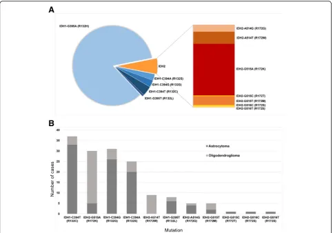
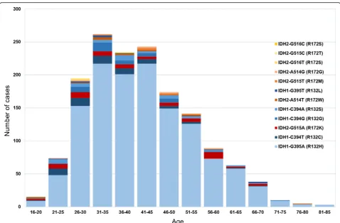
In order to establish if an upper age limit can be justified for the testing ofIDH1/IDH2mutations beyond the use of immunostaining with the IDH1 R132H mutation-specific antibody, we analysed the age distribution in 1546 tu-mours. The frequency distribution of the distinct IDH1 andIDH2mutations is comparable to previous reports of similar scale [13] (Fig. 2). In addition to mutations de-scribed in previous series [7,14,26] we found rare muta-tions such as IDH2-G515C (R172T), IDH2-G516C (R172S), IDH2-G516T (R172S), IDH2-A514G (R172G),
and IDH2-G515T (R172M) in astrocytomas and
55 years and older (Fig. 3), of which 31 (13%) would not have been detected by IDH1 R132H IHC alone. In our opinion, this justifies molecular testing for these rarer
IDH1/IDH2 mutations in patients over 55 years, in
con-trast to previous recommendations [9,22].
As previously shown [20] and in our experience, there is a small subset of IDH-mutant, 1p/19q non-co-deleted tumours with retained ATRX protein expression. These tumours correspond to methylation class “IDH-mutant astrocytoma, high-grade”, in keeping with earlier studies [18]. Importantly, mutations inTERT promoter may be observed in these tumours [18].
We and most other centres use ATRX IHC as a surro-gate marker for ATRX mutations, however, the loss of function mutations with retained protein expression are not detected [21]. As shown in our cohort and other studies [21], such mutations are relatively infrequent but provide an explanation for the discrepancies between ATRX sequencing and immunohistochemistry. Centres
with access to affordable next-generation sequencing may consider testing such cases for the presence of a mutation.
In our cohort, we observed mutations in the TERT promoter in all IDH-mutant, 1p/19q co-deleted oligo-dendrogliomas, where testing was successful. Although the finding of TERT promoter mutation in IDH-mutant glioma is helpful in confirming the diagnosis of an oligo-dendroglial tumour, in our experience the sequencing of TERTpromoter mutation can be technically challenging. If 1p/19q andTERTpromoter testing remains inconclu-sive, sometimes occurring in referred cases that under-went different fixation protocols, we resort to further analysis with the Classifier.
Histone H3-mutant gliomas
As part of the sequencing panel described above, in 2015 we introduced H3F3A testing in all low- and high-grade gliomas. In our practice, all H3 K27M
A
B
Fig. 2a, the frequency of IDH mutations in our cohort (n= 1546). Blue,IDH1mutations; orange and red,IDH2mutations. TheIDH1/IDH2 frequency in our cohort is slightly skewed toward rarer mutations, due to a proportion of referrals received specifically for sequencing studies.
mutant gliomas (n= 49) have been located in the mid-line, its proximity, or there was an anatomical connec-tion to the midline. Occasionally, the proximity to the midline waspost-hoc suggested after detection of the H3 K27M mutation. When a histone mutation is detected (by sequencing or H3 K27M IHC), we currently do not proceed to methylation array testing. Instead, midline tumours with no H3 K27M mutation, with or without ATRX loss, and no other specific findings on conven-tional molecular testing undergo further methylation studies. In our cohort, H3 mutant gliomas (K27M in particular) manifest also in adults over 50 years (Fig.4). In our experience, loss of ATRX protein expression oc-curs in nearly all (18/19; 95%) H3 G34 mutant gliomas but only in a subset (19/45; 42%) of H3 K27M mutant gliomas. Rarely, we observed biphasic patterns of ATRX loss in H3 K27M (1 case) - and H3 G34R mutant gli-omas (1 case) (Fig. 4). In another case of a recurrent high-grade glioma with ATRX loss, the methylation class was “H3 G34 mutant glioblastoma”, but we could not identify any H3 G34 mutation on theH3F3A,HIST1H3B
and HIST1H3C genes. This raises the possibility that
there is a mutation in another, as yet unknown, H3 variant-encoding histone gene.
Brain tumours with loss of ATRX protein expression
In our cohort, the brain tumours with loss of ATRX protein expression are IDH-mutant low- and high-grade astrocytomas, H3 K27M and G34 mutant gliomas, anaplastic pilocytic astrocytomas (methyla-tion class ANA_PA, also termed anaplastic astrocyto-mas with piloid features) [28], and rarely IDH-wildtype glioblastomas, confirmed with the Clas-sifier. Tumours with ATRX loss but no IDH or His-tone mutation (by IHC or sequencing) generally undergo methylation array testing.
Newly established methylation classes representing new entities
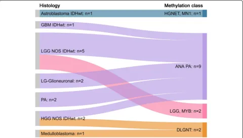
and 33) (Fig. 5). In keeping with previous reports, these tumours had either non-specific low-grade or high-grade astrocytic morphology, primitive small cell histology, or appearances of histologically de-fined entities of astroblastoma and pilocytic astrocy-toma [8, 27, 28].
These new molecular entities were originally identi-fied by DNA-methylation profiling and since then have been found to have characteristic recurrent gen-etic alterations. Therefore, whilst possible to diagnose them with conventional molecular methods involving DNA or RNA sequencing, in routine practice, in our opinion, the DNA methylation-based diagnostic ap-proach is efficient and cost-effective for their identification.
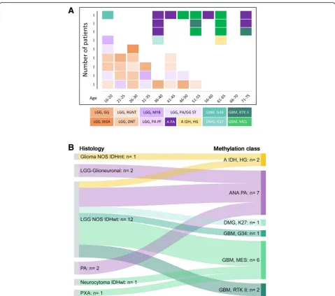
IDH-wildtype gliomas with low-grade morphology
low-grade glial or glioneuronal tumour entities (Fig. 6). The average age for tumours classified as low-grade tumour entities was 30.8 years and for those classified as high-grade tumour entities 53.6 years (Fig. 6).
IDH-wildtype gliomas with high-grade morphology
The majority of IDH-wildtype high-grade gliomas are glioblastomas. Characteristic signatures are chromosome 7 gains and 10 losses, EGFR amplification and TERT promoter mutation [39]. In the cohort of [39] 1788 out of 4284 GBM (41.7%) wereEGFR amplified (methylation classes: GBM_MYCN, GBM_RTK_I, GBM_RTK_II, GBM_RTK_III, GBM_MES, GBM_MID). In our cohort (2010–2018), we found 749 out of 1888 (39.7%) EGFR amplified GBM (6 and more copies), suggesting a nearly identical prevalence. From September 2015 till June 2018, 509 glioblastomas were tested for bothTERT pro-moter mutations and EGFR status (Fig. 7). 121 were
TERT-wildtype and EGFR non-amplified, 211 were
TERT-mutant and EGFR non-amplified, 25 were
TERT-wildtype and EGFR amplified and 152 were TERT-mu-tant andEGFRamplified. These data are largely compar-able with a published dataset [39] (Fig.7). In 41 cases of
IDH-wildtype glioblastoma, EGFR status was assessed with both, methylation arrays and RTPCR. Only two cases showed a discrepant result, where the Illumina-derived plot showed amplification and the RT-PCR result revealed a copy number corresponding to non-amplified status, indicating a 95% concordance be-tween both methods (χ20.21,p= 0.64), (Fig.7).
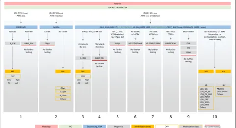
A comprehensive table outlining a suggested diag-nostic test algorithm, combining histology, immuno-histochemical markers, conventional molecular tests and methylation arrays in low-grade and high-grade gliomas is shown in Fig. 8. We choose this approach as the consumable cost for Sanger sequencing and copy number assays are considerably below those of methylation arrays (approximately 10% of the cost) and the turnaround times allow for communication of test results within approximately 7 working days. Therefore, we choose methylation arrays as a first-line approach typically for small biopsies for which we predict an inconclusive outcome with our
“conventional” molecular test portfolio, and for cases with an unusual histological presentation. In our rou-tine practice, the first-line approach is usually as sug-gested in Fig. 8.
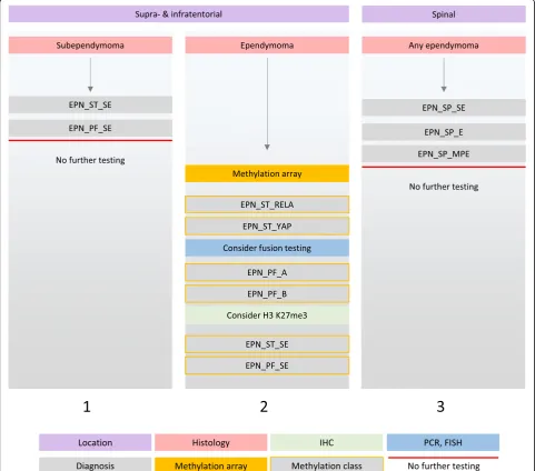
Ependymal tumours
Ependymal tumours located supratentorial or in the posterior fossa are submitted for the methylation ana-lysis specifically for risk stratification purposes [24,
25] (Fig. 9). In our experience, the greatest
a methylation profile of subependymoma. This finding is in line with earlier recommendations [24] that treatment decisions outside of clinical trials should not be based on a histologically assigned WHO grade. The Classifier also has helped in accurately diagnos-ing a subependymoma of which only small amounts of tissue were available, precluding a definitive histo-logical diagnosis. An overview of the changes of
diagnosis for ependymal tumours is given in Fig. 10. In another instance, the Classifier prompted us to change the diagnosis from histologically diagnosed an-aplastic ependymoma to a BRAF V600E-mutant pleo-morphic xanthoastrocytoma. This tumour underwent methylation array analysis specifically for the risk stratification after resection of a recurrence of a pre-sumed ependymoma. The Classifier result (of Fig. 7EGFR amplification in IDH-wildtype glioblastoma:a, comparison of our dataset with a previously published dataset [39] shows that the ratio ofEGFR amplified and non-amplified,TERT-mutant GBM is similar to the published cohort (p= 0.3). Instead, the ratio ofEGFR amplified and non-amplified,TERT-wildtype GBM is different between both cohorts (p= 0.04).b, comparison of the prevalence of EGFR status in GBM in our cohort (London, RT-PCR quantification) with those from the published dataset (“HD”, determined with the copy number readout from the methylation arrays) [39], shows no statistically significant difference (χ22.3,p= 0.13).c, comparison ofEGFR
status in our cohort determined with Illumina arrays and with RT-PCR. There is a 95% concordance between both methods (χ20.21,p=
anaplastic) PXA prompted us to test for, and confirm the BRAF V600E mutation, and the patient under-went treatment with BRAF inhibitors. Another tumour with histological features of anaplastic epen-dymoma was reclassified as H3 K27M-mutant diffuse midline glioma, and this mutation was confirmed sub-sequently by IHC and H3F3A gene sequencing.
Conclusion
We report here a single centre experience of the im-plementation of methylation arrays into routine prac-tice for algorithmic classification of brain tumours. In contrast to a paediatric setting, where a significant proportion of tumours undergoes methylation-based
classification for risk stratification (such as ependy-moma, medulloblastoma), methylation profiling for this purpose constitutes only a minority of our ana-lysis of adult brain tumours. The combination of histological assessment with conventional molecular testing (i.e. IHC, targeted sequencing, copy number assay) is in our practice the first line diagnostic ap-proach and is adequate for the majority of intrinsic tumours, such as IDH-mutant astrocytomas, oligo-dendrogliomas, histone-mutant, or EGFR-amplified, TERT promoter-mutant IDH-wildtype glioblastomas. By far the most common reason for using the methylation-based classification in adult practice is the need to obtain a more accurate and clinically Fig. 8Diagnostic testing algorithm for gliomas in adults. The first layer is the histological assessment. The histological identification of a glial tumour is followed by the standard application of the antibodies IDH1 (R132H) and ATRX. This identifies a majority of IDH-mutant gliomas (column 1, 2). IDH-mutant astrocytomas with ATRX loss are further tested forCDKN2A/Bhomozygous deletion to stratify high risk from lower risk astrocytomas (column 1). Lower risk IDH-mutant astrocytomas are also assessed for copy number variation, a suggested prognostic factor. This is achieved by the readout of the copy number variation (CNV) component of the methylation arrays. IDH-mutant gliomas with retained ATRX expression (column 2) are further tested for 1p/19q co-deletion with a conventional copy number assay (in our practice combined withTERT promoter mutation analysis). Those IDH-mutant tumours which have retained ATRX expression and either no co-deletion or an ambiguous copy number result, are further tested with methylation array. This helps to differentiate IDH-mutant oligodendrogliomas from IDH-mutant
relevant diagnosis for tumours with unusual, non-specific or non-representative histology and where the molecular testing does not yield diagnostic-ally informative results. We found a change of diag-nosis in approximately 25% of patients, refinement in
unnecessary, potentially harmful treatment. Our co-hort further highlights the essential role of the methy-lation array as a diagnostic tool in advanced brain tumour diagnostics, when integrated into the diagnos-tic pathway in a structured fashion as outlined in Fig.
8 and Fig. 9.
Acknowledgements
We thank the biomedical scientists in the Division of Neuropathology, the National Hospital for Neurology and Neurosurgery (NHNN) for excellent technical assistance. We also thank clinicians and neuropathologists for referring cases for molecular analysis. Part of the study was funded by the National Institute for Health Research to UCLH Biomedical research centre (BRC399/NS/RB/101410). SB is also supported by the Department of Health’s NIHR Biomedical Research Centre’s funding scheme.
Declarations
Data were obtained from University College London NHS Foundation Trust as part of the UK Brain Archive Information Network (BRAIN UK) which is funded by the Medical Research Council and Brain Tumour Research. BRAIN UK reference number: 19/002 - Molecular analyses of adult brain tumours by conventional molecular tests and DNA methylation profiling. All authors have seen the manuscript and have approved publication. Datasets can be made available on request.
Authors’contributions
ZJ, NS, MD, and SB generated and analysed data. DC, DTWJ, DS, MS, SMP, and AvD engineered the analysis platform and provided analytical tools. ZJ and SB wrote the manuscript with input from all authors. All authors read and approved the final manuscript.
Competing interests
The authors declare that they have no competing interests.
Publisher’s Note
Springer Nature remains neutral with regard to jurisdictional claims in published maps and institutional affiliations.
Author details
1Division of Neuropathology, National Hospital for Neurology and Neurosurgery, University College London Hospitals NHS Foundation Trust, Queen Square, London WC1N 3BG, UK.2Department of Clinical and Movement Neurosciences, UCL Queen Square Institute of Neurology, Queen Square, London WC1N 3BG, UK.3Department of Neuropathology, Charité Universitätsmedizin Berlin, Corporate Member of Freie Universität Berlin, Humboldt-Universität zu Berlin and Berlin Institute of Health, Berlin, Germany. 4German Cancer Consortium (DKTK), Partner Site Berlin, German Cancer Research Center (DKFZ), Heidelberg, Germany.5Hopp Children’s Cancer Center Heidelberg (KiTZ), Heidelberg, Germany.6Pediatric Glioma Research Group, German Cancer Research Center (DKFZ), Heidelberg, Germany. 7Department of Neuropathology, University Hospital Heidelberg, Heidelberg, Germany.8Clinical Cooperation Unit Neuropathology, German Cancer Consortium (DKTK), German Cancer Research Center (DKFZ), Heidelberg, Germany.9Division of Pediatric Neurooncology, German Cancer Research Center (DKFZ), Heidelberg, Germany.10Department of Pediatric Oncology, Hematology, Immunology and Pulmonology, Heidelberg University Hospital, Heidelberg, Germany.11Department of Neurodegenerative Disease, UCL Queen Square Institute of Neurology, Queen Square, London WC1N 3BG, UK.
Received: 11 January 2019 Accepted: 28 January 2019
References
1. Balss J, Meyer J, Mueller W, Korshunov A, Hartmann C, von Deimling A (2008) Analysis of the IDH1 codon 132 mutation in brain tumors. Acta Neuropathol 116:597–602
2. Capper D, Jones DTW, Sill M, Hovestadt V, Schrimpf D, Sturm D, Koelsche C, Sahm F, Chavez L, Reuss DE, Kratz A, Wefers AK, Huang K, Pajtler KW, Schweizer L, Stichel D, Olar A, Engel NW, Lindenberg K, Harter PN,
Braczynski AK, Plate KH, Dohmen H, Garvalov BK, Coras R, Holsken A, Hewer E, Bewerunge-Hudler M, Schick M, Fischer R, Beschorner R, Schittenhelm J, Staszewski O, Wani K, Varlet P, Pages M, Temming P, Lohmann D, Selt F, Witt H, Milde T, Witt O, Aronica E, Giangaspero F, Rushing E, Scheurlen W, Geisenberger C, Rodriguez FJ, Becker A, Preusser M, Haberler C, Bjerkvig R, Cryan J, Farrell M, Deckert M, Hench J, Frank S, Serrano J, Kannan K, Tsirigos A, Bruck W, Hofer S, Brehmer S, Seiz-Rosenhagen M, Hanggi D, Hans V, Rozsnoki S, Hansford JR, Kohlhof P, Kristensen BW, Lechner M, Lopes B, Mawrin C, Ketter R, Kulozik A, Khatib Z, Heppner F, Koch A, Jouvet A, Keohane C, Muhleisen H, Mueller W, Pohl U, Prinz M, Benner A, Zapatka M, Gottardo NG, Driever PH, Kramm CM, Muller HL, Rutkowski S, von Hoff K, Fruhwald MC, Gnekow A, Fleischhack G, Tippelt S, Calaminus G, Monoranu CM, Perry A, Jones C, Jacques TS, Radlwimmer B, Gessi M, Pietsch T, Schramm J, Schackert G, Westphal M, Reifenberger G, Wesseling P, Weller M, Collins VP, Blumcke I, Bendszus M, Debus J, Huang A, Jabado N, Northcott PA, Paulus W, Gajjar A, Robinson GW, Taylor MD, Jaunmuktane Z, Ryzhova M, Platten M, Unterberg A, Wick W, Karajannis MA, Mittelbronn M, Acker T, Hartmann C, Aldape K, Schuller U, Buslei R, Lichter P, Kool M, Herold-Mende C, Ellison DW, Hasselblatt M, Snuderl M, Brandner S, Korshunov A, von Deimling A, Pfister SM (2018) DNA methylation-based classification of central nervous system tumours. Nature 555:469–474 3. Capper D, Preusser M, Habel A, Sahm F, Ackermann U, Schindler G, Pusch S,
Mechtersheimer G, Zentgraf H, von Deimling A (2011) Assessment of BRAF V600E mutation status by immunohistochemistry with a mutation-specific monoclonal antibody. Acta Neuropathol 122:11–19
4. Capper D, Stichel D, Sahm F, Jones DTW, Schrimpf D, Sill M, Schmid S, Hovestadt V, Reuss DE, Koelsche C, Reinhardt A, Wefers AK, Huang K, Sievers P, Ebrahimi A, Scholer A, Teichmann D, Koch A, Hanggi D, Unterberg A, Platten M, Wick W, Witt O, Milde T, Korshunov A, Pfister SM, von Deimling A (2018) Practical implementation of DNA methylation and copy-number-based CNS tumor diagnostics: the Heidelberg experience. Acta Neuropathol 136:181–210
5. Capper D, Zentgraf H, Balss J, Hartmann C, von Deimling A (2009) Monoclonal antibody specific for IDH1 R132H mutation. Acta Neuropathol 118:599–601
6. Castel D, Philippe C, Calmon R, Le Dret L, Truffaux N, Boddaert N, Pages M, Taylor KR, Saulnier P, Lacroix L, Mackay A, Jones C, Sainte-Rose C, Blauwblomme T, Andreiuolo F, Puget S, Grill J, Varlet P, Debily MA (2015) Histone H3F3A and HIST1H3B K27M mutations define two subgroups of diffuse intrinsic pontine gliomas with different prognosis and phenotypes. Acta Neuropathol 130:815–827
7. Dahlrot RH, Kristensen BW, Hjelmborg J, Herrstedt J, Hansen S (2013) A population-based study of low-grade gliomas and mutated isocitrate dehydrogenase 1 (IDH1). J Neuro-Oncol 114(3):309–317
8. Deng MY, Sill M, Chiang J, Schittenhelm J, Ebinger M, Schuhmann MU, Monoranu CM, Milde T, Wittmann A, Hartmann C, Sommer C, Paulus W, Gartner J, Bruck W, Rudiger T, Leipold A, Jaunmuktane Z, Brandner S, Giangaspero F, Nozza P, Mora J, Morales la Madrid A, Cruz Martinez O, Hansford JR, Pietsch T, Tietze A, Hernaiz-Driever P, Stoler I, Capper D, Korshunov A, Ellison DW, von Deimling A, Pfister SM, Sahm F, Jones DTW (2018) Molecularly defined diffuse leptomeningeal glioneuronal tumor (DLGNT) comprises two subgroups with distinct clinical and genetic features. Acta Neuropathol 136(2):239–253
9. DeWitt JC, Jordan JT, Frosch MP, Samore WR, Iafrate AJ, Louis DN, Lennerz JK (2017) Cost-effectiveness of IDH testing in diffuse gliomas according to the 2016 WHO classification of tumors of the central nervous system recommendations. Neuro-Oncology 19:1640–1650
10. Eckel-Passow JE, Lachance DH, Molinaro AM, Walsh KM, Decker PA, Sicotte H, Pekmezci M, Rice T, Kosel ML, Smirnov IV, Sarkar G, Caron AA, Kollmeyer TM, Praska CE, Chada AR, Halder C, Hansen HM, McCoy LS, Bracci PM, Marshall R, Zheng S, Reis GF, Pico AR, O'Neill BP, Buckner JC, Giannini C, Huse JT, Perry A, Tihan T, Berger MS, Chang SM, Prados MD, Wiemels J, Wiencke JK, Wrensch MR, Jenkins RB (2015) Glioma groups based on 1p/19q, IDH, and TERT promoter mutations in tumors. N Engl J Med 372:2499–2508
11. Fernandez AF, Assenov Y, Martin-Subero JI, Balint B, Siebert R, Taniguchi H, Yamamoto H, Hidalgo M, Tan AC, Galm O, Ferrer I, Sanchez-Cespedes M, Villanueva A, Carmona J, Sanchez-Mut JV, Berdasco M, Moreno V, Capella G, Monk D, Ballestar E, Ropero S, Martinez R, Sanchez-Carbayo M, Prosper F, Agirre X, Fraga MF, Grana O, Perez-Jurado L, Mora J, Puig S, Prat J, Badimon L, Puca AA, Meltzer SJ, Lengauer T, Bridgewater J, Bock C, Esteller M (2012) A DNA methylation fingerprint of 1628 human samples. Genome Res 22:407–419
12. Gessi M, Giagnacovo M, Modena P, Elefante G, Gianno F, Buttarelli FR, Arcella A, Donofrio V, Diomedi Camassei F, Nozza P, Morra I, Massimino M, Pollo B, Giangaspero F and Antonelli M (2019) Role of Immunohistochemistry in the Identification of Supratentorial C11ORF95-RELA Fused Ependymoma in Routine Neuropathology. Am J Surg Pathol 43:56-63
13. Hartmann C, Meyer J, Balss J, Capper D, Mueller W, Christians A, Felsberg J, Wolter M, Mawrin C, Wick W, Weller M, Herold-Mende C, Unterberg A, Jeuken JW, Wesseling P, Reifenberger G, von Deimling A (2009) Type and frequency of IDH1 and IDH2 mutations are related to astrocytic and oligodendroglial differentiation and age: a study of 1,010 diffuse gliomas. Acta Neuropathol 118:469–474
14. Horbinski C (2013) What do we know about IDH1/2 mutations so far, and how do we use it? Acta Neuropathol 125:621–636
15. Hovestadt V, Remke M, Kool M, Pietsch T, Northcott PA, Fischer R, Cavalli FMG, Ramaswamy V, Zapatka M, Reifenberger G, Rutkowski S, Schick M, Bewerunge-Hudler M, Korshunov A, Lichter P, Taylor MD, Pfister SM, Jones DTW (2013) Robust molecular subgrouping and copy-number profiling of medulloblastoma from small amounts of archival tumour material using high-density DNA methylation arrays. Acta Neuropathol 125:913–916
16. Khuong-Quang DA, Buczkowicz P, Rakopoulos P, Liu XY, Fontebasso AM, Bouffet E, Bartels U, Albrecht S, Schwartzentruber J, Letourneau L, Bourgey M, Bourque G, Montpetit A, Bourret G, Lepage P, Fleming A, Lichter P, Kool M, von Deimling A, Sturm D, Korshunov A, Faury D, Jones DT, Majewski J, Pfister SM, Jabado N, Hawkins C (2012) K27M mutation in histone H3.3 defines clinically and biologically distinct subgroups of pediatric diffuse intrinsic pontine gliomas. Acta Neuropathol 124:439–447
17. Koelsche C, Hartmann W, Schrimpf D, Stichel D, Jabar S, Ranft A, Reuss DE, Sahm F, Jones DTW, Bewerunge-Hudler M, Trautmann M, Klingebiel T, Vokuhl C, Gessler M, Wardelmann E, Petersen I, Baumhoer D, Flucke U, Antonescu C, Esteller M, Frohling S, Kool M, Pfister SM,
Mechtersheimer G, Dirksen U, von Deimling A (2018) Array-based DNA-methylation profiling in sarcomas with small blue round cell histology provides valuable diagnostic information. Mod Pathol 31(8):1246–1256 18. Korshunov A, Casalini B, Chavez L, Hielscher T, Sill M, Ryzhova M, Sharma T,
Schrimpf D, Stichel D, Capper D, Reuss DE, Sturm D, Absalyamova O, Golanov A, Lambo S, Bewerunge-Hudler M, Lichter P, Herold-Mende C, Wick W, Pfister SM, Kool M, Jones DTW, von Deimling A and Sahm F (2018) Integrated molecular characterization of IDH-mutant glioblastomas. Neuropathol Appl Neurobiol.https://doi.org/10.1111/nan.12523. Epub ahead of print.
19. Krnajski Z, Geering S, Steadman S (2007) Performance verification of the Maxwell 16 instrument and DNA IQ reference sample kit for automated DNA extraction of known reference samples. Forensic Sci Med Pathol 3: 264–269
20. Leeper HE, Caron AA, Decker PA, Jenkins RB, Lachance DH, Giannini C (2015) IDH mutation, 1p19q codeletion and ATRX loss in WHO grade II gliomas. Oncotarget 6:30295–30305
21. Liu XY, Gerges N, Korshunov A, Sabha N, Khuong-Quang DA, Fontebasso AM, Fleming A, Hadjadj D, Schwartzentruber J, Majewski J, Dong Z, Siegel P, Albrecht S, Croul S, Jones DT, Kool M, Tonjes M, Reifenberger G, Faury D, Zadeh G, Pfister S, Jabado N (2012) Frequent ATRX mutations and loss of expression in adult diffuse astrocytic tumors carrying IDH1/IDH2 and TP53 mutations. Acta Neuropathol 124:615–625
22. Louis DN, Perry A, Reifenberger G, von Deimling A, Figarella-Branger D, Cavenee WK, Ohgaki H, Wiestler OD, Kleihues P, Ellison DW (2016) The 2016 World Health Organization classification of tumors of the central nervous system: a summary. Acta Neuropathol 131:803–820
23. Nobusawa S, Watanabe T, Kleihues P, Ohgaki H (2009) IDH1 mutations as molecular signature and predictive factor of secondary glioblastomas. Clin Cancer Res 15:6002–6007
25. Pajtler KW, Witt H, Sill M, Jones DT, Hovestadt V, Kratochwil F, Wani K, Tatevossian R, Punchihewa C, Johann P, Reimand J, Warnatz HJ, Ryzhova M, Mack S, Ramaswamy V, Capper D, Schweizer L, Sieber L, Wittmann A, Huang Z, van Sluis P, Volckmann R, Koster J, Versteeg R, Fults D, Toledano H, Avigad S, Hoffman LM, Donson AM, Foreman N, Hewer E, Zitterbart K, Gilbert M, Armstrong TS, Gupta N, Allen JC, Karajannis MA, Zagzag D, Hasselblatt M, Kulozik AE, Witt O, Collins VP, von Hoff K, Rutkowski S, Pietsch T, Bader G, Yaspo ML, von Deimling A, Lichter P, Taylor MD, Gilbertson R, Ellison DW, Aldape K, Korshunov A, Kool M, Pfister SM (2015) Molecular classification of ependymal tumors across all CNS compartments, histopathological grades, and age groups. Cancer Cell 27:728–743 26. Preusser M, Capper D, Hartmann C, Euro CNSRC (2011) IDH testing in
diagnostic neuropathology: review and practical guideline article invited by the Euro-CNS research committee. Clin Neuropathol 30:217–230 27. Qaddoumi I, Orisme W, Wen J, Santiago T, Gupta K, Dalton JD, Tang B,
Haupfear K, Punchihewa C, Easton J, Mulder H, Boggs K, Shao Y, Rusch M, Becksfort J, Gupta P, Wang S, Lee RP, Brat D, Peter Collins V, Dahiya S, George D, Konomos W, Kurian KM, McFadden K, Serafini LN, Nickols H, Perry A, Shurtleff S, Gajjar A, Boop FA, Klimo PD Jr, Mardis ER, Wilson RK, Baker SJ, Zhang J, Wu G, Downing JR, Tatevossian RG, Ellison DW (2016) Genetic alterations in uncommon low-grade neuroepithelial tumors: BRAF, FGFR1, and MYB mutations occur at high frequency and align with morphology. Acta Neuropathol 131:833–845
28. Reinhardt A, Stichel D, Schrimpf D, Sahm F, Korshunov A, Reuss DE, Koelsche C, Huang K, Wefers AK, Hovestadt V, Sill M, Gramatzki D, Felsberg J, Reifenberger G, Koch A, Thomale UW, Becker A, Hans VH, Prinz M, Staszewski O, Acker T, Dohmen H, Hartmann C, Mueller W, Tuffaha MSA, Paulus W, Hess K, Brokinkel B, Schittenhelm J, Monoranu CM, Kessler AF, Loehr M, Buslei R, Deckert M, Mawrin C, Kohlhof P, Hewer E, Olar A, Rodriguez FJ, Giannini C, NageswaraRao AA, Tabori U, Nunes NM, Weller M, Pohl U, Jaunmuktane Z, Brandner S, Unterberg A, Hanggi D, Platten M, Pfister SM, Wick W, Herold-Mende C, Jones DTW, von Deimling A, Capper D (2018) Anaplastic astrocytoma with piloid features, a novel molecular class of IDH wildtype glioma with recurrent MAPK pathway, CDKN2A/B and ATRX alterations. Acta Neuropathol 136(2):273–291
29. Reuss DE, Kratz A, Sahm F, Capper D, Schrimpf D, Koelsche C, Hovestadt V, Bewerunge-Hudler M, Jones DT, Schittenhelm J, Mittelbronn M, Rushing E, Simon M, Westphal M, Unterberg A, Platten M, Paulus W, Reifenberger G, Tonn JC, Aldape K, Pfister SM, Korshunov A, Weller M, Herold-Mende C, Wick W, Brandner S, von Deimling A (2015) Adult IDH wild type astrocytomas biologically and clinically resolve into other tumor entities. Acta Neuropathol 130:407–417
30. Reuss DE, Sahm F, Schrimpf D, Wiestler B, Capper D, Koelsche C, Schweizer L, Korshunov A, Jones DT, Hovestadt V, Mittelbronn M, Schittenhelm J, Herold-Mende C, Unterberg A, Platten M, Weller M, Wick W, Pfister SM, von Deimling A (2015) ATRX and IDH1-R132H immunohistochemistry with subsequent copy number analysis and IDH sequencing as a basis for an "integrated" diagnostic approach for adult astrocytoma, oligodendroglioma and glioblastoma. Acta Neuropathol 129:133–146
31. Rohrich M, Koelsche C, Schrimpf D, Capper D, Sahm F, Kratz A, Reuss J, Hovestadt V, Jones DT, Bewerunge-Hudler M, Becker A, Weis J, Mawrin C, Mittelbronn M, Perry A, Mautner VF, Mechtersheimer G, Hartmann C, Okuducu AF, Arp M, Seiz-Rosenhagen M, Hanggi D, Heim S, Paulus W, Schittenhelm J, Ahmadi R, Herold-Mende C, Unterberg A, Pfister SM, von Deimling A, Reuss DE (2016) Methylation-based classification of benign and malignant peripheral nerve sheath tumors. Acta Neuropathol 131:877–887 32. Sahm F, Reuss D, Koelsche C, Capper D, Schittenhelm J, Heim S, Jones DT, Pfister SM, Herold-Mende C, Wick W, Mueller W, Hartmann C, Paulus W, von Deimling A (2014) Farewell to oligoastrocytoma: in situ molecular genetics favor classification as either oligodendroglioma or astrocytoma. Acta Neuropathol 128:551–559
33. Sahm F, Schrimpf D, Olar A, Koelsche C, Reuss D, Bissel J, Kratz A, Capper D, Schefzyk S, Hielscher T, Wang Q, Sulman EP, Adeberg S, Koch A, Okuducu AF, Brehmer S, Schittenhelm J, Becker A, Brokinkel B, Schmidt M, Ull T, Gousias K, Kessler AF, Lamszus K, Debus J, Mawrin C, Kim YJ, Simon M, Ketter R, Paulus W, Aldape KD, Herold-Mende C and von Deimling A (2016) TERT Promoter Mutations and Risk of Recurrence in Meningioma. J Natl Cancer Inst 108(5).https://doi.org/10.1093/jnci/djv377
34. Sahm F, Schrimpf D, Stichel D, Jones DTW, Hielscher T, Schefzyk S, Okonechnikov K, Koelsche C, Reuss DE, Capper D, Sturm D, Wirsching HG, Berghoff AS, Baumgarten P, Kratz A, Huang K, Wefers AK, Hovestadt V, Sill M,
Ellis HP, Kurian KM, Okuducu AF, Jungk C, Drueschler K, Schick M, Bewerunge-Hudler M, Mawrin C, Seiz-Rosenhagen M, Ketter R, Simon M, Westphal M, Lamszus K, Becker A, Koch A, Schittenhelm J, Rushing EJ, Collins VP, Brehmer S, Chavez L, Platten M, Hanggi D, Unterberg A, Paulus W, Wick W, Pfister SM, Mittelbronn M, Preusser M, Herold-Mende C, Weller M, von Deimling A (2017) DNA methylation-based classification and grading system for meningioma: a multicentre, retrospective analysis. Lancet Oncol 18:682–694
35. Schindler G, Capper D, Meyer J, Janzarik W, Omran H, Herold-Mende C, Schmieder K, Wesseling P, Mawrin C, Hasselblatt M, Louis DN, Korshunov A, Pfister S, Hartmann C, Paulus W, Reifenberger G, von Deimling A (2011) Analysis of BRAF V600E mutation in 1,320 nervous system tumors reveals high mutation frequencies in pleomorphic xanthoastrocytoma,
ganglioglioma and extra-cerebellar pilocytic astrocytoma. Acta Neuropathol 121:397–405
36. Schmittgen TD, Livak KJ (2008) Analyzing real-time PCR data by the comparative C(T) method. Nat Protoc 3:1101–1108
37. Schwalbe EC, Lindsey JC, Nakjang S, Crosier S, Smith AJ, Hicks D, Rafiee G, Hill RM, Iliasova A, Stone T, Pizer B, Michalski A, Joshi A, Wharton SB, Jacques TS, Bailey S, Williamson D, Clifford SC (2017) Novel molecular subgroups for clinical classification and outcome prediction in childhood medulloblastoma: a cohort study. Lancet Oncol 18:958–971
38. Shirahata M, Ono T, Stichel D, Schrimpf D, Reuss DE, Sahm F, Koelsche C, Wefers A, Reinhardt A, Huang K, Sievers P, Shimizu H, Nanjo H, Kobayashi Y, Miyake Y, Suzuki T, Adachi JI, Mishima K, Sasaki A, Nishikawa R, Bewerunge-Hudler M, Ryzhova M, Absalyamova O, Golanov A, Sinn P, Platten M, Jungk C, Winkler F, Wick A, Hanggi D, Unterberg A, Pfister SM, Jones DTW, van den Bent M, Hegi M, French P, Baumert BG, Stupp R, Gorlia T, Weller M, Capper D, Korshunov A, Herold-Mende C, Wick W, Louis DN, von Deimling A (2018) Novel, improved grading system(s) for IDH-mutant astrocytic gliomas. Acta Neuropathol 136:153–166
39. Stichel D, Ebrahimi A, Reuss D, Schrimpf D, Ono T, Shirahata M, Reifenberger G, Weller M, Hanggi D, Wick W, Herold-Mende C, Westphal M, Brandner S, Pfister SM, Capper D, Sahm F, von Deimling A (2018) Distribution of EGFR amplification, combined chromosome 7 gain and chromosome 10 loss, and TERT promoter mutation in brain tumors and their potential for the reclassification of IDHwt astrocytoma to glioblastoma. Acta Neuropathol 136:793–803
40. Sturm D, Orr BA, Toprak UH, Hovestadt V, Jones DTW, Capper D, Sill M, Buchhalter I, Northcott PA, Leis I, Ryzhova M, Koelsche C, Pfaff E, Allen SJ, Balasubramanian G, Worst BC, Pajtler KW, Brabetz S, Johann PD, Sahm F, Reimand J, Mackay A, Carvalho DM, Remke M, Phillips JJ, Perry A, Cowdrey C, Drissi R, Fouladi M, Giangaspero F, Lastowska M, Grajkowska W, Scheurlen W, Pietsch T, Hagel C, Gojo J, Lotsch D, Berger W, Slavc I, Haberler C, Jouvet A, Holm S, Hofer S, Prinz M, Keohane C, Fried I, Mawrin C, Scheie D, Mobley BC, Schniederjan MJ, Santi M, Buccoliero AM, Dahiya S, Kramm CM, von Bueren AO, von Hoff K, Rutkowski S, Herold-Mende C, Fruhwald MC, Milde T, Hasselblatt M, Wesseling P, Rossler J, Schuller U, Ebinger M, Schittenhelm J, Frank S, Grobholz R, Vajtai I, Hans V, Schneppenheim R, Zitterbart K, Collins VP, Aronica E, Varlet P, Puget S, Dufour C, Grill J, Figarella-Branger D, Wolter M, Schuhmann MU, Shalaby T, Grotzer M, van Meter T, Monoranu CM, Felsberg J, Reifenberger G, Snuderl M, Forrester LA, Koster J, Versteeg R, Volckmann R, van Sluis P, Wolf S, Mikkelsen T, Gajjar A, Aldape K, Moore AS, Taylor MD, Jones C, Jabado N, Karajannis MA, Eils R, Schlesner M, Lichter P, von Deimling A, Pfister SM, Ellison DW, Korshunov A, Kool M (2016) New brain tumor entities emerge from molecular classification of CNS-PNETs. Cell 164:1060–1072