R E S E A R C H
Open Access
Robust and highly efficient hiPSC
generation from patient non-mobilized
peripheral blood-derived CD34
+
cells using
the auto-erasable Sendai virus vector
Takashi Okumura
1, Yumi Horie
1, Chen-Yi Lai
1, Huan-Ting Lin
1, Hirofumi Shoda
2, Bunki Natsumoto
2, Keishi Fujio
2,
Eri Kumaki
3, Tsubasa Okano
3, Shintaro Ono
3, Kay Tanita
3, Tomohiro Morio
3, Hirokazu Kanegane
4,
Hisanori Hasegawa
5, Fumitaka Mizoguchi
5, Kimito Kawahata
5,6, Hitoshi Kohsaka
5, Hiroshi Moritake
7,
Hiroyuki Nunoi
7, Hironori Waki
8, Shin-ichi Tamaru
8, Takayoshi Sasako
8,9, Toshimasa Yamauchi
8,
Takashi Kadowaki
8,10,11, Hiroyuki Tanaka
12, Sachiko Kitanaka
12, Ken Nishimura
13, Manami Ohtaka
14,15,
Mahito Nakanishi
14,15and Makoto Otsu
1*Abstract
Background:Disease modeling with patient-derived induced pluripotent stem cells (iPSCs) is a powerful tool for elucidating the mechanisms underlying disease pathogenesis and developing safe and effective treatments. Patient peripheral blood (PB) cells are used for iPSC generation in many cases since they can be collected with minimum invasiveness. To derive iPSCs that lack immunoreceptor gene rearrangements, hematopoietic stem and progenitor cells (HSPCs) are often targeted as the reprogramming source. However, the current protocols generally require HSPC mobilization and/or ex vivo expansion owing to their sparsity at the steady state and low reprogramming efficiencies, making the overall procedure costly, laborious, and time-consuming.
Methods:We have established a highly efficient method for generating iPSCs from non-mobilized PB-derived
CD34+HSPCs. The source PB mononuclear cells were obtained from 1 healthy donor and 15 patients and were
kept frozen until the scheduled iPSC generation. CD34+HSPC enrichment was done using immunomagnetic beads,
with no ex vivo expansion culture. To reprogram the CD34+-rich cells to pluripotency, the Sendai virus vector SeVdp-302L was used to transfer four transcription factors:KLF4, OCT4,SOX2, andc-MYC. In this iPSC generation series, the reprogramming efficiencies, success rates of iPSC line establishment, and progression time were recorded. After generating the iPSC frozen stocks, the cell recovery and their residual transgenes, karyotypes, T cell receptor gene rearrangement, pluripotency markers, and differentiation capability were examined.
(Continued on next page)
© The Author(s). 2019Open AccessThis article is distributed under the terms of the Creative Commons Attribution 4.0 International License (http://creativecommons.org/licenses/by/4.0/), which permits unrestricted use, distribution, and reproduction in any medium, provided you give appropriate credit to the original author(s) and the source, provide a link to the Creative Commons license, and indicate if changes were made. The Creative Commons Public Domain Dedication waiver (http://creativecommons.org/publicdomain/zero/1.0/) applies to the data made available in this article, unless otherwise stated. * Correspondence:[email protected]
1Division of Stem Cell Processing/Stem Cell Bank, Center for Stem Cell
(Continued from previous page)
Results:We succeeded in establishing 223 iPSC lines with high reprogramming efficiencies from 15 patients with 8 different disease types. Our method allowed the rapid appearance of primary colonies (~ 8 days), all of which were expandable under feeder-free conditions, enabling robust establishment steps with less workload. After thawing, the established iPSC lines were verified to be pluripotency marker-positive and of non-T cell origin. A majority of the iPSC lines were confirmed to be transgene-free, with normal karyotypes. Their trilineage differentiation capability was also verified in a defined in vitro assay.
Conclusion:This robust and highly efficient method enables the rapid and cost-effective establishment of transgene-free iPSC lines from a small volume of PB, thus facilitating the biobanking of patient-derived iPSCs and their use for the modeling of various diseases.
Keywords:Human-induced pluripotent stem cells, Sendai virus vector, SeVdp-302L, Feeder-free, CD34+ hematopoietic stem and progenitor cells, Peripheral blood, Disease modeling, Biobank
Background
In the past decade, thousands of human-induced pluripotent stem cells (hiPSCs) have been generated from healthy donors and from patients afflicted with various diseases [1–3]. The patient-derived hiPSCs have the potential to infinitely produce specialized disease-associated cells and organoids, enabling researchers to recapitulate some pathological aspects in Petri dishes for human disease modeling. In fact, such models have already contributed to the uncovering of molecular mechanisms of pathogenesis, potentially leading to the development of new treatments for some diseases [3]. Therefore, a simple and efficient method to establish patient-derived hiPSC lines in labora-tories and biobanks is essential to facilitate the further un-derstanding of disease pathogenesis and the development of safe and effective treatments.
Although many different methods for generating hiPSCs have been developed, they all have huge variations in their characteristics, including efficiency, quality, speed, cost, and robustness. Several non-integrating methods are currently available, using episomal DNAs [4, 5], Sendai virus (SeV) [6], adenovirus [7], PiggyBac transposons [8], minicircles [9], synthesized RNAs [10], and recombinant proteins [11] to transfer reprogramming factors into target cells. More-over, many additional enhancers for somatic cell repro-gramming have been identified and used with or without these technologies [12]. However, these technologies gener-ally remain inefficient overall, with their reprogramming efficiencies being less than 1% in many cases [13–15]. An unstable and/or insufficient transgene expression may be a contributory factor. Prolonged residual exogenous factors may also negatively affect the high-quality generation of transgene-free hiPSCs [4,6]. Recently, we reported the use of a novel SeV vector, SeVdp-302L, which delivers a single-stranded RNA genome containing multiple transgenes into somatic cells in a manner suitable for hiPSC generation. The SeVdp-302L vector carrying four Yamanaka fac-tors, named SeVdp(KOSM)-302L, induces stable trans-gene expression and also possesses an auto-erasable
feature by responding to the presence of stem cell-specific microRNA-302. Using this vector, we demonstrated the simple and efficient generation of transgene-free hiPSCs without additional reprogramming enhancers [16].
scarcity at the steady state or to enhance their overall effi-ciencies [19,20,26–37]. These cultures need a lot of cyto-kines, making the procedures costly and labor-intensive. Therefore, it is desirable to develop an hiPSC generation protocol that allows the use of non-mobilized PB-derived CD34+cells without the need for extensive culturing, while achieving high efficiency and robustness.
Previously, we had succeeded in generating disease-specific hiPSCs from patient bone marrow-derived CD34+cells or from freshly obtained PB-derived CD34+ cells using the SeVdp vector (the one-generation-old ver-sion of SeVdp-302L) with on-feeder conditions [38–40]. However, for the purpose of biobanking, we sought to es-tablish a more robust method to fulfill the abovementioned requirements and to allow a feeder-free protocol for sim-plicity. Consequently, in this present paper, we report the successful establishment of a series of hiPSC lines from 15 patients with 8 different types of diseases, using our new protocol. This simple SeV method allows the robust and rapid establishment of transgene-free hiPSC lines from non-mobilized blood samples, with high reprogramming efficiencies (up to > 5%). The scheduled hiPSC gener-ation was feasible because we could start a CD34+ cell-enrichment step from frozen mononuclear cell stocks without hampering the overall establishment proce-dures. No extensive expansion culture was necessary. Characterization of the hiPSC lines verified their utility for downstream applications, with their stable pluripo-tency and intact trilineage differentiation capability. Taken together, the results of this study indicate that this robust and highly efficient method promises the fa-cile establishment of disease-specific hiPSC lines that are compatible with the purpose for biobanking, which will be useful for medical research.
Methods
Preparation of healthy donor and patient PBMC samples A frozen vial of PB mononuclear cells (PBMCs) derived from a healthy donor was purchased from Lonza Walk-ersville. The other PB samples described in this report were obtained from 15 patients with 8 different diseases. Informed consent was obtained in an appropriate man-ner for all the cases according to the Declaration of Helsinki standard ethics procedure, with approval from the Institutional Ethical Committees (see “Ethics ap-proval and consent to participate” section). The non-mobilized fresh PB samples from patients were collected into heparin tubes by venipuncture. Except for 2 cases, all patient PBMCs were purified with Lymphocyte Separation Medium (PromoCell) in 50-mL Leucosep™Tubes (Greiner Bio-One) within 5 h after PB collection and were then cryopreserved in Bambanker™freezing medium (NIPPON Genetics) for up to ~ 1 year in liquid nitrogen until use for hiPSC generation (Table1). In the cases of patients for
TkSCR1 and TkS42-1, who resided far from Tokyo, the purification and cryopreservation of their PBMCs were performed on the next day after long-distance transport of the samples to our laboratory.
Preparation of CD34+-enriched cell population
At day−3, 0.4 × 10⁷to 1.0 × 10⁷PBMCs were thawed with ThawSTAR™ (BioCision) and kept overnight in Embryoid Body (EB) medium in 6-well plates at 37 °C with 5% CO2(Fig.1a and Table2). The EB medium
con-sisted of Iscove’s modified Dulbecco’s medium (Sigma) supplemented with 15% fetal bovine serum (Nichirei Bio-sciences), ITS liquid media supplement (Sigma), penicillin-streptomycin-glutamine (Gibco), 50μg/mLL-ascorbic acid (Sigma), 0.45 mM 1-thioglycerol (Sigma), and the following six cytokines: 50 ng/mL stem cell factor, 50 ng/mL Fms-related tyrosine kinase 3 ligand, 10 ng/mL interleukin-3, 10 ng/mL interleukin-6, 50 ng/mL thrombopoietin, and 20 ng/mL granulocyte colony-stimulating factor (G-CSF) (all from R&D Systems). At day−2, enrichment of the CD34+ cells was performed using the CD34 MicroBead Kit (Miltenyi Biotec) according to the manufacturer’s instructions. The CD34+-enriched PBMCs were kept
Table 1Information on the healthy donor and patient samples
Healthy donor and patients (hiPSC line)
Age (year)/ gender
No. of cells or blood volume donated
No. of purified PBMCs per 1 mL blood sample
Healthy (TkPP2) 33/M 1.0 × 10⁷cells N.A.
SLE (TkSLE3) 59/F 22 mL 0.5 × 10⁶cells/mL
SLE (TkSLE4) 50/F 30 mL 0.5 × 10⁶cells/mL
SLE (TkSLE5) 43/F 30 mL 1.3 × 10⁶cells/mL
PM (TkSPD3) 65/M 30 mL 0.7 × 10⁶cells/mL
PM (TkSPD4) 45/F 30 mL 2.4 × 10⁶cells/mL
PM (TkSPD5) 56/M 30 mL 1.1 × 10⁶cells/mL
X-CGD (TkSCG3) 18/M 38 mL 0.8 × 10⁶cells/mL
X-CGD (TkSCG4) 9/F 20 mL 1.2 × 10⁶cells/mL
PID (TkSPR1) 33/F 30 mL 1.9 × 10⁶cells/mL
PID (TkSPR2) 30/F 22 mL 1.3 × 10⁶cells/mL
JIA (TkSCR1) 3/F 23 mL 2.4 × 10⁶cells/mL
CMS (TkS42–1) 11/F 26 mL 0.7 × 10⁶cells/mL
MD (TkSmD1) 59/M 40 mL 0.5 × 10⁶cells/mL
MD (TkSmD3) 32/M 30 mL 1.1 × 10⁶cells/mL
KCS2 (TkSKCII2) 13/F 22 mL 1.0 × 10⁶cells/mL
The disease/symptom affecting each individual and the name of the derived human induced pluripotent stem cell (hiPSC) line are listed. The age and gender of each donor are also indicated. The number (No.) of peripheral blood mononuclear cells (PBMCs) and total peripheral blood (PB) volume donated are shown for the purchased healthy donor PBMCs and the patient PBMCs, respectively. The right-hand column shows the number of PBMCs purified from 1 mL of patient PB
SLEsystemic lupus erythematosus,PMpolymyositis,X-CGDX-linked chronic granulomatous disease,PIDprimary immunodeficiency,JIAjuvenile idiopathic arthritis,CMScongenital malformation syndrome,MDmitochondrial diabetes,
overnight in EB medium in 96-well plates at 37 °C with 5% CO2 to ensure the recovery of truly viable cells for
the subsequent reprogramming procedures (Fig.1a).
Analysis of enriched CD34+cells derived from PBMCs The purchased healthy donor PBMCs were kept over-night in EB medium after thawing, following which approximately three-quarters of the cells were sub-jected to CD34+ cell enrichment as described above. Both positively selected cells and a flow-through population were obtained. The remaining one-quarter of PBMCs was left untouched as the non-treated con-trol sample. The three samples were stained with allophycocyanin-conjugated anti-human CD34 antibody
(343510, BioLegend), Pacific Blue™ dye-conjugated anti-human CD45 antibody (304022, BioLegend), and a fluor-escein isothiocyanate-conjugated anti-human lineage cocktail (CD3, CD14, CD19, CD20, and CD56) (348701, BioLegend). The samples were also stained with propi-dium iodide to exclude dead cells. Population data were acquired with a fluorescence-activated cell sorting (FACS) Aria II sorter (BD Biosciences) and analyzed using FlowJo X software (Tree Star). A series of serially diluted CD34+
cells was prepared using TkSPD4 donor’s PBMCs by combining the microbead-selected samples and the flow-through population. The immunostaining and flow cy-tometry analysis were performed in the same way as described above.
a
b
d
e
c
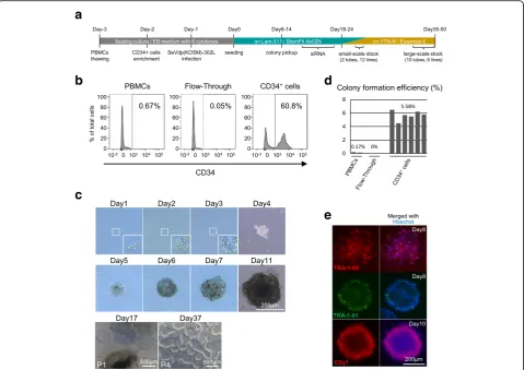
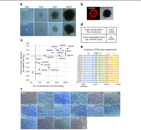
Fig. 1Healthy donor-derived human-induced pluripotent stem cell (hiPSC) generation from non-mobilized peripheral blood (PB)-derived CD34+ hematopoietic stem and progenitor cells (HSPCs) using SeVdp(KOSM)-302L.aSchematic diagram illustrating the schedule of hiPSC generation.b
hiPSC generation
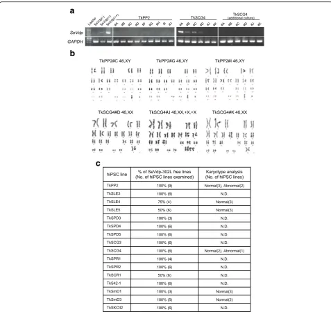
At day − 1, the CD34+-enriched cells were infected with SeVdp(KOSM)-302L at a multiplicity of infection (MOI) of 3–7 (Fig.1a). For the experiment shown in Additional file5: Figure S4, the CytoTune®-iPS2.0 (ID Pharma) was used to infect CD34+-enriched cells at an MOI of > 1, > 3, and > 6, based on the titer described in the instruction manual (> 5.0 × 10⁶ CIU/100 μL). After addition of the viral vector, the cells were left at room temperature for 2 h and then incubated overnight at 37 °C with 5% CO2. At day 0, the
SeVdp(KOSM)-302L-infected cells were seeded into iMatrix-511 (Nippi)-coated 6-well plates (TPP) with StemFit® AK02N medium (REPROCELL) containing 10 μM Y-27632 (Fig.1a). At day 2, half the medium volume was replaced with fresh StemFit AK02N medium, follow-ing which the entire medium was changed on every 2 days until colony pickup. At days 8–14, individual col-onies with an appropriate size of 250–500 μm in
diameter were picked up into iMatrix-511-coated 24-well plates (TPP) containing StemFit AK02N medium supplemented with 10 μM Y-27632 (Fig. 1a). At the medium changes on every 2 days, small interfering RNA (siRNA) treatment against SeVdp(KOSM)-302L was performed 3 times using Lipofectamine™ RNAi-MAX Transfection Reagent (Invitrogen) according to the manufacturer’s instructions (Fig. 1a). The two siR-NAs (siL527 and siL1913), used at 1.6 nM each, were generated from the following single-stranded RNA ol-igonucleotides (Fasmac): siL527-S, 5′ -GGUUCAG-CAUCAAAUAUGAAG-3′; siL527-AS, 5′-UCAU AUUUGAUGCUGAACCAU-3′; siL1913-S, 5′-GGUC CAGACAUGAAUUCAAAG-3′; and siL1913-AS, 5′ -UUGAAUUCAUGUCUGGACCAU-3′.
hiPSC expansion and maintenance culture for storage For expansion and maintenance, hiPSCs were cultured on iMatrix-511- or Vitronectin (VTN-N, Gibco)-coated 12- or 6-well plates in StemFit AK02N medium (with iMatrix-511) or Essential 8™ medium (Gibco) (with VTN-N) at 37 °C with 5% CO2. Daily medium change
was routinely performed. When passaged, the hiPSCs were mechanically and enzymatically detached from the plates using a scraper, 0.5× TrypLE™ Select (Gibco), 0.5 mM EDTA (Invitrogen), and/or Accutase® (Sigma), and plated on fresh iMatrix-511- or VTN-N-coated plates in the respective medium described above (containing 10
μM Y-27632). Before their use in the experiments, all cells were confirmed to be mycoplasma-free, using the MycoAlert™ Mycoplasma Detection Kit (Lonza) and MycoAlert™ Assay Control Kit (Lonza). Luminescence was measured with a GloMax® Explorer system (Pro-mega). Images of hiPSC colonies were captured with a Leica DMI3000B system and their processing was per-formed using the ImageJ (NIH) or GNU Image Manipu-lation Program 2.10 (GIMP2.10). Unless we had a specific need, we generally selected ~ 12 colonies for the subsequent expansion steps that lasted for another 4–5 weeks. The small-scale frozen stocks (2 tubes) of the 12 lines were initially generated as the backup stocks, with the cells kept in the iMatrix-511/StemFit AK02N media (Fig. 1a). Six lines were adapted to culture under the VTN-N/Essential 8 conditions and finally stored as large-scale frozen stocks (10 tubes), where ~ 2.0 × 10⁶ cells per tube were cryopreserved in Bambanker for the hiPSC banking.
RT-PCR verification of SeVdp(KOSM)-302L removal Total RNA was isolated from the hiPSCs using the AllPrep DNA/RNA Mini Kit (Qiagen), RNeasy® Mini Kit (Qiagen), or Monarch® Total RNA Miniprep Kit (New England Biolabs). Complementary DNA (cDNA) was synthesized using the High-Capacity cDNA Reverse Transcription Kit
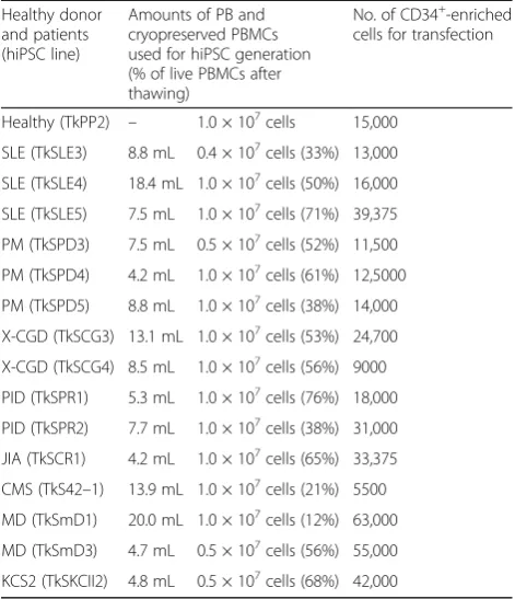
Table 2Amounts of PB, PBMCs, and CD34+-enriched cells used for reprogramming
Healthy donor and patients (hiPSC line)
Amounts of PB and cryopreserved PBMCs used for hiPSC generation (% of live PBMCs after thawing)
No. of CD34+-enriched
cells for transfection
Healthy (TkPP2) – 1.0 × 10⁷cells 15,000
SLE (TkSLE3) 8.8 mL 0.4 × 10⁷cells (33%) 13,000
SLE (TkSLE4) 18.4 mL 1.0 × 10⁷cells (50%) 16,000
SLE (TkSLE5) 7.5 mL 1.0 × 10⁷cells (71%) 39,375
PM (TkSPD3) 7.5 mL 0.5 × 10⁷cells (52%) 11,500
PM (TkSPD4) 4.2 mL 1.0 × 10⁷cells (61%) 12,5000
PM (TkSPD5) 8.8 mL 1.0 × 10⁷cells (38%) 14,000
X-CGD (TkSCG3) 13.1 mL 1.0 × 10⁷cells (53%) 24,700
X-CGD (TkSCG4) 8.5 mL 1.0 × 10⁷cells (56%) 9000
PID (TkSPR1) 5.3 mL 1.0 × 10⁷cells (76%) 18,000
PID (TkSPR2) 7.7 mL 1.0 × 10⁷cells (38%) 31,000
JIA (TkSCR1) 4.2 mL 1.0 × 10⁷cells (65%) 33,375
CMS (TkS42–1) 13.9 mL 1.0 × 10⁷cells (21%) 5500
MD (TkSmD1) 20.0 mL 1.0 × 10⁷cells (12%) 63,000
MD (TkSmD3) 4.7 mL 0.5 × 10⁷cells (56%) 55,000
KCS2 (TkSKCII2) 4.8 mL 0.5 × 10⁷cells (68%) 42,000
The amounts of peripheral blood (PB) and peripheral blood mononuclear cells (PBMCs) used in the generation of these human induced pluripotent stem cell (hiPSC) lines are listed. The PB volume shown here was calculated from the corresponding PBMC numbers contained in each frozen vial (cryopreserved PBMCs). The number of PBMCs represents the value before cryopreservation. After thawing, a certain level of cell death occurs; % live PBMCs are thus shown in parentheses, calculated by counting the actual number of living cells recovered after thawing. The number of CD34+
-enriched cells used for SeVdp(KOSM)-302L transduction is also indicated
SLEsystemic lupus erythematosus,PMpolymyositis,X-CGDX-linked chronic granulomatous disease,PIDprimary immunodeficiency,JIAjuvenile idiopathic arthritis,CMScongenital malformation syndrome,MDmitochondrial diabetes,
(Applied Biosystems). The reverse transcription-polymerase chain reaction (RT-PCR) assay for SeVdp(KOSM)-302L cDNA was performed with TaKaRa Ex Taq® Hot Start Version (TaKaRa). GAPDH was used as the internal control. The PCR products were resolved on 2.5% agarose gels. The primers used in this study are listed in Additional file 1: Table S1.
In vitro differentiation of three germ layers
To analyze the capability of the hiPSCs to differentiate into three germ layers, in vitro differentiation was per-formed with the STEMdiff™ Trilineage Differentiation Kit (STEM CELL Technologies) according to the manu-facturer’s instructions.
Quantification of pluripotency and differentiation markers with real-time PCR
cDNA was prepared as described above for quantifying the pluripotency markers octamer-binding transcription factor 4 (OCT4) and NANOG by multiplex TaqMan real-time PCR, using the Eagle Taq Master Mix with ROX (Roche). Universal probes #34 and #69 (Roche) were used for labeling theOCT4and NANOGproducts, respectively. The HumanG6PDGene Assay (Roche) was used for quantifying the reference gene. Because the uni-versal probe #69 failed to be incorporated into the NANOG transcripts of the TkPP2 lines owing to the presence of a single nucleotide polymorphism, the SYBR Green method was used with the Power SYBR Green PCR Master Mix (Applied Biosystems) for these cell lines only. The relative expression ofOCT4andNANOG was evaluated with reference to their expression levels in our standard hiPSC line TkDA3-4 previously established from a healthy donor [41].
The gene expression of trilineage differentiation markers was evaluated using a real-time PCR SYBR Green method with the Power SYBR Green PCR Master Mix (Applied Biosystems) or THUNDERBIRD® SYBR qPCR Mix (Toyobo). The marker genes analyzed were paired box 6 (PAX6), SRY-box 1 (SOX1), SRY-box 17 (SOX17), Foxhead box A2 (FOXA2), T-box transcription factor T (or Brachyury) (TBXT), and neural cell adhesion molecule (NCAM). G6PD was used as the reference gene. The primers used in this analysis are listed in Additional file 1: Table S1. All the real-time PCR analyses were performed using a CFX96 C1000™thermal cycler (Bio-Rad). Relative expression was calculated by the
ΔΔCt method with CFX™Manager software (Bio-Rad).
Detection of pluripotency characteristics with immunostaining and chemical staining
Live hiPSC colonies were directly stained with the NL557-conjugated anti-human TRA-1-60 antibody (NLLC4770R, R&D Systems), the LN493-conjugated anti-human TRA-1-81
antibody (NLLC16581G, R&D Systems), or the fluor-escent imaging probe CDy1 [42]. Fluorescence images were captured with a Nikon Eclipse Ti microscope system. Images were edited using ImageJ (NIH) or GNU Image Manipulation Program 2.10 (GIMP2.10).
Karyotyping
hiPSCs were expanded in VTN-N-coated 25-cm2flasks (TPP) with Essential 8 medium. Standard G-banded karyotyping was performed by the Chromosome Science Laboratory or NIHON Gene Research Laboratories.
TCRγgene rearrangement analysis
Genomic DNA was isolated using the AllPrep DNA/ RNA Mini Kit. Then, the rearrangement of the T cell re-ceptor gamma (TCRγ) gene segment was analyzed using the T Cell Receptor Gamma Gene Rearrangement Assay Kit for Gel Detection (Invivoscribe) according to the manufacturer’s instructions.
Flow cytometry analysis
Detailed information is provided in Additional file 2: Supplemental Methods.
Results
Establishing hiPSC lines from healthy donor PB-derived CD34+HSPCs
We sought to establish an hiPSC generation method capable of meeting the following requirements: (1) use of non-mobilized PB cells, (2) compatibility with sched-uled procedures, (3) integration-free culture, (4) absence of immunoreceptor gene rearrangement, (5) high effi-ciency, (6) cost-effectiveness, and (7) feeder-free condi-tions. We finally achieved these goals with the protocol summarized in Fig.1a.
2000, and 4000 cells/well) onto iMatrix-511-coated 6-well plates containing StemFit AK02N medium. No more hematopoietic cytokines were needed beyond day 0. After seeding, some adherent cells showed ini-tial proliferation on days 1–3 and started forming colony-like structures on days 4–6 (Fig. 1c). Once formed, the growth of these spherical brown colonies was rapid, with their size reaching > 200 μm at day 11 (Fig. 1c). On the basis of the number of colonies counted at day 7, the colony formation efficiency was calculated to be 5.58% per cell input (Fig. 1d and Table 3), which was extremely high compared with that of previous reports (< 1% in many cases) [43,44]. These primary colonies were confirmed to be TRA-1-60-positive and TRA-1-81-positive (Fig.1e). They also stained positively with CDy1 dye (Fig.1e), demonstrat-ing that their characteristics were compatible with those of pluripotent colonies. When the non-enriched PBMCs and flow-through samples (both containing only a few CD34+ cells (Fig.1b)) were used under the same conditions, colony formation barely occurred (Fig. 1d), suggesting that the CD34+ cells represented a preferable target for highly efficient hiPSC gener-ation in the SeVdp(KOSM)-302L reprogramming sys-tem. The idea that the purified CD34+ cells are
well-suited to hiPSC establishment was later confirmed by the additional experiment, the results of which showed a strong positive correlation between the proportion of CD34+ cells and the colony formation efficiencies (Additional file3: Figure S1).
To establish stable hiPSC lines, we tentatively per-formed a random pickup of 10 colonies larger than the 250-μm diameter on day 11. Their spherical shape under feeder-free conditions allowed for an extremely facile colony pickup procedure. We carried out siRNA treatment against SeVdp-302L during the initial post-pickup culture, with no colonies lost in the subse-quent expansion phase. We divided each line into two feeder-free conditions (i.e., iMatrix-511/StemFit AK02N and VTN-N/Essential 8) and eventually estab-lished all 10 lines as stable hiPSCs named TkPP2 (Fig. 1c and Table 3). The colonies of the TkPP2 lines showed a typical hiPSC monolayer morphology (day 37, Figs. 1c and 2f) and were stable in culture after their recovery from cryopreservation, with their pluripotency characteristics maintained (discussed below). This trial demonstrated the high success rate of hiPSC generation from non-mobilized PB samples with no need for extensive expansion culture, allowing a rapid (5–6 weeks) and robust protocol that is appropriate for the purpose of biobanking.
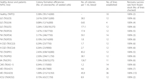
Table 3Colony formation efficiencies and number of established lines
Healthy donor and patients (hiPSC line)
Colony formation efficiencies (No. of colonies/No. of seeded cells)
No. of colonies per 1 mL blood
No. of lines established
Recovery success rate from frozen stock (No. of lines checked)
Healthy (TkPP2) 5.58% (781/14,000) – 10 100% (2)
SLE (TkSLE3) 2.61% (339/13,000) 38.5 12 100% (6)
SLE (TkSLE4) 0.08% (12/16,000) 0.65 4 100% (4)
SLE (TkSLE5) 3.26% (1283/39,375) 171.1 12 100% (6)
PM (TkSPD3) 1.67% (130/7750) 17.4 12 100% (5)
PM (TkSPD4) 3.17% (246/7750) 59.1 14 100% (3)
PM (TkSPD5) 0.10% (16/14,000) 1.8 12 100% (6)
X-CGD (TkSCG3) 0.29% (23/7750) 1.7 12 100% (6)
X-CGD (TkSCG4) 0.26% (23/9000) 2.7 12 100% (6)
PID (TkSPR1) 2.43% (438/18,000) 83.2 12 100% (5)
PID (TkSPR2) 2.50% (294/11,750) 38.1 12 100% (6)
JIA (TkSCR1) 1.59% (530/33,375) 126.7 11 100% (6)
CMS (TkS42–1) 0.34% (17/5000) 1.2 14 100% (6)
MD (TkSmD1) 1.09% (85/7800) 4.6 26 100% (7)
MD (TkSmD3) 1.09% (215/19,763) 45.9 36 100% (13)
KCS2 (TkSKCII2) 0.15% (43/27,750) 8.9 12 100% (2)
The colony formation efficiency of each seeded cell line and the estimated number of colonies per 1 mL of peripheral blood are shown. The ratios of the total number of emerged primary colonies/the total number of seeded cells are indicated in parentheses. The number of human induced pluripotent stem cell (hiPSC) lines established from each sample is also shown. The right-hand column shows the success rate of recovering lines from frozen stock, meaning that there were no failures in the freeze–thaw process for all the established stocks tested
Preparation of non-mobilized PB-derived CD34+HSPCs from patients
We next applied the established method to the generation of patient-derived hiPSC lines. PB samples were obtained from 15 patients of various ages (range 3–65 years) and with different types of diseases (Table1). Among these 15 patients, there were 3 cases of systemic lupus
erythematosus (SLE), 3 cases of polymyositis (PM), 2 cases of X-linked chronic granulomatous disease (X-CGD), 2 cases of primary immunodeficiency (PID), 1 case of juvenile idiopathic arthritis (JIA), 1 case of congenital malformation syndrome (CMS), 2 cases of mitochondrial diabetes (MD), and 1 case of Kenny-Caffey syndrome type 2 (KCS2) (Table 1). The detailed information of their diseases and
a
b
d
e
c
f
causative genetic lesions were not provided for this report since they will be reported in more detail by collaborators elsewhere.
Using a gradient separation method, the PBMCs were obtained within a day of collection of 20–40 mL of fresh PB samples from the patients. In the JIA and CMS cases, PBMC purification was performed 1 day after PB collection owing to the need for long-distance transport of the samples. The number of PBMCs ob-tained ranged between ~ 1.0 × 10⁷and ~ 7.0 × 10⁷cells (0.45 × 10⁶to 2.4 × 10⁶cells/mL). These were all cryo-preserved in aliquots until the start of hiPSC generation (Table1). At day−3, a vial containing 0.4 × 10⁷, 0.5 × 10⁷, or 1.0 × 10⁷PBMCs was thawed for each hiPSC generation attempt, upon which it was shown that ~ 24–88% of PBMCs had been lost during the storage (Table 2). To ensure the survival of CD34+ HSPCs after thawing, the cells were kept overnight in EB medium containing six cy-tokines. At day−2, CD34+cell enrichment was performed as described for TkPP2 (Fig.1a). The number of recovered cells after enrichment varied significantly among the patient samples, ranging from 5500 to 125,000 cells (Table 2). However, we did not experience any unsuccessful cases with these limited materials (median values; 21,350 cells).
Successful establishment of patient-derived hiPSC lines According to the established protocol, we infected the patient-derived CD34+-rich HSPCs with SeVdp(KOSM)-302L at an MOI of 3–7 on day −1 and plated all or a part of them onto iMatrix-511-coated 6-well plates at various cell densities (i.e., 250–20,000 cells/well). Similar to the observations made with TkPP2 establishment, the attached cells gradually formed spherically shaped brown colonies containing pluripotent cells (Fig.2a, b). The col-ony formation efficiencies per seeded cells showed some variation, with the average value being 1.38 ± 1.17% (mean ± SD) (Fig.2c and Table3). In 9 out of the 15 cases, the efficiencies varied between 1.09% and 3.26%, which trans-lated to yields of 4.6–127.7 colonies per 1 mL of PB (Fig.2c and Table3). However, for the other 6 cases, the efficiency values were between 0.08% and 0.34% per seeded cells, translating to 0.65–8.9 colonies per 1 mL of PB (Fig. 2c and Table 3). The lower efficiencies in the latter group may be attributable to the putative low titer of the SeVdp(-KOSM)-302L stock used for the period corresponding to the establishment, because when tested later, it was discovered that a higher MOI (i.e., 7 instead of 3) of the same virus was needed to obtain efficiencies above 1%. Nevertheless, the hiPSC generation efficiencies obtained with our method can be considered as being markedly high, even when compared with a previous report that showed an efficient reprogramming (~ 0.02%) of non-mobilized PBMCs using SeVdp(KOSM)-302L in similar feeder-free conditions [45].
Since all colonies that were randomly picked up led to the successful establishment of TkPP2 lines as described above, we performed colony pickup for the establishment of the patient-derived hiPSC lines in a similar manner, mostly on day 11 (based simply on observations under the microscope). Consequently, out of 294 colonies picked up, all but one (0.34% pickup failure) showed initial attach-ment and growth, enabling subsequent expansion culture. Once subjected to culture for stable hiPSC line establish-ment, all 223 selected colonies proved eventually to be expandable, leading to the completion of the freezing stor-age (Fig.2d). The progress of the establishment processes was similar in all hiPSC lines (Fig. 2e). Furthermore, the overall process was fast, as we were able to end the culture for each line routinely within ~ 5–6 weeks with the stor-age of a sufficient number of hiPSCs showing typical pluripotent colonies (Fig. 2f ). So far, we have not found any cases where we failed to recover viable hiPSC colonies from their frozen stocks (Table 3). Collectively, these re-sults demonstrate that our method enables the scheduled establishment of disease-specific hiPSC lines from patient PB samples with a high success rate.
Confirmation of the transgene-free status and karyotypes To confirm the removal of SeVdp(KOSM)-302L in the established hiPSC lines, we performed RT-PCR analysis for the presence of vector genome sequences. As shown in Fig. 3a, the amplicons representing the residual exist-ence of the SeV genome were infrequently found. This, however, occurred generally when assessed at an early passage before the initial storage. The same line showed the disappearance of detectable SeV sequences consist-ently with a few additional passages (Fig.3a), most likely due to the auto-erasable function equipped in the SeVdp-302L system [16]. Most of the hiPSC lines tested were shown to be transgene-free at the time of storage (81 out of 88 lines, Fig.3c). Karyotyping analysis has so far been conducted for a few of the patient-derived hiPSC lines and is ongoing for some others, showing mostly normal karyo-types (Fig.3b, c). Taken together, the results indicate that our method is compatible with the measure to obtain integration-free hiPSC lines with a normal karyotype.
was detectable in the T lymphoid cell-derived hiPSC line TkSST2-2, which we reported previously [46], confirming the validity of this assay (Fig.4a). As B cells were thought to be extremely refractory to reprogramming in our system, necessitating enhancement by the coexpression of other factors (e.g., C/EBPα), an analysis of immuno-globulin genes was not conducted [47, 48]. Although the origin of monocytes cannot be excluded, these results—taken together with the great enhancement in
generation efficiencies observed after CD34+
cell en-richment—suggest that the hiPSC lines have likely been derived from HSPCs (Fig.1d).
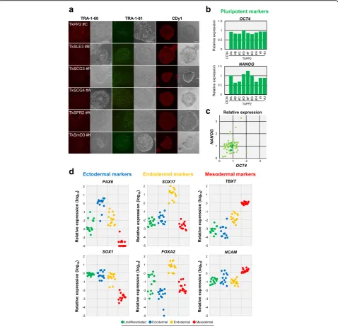
Stable pluripotency and trilineage differentiation capability
All hiPSC lines were in a stable pluripotent state in culture, showing the retention of pluripotency character-istics, i.e., positive staining with the antibodies for
c
b
a
TRA-1-60 or TRA-1-81 and the CDy1 dye (Fig.5a). Ex-pression of the marker genes OCT4 and NANOG was quantified in the 85 hiPSC lines using RT-qPCR to ver-ify their pluripotency features. As shown in Fig. 5b, c, all the lines exhibited the expression of these genes, demonstrating their pluripotent status. Expression of these markers was also confirmed at a single cell level when the samples were assessed by flow cytometry analysis (Additional file4: Figure S2).
Finally, we examined the capability of the 14 selected hiPSC lines to differentiate into three germ layers with an easy-to-use in vitro differentiation assay. After cultivation under different conditions (i.e., either in an undifferenti-ated state or with differentiation for an ectodermal, an endodermal, or a mesodermal lineage), the hiPSC samples were examined by RT-qPCR for their relative gene expres-sion of three germ-layer markers: PAX6andSOX1 (ecto-derm), SOX17 and FOXA2 (endoderm), and TBXT and NCAM(mesoderm). Selective upregulation patterns were detectable for each marker gene in accordance with the corresponding culture condition in all the hiPSC lines, except for the ectodermal marker SOX1, the expression levels of which were also high in the undifferentiated state (Fig.5d). Increased expression of lineage-markers was also observed at a protein level in the differentiated TkPP2 samples by flow cytometry analysis, demonstrating the utility of this in vitro differentiation assay system
(Additional file 5: Figure S3). These results confirmed that the patient-derived hiPSC lines established by our method could be stably maintained in a pluripotent cul-ture and retain their trilineage differentiation capability, both conditions of which are mandatory for these cell lines to be truly valuable.
Discussion
Herein, we have described a rapid and simple method for the robust establishment of transgene-free hiPSC lines using the auto-erasable SeVdp(KOSM)-302L vector [16]. In this method, we targeted non-mobilized PB-derived CD34+ HSPCs, with no need for cell mobilization and ex vivo ex-pansion culture, and achieved highly efficient cell repro-gramming under feeder-free conditions. Primary colonies appeared rapidly, were easily manageable for pickup, and showed robustness in the subsequent expansion steps. Using this method, we succeeded in the scheduled establishment of 223 hiPSC lines from PB samples obtained from 15 pa-tients with 8 different diseases, generating a sufficient num-ber of frozen stocks within 5–6 weeks. These hiPSC lines showed consistent recovery from frozen stocks, a transgene-free status, normal karyotypes, non-T cell derivation, stable pluripotency, and trilineage differentiation cap-ability. This method is therefore valuable to robustly and efficiently generate patient hiPSC lines, allowing
b
a
reductions in the cost, time, and workload for the en-tire procedure, which is advantageous for biobanking.
In our hiPSC banking, PB samples were used because they were easily collected from patients, with less
invasiveness than that of other sources. PB can also be held at room temperature for ~ 48 h, and the derived PBMCs can be cryopreserved for a long period [19], enabling long-distance sample transport and scheduled hiPSC generation.
a
b
d
c
Fig. 5Pluripotency marker expression and trilineage differentiation capability confirmed in the established human induced pluripotent stem cell (hiPSC) lines.aFluorescent images of live colonies stained with anti-TRA-1-60 antibody (red, left), anti-TRA-1-81 antibody (green, middle), or the CDy1 dye (red, right). Phase-contrast images are also shown in a side-by-side fashion for each picture. Scale bars indicate 200μm.bGraph demonstrating the quantification results ofOCT4andNANOGtranscripts in the TkPP2 lines, assessed by RT-qPCR analysis. The relative expression values for each gene, normalized to the level observed in the standard TkDA3-4 hiPSC sample, are shown. CD34+-enriched cells derived from the
In fact, we succeeded in generating hiPSCs from all 15 patient-derived PBMC samples that had been cryopre-served for a few months to 1 year. Of note is that despite the TkSCR1 and TkS42-1 PB samples required long-distance transport of over 24 h, they still resulted in the es-tablishment of hiPSC lines without any detectable loss of generation efficiency (Table3).
Among the hematopoietic cells, CD34+ HSPCs are often the preferred target cells for reprogramming, as they generally allow relatively highly efficient reprogram-ming [17, 20, 49–51]. Although CD34+ HSPCs can be collected from cord blood and bone marrow as well, PB is the source most suitable for patient hiPSC banking, be-cause of its general availability. Their scarcity, however, has made it difficult to utilize PB-derived CD34+cells as a regu-lar target. To overcome this limitation, either mobilization or ex vivo expansion culture or both have been adopted for PB-derived HSPC-targeted reprogramming [22–25]. G-CSF-based mobilization, however, is considered too invasive for patient hiPSC generation. When non-mobilized PB samples are used, many protocols include cultivation steps with various combinations of cytokines to increase the tar-get progenitor cells and thus the reprogramming efficiency [19, 21, 26–28, 30–33, 35–37, 52–54]. After expansion culture (typically 4–10 days), approximately 1.0 × 10⁵to 1.0 × 10⁷target cells are obtained and used for the reprogram-ming studies. Considering these facts, it is a marked feature that our protocol eliminates the need for both mobilization and expansion culture (Fig. 1). At present, however, we have not extensively tried to make the protocol completely cytokine-free. After immunomagnetic enrichment, the re-covery of CD34+-rich HSPCs should be expected to be very limited because of their extremely small numbers in non-mobilized PB samples. As we continue to use frozen PBMCs for the scheduled hiPSC establishment, the ex-pected yields would become even lower. We therefore in-cluded into the culture medium 6 cytokines that favored HSPC survival during the first 3 days. Although this treat-ment did not lead to a massive expansion of cells, it was nevertheless enough for successful hiPSC generation. It should be noted that our culture scale was small, thus limiting the required volume of medium (3 mL maximum on day−3, and only 200μL for culturing CD34+
-enriched cells thereafter) and the total amounts of cytokines made available. To cut the cost further, we may be able to modify the cytokine-related steps in the future.
As the number of target cells was limited, our method needed reprogramming efficiencies high enough to pro-duce a sufficient number of pluripotent colonies. To target this small number of cells (< 10,000 cells in some cases), the use of episomal vectors that have shown util-ity in many applications [5, 28] may not be feasible for the reason of practicality. Viral vectors are advantageous in this context; however, the current methods have
revealed low reprogramming efficiencies (< 1%), even when using another type of SeV vector, and yielded some non-expandable primary colonies [19, 24, 31, 34]. In contrast, our method allowed the highly efficient gener-ation of primary colonies (up to 5.58%) and, more im-portantly, enabled high success rates in the establishment of stable hiPSC lines with almost no fear of failure in the subsequent expansion steps after random pickup (Figs. 1
and2). The hiPSC lines established showed stable pluripo-tency and trilineage differentiation capability (Fig. 5). We thus concluded that the special combination of SeVdp(-KOSM)-302L and non-mobilized PB-derived CD34+ cells represented the best match, leading to the establishment of the highly successful protocol. This report has further ex-tended the utility of SeVdp(KOSM)-302L to hiPSC gener-ation, which has been demonstrated with other human cellular sources [45,46,55–59].
Besides its high efficiency, our protocol reduced the time and workload required for the entire procedure (Figs1and
2). Overall, a sufficient number of hiPSCs for cryopreserved stocks—which proved recoverable—were obtained within 5–6 weeks in all cases. More recently, we generally per-formed colony pickup at day 8 and could end the entire es-tablishment process by day 35 after seeding in many cases. We speculated that the feeder-free condition using iMatrix-511 (Laminin-iMatrix-511-E8 fragment) combined with the StemFit AK02N medium might favor the rapid appearance of pri-mary colonies and subsequent early cell growth, especially due to its great potential in supporting single-cell survival and firm cell adhesion to the plate [28, 60]. We also adapted hiPSCs to Essential 8 medium on VTN-N-coated plates in the late expansion phase for their cryopreservation (Fig.1). This latter culture could help in the distribution of hiPSCs to other investigators, because we have found that the hiPSCs in stock are compatible with various culture conditions after thawing, irrespective of what culture sys-tems other researchers use. The entire feeder-free protocol also benefits researchers by reducing their workload, elim-inating theS23need for feeder cell handling.
Currently, researchers prefer the use of hiPSCs that are free of any genetic materials left behind after reprogramming procedures, because the residual ex-pression of transgenes is known to affect the differen-tiation capability [61]. We had previously demonstrated the generation of a series of hiPSC lines using an older version of the non-auto-erasable Sendai virus vector (SeVdp), which required routine siRNA treatment to erase the SeV genome in the emerging hiPSCs [38–40]. Although we knew that SeVdp(KSOM)-302L was equipped with an auto-erasable function, we carried out siRNA treatment against SeVdp-302L to maximize the removal of the viral genomes (see the “Methods”
were detected, they disappeared spontaneously with a few additional cultures (Fig. 3). From these observa-tions, we concluded that the auto-erasable function of SeVdp-302L worked efficiently in our protocol in a similar way as reported previously [16].
In this report, we described the successful establish-ment of an initial series of 223 hiPSC lines, derived from 15 patients affected by 8 different diseases, which in-cluded autoimmune diseases such as SLE and PD, im-mune deficiencies exemplified by X-CGD, and other diseases such as MD and KCS2 (Table 1). Recently, Gomez Limia et al. [62] reported the successful gener-ation of hiPSCs from a patient, using a very similar method, where non-mobilized PB-derived CD34+HSPCs with no ex vivo expansion (probably from a fresh, un-frozen sample) were reprogrammed to pluripotency under feeder-free conditions using Geltrex with the SeV vector (CytoTune® 2.0). When tested in our protocol, the CytoTune®-iPS 2.0 also yielded primary hiPSC col-onies with very high efficiencies at the given setting (Additional file 6: Figure S4). Although still preliminary, the use of Sendai virus vector systems may be consid-ered commonly compatible with our protocol. By our hands, cell line establishment is currently still ongoing, and the panel of disease-specific hiPSCs is thus expand-ing. Some of the hiPSC lines established in this study are already being used for disease studies by our collabora-tors and our research team, and the results will be re-ported elsewhere in the near future. For example, we have confirmed that neutrophilic cells differentiated from the TkSCG3 and TkSCG4 lines, derived from X-CGD patients, show severe defects in reactive oxygen species production (data not shown), similar to another X-CGD-iPSC line that we reported previously [39]. Such findings indicate the applicability of the established hiPSC lines to disease-modeling studies.
Conclusions
In summary, we have developed an improved feeder-free method for establishing transgene-feeder-free hiPSC lines from non-mobilized PB-derived CD34+ HSPCs, using the auto-erasable SeVdp(KOSM)-302L. With this method, we successfully established a panel of disease-specific hiPSC lines from 15 patients with 8 different disease types. The hiPSC generation was feasible with the use of cryopreserved PBMCs, even after long-distance transport of the sampled PB. This method rapidly gen-erated expandable primary colonies with high effi-ciency, resulting in the establishment of stable hiPSC lines with almost no failure cases. We confirmed that the hiPSC lines were suitable for use in disease model-ing studies by showmodel-ing their efficient transgene re-moval, normal karyotypes, non-T cell origin, and stable pluripotency and differentiation capability. Overall, this
method allows for the scheduled generation of a panel of disease-specific hiPSC lines from patient-derived non-mobilized PB samples in a cost-effective and time-and labor-saving manner.
Additional files
Additional file 1:Table S1.List of primers used in this study. (PDF 106 kb)
Additional file 2:Supplemental methods. (PDF 76 kb)
Additional file 3:Figure S1.Correlation between %CD34+cells of the
source sample and the reprogramming (colony formation) efficiency. (PDF 78 kb)
Additional file 4:Figure S2.Flow cytometry analysis of the marker expression in established iPSC clones. (PDF 46 kb)
Additional file 5:Figure S3.Flow cytometry analysis of the marker expression in the TkPP2 cells after induction of lineage-oriented differentiation. (PDF 86 kb)
Additional file 6:Figure S4.Comparison of CytoTune®-iPS2.0 with SeVdp(KOSM)-302L in the same hiPSC generation protocol. (PDF 87 kb)
Abbreviations
BM:Bone marrow; G-CSF: Granulocyte-colony stimulating factor; hiPSC: Human-induced pluripotent stem cell; HSPC: Hematopoietic stem/ progenitor cell; PB: Peripheral blood; PBMC: Peripheral blood mononuclear cell; SeV: Sendai virus
Acknowledgements
We thank Drs. Toyoki Nishimura, Yoichiro Oda, and Etsuro Tokuhiro for providing patient care, Dr. Mayuko Tamura for blood sampling, and Hiroko Ohkura and Akiko Kikuchi for technical assistance.
Authors’contributions
TaOku performed the hiPSC establishment, experiments, data analyses, and manuscript preparation. YH established the hiPSC lines, performed the experiments, and edited the manuscript. CYL arranged the hiPSC
establishment and supervised the study. HTL discussed the experiments. HS, BN, EK, TsOka, SO, KT, Hka, HH, FM, KK, HM, HN, HW, ST, HT, and SK contributed to the patient recruitment and enrollment. KF, TM, HKo, TS, TY, and TK supported the experiments scientifically. KN, ManOh, and MN prepared the SeVdp(KOSM)-302L. MakOt supervised the study, analyzed the data, and wrote the manuscript. All authors read and approved the final manuscript.
Funding
This study was supported by the JSPS Grant-in-Aid for Scientific Research (B) Grant Numbers JP18H02778, JSPS KAKENHI, and JP26670474, and the Program for Intractable Disease Research Utilizing Disease-specific iPS Cells (Grant Numbers JP16bm0609006, JP16bm0804004, and JP18bm04004h0102) and the Research Project for Practical Applications of Regenerative Medicine (Grant Number JP17bk0104018h0005) funded by the Japan Agency for Medical Research and Development (AMED).
Availability of data and materials
All experimental data and materials obtained and used in this study were described in this article.
Ethics approval and consent to participate
Consent for publication
Not applicable.
Competing interests
The authors declare that have no competing interests.
Author details
1
Division of Stem Cell Processing/Stem Cell Bank, Center for Stem Cell Biology and Regenerative Medicine, The Institute of Medical Science, The University of Tokyo, 4-6-1 Shirokanedai, Minato-ku, Tokyo 108-8639, Japan.
2Department of Allergy and Rheumatology, Graduation School of Medicine,
The University of Tokyo, Tokyo, Japan.3Department of Pediatrics and Developmental Biology, Graduate School of Medical and Dental Sciences, Tokyo Medical and Dental University, Tokyo, Japan.4Department of Child Health and Development, Graduate School of Medical and Dental Sciences, Tokyo Medical and Dental University, Tokyo, Japan.5Department of Rheumatology, Graduate School of Medical and Dental Sciences, Tokyo Medical and Dental University, Tokyo, Japan.6Division of Rheumatology and Allergy, Department of Internal Medicine, St. Marianna University School of Medicine, Kawasaki, Kanagawa, Japan.7Division of Pediatrics, Faculty of Medicine, University of Miyazaki, Miyazaki, Japan.8Department of Diabetes
and Metabolic Diseases, Graduate School of Medicine, The University of Tokyo, Tokyo, Japan.9Department of Molecular Sciences on Diabetes,
Graduate School of Medicine, The University of Tokyo, Tokyo, Japan.
10Department of Prevention of Diabetes and Life-style Related Diseases,
Graduate School of Medicine, The University of Tokyo, Tokyo, Japan.
11Department of Metabolism and Nutrition, Mizonokuchi Hospital, Teikyo
University, Kawasaki, Kanagawa, Japan.12Department of Pediatrics, Graduate School of Medicine, The University of Tokyo, Tokyo, Japan.13Laboratory of
Gene Regulation, Faculty of Medicine, University of Tsukuba, Ibaraki, Japan.
14Biotechnology Research Institute for Drug Discovery, National Institute of
Advanced Industrial Science and Technology, Tsukuba, Ibaraki, Japan.
15TOKIWA-Bio Inc., Tsukuba, Ibaraki, Japan.
Received: 20 March 2019 Revised: 18 May 2019 Accepted: 21 May 2019
References
1. Takahashi K, Tanabe K, Ohnuki M, Narita M, Ichisaka T, Tomoda K, et al. Induction of pluripotent stem cells from adult human fibroblasts by defined factors. Cell. 2007;131:861–72.
2. Park IH, Arora N, Huo H, Maherali N, Ahfeldt T, Shimamura A, et al. Disease-specific induced pluripotent stem cells. Cell. 2008;134:877–86.
3. Shi Y, Inoue H, Wu JC, Yamanaka S. Induced pluripotent stem cell technology: a decade of progress. Nat Rev Drug Discov. 2017;16:115–30. 4. Yu J, Hu K, Smuga-Otto K, Tian S, Stewart R, Slukvin II, et al. Human induced
pluripotent stem cells free of vector and transgene sequences. Science. 2009;324:797–801.
5. Okita K, Matsumura Y, Sato Y, Okada A, Morizane A, Okamoto S, et al. A more efficient method to generate integration-free human iPS cells. Nat Methods. 2011;8:409–12.
6. Fusaki N, Ban H, Nishiyama A, Saeki K, Hasegawa M. Efficient induction of transgene-free human pluripotent stem cells using a vector based on Sendai virus, an RNA virus that does not integrate into the host genome. Proc Jpn Acad Ser B Phys Biol Sci. 2009;85:348–62.
7. Stadtfeld M, Nagaya M, Utikal J, Weir G, Hochedlinger K. Induced pluripotent stem cells generated without viral integration. Science. 2008;322:945–9. 8. Woltjen K, Michael IP, Mohseni P, Desai R, Mileikovsky M, Hämäläinen R,
et al. piggyBac transposition reprograms fibroblasts to induced pluripotent stem cells. Nature. 2009;458:766–70.
9. Jia F, Wilson KD, Sun N, Gupta DM, Huang M, Li Z, et al. A nonviral minicircle vector for deriving human iPS cells. Nat Methods. 2010;7:197–9. 10. Warren L, Manos PD, Ahfeldt T, Loh YH, Li H, Lau F, et al. Highly efficient
reprogramming to pluripotency and directed differentiation of human cells with synthetic modified mRNA. Cell Stem Cell. 2010;7:618–30.
11. Kim D, Kim CH, Moon JI, Chung YG, Chang MY, Han BS, et al. Generation of human induced pluripotent stem cells by direct delivery of reprogramming proteins. Cell Stem Cell. 2009;4:472–6.
12. Takahashi K, Yamanaka S. A decade of transcription factor-mediated reprogramming to pluripotency. Nat Rev Mol Cell Biol. 2016;17:183–93.
13. Zhou YY, Zeng F. Integration-free methods for generating induced pluripotent stem cells. Genomics Proteomics Bioinformatics. 2013;11:284–7. 14. Sharma R. iPS cells-the triumphs and tribulations. Dent J (Basel). 2016;4:E19. 15. Borgohain MP, Haridhasapavalan KK, Dey C, Adhikari P, Thummer RP. An
insight into DNA-free reprogramming approaches to generate integration-free induced pluripotent stem cells for prospective biomedical applications. Stem Cell Rev. 2019;15:286–313.
16. Nishimura K, Ohtaka M, Takada H, Kurisaki A, Tran NVK, Tran YTH, et al. Simple and effective generation of transgene-free induced pluripotent stem cells using an auto-erasable Sendai virus vector responding to microRNA-302. Stem Cell Res. 2017;23:13–9.
17. Loh YH, Agarwal S, Park IH, Urbach A, Huo H, Heffner GC, et al. Generation of induced pluripotent stem cells from human blood. Blood. 2009;113:5476–9. 18. Gore A, Li Z, Fung HL, Young JE, Agarwal S, Antosiewicz-Bourget J, et al.
Somatic coding mutations in human induced pluripotent stem cells. Nature. 2011;471:63–7.
19. Agu CA, Soares FA, Alderton A, Patel M, Ansari R, Patel S, et al. Successful generation of human induced pluripotent stem cell lines from blood samples held at room temperature for up to 48 hr. Stem Cell Rep. 2015;5:660–71. 20. Mack AA, Kroboth S, Rajesh D, Wang WB. Generation of induced pluripotent
stem cells from CD34+ cells across blood drawn from multiple donors with non-integrating episomal vectors. PLoS One. 2011;6:e27956.
21. Merling RK, Sweeney CL, Choi U, De Ravin SS, Myers TG, Otaizo-Carrasquero F, et al. Transgene-free iPSCs generated from small volume peripheral blood nonmobilized CD34+ cells. Blood. 2013;121:e98–107.
22. Chou BK, Mali P, Huang X, Ye Z, Dowey SN, Resar LM, et al. Efficient human iPS cell derivation by a non-integrating plasmid from blood cells with unique epigenetic and gene expression signatures. Cell Res. 2011;21:518–29. 23. Park TS, Huo JS, Peters A, Talbot CC, Verma K, Zimmerlin L, et al. Growth
factor-activated stem cell circuits and stromal signals cooperatively accelerate non-integrated iPSC reprogramming of human myeloid progenitors. PLoS One. 2012;7:e42838.
24. Ye L, Muench MO, Fusaki N, Beyer AI, Wang J, Qi Z, et al. Blood cell-derived induced pluripotent stem cells free of reprogramming factors generated by Sendai viral vectors. Stem Cells Transl Med. 2013;2:558–66.
25. Liu J, Brzeszczynska J, Samuel K, Black J, Palakkan A, Anderson RA, et al. Efficient episomal reprogramming of blood mononuclear cells and differentiation to hepatocytes with functional drug metabolism. Exp Cell Res. 2015;338:203–13.
26. Staerk J, Dawlaty MM, Gao Q, Maetzel D, Hanna J, Sommer CA, et al. Reprogramming of human peripheral blood cells to induced pluripotent stem cells. Cell Stem Cell. 2010;7:20–4.
27. Dowey SN, Huang X, Chou BK, Ye Z, Cheng L. Generation of integration-free human induced pluripotent stem cells from postnatal blood mononuclear cells by plasmid vector expression. Nat Protoc. 2012;7:2013–21.
28. Nakagawa M, Taniguchi Y, Senda S, Takizawa N, Ichisaka T, Asano K, et al. A novel efficient feeder-free culture system for the derivation of human induced pluripotent stem cells. Sci Rep. 2014;4:3594.
29. Tan HK, Toh CX, Ma D, Yang B, Liu TM, Lu J, et al. Human finger-prick induced pluripotent stem cells facilitate the development of stem cell banking. Stem Cells Transl Med. 2014;3:586–98.
30. Chou BK, Gu H, Gao Y, Dowey SN, Wang Y, Shi J, et al. A facile method to establish human induced pluripotent stem cells from adult blood cells under feeder-free and xeno-free culture conditions: a clinically compliant approach. Stem Cells Transl Med. 2015;4:320–32.
31. Zhou H, Martinez H, Sun B, Li A, Zimmer M, Katsanis N, et al. Rapid and efficient generation of transgene-free iPSC from a small volume of cryopreserved blood. Stem Cell Rev. 2015;11:652–65.
32. Li Y, Liu T, Van Halm-Lutterodt N, Chen J, Su Q, Hai Y. Reprogramming of blood cells into induced pluripotent stem cells as a new cell source for cartilage repair. Stem Cell Res Ther. 2016;7:31.
33. Táncos Z, Varga E, Kovács E, Dinnyés A, Kobolák J. Establishment of induced pluripotent stem cell (iPSC) line from a 75-year old patient with late onset Alzheimer’s disease (LOAD). Stem Cell Res. 2016;17:81–3.
34. Wen W, Zhang JP, Xu J, Su RJ, Neises A, Ji GZ, et al. Enhanced generation of integration-free iPSCs from human adult peripheral blood mononuclear cells with an optimal combination of episomal vectors. Stem Cell Rep. 2016; 6:873–84.
36. Taylor JP, Cash MN, Santostefano KE, Nakanishi M, Terada N, Wallet MA. CRISPR/Cas9 knockout of USP18 enhances type I IFN responsiveness and restricts HIV-1 infection in macrophages. J Leukoc Biol. 2018;103:1225–40. 37. Ye L, Peng Y, Mo J, Yao Y. MiR-126 enhances VEGF expression in induced pluripotent stem cell-derived retinal neural stem cells by targeting spred-1. Int J Clin Exp Pathol. 2018;11:1023–30.
38. Nishimura K, Sano M, Ohtaka M, Furuta B, Umemura Y, Nakajima Y, et al. Development of defective and persistent Sendai virus vector: a unique gene delivery/expression system ideal for cell reprogramming. J Biol Chem. 2011; 286:4760–71.
39. Lin HT, Masaki H, Yamaguchi T, Wada T, Yachie A, Nishimura K, et al. An assessment of the effects of ectopic gp91phox expression in XCGD iPSC-derived neutrophils. Mol Ther Methods Clin Dev. 2015;2:15046.
40. Kubara K, Yamazaki K, Ishihara Y, Naruto T, Lin HT, Nishimura K, et al. Status of KRAS in iPSCs impacts upon self-renewal and differentiation propensity. Stem Cell Reports. 2018;11:380–94.
41. Takayama N, Nishimura S, Nakamura S, Shimizu T, Ohnishi R, Endo H, et al. Transient activation of c-MYC expression is critical for efficient platelet generation from human induced pluripotent stem cells. J Exp Med. 2010; 207:2817–30.
42. Kang NY, Yun SW, Ha HH, Park SJ, Chang YT. Embryonic and induced pluripotent stem cell staining and sorting with the live-cell fluorescence imaging probe CDy1. Nat Protoc. 2011;6:1044–52.
43. Schlaeger TM, Daheron L, Brickler TR, Entwisle S, Chan K, Cianci A, et al. A comparison of non-integrating reprogramming methods. Nat Biotechnol. 2015;33:58–63.
44. Ye H, Wang Q. Efficient generation of non-integration and feeder-free induced pluripotent stem cells from human peripheral blood cells by Sendai virus. Cell Physiol Biochem. 2018;50:1318–31.
45. Trokovic R, Weltner J, Nishimura K, Ohtaka M, Nakanishi M, Salomaa V, et al. Advanced feeder-free generation of induced pluripotent stem cells directly from blood cells. Stem Cells Transl Med. 2014;3:1402–9.
46. Iizuka-Koga M, Asashima H, Ando M, Lai CY, Mochizuki S, Nakanishi M, et al. Functional analysis of dendritic cells generated from T-iPSCs from CD4+ T cell clones of Sjögren’s syndrome. Stem Cell Rep. 2017;8:1155–63. 47. Bueno C, Sardina JL, Di Stefano B, Romero-Moya D, Muñoz-López A, Ariza L,
et al. Reprogramming human B cells into induced pluripotent stem cells and its enhancement by C/EBPα. Leukemia. 2016;30:674–82.
48. Muñoz-López Á, van Roon EH, Romero-Moya D, López-Millan B, Stam RW, Colomer D, et al. Cellular ontogeny and hierarchy influence the reprogramming efficiency of human B cells into induced pluripotent stem cells. Stem Cells. 2016;34:581–7.
49. Eminli S, Foudi A, Stadtfeld M, Maherali N, Ahfeldt T, Mostoslavsky G, et al. Differentiation stage determines potential of hematopoietic cells for reprogramming into induced pluripotent stem cells. Nat Genet. 2009;41:968–76. 50. Haase A, Olmer R, Schwanke K, Wunderlich S, Merkert S, Hess C, et al.
Generation of induced pluripotent stem cells from human cord blood. Cell Stem Cell. 2009;5:434–41.
51. Ye Z, Zhan H, Mali P, Dowey S, Williams DM, Jang YY, et al. Human-induced pluripotent stem cells from blood cells of healthy donors and patients with acquired blood disorders. Blood. 2009;114:5473–80.
52. Kunisato A, Wakatsuki M, Shinba H, Ota T, Ishida I, Nagao K. Direct generation of induced pluripotent stem cells from human nonmobilized blood. Stem Cells Dev. 2011;20:159–68.
53. Su RJ, Baylink DJ, Neises A, Kiroyan JB, Meng X, Payne KJ, et al. Efficient generation of integration-free ips cells from human adult peripheral blood using BCL-XL together with Yamanaka factors. PLoS One. 2013;8:e64496. 54. Sharma A, Mücke M, Seidman CE. Human induced pluripotent stem cell
production and expansion from blood using a non-integrating viral reprogramming vector. Curr Protoc Mol Biol. 2018;122:e58. 55. Kawagoe S, Higuchi T, Otaka M, Shimada Y, Kobayashi H, Ida H, et al.
Morphological features of iPS cells generated from Fabry disease skin fibroblasts using Sendai virus vector (SeVdp). Mol Genet Metab. 2013;109:386–9. 56. Nishimura T, Kaneko S, Kawana-Tachikawa A, Tajima Y, Goto H, Zhu D, et al.
Generation of rejuvenated antigen-specific T cells by reprogramming to pluripotency and redifferentiation. Cell Stem Cell. 2013;12:114–26. 57. Wakao H, Yoshikiyo K, Koshimizu U, Furukawa T, Enomoto K, Matsunaga
T, et al. Expansion of functional human mucosal-associated invariant T cells via reprogramming to pluripotency and redifferentiation. Cell Stem Cell. 2013;12:546–58.
58. Nishimura K, Kato T, Chen C, Oinam L, Shiomitsu E, Ayakawa D, et al. Manipulation of KLF4 expression generates iPSCs paused at successive stages of reprogramming. Stem Cell Rep. 2014;3:915–29.
59. Yamasaki S, Hamada A, Akagi E, Nakatao H, Ohtaka M, Nishimura K, et al. Generation of cleidocranial dysplasia-specific human induced pluripotent stem cells in completely serum-, feeder-, and integration-free culture. In Vitro Cell Dev Biol Anim. 2016;52:252–64.
60. Miyazaki T, Futaki S, Suemori H, Taniguchi Y, Yamada M, Kawasaki M, et al. Laminin E8 fragments support efficient adhesion and expansion of dissociated human pluripotent stem cells. Nat Commun. 2012;3:1236. 61. Ramos-Mejía V, Montes R, Bueno C, Ayllón V, Real PJ, Rodríguez R, et al.
Residual expression of the reprogramming factors prevents differentiation of iPSC generated from human fibroblasts and cord blood CD34+ progenitors. PLoS One. 2012;7:e35824.
62. Gomez Limia CE, Devalle S, Reis M, Sochacki J, Carneiro M, Madeiro da Costa R, et al. Generation and characterization of a human induced pluripotent stem (iPS) cell line derived from an acute myeloid leukemia patient evolving from primary myelofibrosis carrying the CALR 52bp deletion and the ASXL1 p.R693X mutation. Stem Cell Res. 2017;24:16–20.
Publisher’s Note

![Di μ methacrylato κ4O:O′ bis[aquabis(1,10 phenanthroline κ2N,N′)copper(II)] dinitrate dihydrate](data:image/gif;base64,R0lGODlhAQABAIAAAP///wAAACH5BAEAAAAALAAAAAABAAEAAAICRAEAOw==)