GENOME REPORT
Complete genome sequence
of
Enterococcus durans
KLDS6.0933, a potential
probiotic strain with high cholesterol removal
ability
Bailiang Li
1, Smith Etareri Evivie
1,2, Da Jin
1, Yueyue Meng
1, Na Li
1, Fenfen Yan
1, Guicheng Huo
1,3*and Fei Liu
1*Abstract
Background: Enterococci are commensal bacteria in the mammalian gastrointestinal tract which play an important role in the production of various fermented foods. Thus, certain enterococcal strains are commonly used as probiotics to confer health benefits to human and animals. Enterococcus durans KLDS6.0933 is a potential probiotic strain with high cholesterol removal ability, which was isolated from traditional naturally fermented cream in Inner Mongolia of China. To better understand the genetic basis of the probiotic properties of this strain, the whole-genome sequence was performed using the PacBio RSII platform.
Results: Enterococcus durans KLDS6.0933 contains a circular chromosome of 2,867,028 bp, two plasmids of
163,286 bp and 41,490 bp, respectively. Within the 2704 predicted genes, genes involved with acid, bile and oxidative stress resistance were identified. Bile salt hydrolase (BSH, LIANG_RS13510), a cholesterol removal enzyme identified in the E. durans KLDS6.0933 genome is different from that of other Enterococcus strains. Furthermore, unlike other Entero-coccus strains, E. durans KLDS 6.0933 can facilitate the complete biosynthesis pathway of l-tryptophan.
Conclusions: In silico analysis confirmed the probiotic properties of E. durans KLDS6.0933 and may help us exploit the potential applications of E. durans KLDS6.0933 as an industrially important strain.
Keywords: Enterococcus durans, Genome, Probiotic, Stress, l-Tryptophan, Cholesterol removal
© The Author(s) 2018. This article is distributed under the terms of the Creative Commons Attribution 4.0 International License (http://creat iveco mmons .org/licen ses/by/4.0/), which permits unrestricted use, distribution, and reproduction in any medium, provided you give appropriate credit to the original author(s) and the source, provide a link to the Creative Commons license, and indicate if changes were made. The Creative Commons Public Domain Dedication waiver (http://creat iveco mmons .org/ publi cdoma in/zero/1.0/) applies to the data made available in this article, unless otherwise stated.
Introduction
Enterococci are Gram-positive lactic acid bacteria (LAB) and comprise 54 species [1], which are ubiquitously pre-sent in the environment, food and the gastrointestinal tracts of diverse hosts. Enterococci may have important roles in various fermented food as they contribute to the sensory properties and ripening of sausages or certain cheeses, presumably through proteolysis, lipolysis, and citrate utilization [2, 3]. As a prominent member of nor-mal flora, Enterococci play a helpful part in the balance between the gut microbiota and the host.
Enterococci are commonly used as probiotics to con-fer health benefits to human and animals. These bacteria can be used in the treatment of irritable bowel syndrome and antibiotic-associated diarrhea in humans as well as in lowering cholesterol levels or regulating immune system to improve health [4–7]. Likewise, the antioxidant poten-tial of Enterococci has been studied [8].
Enterococcus durans KLDS6.0933 was originally iso-lated from traditional naturally cream samples collected in Inner Mongolia of China. It has been demonstrated that E. durans KLDS6.0933 had the potential to resist acid and bile salt, and assimilate cholesterol in a recent in vitro study [6]. In order to analyze these characteris-tics and mine probiotic properties of this strain from genomic insights, the whole-genome sequence of E. durans KLDS6.0933 was carried out and analyzed in
Open Access
*Correspondence: [email protected]; [email protected]
1 Key Laboratory of Dairy Science, Ministry of Education, Northeast
silico. Comparison of genomic data from E. durans
KLDS6.0933 with other Enterococcus strains may improve our understanding of the traits of E. durans
KLDS6.0933.
Methods
Strain isolation and DNA extraction
Enterococcus durans KLDS6.0933 was isolated from traditional naturally fermented cream in Inner Mon-golia of China and was available at the Key Laboratory of Dairy Science (KLDS), Northeast Agricultural Uni-versity (NEAU), Harbin, China. Before use, E. durans
KLDS6.0930 was activated through three propagation steps in M17 broth (Oxoid Ltd, Hampshire, UK) at 37 °C for 24 h. The genomic DNA of E. durans KLDS6.0933 was extracted using the DNeasy Tissue kit (Qiagen, Ger-many) following the manufacturer’s instruction.
Genome sequencing, assembly, and analysis
The quantity and purity of total DNA were determined by 2% agarose gel electrophoresis and a NanoDrop™ spectrophotometer. The whole-genome sequence of
E. durans KLDS6.0933 was carried out on the single molecule real-time by the Pacbio RSII platform (Pacific Biosciences, USA). A 20 K template library was gener-ated and sequenced by P4-C2 chemistry on two cells. The raw data was obtained as 59,078 pair-end reads (484 MB) with an average read length of 8198 bp. The filtered paired-end reads were de novo assembled by using the hierarchical genome assembly process protocol version 3.0 and polished using Quiver [9]. Gene annotation was determined by Annotation NCBI Prokaryotic Genome Annotation Pipeline [10]. Ribosomal RNA genes were identified using RNAmer 1.2 [11] and tRNA genes were detected using tRNAscan SE v. 2.0 [12]. Functional cat-egories of coding sequences (CDSs) were classified by WebMGA, using RPSBLAST program (applied threshold 1e−5) for clusters of orthologous groups (COG) anno-tation [13]. The circular genomic map was constructed using CGView Server [14]. Functional annotation was performed with the Kyoto Encyclopedia of Genes and Genomes (KEGG) database using Bi-directional Best Hit method by KAAS [15] for analyzing l-tryptophan biosynthetic pathway. Using the BSH sequence from the complete genomes of representative strains, a phyloge-netic tree was constructed using MEGA 7.0 software with the neighbor-joining method [16]. Comparative genomic analysis was performed between E. durans KLDS6.0933 and other representative Enterococcus strains that are rel-atively close to E. durans KLDS6.0933 based on the phy-logenetic tree of 16S rRNA gene.
Quality assurance
The genomic DNA used for sequencing was isolated from a single colony of the E. durans KLDS6.0933. The 16S rRNA gene was sequenced and BLAST was con-ducted against the NCBI database, then the phylogenetic tree based on the 16S rRNA was constructed by MEGA 7.0 software with the Neighbour-joining method. The result clearly indicated this strain belonged to the spe-cies E. durans (Additional file 1: Figure S1). In addition, the average nucleotide identity (ANI) of the genomic sequences between E. durans KLDS6.0933 and E. durans
ATCC6056 was evaluated by the ANI calculator using the OrthoANIu algorithm at the genomic level. Here, we reported that the value of their ANI was 99.66% (Addi-tional file 1: Table S1).
Results and discussion General features
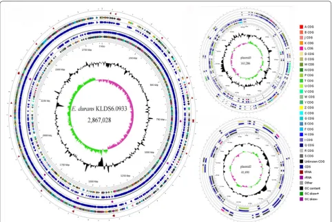
As shown in Fig. 1, the complete genome of E. durans
KLDS6.0933 is composed of a 2,867,028 bp chromosome with GC content of 38% and two plasmids—a plasmid of 163,286 bp with a GC content of 35.5% and another plasmid of 41,490 bp with a GC content of 35.3%. Among the 2704 predicted genes, 2393 CDSs, 86 RNAs and 225 pseudogenes were found in the chromosome of E. durans
KLDS6.0933 (Additional file 1: Table S2). Of the identi-fied CDSs, 2024 genes can be classiidenti-fied into COG classes (Additional file 1: Figure S2). The highest number of genes in this strain was found in the functional groups related to carbohydrate metabolism (212).
Identification of genes coding stress resistance and cholesterol removal
A recent study reported that E. durans KLDS6.0933 was highly tolerant to acid [6]. In order to mine the genetic elements contributing to acid tolerance, proton motive force F1F0ATPase subunits, Na+/H+ antiporters, K+ uptake transporter and cation-transporting ATPase were analyzed in the genome of E. durans KLDS6.0933,
transcarbamylase, carbamate kinase and arginine-orni-thine transporter [18]. These genes are presented in E. durans KLDS6.0933 (Additional file 1: Table S3).
The effect of bile on the growth of E. durans
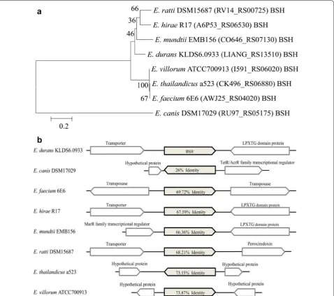
KLDS6.0933 showed that this strain had bile tolerance property [6], a gene encoding BSH (LIANG_RS13510), a member of cholylglycine hydrolase family, was identified in the genome of E. durans KLDS6.0933, which catalyzes the hydrolysis of glycine- and taurine-conjugated bile salts into amino acid residues and free bile acids [19]. Bile salt deconjugation by this enzyme can lower serum cho-lesterol level [20]. Supporting this, E. durans KLDS6.0933 showed high cholesterol removal ability in our previous study [6]. Phylogenetic relationship among the selected BSH sequences of E. durans KLDS6.0933, E. canis
DSM17029, E. faecium 6E6, E. hirae R17, E. mundtii
EMB156, E. ratti DSM15687, E. thailandicus a523 and E. villorum ATCC700913 that had more homology in 16S RNA gene with each other were represented on a neigh-bor-joining tree, which was constructed using amino acid sequences of BSH with bootstrap replication of 1000 in
MEGA 7.0 software. Phylogenetic tree analysis (Fig. 2a) showed that the BSH (LIANG_RS13510) of E. durans
KLDS 6.0933 is more closely related to that of E. mundtii
EMB156 than other Enterococcus strains. However, they are still evolutionarily distant. In addition, alignment and comparison of BSH sequences and their contexts were studied using BLASTp, as shown in Fig. 2b. We found that BSH sequences showed low sequence identities with that of E. durans KLDS6.0933 and the contexts of BSH sequences were different, these imply that BSH is specific to genus and it is significant to further study the relation-ship between gene structure and enzymatic activity of BSH.
Identification of genes coding antioxidant system
Oxidative stress occurs when abnormally high levels of reactive oxygen species (ROS) are generated, result-ing in nucleic acid, protein and lipid damage [21]. The antioxidant mechanisms of probiotics were associated with enzymatic and non-enzymatic antioxidative sys-tem. Among enzymatic antioxidative system, the most
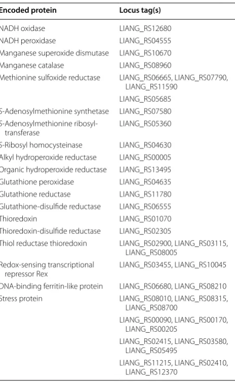
conserved oxidative resistance mechanism in the LAB is that oxygen is reduced indirectly to water by coupling of NADH oxidase and NADH peroxidase oxidative [22]. Another important player in resistance to oxidative stress is superoxide dismutase (SOD), which scavenges superoxide anion radicals [21]. Bacteria can also coun-teract the negative effects of oxidation through catalase, an enzyme catalyzing the detoxification of H2O2 [23]. Genomic insights into antioxidant activity revealed that the genes encoding NADH oxidase, NADH peroxidase, SOD, catalase and glutathione peroxidase were found in the genome of E. durans KLDS6.0933 (Table 1), which
are the main components of ROS resistome in the LAB. To cope with oxidative stress, E. durans KLDS6.0933 car-ries diverse genes as shown in Table 1, namely methio-nine sulfoxide reductase, S-methyltransferase, S-ribosyl homocysteinase and S-adenosylmethionine synthetase, alkyl hydroperoxide reductase and organic hydroperox-ide reductase [24].
The non-enzymatic antioxidative system has been sug-gested to be mainly composed of mercapto peptides and regulators. E. durans KLDS6.0933 harbors complete glutathione and thioredoxin systems. The presence of glutathione reductase, glutathione-disulfide reductase,
thioredoxin, thiol reductase thioredoxin and thiore-doxin-disulfide reductase in the genome confers it with antioxidant capacity (Table 1). Regulators and stress pro-teins contribute to triggering different stress responses to protect against oxidative damage when bacterial cells encounter a specific stress condition [22]. These genes were found in the genome of E. durans KLDS6.0933 (Table 1).
l‑Tryptophan biosynthesis pathway
Comparison of the genomes of E. durans KLDS6.0933,
E. canis DSM17029, E. faecium 6E6, E. hirae R17, E. mundtii EMB156, E. ratti DSM15687, E. thailandicus
a523 and E. villorum ATCC700913 revealed that only E. durans KLDS6.0933 can synthesize l-tryptophan, which
is an essential amino acid for humans and other animals and widely used in food, animal feed, and pharmaceuti-cal industries [25, 26]. E. durans KLDS6.0933 presents a complete l-tryptophan biosynthetic pathway (Fig. 3) and uses phosphoenolpyruvate as an intermediate, which can be formatted from the pathway of glycolysis. The part of this gene set was also found in other selected Enterococ-cus strains, but these strains lack genes encoding anthra-nilate synthase, anthraanthra-nilate phosphoribosyltransferase, phosphoribosylanthranilate isomerase, indole-3-glycerol phosphate synthase and tryptophan synthase, which are essential for producing l-tryptophan from chorismate. Furthermore, the genome of E. durans KLDS6.0933 does not carry any genes related to the l-tryptophan degrada-tion pathway, thus indicating that E. durans KLDS6.0933 can biosynthesize l-tryptophan de novo.
In conclusion, the complete genome sequence of E. durans KLDS6.0933 allows us to better understand the genetic basis of its probiotic potentials. These data will help us explore its potential applications as an important strain in the food industry. However, more in vivo and in vitro researches need to be done to verify the probiotic properties and evaluate its safety status.
Table 1 Putative genes for antioxidative response in Enterococcus durans KLDS6.0933
Encoded protein Locus tag(s)
NADH oxidase LIANG_RS12680
NADH peroxidase LIANG_RS04555
Manganese superoxide dismutase LIANG_RS10670
Manganese catalase LIANG_RS08960
Methionine sulfoxide reductase LIANG_RS06665, LIANG_RS07790, LIANG_RS11590
LIANG_RS05685
S-Adenosylmethionine synthetase LIANG_RS07580
S-Adenosylmethionine
ribosyl-transferase LIANG_RS05360
S-Ribosyl homocysteinase LIANG_RS04630 Alkyl hydroperoxide reductase LIANG_RS00005 Organic hydroperoxide reductase LIANG_RS13495 Glutathione peroxidase LIANG_RS04635
Glutathione reductase LIANG_RS11780
Glutathione-disulfide reductase LIANG_RS06555
Thioredoxin LIANG_RS01070
Thioredoxin-disulfide reductase LIANG_RS02305
Thiol reductase thioredoxin LIANG_RS02900, LIANG_RS03115, LIANG_RS08005
Redox-sensing transcriptional
repressor Rex LIANG_RS03455, LIANG_RS10045
DNA-binding ferritin-like protein LIANG_RS06680, LIANG_RS08210
Stress protein LIANG_RS08010, LIANG_RS08315,
LIANG_RS08700
LIANG_RS00090, LIANG_RS00170, LIANG_RS00205
LIANG_RS02415, LIANG_RS03580, LIANG_RS05495
LIANG_RS11215, LIANG_RS02410, LIANG_RS12370
Fig. 3 Overview of the l-tryptophan biosynthetic pathway in
Abbreviations
BSH: bile salt hydrolase; LAB: lactic acid bacteria; COG: clusters of orthologous groups; KEGG: Kyoto Encyclopedia of Genes and Genomes; CDSs: coding sequences; ANI: average nucleotide identity; ADI: arginine deiminase; ROS: reactive oxygen species.
Authors’ contributions
GH and BL designed the study; BL, DJ, FL, FY, YM and NL analyzed data; SM and BL wrote the manuscript. All authors read and approved the final manuscript.
Author details
1 Key Laboratory of Dairy Science, Ministry of Education, Northeast
Agricul-tural University, Harbin 150030, People’s Republic of China. 2 Food Science
and Nutrition Unit, Department of Animal Science, Faculty of Agriculture, University of Benin, PMB 1154, Benin City, Nigeria. 3 Food College, Northeast
Agricultural University, Harbin 150030, People’s Republic of China.
Acknowledgements Not applicable.
Competing of interests
The authors declare that they have no competing interests.
Availability of data and materials
The completed genome sequence has been deposited in GenBank database with accession number CP012366.1 (chromosome), CP012367.1 (plasmid 1) and CP012368.1 (plasmid 2), respectively.
Consent for publication Not applicable.
Ethics approval and consent to participate Not applicable.
Funding
This research was supported by the National Key Research and Development Program of China (Grant No. 2017YFD0400303).
Publisher’s Note
Springer Nature remains neutral with regard to jurisdictional claims in pub-lished maps and institutional affiliations.
Received: 10 May 2018 Accepted: 16 July 2018
References
1. Parte AC. LPSN—list of prokaryotic names with standing in nomencla-ture. Nucleic Acids Res. 2014;42:613–6.
2. Moreno MFR, Sarantinopoulos P, Tsakalidou E, Vuyst L. The role and application of enterococci in food and health. Int J of Food Microbiol. 2006;106:1–24.
Additional file
Additional file 1: Figure S1. Neighbour-joining tree based on the 16S rRNA gene sequences of strain KLDS6.0933 and phylogenetically related
Enterococcus strains. Bootstrap values based on 1000 resampled datasets are shown at branch nodes. Figure S2. Clusters of orthologous groups (COG) functional categories in the complete genome of Enterococcus durans KLDS6.0933. Table S1. Average nucleotide identity (ANI) of the genomic sequences between Enterococcus durans KLDS6.0933 and
Enterococcus durans ATCC6056. Table S2. General genome features of
Enterococcus durans KLDS6.0933. Table S3. Putative genes for acid stress response in Enterococcus durans KLDS6.0933.
3. Bhardwaj A, Malik RK, Chauhan P. Functional and safety aspects of entero-cocci in dairy foods. Indian J of Microbiol. 2008;48:317–25.
4. Bybee SN, Scorza AV, Lappin MR. Effect of the probiotic Enterococcus fae-cium SF68 on presence of diarrhea in cats and dogs housed in an animal shelter. J Vet Int Med. 2011;25:856.
5. Cao GT, Zeng XF, Chen AG, Zhou L, Zhang L, Xiao YP, Yang CM. Effects of a probiotic, Enterococcus faecium, on growth performance, intestinal morphology, immune response, and cecal microflora in broiler chickens challenged with Escherichia coli K88. Poultry Sci. 2013;92:2949. 6. Guo L, Li T, Tang Y, Yang L, Huo G. Probiotic properties of Enterococcus
strains isolated from traditional naturally fermented cream in China. Microb Biotechnol. 2016;9:737–45.
7. Franz CM, Huch M, Abriouel H, Holzapfel W, Gálvez A. Enterococci as probiotics and their implications in food safety. Int J Food Microbiol. 2011;151:125–40.
8. Pieniz S, Andreazza R, Anghinoni T, Camargo F, Brandelli A. Probiotic potential, antimicrobial and antioxidant activities of Enterococcus durans
strain LAB18s. Food Control. 2014;37:251–6.
9. Chin CS, Alexander DH, Marks P, Klammer AA, Drake J, Heiner C, Clum A, Copeland A, Huddleston J, Eichler EE. Nonhybrid, finished microbial genome assemblies from long-read SMRT sequencing data. Nat Meth-ods. 2013;10:563.
10. Pruitt KD, Tatusova T, Brown GR, Maglott DR. NCBI reference sequences (RefSeq): current status, new features and genome annotation policy. Nucleic Acids Res. 2012;40:130–5.
11. Lagesen K, Hallin P, Rødland EA, Staerfeldt HH, Rognes T, Ussery DW. RNAmmer: consistent and rapid annotation of ribosomal RNA genes. Nucleic Acids Res. 2007;35:3100.
12. Lowe TM, Chan PP. tRNAscan-SE On-line: integrating search and context for analysis of transfer RNA genes. Nucleic Acids Res. 2016;44:W54–7. 13. Wu S, Zhu Z, Fu L, Niu B, Li W. WebMGA: a customizable web server for
fast metagenomic sequence analysis. BMC Genomics. 2011;12:1–9. 14. Grant JR, Stothard P. The CGView Server: a comparative genomics tool for
circular genomes. Nucleic Acids Res. 2008;36:181–4.
15. Moriya Y, Itoh M, Okuda S, Yoshizawa AC, Kanehisa M. KAAS: an automatic genome annotation and pathway reconstruction server. Nucleic Acids Res. 2007;35:182–5.
16. Kumar S, Stecher G, Tamura K. MEGA7: molecular evolutionary genetics analysis version 7.0 for bigger datasets. Mol Biol Evol. 2016;33:1870. 17. Perez M, Calles-Enríquez M, Nes I, Martin MC, Fernandez M, Ladero V,
Alvarez MA. Tyramine biosynthesis is transcriptionally induced at low pH and improves the fitness of Enterococcus faecalis in acidic environments. Appl Microbiol Biotechnol. 2015;99:3547–58.
18. Chou L, Weimer BC, Cutler R. Relationship of arginine and lactose utiliza-tion by Lactococcus lactis ssp. lactis ML3. Int Dairy J. 2001;11:253–8. 19. Kim GB, Yi SH, Lee BH. Purification and characterization of three different
types of bile salt hydrolases from Bifidobacterium strains. J Dairy Sci. 2004;87:258–66.
20. Begley M, Hill C, Gahan CGM. Bile salt hydrolase activity in probiotics. Appl Environ Microbiol. 2006;72:1729.
21. Papadimitriou K, Alegría Á, Bron PA, De AM, Gobbetti M, Kleerebezem M, Lemos JA, Linares DM, Ross P, Stanton C. Stress physiology of lactic acid bacteria. Microbiol Mol Biology Rev (MMBR). 2016;80:837.
22. Miyoshi A, Rochat T, Gratadoux JJ, Le LY, Oliveira SC, Langella P, Azevedo V. Oxidative stress in Lactococcus lactis. Genet Mol Res. 2003;2:348–59. 23. Frankenberg L, Brugna M, Hederstedt L. Enterococcus faecalis
heme-dependent catalase. J Bacteriol. 2002;184:6351–6.
24. Senan S, Prajapati JB, Joshi CG. Whole-genome based validation of the adaptive properties of Indian origin probiotic Lactobacillus helveticus
MTCC 5463. J Sci of Food Agric. 2015;95:321–8.
25. Ikeda M. Towards bacterial strains overproducing l-tryptophan and other aromatics by metabolic engineering. Appl Microbiol Biotechnol. 2006;69:615.