R E S E A R C H A R T I C L E
Open Access
Persistent post-traumatic headache vs.
migraine: an MRI study demonstrating
differences in brain structure
Todd J. Schwedt
1*, Catherine D. Chong
1,3, Jacob Peplinski
3, Katherine Ross
2and Visar Berisha
3Abstract
Background:The majority of individuals with post-traumatic headache have symptoms that are indistinguishable from migraine. The overlap in symptoms amongst these individuals raises the question as to whether post-traumatic headache has a unique pathophysiology or if head trauma triggers migraine. The objective of this study was to compare brain structure in individuals with persistent post-traumatic headache (i.e. headache lasting at least 3 months following a traumatic brain injury) attributed to mild traumatic brain injury to that of individuals with migraine. Methods:Twenty-eight individuals with persistent post-traumatic headache attributed to mild traumatic brain injury and 28 individuals with migraine underwent brain magnetic resonance imaging on a 3 T scanner. Regional volumes, cortical thickness, surface area and curvature measurements were calculated from T1-weighted sequences and compared between subject groups using ANCOVA. MRI data from 28 healthy control subjects were used to interpret the differences in brain structure between migraine and persistent post-traumatic headache.
Results:Differences in regional volumes, cortical thickness, surface area and brain curvature were identified when comparing the group of individuals with persistent post-traumatic headache to the group with migraine. Structure was different between groups for regions within the right lateral orbitofrontal lobe, left caudal middle frontal lobe, left superior frontal lobe, left precuneus and right supramarginal gyrus (p< .05). Considering these regions only, there were differences between individuals with persistent post-traumatic headache and healthy controls within the right lateral orbitofrontal lobe, right supramarginal gyrus, and left superior frontal lobe and no differences when comparing the migraine cohort to healthy controls.
Conclusions:In conclusion, persistent post-traumatic headache and migraine are associated with differences in brain structure, perhaps suggesting differences in their underlying pathophysiology. Additional studies are needed to further delineate similarities and differences in brain structure and function that are associated with post-traumatic headache and migraine and to determine their specificity for each of the headache types.
Keywords:Post-traumatic headache, Migraine, Traumatic brain injury, Magnetic resonance imaging, Brain structure, Brain volume, Brain curvature, Brain surface area, Cortical thickness
* Correspondence:[email protected]
1Mayo Clinic Arizona, 5777 East Mayo Boulevard, Phoenix, AZ 85255, USA
Full list of author information is available at the end of the article
Background
The majority of individuals with post-traumatic headache (PTH) have headache characteristics that are consistent with a migraine phenotype [1–4]. The only clinical feature that then differentiates PTH from migraine is the head in-jury itself. The International Classification of Headache Disorders (ICHD) 3 beta criteria stipulate that a PTH must begin within 7 days of the head injury or within 7 days of being able to detect headaches following a head injury [5]. If the PTH persists for at least 3 months, it is classified as“persistent PTH”(PPTH). The ICHD criteria do not include any headache characteristics that differen-tiate PTH from other headache types such as migraine or tension-type headache. For the patient without a history of migraine who has head trauma and develops headache immediately following the trauma, the diagnosis of PTH is rather straightforward. However, even in such a situation, it is not clear if the head trauma caused a unique headache type (i.e. PTH) with a unique underlying pathophysiology or if the trauma unmasked an underlying propensity toward the development of migraine. Identification of dif-ferences in the pathophysiology of migraine and PTH would support the notion that migraine and PTH are truly distinct headache types.
The objective of this study was to compare measures of brain regional volume, cortical thickness, surface area, and brain curvature in a cohort of patients with PPTH attributed to mild traumatic brain injury (mTBI) to a co-hort of patients with migraine. Differences in brain structure would serve as evidence that PPTH and mi-graine might be associated with different underlying mechanisms and thus should indeed be considered dis-tinct headache types.
Methods
Research participants
Men and women between the ages of 18 and 65 were enrolled. A certified headache specialist assigned head-ache diagnoses according to the criteria in the ICHD 3 beta [5]. Individuals with PPTH attributed to mTBI were excluded if they had a personal history of moderate or severe TBI, migraine or any other headache type prior to their head injury (infrequent episodic tension-type head-ache was allowed). Individuals with migraine were ex-cluded if they had a personal history of TBI. Individuals with PPTH or migraine were eligible for this study even if they used headache treatments (abortive, preventive) and even if they had comorbid conditions such as de-pression, anxiety, and post-traumatic stress disorder. Healthy control subjects had no history of migraine or other headache type (other than infrequent tension-type headache) and no history of TBI. Participants were en-rolled from patient clinics at Mayo Clinic and at the Phoenix VA Health Care System. Healthy controls were
enrolled from the Phoenix area via advertisements and word-of-mouth referrals.
Questionnaires
All research participants provided information regarding their demographics and were screened for TBI using the Ohio State TBI Identification method [6]. Individuals with mTBI provided information about the injury mech-anism. Participants with migraine and those with PPTH provided detailed information about their headaches. All participants completed the Beck Depression Inventory and the State-Trait Anxiety Inventory [7–9].
Magnetic resonance imaging
All participants were imaged on a 3-Tesla MAGNETOM Skyra MRI scanner located at the Mayo Clinic Arizona Hospital. Structural sequences included a high-resolution 3D T1-weighted sagittal magnetization prepared rapid gra-dient echo (MP-RAGE) series (TE (echo time) =3.03 ms; TR (repetition time) = 2.4 s; 1x1x1.3 mm voxels; 256 × 256 mm field-of-view (FOV), acquisition matrix 256 × 256) and T2-weighted images in axial plane (TE = 84 ms; TR = 6800 ms; 1x1x4 mm voxels; 256x256mm FOV, acquisition matrix 256 × 256). T1 data were used for calculating brain regional volumes, surface area, cortical thickness and curvature. T2 images were used in conjunc-tion with T1 images to rule-out gross anatomical abnor-malities. Three participants were excluded due to gross anatomical abnormalities (one PPTH participant with white matter hyperintensities, one migraine participant with generalized cerebral atrophy, and one healthy control with focal parietal atrophy).
amongst individuals, volume measurements were normal-ized using total intracranial volume, and area measure-ments were normalized using total surface area.
Statistical analyses
Descriptive statistics were used to describe demographic data, scores on questionnaires, and headache characteris-tics. Two-tailed t-tests or Fisher’s exact tests were utilized for comparing these data between subject cohorts. An in-dependent samples t-test was performed on each MRI brain metric (i.e. regional volumes, cortical thicknesses, area, curvature) to identify significant differences between PPTH and migraine cohorts. To control for covariates (nuisance variables) that could contribute to the variances between PPTH and migraine, an analysis of covariance (ANCOVA) was performed on each brain MRI metric that significantly differed between patient groups via t-test. For the PPTH vs. migraine analysis, ANCOVA controlled for sex, age, depression scores, state and trait anxiety scores, and the number of years a subject had been having head-aches, since these were parameters for which there were differences between the PPTH and migraine groups. For brain regions in which a structural measure differed be-tween PPTH and migraine cohorts after controlling for nuisance variables in the initial ANCOVA: 1) correlations between the measurement with the number of years with PTH and with the number of total lifetime TBIs were cal-culated; 2) ANCOVA was performed to determine if there were differences in brain structure of these regions be-tween PPTH and healthy controls; and 3) ANCOVA was performed to determine if there were differences in brain structure of these regions between migraine and healthy controls. In the ANCOVA that compared PPTH with healthy controls, we controlled for sex, depression scores, and anxiety scores, since the PPTH and healthy control cohorts differed on these parameters. In the ANCOVA that compared migraine with healthy controls, we con-trolled for depression and anxiety scores, since the mi-graine and healthy control cohorts differed on these
variables. For all analyses, the statistical significance threshold was set top< 0.05.
Results
Eighty-four subjects, consisting of 28 subjects with PPTH, 28 with migraine, and 28 healthy controls were included (Table 1). Average age of subjects was 35.9 +/− 9 years and 56% were female. Comparing the patients with PPTH to those with migraine, there were no differ-ences in age (PPTH: 35.1 +/− 9.6 years vs. Migraine: 37.5 +/−8.5 years,p= .33) or headache frequency (16.6 +/−7.8 days per month vs. 16.4 +/−8.1 days per month,
p = .93). There were differences in sex (PPTH: females 32% vs. Migraine: 68%, p = .02), number of years with headache (8.5 +/− 7.8 years vs. 17.1 +/− 9.3 years, p = <.001), depression scores (18.6 +/−9 vs. 6.1 +/−5.3, p = <.001), state anxiety scores (39.3 +/−14.4 vs. 32 +/− 7.9, p = .02) and trait anxiety scores (47.2 +/− 12.6 vs. 36.4 +/−9.8, p = <.001). The healthy control group had an average age of 35.2 +/− 9.1 years, 68% were female, average depression score was 1.5 +/−1.9, average state anxiety score was 24.9 +/− 6, and average trait anxiety score was 27.4 +/−5.5. Compared to the PPTH cohort, the healthy control cohort had similar age (p = .97), fewer females (p= .02), and lower depression (p< .001), state anxiety (p < .001), and trait anxiety scores (p< .001). Compared to the migraine cohort, the healthy control cohort had similar age (p= .33), the same num-ber of females, and lower depression (p < .001), state anxiety (p< .001), and trait anxiety scores (p< .001).
Amongst the 28 individuals with PPTH, 21 (75%) had a phenotype that was consistent with a diagnosis of mi-graine (had the symptoms not been initiated by TBI), 4 with probable migraine, 2 with tension-type headache, and 1 was unclassifiable. Twenty-seven reported that their headaches were of moderate or severe intensity, 25 reported sensitivity to sound, 24 sensitivity to light, 22 nausea, 21 throbbing quality of headache, 19 worsening of headache with physical activity, and 11 vomiting. Five
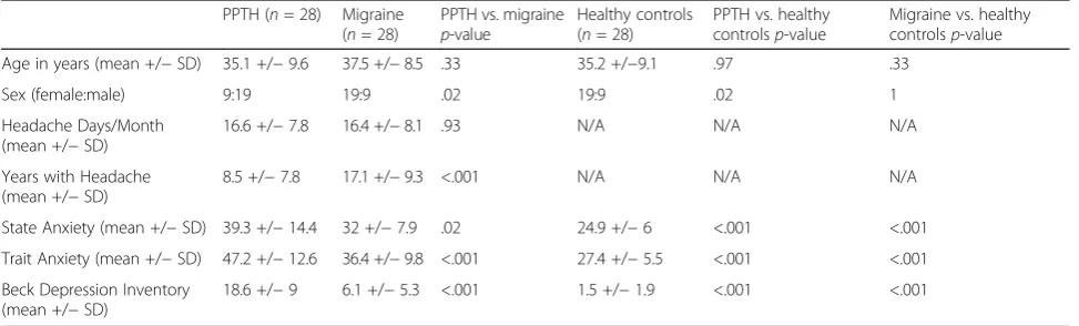
Table 1Participant characteristics
PPTH (n= 28) Migraine (n= 28)
PPTH vs. migraine p-value
Healthy controls (n= 28)
PPTH vs. healthy controlsp-value
Migraine vs. healthy controlsp-value
Age in years (mean +/−SD) 35.1 +/−9.6 37.5 +/−8.5 .33 35.2 +/−9.1 .97 .33
Sex (female:male) 9:19 19:9 .02 19:9 .02 1
Headache Days/Month (mean +/−SD)
16.6 +/−7.8 16.4 +/−8.1 .93 N/A N/A N/A
Years with Headache (mean +/−SD)
8.5 +/−7.8 17.1 +/−9.3 <.001 N/A N/A N/A
State Anxiety (mean +/−SD) 39.3 +/−14.4 32 +/−7.9 .02 24.9 +/−6 <.001 <.001
Trait Anxiety (mean +/−SD) 47.2 +/−12.6 36.4 +/−9.8 <.001 27.4 +/−5.5 <.001 <.001
Beck Depression Inventory (mean +/−SD)
18.6 +/−9 6.1 +/−5.3 <.001 1.5 +/−1.9 <.001 <.001
subjects had one TBI in their lifetime, 13 had 2 TBIs, two had 3 TBIs, one had 4 TBIs, two had 5 TBIs, and five had 6 or more TBIs. Of the TBIs leading to PPTH, 14 were due to explosions/blasts, 7 were due to sports injuries, 4 were due to motor vehicle accidents, and 3 were due to falls.
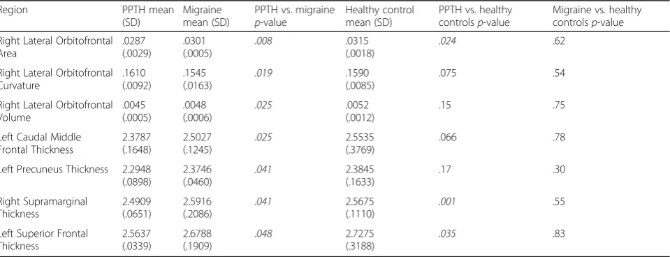
Comparison of the PPTH cohort to the migraine co-hort revealed differences in area of the right lateral orbi-tofrontal area (p = .008), curvature of the right lateral orbital frontal area (p= .019), volume of the right lateral orbital frontal area (p = .025), and thickness of the left caudal middle frontal region (p = .025), left precuneus (p= .041), right supramarginal gyrus (p= .041), and left superior frontal region (p = .048). (Table 2, Fig. 1) For each of these regions, thickness, area and/or volume measurements in those with PPTH were less than those within the migraine cohort. Lateral orbital frontal region curvature was greater in the PPTH cohort compared to the migraine cohort, suggesting underlying structural damage. The significance of each variable within the ANCOVA is reported in Table 3. Amongst those regions that differed in structure when comparing PPTH to mi-graine, there were no significant correlations between the numbers of lifetime TBIs or years with PTH with brain structure.
To better interpret the differences in brain structure identified between PPTH and migraine, the seven re-gional structural measures that differed between the PPTH and migraine cohorts were compared between the PPTH group to a group of healthy controls and be-tween the migraine group and the healthy controls. Amongst these regions, there were differences between the PPTH group and healthy controls for thickness of the right supramarginal gyrus (p = .001), area of the
right lateral orbitofrontal region (p = .024), and thick-ness of the left superior frontal region (p= .035). For all of these comparisons, PPTH was associated with less area and cortical thickness compared to the healthy con-trols. Comparisons between the migraine group and the healthy control group demonstrated no significant differ-ences in the seven structural measurements that differed when comparing the PPTH group to the mi-graine group.
Discussion
The phenotypic similarities between migraine and PPTH justify the need for studies that investigate mechanistic similarities and differences between migraine and PPTH. The main finding of this study is that there are differ-ences in brain structure between patients who have PPTH and those with migraine, perhaps suggesting that these two headache types are associated with distinct underlying pathophysiology despite their substantial similarities in symptoms.
Brain areas that differed between PPTH and migraine were located in lateral orbitofrontal, superior and middle frontal, precuneus and supramarginal gyrus regions. The observation that only three of the seven brain re-gional measurements differed when comparing PPTH to healthy controls and none of the seven brain regional measurements differed when comparing migraine to healthy controls suggests specificity of some brain struc-tural differences to the comparison of PPTH vs. mi-graine. We are not aware of prior studies that have compared brain structure or function in a group of indi-viduals with PPTH to a group with migraine and thus are unable to compare our findings to others. However, there are a few studies that have compared brain
Table 2Brain regions with structural differences when comparing individuals with persistent post-traumatic headache (PPTH) to those with migraine
Region PPTH mean
(SD)
Migraine mean (SD)
PPTH vs. migraine p-value
Healthy control mean (SD)
PPTH vs. healthy controlsp-value
Migraine vs. healthy controlsp-value
Right Lateral Orbitofrontal Area .0287 (.0029) .0301 (.0005) .008 .0315 (.0018) .024 .62
Right Lateral Orbitofrontal Curvature .1610 (.0092) .1545 (.0163) .019 .1590 (.0085) .075 .54
Right Lateral Orbitofrontal Volume .0045 (.0005) .0048 (.0006) .025 .0052 (.0012) .15 .75
Left Caudal Middle Frontal Thickness 2.3787 (.1648) 2.5027 (.1245) .025 2.5535 (.3769) .066 .78
Left Precuneus Thickness 2.2948 (.0898) 2.3746 (.0460) .041 2.3845 (.1633) .17 .30 Right Supramarginal Thickness 2.4909 (.0651) 2.5916 (.2086) .041 2.5675 (.1110) .001 .55
Left Superior Frontal Thickness 2.5637 (.0339) 2.6788 (.1909) .048 2.7275 (.3188) .035 .83
Volume and area measurements are normalized. Thickness measurements are in mm. Curvature measurements are in mm−1
structure and metabolism in individuals with PTH com-pared to healthy controls. For example, a longitudinal voxel-based morphometry study by Obermann and col-leagues compared 32 patients with PTH attributed to whiplash injury to healthy controls [15]. During the first 14 days post-TBI, there were no differences in brain structure. However, at 3 months, PTH was associated with decreased gray matter density in the anterior cingu-late cortex and dorsocingu-lateral prefrontal cortex. As head-aches subsided by 1 year, the gray matter density normalized in these brain areas. Amongst those who de-veloped PPTH, at 1 year there was in increase in gray matter in the midbrain, thalamus and cerebellum. As-suming that the patients who developed PPTH and those who did not had similar brain injuries, this study supports the notion that brain structural changes in pa-tients with PPTH are related to the continued headaches
and are not due solely to the inciting brain injury. A magnetic resonance spectroscopy study of 17 individuals with PTH attributed to mTBI (9 with acute PTH and 8 with PPTH) found that compared to healthy controls those with PTH had metabolic abnormalities in the anter-ior frontal lobes, anteranter-ior and posteranter-ior medial frontal lobes, and medial parietal lobes [16]. Although there was little power in this study to detect differences between pa-tients with acute PTH compared to those with PPTH, those with PPTH had non-significant but lower N-acetylaspartate values (a marker for neuronal health). In our study, there were no significant correlations between the structural measurements of regions that differed when comparing PPTH to migraine with the number of years that individuals had PPTH. However, our patient popula-tion differed than those in prior publicapopula-tions in that our patients had a longer duration of PPTH at the time of
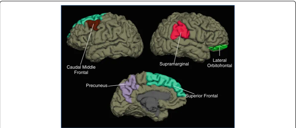
Caudal Middle Frontal
Precuneus
Superior Frontal Supramarginal Orbitofrontal Lateral
Fig. 1Regions with structural differences when comparing individuals with persistent post-traumatic headache (PPTH) to those with migraine. When comparing structural measurements of entire brain regions in patients with PPTH to patients with migraine, the right lateral orbital frontal region differed in area, volume, and curvature. The left caudal middle frontal, precuneus, and superior frontal regions and the right supramarginal gyrus region differed in cortical thickness
Table 3Significance of each variable in the ANCOVA for brain region measurements that differed between PPTH and migraine
Region Age Depression State anxiety Trait anxiety Years with PTH Sex
Right Lateral Orbitofrontal Area .898 .043 .674 .580 .448 .810
Right Lateral Orbitofrontal Curvature
.419 .847 .374 .117 .183 .746
Right Lateral Orbitofrontal Volume
.067 .130 .985 .340 .187 .033
Left Caudal Middle Frontal Thickness .029 .521 .065 .004 .266 .140
Left Precuneus Thickness .055 .440 .205 .023 .065 .874
Right Supramarginal Thickness .000 .401 .371 .072 .612 .914
Left Superior Frontal Thickness .010 .547 .586 .093 .567 .251
imaging (mean of 8.5 years). Studies of mTBI have also demonstrated abnormalities of brain structure in several of the regions that we identified to have different structure in PPTH compared to migraine [17–19]. Most of these previously published studies have not reported whether the participants had PTH and they did not attempt to dis-entangle the effects of the brain injury itself on brain structure vs. the effects of post-TBI symptoms on brain structure.
The brain regions that differed in structure when com-paring individuals with PPTH to those with migraine in this study have all been previously demonstrated to par-ticipate in pain processing. Frontal regions play roles in the affective and cognitive evaluation of pain [20, 21]. Frontal regions have previously been found to have ab-normal structure, function, and functional connectivity in individuals with different headache types including migraine, cluster headache, and medication overuse headache [22–26]. The precuneus, a core region of the default mode network that is responsible for self-referential processing and interoception, participates in the determination of pain sensitivity and pain thresholds [27–30]. Prior studies have implicated the precuneus and the default mode network in headache disorders in-cluding migraine, medication overuse headache, and cluster headache [22, 30–33]. The supramarginal gyrus is likely involved in cognitive evaluation of pain includ-ing pain empathy [34, 35]. The supramarginal gyrus has previously been demonstrated to have atypical function and structure in groups of individuals with migraine and medication overuse headache [36, 37]. Although there are sufficient data to support a role for these brain re-gions in pain processing and headache, the explanation as to why the structure of these regions differs in indi-viduals with PPTH compared to those with migraine is yet to be elucidated. It is plausible that these are brain regions that are simply more susceptible to the effects of mTBI and once damaged they contribute to the initi-ation and persistence of PTH.
The different measures used in this study (volume, area, cortical thickness, curvature) provide complementary in-formation about brain structure. Regional volumes, sur-face area, cortical thickness and curvature are plastic measures of brain structure that commonly show alter-ations in the presence of aging, learning, and neurodegen-eration related to disease. Brain curvature provides a measure of cortical folding, with increased curvature indi-cating areas of sharper cortical folds. Increased cortical curvature has been associated with white matter damage, aging, neurodegenderative disease, and mild TBI [38]. Al-though regional volumes have been most commonly used to measure brain structure and compare brain structure between subject cohorts, measurements of area and cor-tical thickness are likely to be more sensitive to small
changes in brain structure. Regional brain area, cortical thickness, and volume have been previously identified to differ in cohorts of individuals with headache compared to healthy controls and to contribute to subclassification of headache types [39–41].
Limitations: Similar to individuals with migraine and PPTH in the community and in clinical practice, our re-search participants had co-morbid medical conditions, and they were utilizing medications. For example, pa-tients with PPTH had higher anxiety and depression scores than patients with migraine and healthy controls. Although we controlled for many potentially confound-ing variables in our ANCOVA, includconfound-ing depression and anxiety scores, it is not entirely possible to decouple the effects of these co-morbid illnesses on brain structure from the effects of migraine and PPTH on brain struc-ture. However, study results would be of little value in understanding migraine and PPTH if we excluded pa-tients with typical disease and only enrolled the very rare individual who has migraine or PPTH in isolation. Inclu-sion of typical patients with migraine and PPTH makes the results from this study generalizable to the typical individual with migraine or PPTH. Furthermore, the subject cohorts were not exactly matched for age and sex. Although we attempted to do so, the realities of our patient populations resulted in enrolling more men with PPTH (mostly armed forces veterans) and more women with chronic migraine. Sex was controlled for in our ANCOVA and was significant for only one of the brain measurements that differed between migraine and PPTH, but it still could have impacted our results to some extent. Perhaps the biggest limitation of this ana-lysis is the inability to differentiate the effects of TBI from that of PPTH on brain structure. It is not clear if the differences in brain structure identified in this study are due to PPTH or if the differences would be similar in individuals who have persistent post-TBI symptoms without headache. We are currently trying to enroll those rare patients who do not have headache but do have other persistent symptoms following a mild TBI for comparison to the PPTH group.
Conclusions
Abbreviations
ANCOVA:Analysis of covariance; FOV: Field of view; ICHD: International classification of headache disorders; MP-RAGE: Magnetization prepared rapid gradient echo; mTBI: Mild traumatic brain injury; N/A: Not applicable; PPTH: Persistent post-traumatic headache; PTH: Post-traumatic headache; SD: Standard deviation; TBI: Traumatic brain injury; TE: Echo time; TR: Repetition time
Acknowledgements
This work was supported by the National Institutes of Health (NIH) grant NIH K23NS070891 and by the Office of the Assistant Secretary of Defense for Health Affairs, through the Peer Reviewed Medical Research Program under Award No. W81XWH-15-1-0286. Opinions, interpretations, conclusions, and recommendations are those of the authors and are not necessarily endorsed by the Department of Defense. The U.S. Army Medical Research Acquisition Activity, 820 Chandler Street, Fort Detrick MD 21702–5014 is the awarding and administering acquisition office.
Availability of data and materials
The dataset supporting the conclusions of this article is available via the Federal Interagency Traumatic Brain Injury Research Informatics System (FITBIR). fitbir.nih.gov.
Authors’contributions
TJS conceived the project, participated in data analyses, participated in the manuscript writing process, read and approved the final manuscript. CDC conceived the project, participated in data analyses, participated in the manuscript writing process, read and approved the final manuscript. JP participated in data analyses, participated in the manuscript writing process, read and approved the final manuscript. KR conceived the project, participated in data analyses, participated in the manuscript writing process, read and approved the final manuscript. VB conceived the project, participated in data analyses, participated in the manuscript writing process, read and approved the final manuscript.
Ethics approval and consent to participate
Mayo Clinic Institutional Review Board, Phoenix VA Health Care System Institutional Review Board, and U.S. Department of Defense Human Research Protection Office approvals were obtained prior to initiation of study procedures. Each research participant underwent an informed consent process during which the potential risks and benefits of study participation were discussed.
Competing interests
Within the last 12 months, TJS has received compensation for consulting/ advisory boards from Allergan, Amgen, ATI, Avanir, Dr. Reddy’s, Nocira, and Novartis; he has options in Nocira and Second Opinion; and has received royalties from UpToDate.
Publisher’s Note
Springer Nature remains neutral with regard to jurisdictional claims in published maps and institutional affiliations.
Author details
1Mayo Clinic Arizona, 5777 East Mayo Boulevard, Phoenix, AZ 85255, USA. 2
Phoenix VA Health Care System, Phoenix, USA.3Arizona State University, Phoenix, USA.
Received: 26 June 2017 Accepted: 8 August 2017
References
1. Erickson JC (2011) Treatment outcomes of chronic post-traumatic headaches after mild head trauma in US soldiers: an observational study. Headache 51(6):932–944
2. Eskridge SL, Macera CA, Galarneau MR, Holbrook TL, Woodruff SI, MacGregor AJ et al (2012) Injuries from combat explosions in Iraq: injury type, location, and severity. Injury 43(10):1678–1682
3. Ruff RL, Ruff SS, Wang XF (2008) Headaches among veterans of Operations Iraqi Freedom and Enduring Freedom with mild traumatic brain injury associated with exposures to explosions. J Rehabil Res Dev 45:941–953
4. Theeler BJ, Erickson JC (2009) Mild head trauma and chronic headaches in returning US soldiers. Headache 49(4):529–534
5. Classification Committee of the International Headache Society (2013) The International Classification of Headache Disorders, 3rd edition (beta version). Cephalalgia 33(9):629–808
6. Corrigan JD, Bogner J (2007) Initial reliability and validity of the Ohio State University TBI Identification Method. J Head Trauma Rehabil 22(6):318–329 7. Beck AT, Steer RA, Brown GK (1996) Manual for Beck Depression Inventory II
(BDI-II). Pscyhology Corp; San Antonio, TX
8. Spielberger C, Gorsuch R (1983) State trait anxiety inventory for adults: sampler set, manual, test, scoring key. Redwood City, California: Mind Garden 9. Spielberger C (1983) Manual for the state/trait anxiety inventory (form Y):
self-evaluation questionnaire. Consulting Psychologists Press, Palo Alto 10. Gronenschild EH, Habets P, Jacobs HI, Mengelers R, Rozendaal N, van Os J
et al (2012) The effects of FreeSurfer version, workstation type, and Macintosh operating system version on anatomical volume and cortical thickness measurements. PLoS One 7(6):e38234
11. Dale AM, Fischl B, Sereno MI (1999) Cortical surface-based analysis. I. Segmentation and surface reconstruction. NeuroImage 9(2):179–194 12. Fischl B, Liu A, Dale AM (2001) Automated manifold surgery: constructing
geometrically accurate and topologically correct models of the human cerebral cortex. IEEE Trans Med Imaging 20(1):70–80
13. Fischl B, Salat DH, Busa E, Albert M, Dieterich M, Haselgrove C et al (2002) Whole brain segmentation: automated labeling of neuroanatomical structures in the human brain. Neuron 33(3):341–355
14. Segonne F, Dale AM, Busa E, Glessner M, Salat D, Hahn HK et al (2004) A hybrid approach to the skull stripping problem in MRI. NeuroImage 22(3):1060–1075
15. Obermann M, Nebel K, Schumann C, Holle D, Gizewski ER, Maschke M et al (2009) Gray matter changes related to chronic posttraumatic headache. Neurology 73(12):978–983
16. Sarmento E, Moreira P, Brito C, Souza J, Jevoux C, Bigal M (2009) Proton spectroscopy in patients with post-traumatic headache attributed to mild head injury. Headache 49(9):1345–1352
17. Meier TB, Bellgowan PS, Bergamino M, Ling JM, Mayer AR (2016) Thinner Cortex in Collegiate Football Players With, but not Without, a Self-Reported History of Concussion. J Neurotrauma 33(4):330–338
18. Sussman D, da Costa L, Chakravarty MM, Pang EW, Taylor MJ, Dunkley BT (2017) Concussion induces focal and widespread neuromorphological changes. Neurosci Lett 650:52–59
19. Tremblay S, De Beaumont L, Henry LC, Boulanger Y, Evans AC, Bourgouin P et al (2013) Sports concussions and aging: a neuroimaging investigation. Cereb Cortex 23(5):1159–1166
20. Kong J, White NS, Kwong KK, Vangel MG, Rosman IS, Gracely RH et al (2006) Using fMRI to dissociate sensory encoding from cognitive evaluation of heat pain intensity. Hum Brain Mapp 27(9):715–721
21. Becker S, Gandhi W, Schweinhardt P (2012) Cerebral interactions of pain and reward and their relevance for chronic pain. Neurosci Lett 520(2):182–187
22. Chanraud S, Di Scala G, Dilharreguy B, Schoenen J, Allard M, Radat F (2014) Brain functional connectivity and morphology changes in medication-overuse headache: Clue for dependence-related processes? Cephalalgia 34(8):605–615
23. Jin C, Yuan K, Zhao L, Yu D, von Deneen KM, Zhang M et al (2013) Structural and functional abnormalities in migraine patients without aura. NMR Biomed 26(1):58–64
24. Xue T, Yuan K, Cheng P, Zhao L, Yu D, Dong T et al (2013) Alterations of regional spontaneous neuronal activity and corresponding brain circuit changes during resting state in migraine without aura. NMR Biomed 26(9):1051–1058
25. Yang FC, Chou KH, Fuh JL, Lee PL, Lirng JF, Lin YY et al (2014) Altered hypothalamic functional connectivity in cluster headache: a longitudinal resting-state functional MRI study. J Neurol Neurosurg Psychiatry 86(4):437–445. 26. Jia Z, Yu S (2017) Grey matter alterations in migraine: A systematic review
and meta-analysis. Neuroimage Clin 14:130–140
27. Emerson NM, Zeidan F, Lobanov OV, Hadsel MS, Martucci KT, Quevedo AS et al (2014) Pain sensitivity is inversely related to regional grey matter density in the brain. Pain 155(3):566–573
29. Schwedt TJ, Chong CD (2014) Correlations between brain cortical thickness and cutaneous pain thresholds are atypical in adults with migraine. PLoS One 9(6):e99791
30. Zhang J, Su J, Wang M, Zhao Y, Yao Q, Zhang Q et al (2016) Increased default mode network connectivity and increased regional homogeneity in migraineurs without aura. J Headache Pain. 17(1):98
31. Chou KH, Yang FC, Fuh JL, Kuo CY, Wang YH, Lirng JF et al (2016) Bout-associated intrinsic functional network changes in cluster headache: A longitudinal resting-state functional MRI study. Cephalalgia. Epub ahead of print.
32. Michels L, Christidi F, Steiger VR, Sandor PS, Gantenbein AR, Landmann G et al (2017) Pain modulation is affected differently in medication-overuse headache and chronic myofascial pain - A multimodal MRI study. Cephalalgia 37(8):764–779.
33. Tessitore A, Russo A, Giordano A, Conte F, Corbo D, De Stefano M et al (2013) Disrupted default mode network connectivity in migraine without aura. J Headache Pain 14(1):89
34. Lamm C, Decety J, Singer T (2011) Meta-analytic evidence for common and distinct neural networks associated with directly experienced pain and empathy for pain. NeuroImage 54(3):2492–2502
35. Moulton EA, Pendse G, Becerra LR, Borsook D (2012) BOLD responses in somatosensory cortices better reflect heat sensation than pain. J Neurosci 32(17):6024–6031
36. Chiapparini L, Grazzi L, Ferraro S, Mandelli ML, Usai S, Andrasik F et al (2009) Functional-MRI evaluation of pain processing in chronic migraine with medication overuse. Neurol Sci 30(Suppl 1):S71–S74
37. Ferraro S, Grazzi L, Mandelli ML, Aquino D, Di Fiore D, Usai S et al (2012) Pain processing in medication overuse headache: a functional magnetic resonance imaging (fMRI) study. Pain Med 13(2):255–262
38. King JB, Lopez-Larson MP, Yurgelun-Todd DA (2016) Mean cortical curvature reflects cytoarchitecture restructuring in mild traumatic brain injury. Neuroimage Clin 11:81–89
39. Schwedt TJ, Chong CD (2017) Medication Overuse Headache: Pathophysiological Insights from Structural and Functional Brain MRI Research. Headache 57(7):1173–1178
40. Schwedt TJ, Chong CD, Wu T, Gaw N, Fu Y, Li J (2015) Accurate Classification of Chronic Migraine via Brain Magnetic Resonance Imaging. Headache 55(6):762–777