RENAL TUBULAR DYSFUNCTIONCOMPLICATING
THE NEPHROTICSYNDROME
Gunnar B. Stickler, M.D., Ph.D., Alvin B. Hayles, M.D., Marschelle H. Power, Ph.D., and John A. Ulrich, Ph.D.
Sections of Pediatrics (G.B.S., A.B.H.), Biochemistry (M.H.P.) and Bacteriology (J.A.U.), Mayo Clinic and Mayo Foundation, Rochester, Minnesota
vitamin D in relatively large doses resulted in healing of the rickets. A metabolic study
of one patient (Case 2) showed that the
administration of adrenocorticosteroids was
followed by a large reduction in the loss
of protein in the urine and in improved
renal tubular function.
Case 1
P ROCRESSIVEglomerular failure as mani
fested by azotemia and hypertension
not infrequently occurs as a terminal event among patients with nephrosis. Renal tubu
lar insufficiency as manifested by amino
aciduria, glycosuria, hypophosphatemic
rickets and hypocalcemic tetany is a rare
consequence or sequela of long-standing
nephrosis. Stanbury and Macaulay1 ob
served a boy in whom nephrosis had de
veloped at the age of 2Vi2 years, and at the
age of 4 years glycosuria and amino
aciduria were present. Tegelaers and Tid
dens2 reported their observations on two
children with similar clinical courses. Tet any, rickets, growth failure, hypokalemia
and hypocalcemia had developed in their
patients and in the patient of Stanbury and Macaulay.
During recent years two girls have been
observed who have manifested evidence of
renal tubular insufficiency subsequent to
nephrosis. In each instance the classic pic
ture of the nephrotic syndrome appeared at
an early age and was followed in the en
suing 8 years by the gradual disappearance
of edema in spite of persistent protein
uria and hypoproteinemia. Retardation of
growth, polyuria, polydipsia, intermittent
attacks of tetany and evidence of rickets
soon appeared. Moderately severe metabolic
acidosis, azotemia and hypocalcemia were
observed before therapy was instituted.
Amino-aciduria and glycosuria in addition
to proteinuria and cylindruria have been
persistent findings. The administration of
CASE REPORTS
HIsToRY: An 8@M2-year-old white girl was ad
mitted to the Mayo Clinic in August, 1953, because of tetany, rickets and dwarfism. The prenatal, birth and family histories were not significant. The illness had its onset at the age
of 18 months when an acute upper respira
tory infection developed and was followed by edema and rapid gain of weight. The diagnosis of nephrotic syndrome was established at that
time. Repeated abdominal paracenteses were
necessary to relieve ascites, when she was 3 years of age. Numerous transfusions of blood were given. Massive proteinuria and intermit tent erythrocyturia persisted, glvcosuria ap peared, and edema disappeared when the child was 33@years of age.
At the age of 4 years retardation of growth was apparent and a diagnosis of rickets was established. During the patient's fifth and sixth years repeated laboratory studies were made and persistent hypoproteinemia and hyper cholesterolemia were observed. Concentration of calcium in the serum was 9.5 mg/lOO ml; phosphorus, 4 mg/100 ml; and nonprotein ni trogen, 16.5 mg/100 ml. When the patient was 8 years of age diarrhea developed and was fol
lowed by tetany which responded to intra
venously administered calcium gluconate. Sub
This investigation was supported in part by research grant A-2011 from the Public Health Service.
The Mayo Foundation, Rochester,Minnesota, is a part of the Graduate School of the Universityof
Minnesota.
ADDRESS FOR REPRINTS: Section of Publications, Mayo Clinic, Rochester, Minnesota.
PEDIATRICS, July 1960
76 NEPHROTIC SYNDROME
scopic examination disclosed 3 to 4 diopters of chronic papilledema of each optic disk. The papilledema was considered to be associated with hypocalcemia. Slit-lamp examination of the cornea did not disclose any abnormality.
LABORATORY FINDINGS : Roentgenograms of
the thorax and of the skull were considered normal while roentgenograms of the long bones revealed evidence of changes consistent with active rickets. There were cystic changes in the proximal left humerus and pseudofractures
‘¿, of the proximal left fibula and the necks of the
femurs. Bone maturation was compatible with the chronologic age of approximately 33@years. A summary of the laboratory data is shown in
TableI.
THERAPY AND COURSE: A tentative diagno
sis of chronic renal insufficiency secondary to nephrotic syndrome was established and cal cium lactate solution was administered orally. A regular diet was allowed. The concentration of calcium in the serum increased to 7 to 8 mg/100 ml with this therapy and the patient had no further attacks of tetany.
The patient was re-examined in July, 1954, at the age of 9342 years. She had been asymp tomatic during the interval since her last visit except for scarlet fever in April, 1954. The
genu valgum deformity of the knees had be
come more prominent. Roentgenograms of the
extremities disclosed more evidence of rachitic activity (Fig. 2a). Vitamin D, in doses of 20,000 units daily, was added to the program of treat ment. Within 3 months there was radiographic evidence of healing of the rickets and the con centration of calcium in the serum was normal. With this treatment the patient remained free of symptoms except for polyuria. Glycosuria and proteinuria persisted.
In August, 1955, at the age of 10@42years
the child was examined again. She had re
mained normotensive and the degree of papil ledema had definitely decreased. The genu val gum deformity of the legs had not changed, but roentgenograms disclosed evidence of heal ing of the rickets (Fig. 2b). Laboratory studies
are summarized in Table I. Amino-aciduria
was present (Table II). Glycosuria and prote inuria persisted, but tetany had not recurred
and the rachitic condition was considered
healed.
A recent follow-up examination indicates that the child has continued to grow and has taken part in school activities. In 1957 the medial edges of the femoral epiphyses were immobil
I
70
goodiwso-N50
mg/mL 30
10
Fic. 1. Case 1. Height, weight and findings in the
urine in relation to age.
sequent determinations of the concentrations
of calcium disclosed hvpocalcemia, the lowest
value being 4.7 mg/100 ml. The concentration
of phosphorus in the serum varied between
4.0 and 4.9 mg/100 ml. In January ‘¿53,the oral administration of a medication containing cal
cium was started; thereafter the number of
attacks of tetany decreased.
Three months before the child's admission to the clinic she complained of increasingly fre
quent headaches and dimness of vision. Her
admission to the clinic was prompted by the recurrence of tetany, in spite of continued ad ministration of the calcium solution. A graphic representation of some of the laboratory data and the patient's height and weight at differ ent ages is shown in Figure 1.
PHYSICAL FINDINGS: Physical examination at
the time of admission disclosed a dwarfed girl whose height was 100.5 cm and weight, 15.4 kg. The skin had a brownish tinge. There was
no evidence of edema. The epiphyseal ends
of the long bones were prominent and there
was genu valgum deformity of the legs. The
blood pressure was 95/60 mm Hg. Fundu
Ag.
1011i@
i@
WY@\WNT@
I I I II I I I
Case 1Case 28,@
yr9T7T IP'1o@% yr9i% @ir10
yrHemoglobin
(grn/100 ml)13.59.810.311.1Leukocytes
(/mm')10,2007,2005,800Serum
cholesterol (mg/100 ml)282Blood
urea (mg/100 nil)63100112124114Blood
sugar (mg/100 ml)909990Serum
albulnin (gm/100 ml)1 .651
.352.00Alphai-globulin
(gm/100 ml)I
2 .880.30.28Alpha,-globulii@
(gm/IO0 ml)
—¿1
.410.90Beta-globulin
(gm/100 ml)—
—¿
1 .040.72Gamma-globulin
(gm/100 ml)0.510.59Sodium
(meq/l)132127138136Potassium
(meq/l)3 .93
.43-9Chloride
(meq/l)97105.6117.9112Carbon
dioxide content (meq/l)16 . 114 .621 .320.5Serum
phosphatase (King-Armstrong units)52 .5103 .421 .771 .1Calciu;n(ing/lOOml)5.8648.95.48.0Phosphorus
(mg/100 ml)3 .44 .64 .94
.75.4Urinalysis:
Spécificgravity1.0121.0081.0091.0111.011ReactionAlkalineAlkalineAlkalineAlkalineAlkalineProtein
(grade)44444Sugar(grade)11111Erythrocytes
(grade)A few12A fewA
fewBence
Jones proteinEquivocalEquivocalEquivocal
ARTICLES
TABLE I
LABORATORY DATA ON Two PATIENTS WITH RENAL TUBULAR DYSFUNCrION
77
ized with staples to correct genu valgum. The staples were removed in November, 1958, after almost complete correction of the deformities of the knees. She continues to take 50,000 units of vitamin D every fourth day, 10 ml of cal
cium gluconate twice daily and a multiple
vitamin preparation. During recent months the
concentration of calcium in the serum has
varied between 5.8 and 8.2 mg/100 ml. The
78 NEPHROTIC SYNDRO\IE
complaints of dwarfism and rickets. The family
history and the prenatal and perinatal histories were noncontributorv. At the age of 15 months puffiness of the eyelids and swelling of the face had developed. The diagnosis of nephrotic syndrome @s'asestablished by the presence of
anasarca and persistent massive protei1s@;ria.
The patient's blood pressure was said to have been IU)rnlal and remained norn@al during the period of her observation. The edema which persiste(l for several months was not affected by restriction of salt in the diet.
The patieiit had measles at the age of 3 @‘¿ears and soon thereafter meningitis developed due
to Hemophilus influenzae. The patient was
treated successfully for meningitis with peni cillin and streptomvcin. During the course of these two illnesses much of the edema sub sided and the proteinuria decreased for a short time. Since then, the patient has had no fur
ther episodes of massive edema, but has con
tinned to have I)rOteinuria . \Iininial ervthro
cyturia was noted on a few occasions.
Glvcosuria, growth retardation, and rickets were noted when the child was 5 \ears of age. The patient was treated with various medica
tions, including vitamin D in moderate amounts, thyroid extract, norethandrolone (Nilevar@), adrenocorticotropic hormone, cortisone and
sitosterols (Cvtellin @),without apparent bene
fit. At the age of 9 years she experienced re
TABLE 11
Fic. 2. Case 1. (a, left) View of knee to demonstrate rickets before vitamin I) was taken; (b, right) after
use of Vitamin 1).
tween 47 and 87 rng 100 ml, with the lower concentrations being observed during recent months. The patient continues to have poiv uria, proteinuria and glvcosuria.
Case 2
HIsTORY: A girl, 9@'@ years of age, was ad
mitted to the clinic on October 21, 1957, with
EXI;ETIorc OF “¿FIIEEAMINo Acm― (Mo/@24 lilt) IN Iwo PATIENTS WITh RENAL
‘¿1'ti@vi@ioI)ysm';No'TIoN
lrn,no Acid Excreted (rng/24 br)
(‘use2 (Study Periool.s)
1
@ 2
@ .1
@
@ I 5
@ (@
@
129 119 137 117 86 95 J 41
163 1(13 299 106
@ 100 1 1 1 59
84 86 146 193
@ 170
@ 147 114
I (166 1 316 1 , lU; 588
@ 280
@ 119
@ 167
202 170 244 237 I 366 200
@ 162
133 154 147 77
@ (i6 68
@ 29
505 .563 274 199 164 140
@ 81
365 452 421 392
@ 336
@ 274
@ 172
41 39 44 43
@ 35
@ 32 11
139 149 148 118
@ 103 106
@ 44
238 207 287 146
@ 110 79 69
192 177 294 216 I 157
@ 162
@ 155
36 42 36 37 33
@ 26 I 17
185 183 121 119 122
@ 118 54
413 405 362 266 233 ‘¿214
@ 92 (a.se 1 218 264 76 896 217 124 623 480 38 226 170 224 138 431 Amino ACi(l Argiimimme @@s1)artic:;cil (‘ystine (lutamnimmc Ilistidine Isoleucine Leucim;e I@ySiIIe \lethionim;e I'heIlylIh hi nine Prolino ‘¿I'l;reonine Iryptopha n 1'yrosine Valine “¿,ormal
20 ± 35
4@±5.1
‘¿.P)± 5.1
138 ±21
125 ±16.4
3.7± 0.4 17 ± 2
40 ± 4.6
1.7± 0.4 13 ± 1.9
33 ± 4.9
ARTICLES 79
l)e@1ted attacks of tetanv which sui)sicled spon taneouslv within a few months. A progressive
genii valgum deformity also developed and walking became difficult. A graphic presenta tion of her history and development is shown in Figure 3.
PHYSICAL FINDINGS: The patient was a
dwarfed, dejected girl whose height was 106.7 Clii and weight, 19.5 kg. The blood pressure was 95 60 mm Hg. The face was slightly puffy and the abdomen was prominent but ascites could not be demonstrated on examination. The edge of the liver was palpable 2 CITIbelow the right costal margin . Genu valguni deformity of the legs @s'asprominent.
LABORATORY FINDINGS: Laboratoiy studies o)btailled On this and the next visit are sum
marized in Table I. Roentgenograms of the thorax and of the extremities were interpreted as normal except for the presence of active rickets (Fig. 4a).
THERAPY AND COURSE: The administration of vitamin D in (loses of 50,000 units daily was started on October 29, 1957, and was followed by considerable improvement in the child's ability to walk. She was able to paiticipate in no)rInal activities including roller skating.
FIG. 3. Case 2. Height, weight and findings in the urine in relation to age.
—¿@
F—
I@1
FIG. 4. Case 2. (a, left) View of knee to demonstraterickets before patient took vitamin 1); (h, right)
after use of vitalilin I).
The patient returned to the clinic for met abolic stud@ 00 March 3, 1958, at the age of 10 years, at which time her height was 1C6.7 cm and weight, 18.8 kg. The blood pressure
was normal. The results of 1)h'sical examina
tion were IlOt significantly different from those previously noted. Roentgenograms of the knees showed healing of the rickets (Fig. 4b).
SPECIAL STUDIES OF CASE 2
Methods
After 6 days of @)relin-Iinary ol)servation, the child was studied during six sequential balance periods, each of 6 days' duration. An additional study of a 6-day balance period was made 6
months later. The diet remained constant
throughout the entire studs'. The patient was allowed to determine her caloric requirements (luring the 6 days preceding the first balance period. The diet consisted of three daily menus which were used in rotation. l)istilled water was used as drinking water, and the amount needed was deterniined by the patient and re cOr(led; the teeth also were cleaned with cbs tilled water. She was weighed daily.
Administration of vitamin D was discon tinued 3 weeks before the metabolic study. Balance Periods 1 and 2 were control periods. During Periods 3 and 4, 60 mg of prednisone was given daily; during Periods 5 and 6 the
patient received 20 mg of prednisone cvei-v
HEIGHT
160
3@
* /97
-60
-50
‘¿50
@40 @
@30 @.
20
@10
* Percentiles
3 4 5 6 7 8 9 10 11 12
140
-.@
120-cJ
100
80 F
Age
@
@ .5 0— - - -- - -@
@
—¿-@ Trace
:@ Glucose o a o a o
@ @‘¿+ + ++
.
@ 1020 1008 1012
Specificgravity 1015 1015 1010
@
@ - —¿â€”@I
@
Prednisone
2Omqidoy 3 times per week NEPHROTIC SYNDROME
80
third, fourth and fifth day. During the sixth metabolic period 9 gm of calcium lactate was dissolved in the drinking water because of the appearance of tetany. After the sixth metabolic period the patient was dismissed on the daily dose of 50,000 units of vitamin D in addition to 20 mg of prednisone daily for S successive days of each week. These same medications were continued during the seventh metabolic period which followed 6 months after the sixth period.
Carmine markers were given at the beginning
of each period and at the end of the sixth
period. A similar marker was used at the be ginning and at the end of the seventh period. The stools were collected and pooled for each 6-day period and analyzed as a single speci
men. Urine was collected and analyzed each
24 hours. Venous blood for the determination of electrolytes was drawn under oil into a tube containing purified heparin. The methods used for the determination of sodium, potassium, chloride, carbon dioxide content of the plasma, blood urea, blood sugar, inorganic phosphate and calcium in the serum, and sodium, potas sium, calcium, phosphorus, nitrogen, titratable acidity and ammonia in the urine have been
described previously.3 The pH of the serum
was measured by the method of Van Slyke et
al.@The methods for electrophoretic separa
tion of the serum and urinary proteins are de scribed in detail elsewhere.5 Individual “¿free amino acids―were measured in the urine by a modification of Henderson and Snell's micro biologic method.6
Results
The patient excreted approximately 7 gm
of protein per 24 hours (Fig. 5) during the
control periods. The major part of the un
nary protein was albumin. Hypoalbumi
nemia, relatively low concentrations of
gamma-globulin in the serum, and elevated concentrations of alpha2-globulin as well as
serum cholesterol were present. The con
centrations of calcium and potassium in the serum were low while the concentration of
phosphorus was normal. The concentration
of the blood urea was elevated (Fig. 6), that
of the serum sodium was normal, that of
the serum chloride was elevated, and the
carbon dioxide content was reduced. The pH of the serum was 7.21 during the control
Vitamin D
50@D00untts/day
PrednisoneI
eOmg/doyJ
Months of observation
6-day periods
Fic. 5. Case 2. Serum cholesterol, serum protein fractions and protein excretion in the urine during periods of observation and metabolic study.
periods. There was decreased excretion of titratable acidity and ammonia in the urine
(Table III). The value for blood sugar re
mained normal during the control period,
but the patient excreted approximately 4
gm of glucose per 24 hours in the urine. In
creased amino-aciduria was present, with
the excretion of glutamine being greatest
(Table II). As indicated in Figure 7, the
nitrogen, phosphorus, calcium, sodium and
chloride balances, which were plotted ac
cording to the method of Albnight and
Reifenstein,7 were positive while the patient was in negative potassium balance during the control periods.
Administration of prednisone was fol
lowed by a decrease in the proteinuria and
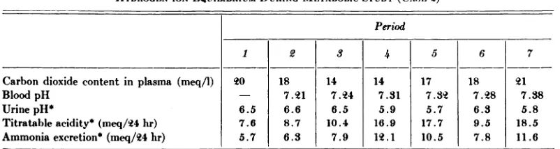
Period123567Carbon
dioxide content in plasma (meq/1) Blood pH
Urine pH*
Titratableacidity*(meq/24hr)
Ammonia excretion* (meq/24 hr)20
—¿
6.5 7.6 5.718
7.21 6.6 8.7 6.314
7.24 6.5 10.4
7.914 7.31 5.9 16.9 12.117
7.32 5.7 17.7 10.518
7.28 6.3 9.5 7.821
7.38 5.8 18.5 11.6
ARTICLES 81
Prednisone 2Omç./doy3 times per week
Calciumlactate
Prednisone 9gm/do Vitamin 0
4;
tI, i-i 50,000
units/day
Monthsof observation
I 3 5 —¿â€” 6 - —¿.‘¿â€”———7 —¿â€”‘8 10 12 13 14 16 i1iir
@
@ ii
@ °@ @_.
@ @ @
@ .@
@ j
@ 0%
@ 100 @0
80
140 Na .--.o
l3oE
@
@
—¿.-
.-o---.'———o.---.
.._—¿Â°
120
116
114
112
110
K
3.9 @7
3.5 @@‘¿@0.——¿0
-3.3 I .L:@z.;@
&0 Ca
@_0@,_@ Tetany
-@ 7.0 @
5.0
@ I
7.0 I
P
@ ‘¿P'.
____1 @0@@‘¿
5.0 __
1 2 3 4 5 6 7
6-day periods
FIG. 6. Case 2. Blood urea and serum electrolytes during periods of observation and metabolic study.
TABLE III
HYDROGEN-ION EQUILIBRIUM DURING METABOLIC STUDY (CASE 2)
82 NEPHROTIC SYNDROME
Prednisone 20mg /doy, 3times per week
€¿42mIda Soooounts/doy
F—i
Predrisore 20mg/day, 3 times per week
Calcium lactate 9gm /day
Prednisone Vitamin 0
60m Ida 50,000 un3rJday
LI]
[Ii
123 4 5 6 7
6day periods 6doy periods
chloride was noted initially as well as an in crease in the severity of the metabolic acido sis and azotemia. The value for serum potas sium fell and the potassium balance became more negative.8 An increase in the concen
tration of blood sugar was accompanied by
increased excretion of glucose in the urine
(7.1 gm/24 hr). Diuresis was accompanied
by decreased amino-aciduria, the most strik ing decrease being in the excretion of gluta mine. Excretion of hydrogen ions increased and a correction of the metabolic acidosis
was associated with a rise in the serum pH
to 7.32.
Most of the effects of administration of
o No additional potassium was given during the metabolic study.
prednisone noted during Periods 3 and 4
were also observed during Periods 5 and 6
when prednisone was administered inter
mittently. Urinary excretion of protein and
amino acids remained low while the excre
tion of hydrogen iOnS by the kidney was not so great as it had been with the continuous
administration of prednisone. There was no
recurrence of the metabolic acidosis. During Period 5 the excretion of calcium and phos
phorus into the bowel increased and was
associated with a negative balance of cal
cium and phosphorus, a decrease in the con
centration of calcium in the serum and a
recurrence of tetany. Calcium lactate was
added to the drinking water during Period
6 in order to avoid tetany.
The seventh period of metabolic studies,
456
2 @198
@ 0
,+
+0204 06
FIG. 7. Case 2. Data from the balance studies are plotted according to the method of Aibright and Reifenstein.7 Each column represents a 6-day period. Intake is plotted downward and excretion (fecal, hatched; urine, clear) is plotted upward. The data on the daily balances of potassium, sodium and chloride are superim
posed on the plots of the 6-day period.
0
E +2
I
ARTICLES 83
authors1° have encountered glycosuria and
amino-aciduria in patients with nephrotic
syndrome.
Considerable evidence has been presented to indicate that the increased loss of protein in the urine of patients with nephrotic syn
drome is due to increased glomerular per
ty11 The cause of renal tubular
failure which occasionally develops after
nephrotic syndrome is not known, but it is interesting to speculate that the continued
reabsorption of protein by the renal tubular
epithelium may have a damaging effect on
the function of the cells of the tubules.12
Such a possibility is suggested by the ex
periments reported by Rhodin.'3 This possi
bility is further supported by the descrip
tion of renal tubular insufficiency (de Toni
Debre-Fanconi syndrome) occurring among
three patients with Bence Jones protein in
the urine as the result of multiple mye
loma.14'6 In each of the patients in the pres
ent report the tests for Bence Jones protein in the urine gave equivocal results. One
patient (Case 2) showed decreased excre
tion of protein in the urine associated with improved renal tubular function after the
administration of prednisone. It is of interest
to note that glutamine, supposedly the
source of ammonia in the urine, was excreted
in large amounts in the urine of both patients
before steroid therapy was instituted (in
one patient); as this amino acid was more
completely reabsorbed, the production of
ammonia in the urine increased.
An alternative hypothesis is that the pri
mary disturbance producing the nephrotic
syndrome also may have produced renal
tubular damage. This seems unlikely in
view of the rarity of the occurrence of this complication. We cannot exclude the co
existence of two separate renal diseases. Administration of vitamin D was followed by additional improvement in renal tubular
function as indicated by a further decrease in excretion of amino acid and by improved excretion of hydrogen ion in the urine.
The mechanism whereby rickets develops
in patients with this type of renal tubular
dysfunction is not clearly understood. The
which followed 6 months after Period 6,
was carried out while the patient was re
ceiving vitamin D and intermittent doses of prednisone. During this period the value for
blood urea was 144 mg/100 ml. The amino
aciduria had decreased further; glycosuria
had decreased somewhat (to 3.2 gm/24 hr),
and the ability to excrete hydrogen ions had improved as evidenced by an increase in the
titratable acidity and production of am
monia in the urine. The ability of the kid ney to concentrate the urine was still im@ paired. The calcium and phosphorus bal
ances were now more noticeably positive,
but the potassium balance remained nega
tive.
The patient has continued on the therapy
described in Period 7 of the metabolic
studies and according to the last report she
has continued to grow and to make satis
factory progress. The concentration of blood
urea has decreased to 83 mg/100 ml, while
the serum calcium measured 10 mg/l00 ml
and the phosphorus, 5.5 mgIlOO ml.
COMMENT
Studies by Bruck et al.8 have shown that most patients suffering from the nephrotic
syndrome have normal tubular function. It
is also known that amino-acidunia is an un
common finding in the uncomplicated ne
phrotic syndrome.9 No data are available on the excretion of amino acid in the urine of the present patients during the early part of their disease; .nevertheless, it is known that they did not have glycosuria, rickets, or signs of acidosis until many months after the diagnosis of nephrotic syndrome was estab lished. It is interesting that these patients
had become free of edema in spite of mas
sive proteinuria, and it may be postulated that they had reached a state of equilibrium
in which production of protein was com
parable to the loss of protein.
The occurrence of glycosuria, growth fail ure, rickets, metabolic acidosis and the in ability to concentrate urine are considered to be indications of renal tubular dysfunc
tion. Identical sequences of events have
84 NEPHROTIC SYNDROME
rickets was clearly of the hypophosphatemic
type. It should be noted that during the
two control periods the patient (Case 2)
excreted 46% of the total intake of phos
phorus in the urine, while 39 and 42% dur
ing these two periods, respectively, were
lost in the stool. It has been showni? that
normal children excrete 56 to 65% of the
daily intake of phosphorus in the urine and only 26 to 27% of the intake in the stool. In
Case 2, after the administration of vitamin
D (Period 7), 63sf of the total intake of
phosphorus was excreted in the urine and
only 10% of the phosphorus was excreted in
the stool. During this time phosphorus reten tion increased. The finding of relatively low
absorption of phosphorus in the gastroin
testinal tract has been described18 among
patients with de Toni-Debre-Fanconi syn
drome with or without cystinosis. Thus it
appears that the primary cause for the low
concentration of serum phosphorus in these patients is poor absorption rather than in
creased excretion of phosphorus in the
urine.
The effects of continuous administration
of large doses of prednisone consisted of
increased concentrations of serum glucose, increased loss of glucose in the urine, reten
tion of sodium chloride, and phosphate
diuresis. The increased excretion of calcium
and phosphorus in the feces has also been
observed to follow administration of adrenal steroids.19 Other effects of administration of adrenal steroids, namely, decreased loss of protein in the urine, elevated concentration
of serum albumin and decreased concen
tration of serum cholesterol, have been ob
served among patients with nephrotic syn
drome who were treated with steroids.
There was no improvement in azotemia or
in hyposthenuria, an indication of the severe renal damage.
The similarity of the clinical pictures pre sented by these patients and the patients described by others,1'2 and by patients with
de Toni-Debre-Fanconi syndrome without
cystinosis is so striking that one may specu
late that certain patients who are found to
have de Toni-Debre-Fanconi syndrome in
later life may have had mild or subclinical nephrotic syndrome at an earlier date.
SUMMARY AND CONCLUSIONS
Observations on two girls in whom an
unusual type of chronic renal insufficiency
developed many months after the onset of
nephrotic syndrome are reported. Each pa
tient became free of edema in spite of per sistent massive proteinuria. Growth was re tarded and rickets and attacks of tetany de veloped.
The chemical disturbances of the blood
were characterized by hypocalcemia, hy
pokalemia, azotemia and metabolic acidosis. Hyposthenuria, proteinuria, amino-aciduria, and minimal erythrocyturia, cylindruria and glycosuria were present.
Healing of the rickets and cessation of
attacks of tetany followed the administra
tion of vitamin D and calcium salts. Pred
nisone was administered to one patient and thereafter proteinuria decreased and renal tubular function improved. Both girls are relatively asymptomatic 11 and 9 years after the onset of nephrotic syndrome, although they are rather small and still have evidence of renal disease.
It is possible that cells of the renal tubules
have been damaged as a result of prolonged massive proteinuria.
Acknowledgment
We are indebted to Dr. Frederic C. Bartter, Chief, Section on Clinical Endocrinology, Na tional Heart Institute, National Institutes of Health, Bethesda, Maryland, for follow-up data on Case 1.
Addendum
Since completion of this manuscript, we have had the opportunity to study two additional pa tients with tubular dysfunction in the course of nephrotic syndrome, and we have found an additional report of a patient with a similar condition. (Hooft, C., and Vermassen, A.: Syn
drome de De Toni-Debré-Fanconi chez un
85
ARTICLES
REFERENCES
1. Stanbury, S. W., and Macaulay, D. : De fects of renal tubular function in the nephrotic syndrome. Quart. J. Med., 26: 7, 1957.
2. Tegelaers, W. H. H., and Tiddens, H. W. : Nephrotic-glucosuric-aminoaciduric dwarfism and electrolyte metabolism. Helvet. paediat. acta, 10:269, 1955. 3. Salassa, R. M., Power, M. H., Ulrich, J. A.,
and Hayles, A. B. : Observations on the metabolic effects of vitamin D in Fan coni's syndrome. Proc. Staff Meet. Mayo Clin., 29:214, 1954.
4. Van Slyke, D. D., Weisiger, J. R., and Van Slyke, K. K.: Photometric measurement of plasma pH. J. Biol. Chem., 179:743, 1949.
5. Wakim, K. G., and McKenzie, B. F.: The influence of streptococcal toxin on the course of canine experimental nephrosis.
J. Lab.&Clin.Med.,50:410,1957.
6. Ulrich, J. A.: Urinary excretion of amino acids by human subjects on unrestricted diets. Proc. Staff Meet. Mayo Clin., 29:
210, 1954.
-7. Albright, F., and Reifenstein, E. C., Jr.: The Parathyroid Glands and Metabolic Bone Disease. Baltimore, Williams & Wilkins, 1948, p. 309.
8. Bruck, E., Rapoport, M., and Rubin, M. I.: Renal functions in the course of the nephrotic syndrome in children. J. Clin.
Invest.,33:699, 1954.
9. Slater, R., Kretchmer,N., McNamara,H.,
and Barnett, H. L.: Protein metabolism in nephrosis: studies on proteinuria. Am.
J. Dis.Child.,90:611,1955.
10. Woolf, L. I., and Giles, H. McC.: Urinary
excretion of amino-acids and sugar in the nephrotic syndrome. Acta paediat.,
45:489,1956.
1 1. Squire, J. R. : The nephrotic syndrome. Brit.M. J.,2:1389, 1953.
12. Rather, L. J. : Filtration, resorption and ex cretion of protein by the kidney. Medi cine, 31:357, 1952.
13. Rhodin, J. : Correlation of Ultrastructural
Organization and Function in Normal
and Experimentally Changed Proximal Convoluted Tubule Cells of the Mouse Kidney. Thesis, Stockholm, Aktiebolaget Godvil, 1954, 76 pp.
14. Sirota, J. H., and Hamerman, D. J. : Renal function studies in adult subject in Fanconi syndrome. Am. J. Med., 16:138, 1954.
15. Dragsted, P. J., and Hjorth, N.: The as sociation of the Fanconi syndrome with malignant disease. Danish M. Bull., 3:
177, 1956.
16. Engle, R. L., Jr., and Wallis, L. A.: Multi ple myeloma and the adult Fanconi svn drome. I. Report of a case with crystal like deposits in the tumor cells and in the epithelial cells of the kidney. Am. J. Med., 22:5, 1957.
17. Macy, I. G.: Nutrition and Chemical
Growth in Childhood, Vol. 1. Spring
field, Thomas, 1942.
18. Bickel, H., et al.: Cystine storage disease with aminoaciduria and dwarfism (Lig nac-Fanconi disease). Acta paediat., 42: suppl. 90, 1952.
19. Dent, C. E.: Cortisone test for hyper
1960;26;75
Pediatrics
Gunnar B. Stickler, Alvin B. Hayles, Marschelle H. Power and John A. Ulrich
SYNDROME
RENAL TUBULAR DYSFUNCTION COMPLICATING THE NEPHROTIC
Services
Updated Information &
http://pediatrics.aappublications.org/content/26/1/75
including high resolution figures, can be found at:
Permissions & Licensing
http://www.aappublications.org/site/misc/Permissions.xhtml
entirety can be found online at:
Information about reproducing this article in parts (figures, tables) or in its
Reprints
http://www.aappublications.org/site/misc/reprints.xhtml
1960;26;75
Pediatrics
Gunnar B. Stickler, Alvin B. Hayles, Marschelle H. Power and John A. Ulrich
SYNDROME
RENAL TUBULAR DYSFUNCTION COMPLICATING THE NEPHROTIC
http://pediatrics.aappublications.org/content/26/1/75
the World Wide Web at:
The online version of this article, along with updated information and services, is located on
American Academy of Pediatrics. All rights reserved. Print ISSN: 1073-0397.