© 2014 Prithwijit Sarkar et al. This is an open access article distributed under the terms of the Creative Commons Attribution License -NonCommercial-ShareAlike Unported License (http://creativecommons.org/licenses/by-nc-sa/3.0/).
Journal of Applied Pharmaceutical Science Vol. 4 (01), pp. 061-065, January, 2014 Available online at http://www.japsonline.com
DOI: 10.7324/JAPS.2014.40110 ISSN 2231-3354
A brief research study on novel antibiotic producing isolate from VIT
Lake, Vellore, Tamil Nadu
Prithwijit Sarkar
1*, Shreya Bhagavatula
1, Suneetha V
11
Industrial Biotechnology Division, School of Bio-Sciences and Technology, VIT University, Vellore, Tamil Nadu-632 014, India.
ARTICLE INFO ABSTRACT
Article history:
Received on: 27/10/2013 Revised on: 04/12/2013 Accepted on: 02/01/2014 Available online: 30/01/2014
The soil ecosystem has an enormous amount of microorganisms, and some of these microorganisms can potentially be useful to us. Bacillus and Actinomycetes are the most abundant microorganisms present in soil and both are known to produce antibiotics and inhibit the growth of other microorganisms. However, there are other potential microorganisms which may have such beneficial properties and are unknown to us. In this study, various microorganisms in the soil were screened to isolate an antibiotic producing microorganism. Soil was collected from VIT lake and serially diluted followed by crowded plate technique to screen for antibiotic producing colonies. The colony which was suspected to produce antibiotics was selected and sub-cultured by streaking. Antibiotic-sensitivity test was performed on Mueller-Hinton Agar and the inhibitory effect of the isolated microorganism towards the growth of bacteria was observed. The growth curve of the isolated microorganism was found out. Characterization was done by performing various biochemical tests and staining methods. Antibiotic production media was prepared, and the antibiotic production of the microorganism was plotted against time. From the antibiotic production curve, it was found that the 6th day showed the maximum antibiotic productivity.
Key words:
Soil, Anti-microbial activity, Crowded-plate, Antibiotic Sensitivity Test, Zone of inhibition.
INTRODUCTION
Soil is a very diverse ecosystem which supports the growth of countless organisms, be it plants, animals, humans, or microbes. The major biotic component of soil comprises of the microbiota. It harbors millions of microorganisms like bacteria,
fungi and Actinomycetes. They are the an integral part of every
food chain, as they decompose the dead and decaying organic matter in the soil, into simple molecules, which they use for their nutrition. Soil provides optimum conditions such as moisture, temperature, pH and organic matter which are favorable for the growth of microorganisms. These microorganisms produce primary metabolites which are required for various cellular functions like growth and metabolism. They also produce secondary metabolites in the latter stages of their growth, which are needed for their defense and survival. Antibiotics are important
secondary metabolites that are produced by certain
microorganisms.
.
* Corresponding Author
Prithwijit Sarkar, Mailing address: L-342, Men’s Hostel, VIT University, Vellore, Tamil Nadu – 632 014, India. Ph: +91 9566820025
Email address: [email protected]
They have immense importance for us humans as they help fight
many bacterial infections and diseases. Predominantly, Bacillus and
Actinomycetes are present in soil. They are known for producing
inhibitory substances like Bacitracin and Actinomycin, respectively (Abdulkadir and Waliyu, 2012).
Another important soil microorganism is Streptomyces
griseus, which produces the bactericidal compound Streptomycin
which combats several pathogenic bacteria like Mycobacterium
tuberculosis (Thakur et al., 2007). They have been widely utilized
MATERIALS AND METHODS
Collection and preparation of soil sample
Soil sample was collected from the lake area in VIT University, Vellore, Tamil Nadu, India. The moist soil near the plants was collected. About 30g of this soil was taken and it was stored in sterile plastic bag.
Then, 9 test tubes were taken, marked and labeled, and 9ml of saline was poured in each test tube. The tubes were subject to sterilization in an autoclave, 121°C for 15 minutes. 1g of the soil sample was dissolved in 10 ml of water, and from 1 ml of this solution was transferred to the first test tube. Similarly, 1 ml of the solution in the first test tube was transferred to the second, second to third and so on. Hence the soil sample was serially diluted. This was done entirely in sterile conditions inside a laminar air flow.
Isolation of the microorganism
Chemicals and glassware were provided by the general laboratories of VIT University, Vellore. Nutrient agar medium was prepared by adding 2.8g of nutrient agar and 2g of agar powder to 100ml of distilled water. The medium along with 2 glass petri dishes were sterilized in an autoclave at 121°C for 15-20 minutes. After sterilization, the media was slowly cooled to 50°C. Meanwhile, two sterilized glass petri dishes were taken and labeled. Crowded plate technique (Fig1) was used to isolate the antibiotic producing colonies. 0.5 ml of the 0.1 and 0.01 dilution test tubes (the first and second test tubes) were taken and added to the two labeled petri dishes. 20 ml of the media was poured into each of these petri dishes. Then, after the media solidified, the petri dishes were inverted and kept for incubation at 37 °C for 24 hours.
After the period of incubation was over, the petri dishes were observed for clear zone of inhibitions. The microorganism colony responsible for the clear zone was then picked up and sub-cultured on nutrient agar plates and slants by the method of streaking (Fig. 2). Streaking was done using a sterile nichrome loop inside a laminar air flow. After the streaks were done, the plates and slants were kept for incubation at 37 °C for 24 hours.
Antibiotic Sensitivity test
Mueller Hinton Agar was prepared, sterilized, and poured into two sterilized petri dishes. After the media solidified,
culture of Escherichia coli and Staphylococcus aureus were
swabbed onto the two plates separately with a sterile cotton bud.
Discs of Whatman filter paper no. 3 were made and sterilized, and the isolated microorganism was coated onto the
discs. The discs were placed in each petri dish, along with discs of other standard antibiotics such as Streptomycin, Rifampicin, Bacitracin and Ampicillin. In another Mueller Hinton
agar plate, Escherichia coli were swabbed and the isolated
microorganism was directly streaked onto the swabbed culture. The plates were incubated at 37 °C for 24 hours. After the incubation period, the plates were observed for zone of inhibitions.
Characterization and Staining methods
Gram staining and spore staining were performed on the microorganism that was isolated and sub-cultured. In Gram staining, a smear was prepared on a clean glass slide by air drying and heat fixing it. To the smear, a drop of Crystal violet solution was added and allowed to stand for 60 seconds, and washed with distilled water. A few drops of Gram’s Iodine was added and left for 30 seconds, and decolorizer was added and the slide was tilted. Finally, Safranine was added and the slide was allowed to stand for 60 seconds. Similarly, spore staining was done by adding Malachite green on a smear, followed by exposure to steam for 5 to 7 minutes and then finally adding Safranine as the counter stain for 60 seconds. Then the slide was washed, dried and viewed under the microscope under 10X and 40X magnifications. Hanging-drop method was performed to check whether the microorganism was motile or not. A drop of the culture was placed on a cover slip, with the sides coated with petroleum jelly. The cavity slide was then placed on it in such a way that the drop was in the center of the cavity. The slide was then rapidly inverted, leaving the drop to “hang” from the cover slip. This was then viewed under 10X magnification under the light microscope.
Plotting the growth curve
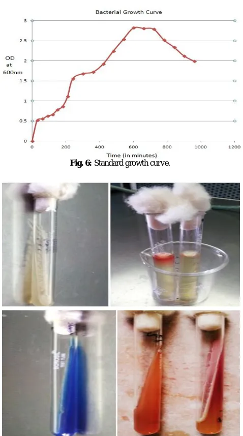
Nutrient broth was prepared in two Erlenmeyer flasks and was then subjected to sterilization by autoclaving. After the media was cooled to room temperature, one colony from the isolated culture was picked up using a sterile inoculating loop and dropped into one of the flasks. The flask which was not inoculated served as the control as well as the blank for colorimeter. Then it was incubated in a shaker incubator for the next 24 hours. The colorimeter was set to 600 nm and the OD was recorded, first 3 times after every half hour, followed by recording the OD after every one hour for the next 24 hours. Then the OD values were plot against the time (in hours) to obtain the growth curve for the isolated microorganism.
Biochemical Tests
incubation, the slants were observed for color change. The culture was also streaked on Macconkey agar to check if it was a lactose fermenter or not.
Antibiotic Production
Antibiotic production media was prepared by addition of 3g glucose, 0.6g sodium nitrate, 0.1g potassium dihidro-phosphate, 0.5g of potassium chloride, 0.02g of magnesium sulphate, 0.01g of ferrous sulphate, 5g of peptone and 3g of beef extract in 100 ml of distilled water. This was then sterilized by autoclaving at 121°C for 15-20 minutes. After sterilization, the broth was cooled and inoculated with the isolated colony and then kept in a shaker incubator for 24 hours. The spectrophotometric method was used to obtain the optical density of the inoculated broth. The optical density of the mixture, taken in a 1:64 dilution with distilled water, was measured every day for seven days using a UV-visible spectrophotometer at 280 nm wavelength.
RESULTS AND DISCUSSION
The soil was collected from the lake soil of VIT University. Vellore has a semi-arid climate with high temperatures and little rainfall. The soil sample that was used was moist, loamy
soil near the banks of the lake (Vuppu et al., 2012).
The colonies that were observed in the crowded plate were counted using a colony counter and the morphology of the colonies was observed. The clear zones were observed in the crowded plate, and the colony near one of the clear zones was selected and then sub-cultured onto a fresh agar medium by the streaking method. The streak-plate was incubated for 24 hours at 37°C. The next day, the streaks of the selected culture were obtained, and the antibiotic sensitivity test was performed. The further characterization methods like staining and biochemical tests were performed.
Fig. 1: Crowded plate method.
The zones of inhibition were observed when the antibiotic sensitivity test was performed (Fig 3). The isolated microorganism successfully grew on a media swabbed with a test
microbe Escherichia coli and inhibited the latter’s growth. When
directly streaked on a Mueller-Hinton agar plate swabbed with E.
coli, a distinct clear zone could be seen in the area that was
streaked, hence confirming the antibiotic producing nature of the isolated microorganism (Fig 4).
Fig. 2: Sub-culture of colony by streaking.
Fig. 3: Antibiotic sensitivity test.
Fig. 4: Clear zone on lawn of E. coli.
Fig. 5: Gram staining; Gram negative rods.
Fig. 6: Standard growth curve.
Fig. 7: Biochemical tests: Indole, Methyl red, Voges-Proskauer, Citrate Utilization and TSI tests.
The various biochemical tests (Fig 7) that were performed gave the following results – Indole (-), MR (+), VP (-), and Citrate Utilization (+). There was no color change after the Kovac’s reagent was added for the Indole test. Upon addition of Methyl red, a red ring was formed in the test tube, and the test was
negative for VP as no color change was observed. Simon’s citrate agar slants turned from green to blue upon incubation with the isolated bacteria, and hence it was positive for citrate utilization. TSI agar slants showed a red slant/yellow bottom and there was no growth on Macconkey agar, indicating that the microorganism was not a lactose fermenter. The results of the biochemical tests were cross-checked with the Bergey’s Manual of Systematic
Bacteriology and the probable identification of the isolated
bacteria was Proteus mirabilis. The antibiotic production media
was made and the microorganism was inoculated, and the optical density of the medium was measured every day for seven days using a UV-vis spectrophotometer. The values were plot against the time (in hours) and the antibiotic production curve was plotted (Fig8). It was observed that there was high production of
secondary metabolites on the 6th day, indicating that this stage was
the antibiotic producing stage of the bacteria’s life cycle.
Fig. 8: Graph of antibiotic production plotted against time.
CONCLUSION
New antibiotics extracted from potential microorganisms are the key to fighting the multi-drug resistant bacteria that are
emerging as of today’s scenario (Bizuye et al., 2013). There are
already many existing Actinomycetes derived antibiotics available
(Gebreyohannes et al., 2013), and hence new soil microbiota that
has the ability to produce antibiotics should be screened and
isolated. The soil environment harbors a variety of
microorganisms, and they need to me explored to find such
potential novel antibiotic producers (D’Costa et al., 2007).
ACKNOWLEDGMENTS
We would like to thank our beloved and honorable Chancellor, Dr. G. Viswanathan for giving us this platform to encourage research among us students, by establishing this institution and for giving us this opportunity to do this project, for the constant support and encouragement, and for providing us the basic knowledge of techniques that we performed in this project. Also, we would like to thank all the lab assistants and attenders, especially Mr. R. Velu and Mr. V.M. Karthikeyan, who helped us with the materials, chemicals and instruments required to complete this project. Lastly, we would like to thank our parents for their constant moral support.
REFERENCES
Abebe B,Feleke M, Berhanu A, Isolation and screening of antibiotic producing actinomycetes from soils in Gondar town, North West Ethiopia, Asian Pacific Journal of Tropical Disease, 2013; 3(5):375–381
Chari N, Shunmugiah KP, Isolation of heterotrophic bacteria from Palk Bay sediments showing heavy metal tolerance and antibiotic production, Microbiological Research, 2010; 165:578—593.
D’Costa VM, Griffiths E, Wright GD, Expanding the soil antibiotic resistome: exploring environmental diversity, Current Opinion in Microbiology, 2007; 10(5):481–489
D. Thakur , A. Yadav, B.K. Gogoi, T.C. Bora, Isolation and screening of Streptomyces in soil of protected forest areas from the states of Assam and Tripura, India, for antimicrobial metabolites, Journal de Mycologie Médicale, 2007; 17:242—249
Eddie C, The soil resistome: The anthropogenic, the native, and the unknown, Soil Biology & Biochemistry, 2013; 63:18-23.
Gebreselema G, Feleke M, Samuel S, Naggapan R, Isolation and characterization of potential antibiotic producing actinomycetes from water and sediments of Lake Tana, Ethiopia, Asian Pac J Trop Biomed, 2013; 3(6):426-435.
Musliu A, Salawudeen W, Screening and Isolation of the Soil Bacteria for Ability to Produce Antibiotics, European Journal of Applied Sciences, 2012; 4(5):211-215.
Suneetha V, Bishwambhar M and Ching L, A Short Period
Study On Vellore Enriched Soils-A Source For Potential Microbial Enzymes Which Degrades The Waste Generated From Various
Industries And Byproducts Can Be Used In Pharmaceuticals, International Journal of Pharmaceutical Sciences and Research, 2012; 3(11):4242-4246.
How to cite this article: