Introduction
The main objective of root canal treatment is thorough mechanical and chemical cleaning of all the canals before obturation with an inert filling material.[1-3] Endodontic failure on a long term may occur due to improper cleaning and shaping of canals, lack of understanding of root canal morphology, failure of establishment of hermetic seal, underfilling, overfilling, and also due to improper identification of canals.
This paper highlights on radix entomolaris (RE) and radix paramolaris (RP), which are developmental variation occurring in mandibular molars which are associated with an extra root and so the extra canal. An extra canal may be present even without an extra root. The presence of an extra root on the distolingual side of mandibular molar is known as RE[4] but not all the teeth with extra roots. The other variant of radix is RP which indicates the presence of extra root on the mesiobuccal side.[5]
ABSTRACT
Endodontic treatment's successrelies on the proper identification of all the canals, complete chemomechanical preparation along with three-dimensional obturation with hermetic seal. Unusual tooth morphology may result in the mentioned steps' failure. Having two roots along with three canals (mesiobuccal, mesiolingual, and distal) are common features of mandibular molars, which might vary only in a few teet. The mentioned variation with regard to the number of roots is known as radix. This article presents case series of mandibular molar with an extra root. Moreover, the modifications concerning the canal preparation, problems encountered during the treatment, common iatrogenic errors which occur during the treatment, and factors which affect the prognosis, are provided.
Keywords: Radix entomolaris, radix paramolaris, variations, extra roots, extra canals
Management of radix endomolaris and paramolaris in
mandibular molars: A case series
S. G. Khirtika, Sindhu Ramesh
Department of Conservative Dentistry and Endodontics, Saveetha Dental College, Saveetha University, Chennai, Tamil Nadu, India
Correspondence: Sindhu Ramesh, Department of Conservative Dentistry and Endodontics, Saveetha Dental College, Saveetha University, 162, Poonamallee High Road, Chennai, Tamil Nadu - 600077, India. Phone: +91-9840136543. E-mail: [email protected]
Case Report 1
A chief complaint of decayed tooth in the right lower back tooth region for 3 months was reported to the department of conservative dentistry and endodonticby a 23-year-old male patient. A diagnostic radiograph was taken which showed caries involving pulp. On keen observation, there appears to be an additional root. Another radiograph has been taken with
same-lingual, opposite-buccal (SLOB) technique which suggested of RE (Figure 1).
Local anesthesia was administered and the tooth was isolated under rubber dam. Access preparation was done with an endo access bur no.1 (Dentsply, Switzerland). The first distal canal has been found slightly away from the center (buccally), and indicating that the other canal will be on the lingual side. As such modification of the access cavity preparation was done transfering from a triangular shape to a trapezoidal form and the fourth canal was located. The root canals were located with DG-16 endodontic explorer and patency of canals was made with #10 K-file (Mani, Japan). Working length was determined using electronic apex locator.Cleaning and shaping were done with rotary files in a step-down manner. Ethylenediaminetetraacetic acid (EDTA) was used as a lubricant and sodium hypochlorite and normal saline were the irrigants used. Obturation was performed with cold lateral condensation (Figure
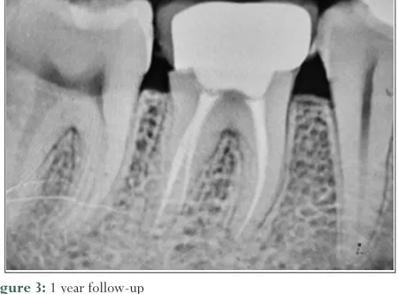
2). Access cavity was restored with composite resin and a postobturation radiograph was taken. Follow-up was done up to 1 year and the radiographs showed no evidence of pathology (Figure 3).
How to cite this article: Khirtika SG, Ramesh S. Management of radix endomolaris and paramolaris in mandibular molars: A case series.J Adv Pharm Edu Res 2017;7(3):358-362. Source of Support: Nil, Conflict of Interest: None declared.
Access this article online
Website: www.japer.in
This is an open access journal, and articles are distributed under the terms of the Creative Commons Attribution-NonCommercial-ShareAlike 4.0 License, which allows others to remix, tweak, and build upon the work non-commercially, as long as appropriate credit is given and the new creations are licensed under the identical terms.
Case Report 2
A chief complaint of decayed tooth in the right lower back tooth region past 6 months was reported by a 20-year-old male patient to the department of conservative dentistry and endodontics. A diagnostic radiograph was taken which revealed radiolucency involving pulp and periradicular aspect of 46 and on observation, there appeared to be an additional root on the mesiobuccal aspect of 46. Another radiograph has been taken with SLOB technique which suggested of RP (Figure 4).
Local anesthesia was administered and the tooth was isolated under rubber dam. Access preparation was done with an endo access bur no.1 (Dentsply, Switzerland).The first distal canal has been found slightly away from the center (buccally), and indicating that the other canal will be on the lingual side. An additional canal was presented middistal between distobuccal and distolingual canal. Modification of the access cavity preparation was done transfering from a triangular shape to a trapezoidal form and the fourth canal was located. The root canals were located with DG-16 endodontic explorer and patency of canals was made with #10 K-file (Mani, Japan). Working length was determined using electronic apex locator.
Cleaning and shaping were done with rotary M-two rotary files in a step-down manner. EDTA was used as a lubricant and odium hypochlorite and normal saline were used as the irrigants. Obturation was performed with cold lateral condensation (Figure 5). Metal Endocrown was luted with glass ionomer cement (Figure 6) and follow up was done up to 6 months (Figure 7).
Figure 1: Pre-operative
Figure 2: Obturation was done by cold lateral condensation technique
Figure 3: 1 year follow-up
Figure 4: Pre-operative
Case Report 3
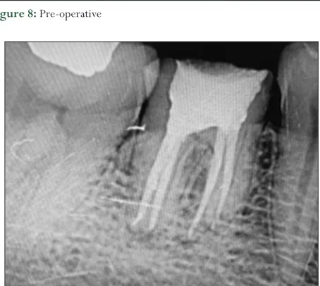
A chief complaint of pain in the right lower back tooth region of the mouth for past 2 weeks was reported by a 25-year-old female patient to the department of conservative dentistry and endodontics with. The pain was continuous, localized and aggravated on chewing, and relieved on medication. Patient gave a history of root canal treatment done 6 years back. On clinical examination, there was presence of temporary filling in 46 with tenderness on palpation and percussion. A diagnostic radiograph was taken which revealed radiopaque filling like material in the root canals and radiolucency involving periradicular aspect of 46. On clinical observation, there appeared to be an additional root on the mesiobuccal aspect of 46. Another radiograph has been taken with SLOB technique which suggested of RP (Figure 8). Local anaesthesia was administered and the tooth was isolated under rubber dam.
Access preparation was done with an endo access bur no.1 (Dentsply, Switzerland).The first mesial canal was found on the buccal aspect, indicating that the other canal should be on the lingual side. Modification was done on the access cavity preparation transfering from a triangular shape to a trapezoidal form and the fourth canal was located. The root canals were located with DG-16 endodontic explorer (Dispodent) and patency of canals was established with #10 K-file (Mani, Japan).
Using electronic apex locator, the working length was determined (Propex Pixi, Dentsply Maillefer). Cleaning and shaping were done with rotary M-two rotary files in a step-down manner. EDTA was used as a lubricant and sodium hypochlorite and normal saline were the irrigants used. The performance of obturation was carried out with cold lateral condensation (Figure 9). Ceramic Endocrown was luted with RelyX Arc resin cement (Figure 10) and follow up was done up to 5 months (Figure 11).
Discussion
Presence of RE in the first mandibular molar is associated with various ethnic groups. Among African population, 3% prevalence is found,[6] while Indians and Eurasians, there is a prevalence of < 5%.[7] An increased prevalence of 5–30% is seen among the Mongoloid population (Chinese, Eskimo, and American Indians).[7-13] Due to its significantly increased prevalence among their population, it is considered a normal variant (eumorphic root morphology). RE is not very common among Caucasians with a frequency of about 3.4–4.2%[14,15] and is thereby considered unusual (dysmorphic root morphology).
Figure 6: Metal endocrown cemented in 46
Figure 7: 6 Months follow-up
Figure 8: Pre-operative
RE can be found in the first, second, and third mandibular molars, with a least frequency among the second molar.[14] Studies have reported bilateral occurrence of 50–67%.[11,16,17]
RP has been reported to be very rare and is said to occur less frequently than RE. RP is observed in about 0% in the first mandibular molar, 0.5% in the second, and 2% in the third molar.[17,18] However, other studies have reported RP to be prevalent in the first mandibular molars.[6,19]
The exact cause of RE is still not known. Authors say that it may be due to disturbance during odontogenesis or may be due to an atavistic gene.[1] To achieve a correct diagnosis, a minimum of two diagnostic radiographs are necessary using buccal object rule (30°).[1] Even the presence of an extra cusp may sometimes indicate be associated with additional root and canals.[6,20,21]
Access cavity preparation should be modified usually from a triangular to a trapezoidal shape. The modification should be done following the dentinal map. Advanced diagnostic aids help in the better identification and visualization of all the canals.[1]
A classification was given by Carlsen and Andersen based on the location of the cervical part of the root division. They are Types A,
B, C, and AC. Type A and B refers to a distally located cervical part, Type C refers to a mesially located cervical part, and Type AC refers to the location of the cervical part in the central location in between the mesial and distal components.[19] DeMoor et al. had given other classification based on the curvature RE variants in the buccolingual direction. They are Type I refers to straight root/canals, Type II refers to a curvature at the entrance of the orifice, and Type III refers to RE with two curvatures; one at the coronal level and the other at the middle third.[22]
Some of the common problems encountered during the treatment of RE and RP are (1) difficulty in radiographic interprétation, (2) inability to locate the fourth canal, (3) modification in access cavity preparation, and (4) confusion in working length determination. Apart from these difficulties, clinicians are prone to commit some iatrogenic errors such as straightening of a root canal resulting in loss of working length, ledge formation, zipping, transportation, or even perforation.
Conclusion
Teeth are not alike. Variations can occur, which tend to pose as a challenge to clinicians. These variations, RE and RP may be a challenge to those who do not have proper diagnostic aids and lack in proper knowledge of root canal anatomy. Correct diagnosis should be made with two pre-operative radiographs taken at two different angles before commencement of the treatment. Hence, thorough knowledge of root canal anatomy and awareness of the variations make the treatment more successful and if one exhibits proper skill, these variations can be handled with ease.
References
1. Calberson FL, De Moor RJ, Deroose CA. The radix entomolaris and paramolaris: Clinical approach in endodontics. J Endod 2007;33:58-63. 2. de Deus GA, Martins F, Lima AC, Gurgel-Filho ED, Maniglia CF,
Coutinho-Filho T. Analysis of the film thickness of a root canal sealer following three obturation techniques. Pesqui Odontol Bras 2003;17:119-25.
3. Ricucci D, Siqueira JF Jr. Anatomic and microbiologic challenges to achieving success with endodontic treatment: A case report. J Endod 2008;34:1249-54.
4. Bolk L. Comments on root variations on the human lower molar. Z Morphol
Anthropol 1915; 17: 605-10.
5. Carlsen O, Alexandersen V. Radix paramolaris in permanent mandibular molars: Identification and morphology. Scand J Dent Res 1991;99:189-95. 6. Sherbet GH, Moreau JL. Study of the number of roots and canals in Senegalese
first permanent mandibular molars. Int Endod J 1998;31:112-6.
7. Tratman EK. Three-rooted lower molars in man and their racial distribution. Br Dent J 1938;64:264-74.
8. Pederson PO. The East Greenland eskimo dentition numerical variations and anatomy. A Contribution to Comparative Ethnic Odontography. Vol. 104. Copenhagen: Meddeleser Om Gronland; 1949. p. 140-4.
9. Turner CG nd. Three-rooted mandibular first permanent molars and the question of American Indian origins. Am J Phys Anthropol 1971;34:229-41. 10. Curzon ME, Curzon JA. Three-rooted mandibular molars in the Keewatin
Eskimo. J Can Dent Assoc (Tor) 1971;37:71-2.
11. Yew SC, Chan K. A retrospective study of endodontically treated mandibular first molars in a Chinese population. J Endod 1993;19:471-3.
12. Reichart PA, Metah D. Three-rooted permanent mandibular first molars in the Thai. Community Dent Oral Epidemiol 1981;9:191-2.
Figure 10: Ceramic endocrown cemented in 46
13. Walker RT, Quackenbush LE. Three-rooted lower first permanent molars in Hong Kong Chinese. Br Dent J 1985;159:298-9.
14. Curzon ME. Three-rooted mandibular permanent molars in English Caucasians. J Dent Res 1973;52:181.
15. Ferraz JA, Pécora JD. Three-rooted mandibular molars in patients of Mongolian, Caucasian and Negro origin. Braz Dent J 1993;3:113-7. 16. Visser JB. Beitrag zur Kenntnis der Menschlichen Zahnwurzelformen.
Hilversum: Rotting; 1948. p. 49-72.
17. Steelman R. Incidence of an accessory distal root on mandibular first permanent molars in Hispanic children. ASDC J Dent Child 1986;53:122-3.
18. Bolk L. Welcher Gebi reihe gehören die Molarenaz. Z Morphol Anthropol 1914;17:83-116.
19. Carlsen O, Alexandersen V. Radix entomolaris: Identification and morphology. Scand J Dent Res 1990;98:363-73.
20. Carlsen O, Alexandersen V. Radix paramolaris and radix distomolaris in Danish permanent maxillary molars. Acta Odontol Scand 1999;57:283-9.
21. Brabant H, Klees L, Werelds RJ. Anomalies, Mutilations et Tumeurs des Dents Humaines. Paris. France: Editions Julien Prelat; 1958.