[Epub ahead of print]
Resumen: El estudio tuvo como objetivo presentar una perspectiva de la razón esencial por la cual se desarrolla microfiltraciones independientemente del tipo de resina compuesta, el sistema de unión aplicado, especialmente en el piso gingival de cavidades de clase II. Materiales y Métodos: Se utilizaron tres tipos de resina compuesta (CharmfilTM, ParaFillTM
y ProMedica®) para evaluar la microfiltración de restauraciones de clase II utilizando dos enfoques restaurativos. Veinticuatro dientes bicúspides recién extraídos se dividieron en dos grupos (n=12 cada uno) de acuerdo con la técnica de restauración (técnicas de sándwich abierto o cerrado). Los dientes de cada grupo se dividieron en 3 sub-grupos (n=4 cada uno) de acuerdo con el tipo de resina compuesta utilizada. Las restauraciones se sometieron después a un proceso de termociclado y finalmente se sumergieron en una solución de azul de metileno durante 12 horas. Se realizaron cortes en la sección mesiodistal a lo largo de los surcos centrales y se evaluaron bajo lupa para determinar la ocurrencia de microfiltración marginal. Los datos se analizaron estadísticamente con un valor de p<0,05 considerado significativo. Resultados: No hubo diferencias estadísticamente significativas en la microfiltración marginal entre los tres grupos examinados utilizando ambas técnicas (p>0.05). Bajo la lupa la microfiltración marginal fue más obvia en la región cervical que en la región oclusal. Conclusión: No hubo efecto del tipo compuesto o la técnica de aplicación utilizada en la aparición de microfiltración marginal. La primera porción del material aplicado el piso de la cavidad es el factor principal involucrado en la posibilidad de minimizar la microfiltración marginal.
Palabras Clave: Restauración dental, materiales dentales, resinas compuestas, micro-filtración.
Abstract: The study aimed to introduce a perspective of the essential reason behind why marginal microleakage develops regardless of the composite type, the technique, or the bonding system applied, especially in gingival floor of class II cavities. Materials and Methods: Three types of composite resin materials (CharmfilTM, ParaFillTM, and ProMedica®)
were used to evaluate microleakage of class II restorations using two restorative approaches. Twenty four newly extracted bicuspid teeth were divided into two main groups (n=12 each) according to the restoration technique (open or closed sandwich techniques). Teeth of each group were then divided into 3 groups (n= 4 each) according to the type of the composite resin used. The restorations were then subjected to a thermocycling process and then were immersed into methylene blue solution for 12 hours. Mesiodistal sectional cuts were made along the central grooves and assessed under stereomicroscope for marginal microleakage. The data were statistically analyzed with a p-value <0.05 considered significant. Results: There were no statistically significant differences in marginal microleakage between the three examined groups using both techniques (p>0.05). Under the microscope, the marginal microleakage was more obvious at the cervical region than at the occlusal region.Conclusion: There was no effect of the composite type or the application technique used on the occurrence of marginal microleakage. The first portion of the material applied against the cavity floor is the primary factor involved in possibly minimizing marginal microleakage.
Keywords:Dental leakage; marginal microleakage; composite resins; sandwich technique; dental restoration.
Bassam Alasbahi.1
Una perspectiva de la microfiltración marginal en restauraciones de
resina compuesta de clase II usando diferentes tipos y
técnicas: un estudio in-vitro.
A perspective of marginal microleakage in class II
composite resin restorations using different types
and techniques: an
in-vitro
study.
Affiliations: 1Al-Rowad Dental College, Sana’a, Yemen.
Cite as:
Alasbahi B. A perspective of marginal microleakage in class II composite resin restorations using different types and techniques: an in-vitro study. J Oral Res 2019; 8(1):22-29.
doi:10.17126/joralres.2019.006
Corresponding author: Bassam Alasbahi. Al-Rowad Dental College, Sana’a, Yemen. E-mail: [email protected]
[Epub ahead of print]
INTRODUCTION.
Marginal microleakage has always been the major issue with class II composite restorations. It has become the prime concern of research, and the contributing factors have been reviewed, and clinically and experimentally tested. Several suggestions have been made to reduce shrinkage stress. A new development in low-shrink dental materials has also been discussed.
Some studies claim that marginal microleakage is caused by polymerization shrinkage of composite resins, suggesting that the improvement in the composition of the dental composite could reduce microleakage. Two directions for improvements have been identified: one suggests focusing on the improvement of fillers size and shape to improve marginal microleakage resistance; the other suggests improving the material matrix.
It has been assumed that improvements in filler particles can increase microleakage resistance besides the esthetic demands. Composite resin materials, therefore, have been developed in different filler sizes from macro to micro, then hybrids, and finally the nano-fillers.1-6
Despite this advancement, there is still no improvement in the marginal adaptation observed with the addition of filler particles, particularly with light-cured resins.7
Two composite resins are commonly used to formulate dental composite matrices: Bis-GMA (bisphenol A-glycidyl methacrylate) and urethane dimethacrylate (UDMA). Factors that can influence stress formation in the matrix include volumetric polymerization shrinkage, adherence of the resin composite to the cavity walls, the elastic modulus, the flowability of the resin composite, and the cavity configuration factor (C-factor).8
A new silorane-based composite resin-system based on the reaction between oxirane and siloxane molecules has been introduced.9 The silorane system uses ring-opening
polymerization instead of free-radical polymerization of dimethacrylate monomers.
Ring-opening polymerization (ROP) is a form of chain-growth polymerization: the terminal end of a polymer chain acts as a reactive center where cyclic monomers can react by opening its ring system, thus lengthening the polymer chain. It has been reported that silorane has <1% volumetric shrinkage and its mechanical properties were clinically successful and comparable to
methacrylate-based composite materials, suggesting its use for dental restorations.6,8,10,15
Many dental adhesive systems are being used to decrease unwanted effects of polymerization shrinkage and gaps formation. However, restorative dental materials can be affected by marginal microleakage at different levels especially in adhesion regions with enamel, dentin and cementum.16
Seven generations of bonding systems have been developed during sixteen years, many of which failed, but the fourth-generation that appeared in the early 1990's is still widely used. Most of these systems are based on the
"total-etch" technique or simultaneous etching of enamel and dentin, typically with phosphoric acid.
The difficulties associated with class II restorations lead to the development of techniques so-called open and close sandwich restoration techniques, where glass ionomer cement (GIC) is placed as a layer between the gingival dentin margin and occlusion composite restoration.
It is thought that the sandwich technique is less prone to microleakage than direct application of composite, and can decrease gaps formation along the conjunction line with dentin.17 Some studies have focused on dental
composite application techniques to decrease marginal microleakage, these techniques are:
1) bulk technique,
2) oblique incremental insertion technique, 3) centripetal incremental insertion technique, 4) split horizontal incremental insertion, 5) centripetal buildup and,
6) snowplow technique.18,19
However, none has been able to completely prevent marginal microleakage. The aim of this study was to introduce a perspective of the essential reason behind the occurrence of marginal microleakage regardless of the type of composite used, bonding system applied, or the technique used to restore the cavity.
MATERIALS AND METHODS.
Specimen Preparation
[Epub ahead of print]
salvaged. To be included, all teeth should be intact, permanent, and extracted for orthodontic/periodontal reasons with fully formed roots. The selected teeth were stored in 2.5% sodium hypochlorite for three days.5
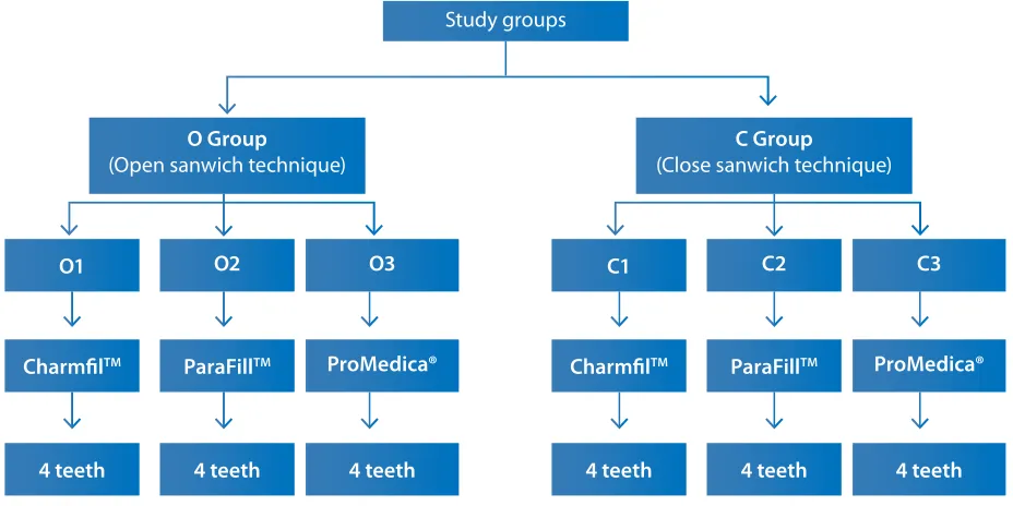
After that, teeth were rinsed and dried to remove any remaining debris. Teeth were randomly divided into two main groups representing the restoration techniques (O group for open sandwich technique and C group for close sandwich technique). Each main group was divided into three sub-groups (4 each) according to the type of composite resin used for restoration. (Figure 1). Three types of composite resins were used: CharmfilTM
(Dentkist, Gunpo, South Korea), ParaFillTM
(Prime-Dental, Chicago, IL), and ProMedica® (Promedica, Neumuenster, Germany).
Class II cavities were prepared to be restored with either open or close sandwich techniques. For open sandwich technique, straight bur No.10 with high speed turbine under water cooling was used. The following dimensions were considered during preparation: the proximal box 4mm in buccolingual direction, 2mm for pulpal depth, and the gingival floor located under the cemento-enamel junction (CEJ) by 1mm.7 Twelve
cavities were prepared with these specifications, four teeth from each sub-group, and restored with open sandwich technique. The GIC was placed over the whole gingival floor up to third or half of the proximal wall. After GIC polymerization, composite layers were added to complete the restoration process and reestablishing the anatomical landmarks of the tooth. In this technique, a multiple increment of GIC was applied in proximal box to cover all the gingival floor up to the CEJ and was left for 10 minutes until primary hardening took place.
Then, acid etching was applied to the cavity using 35% phosphoric acid for 20 seconds, the cavity was air-dried and the bonding agent was rubbed in for 10 seconds, and light cure was applied for 20 seconds. The composite resin was applied as multiple increments with 2mm thickness and each increment was light-cured for 20 seconds. For the close sandwich technique, the same procedures for open sandwich technique were followed except that the gingival floor located above the CEJ by 1mm.7
The manufacturer’s instructions were followed for all materials. The specimens were then exposed to 500 cycles of thermocycling at 5ºC-55ºC (±2ºC) and the dwell time was 15 sec.5,7 After thermocycling finished,
the specimens were dried and the apexes were sealed with sticky wax and the teeth were coated with a double layer of nail varnish with different colors for each group except for 1mm window around the restoration margins. The specimens were immersed into 2% methylene blue dye solution for 12 hours at 37ºC. After that, specimens were dried for 24 hours on a controlled environment.
Microleakage inspection
Sectioning was done along the mesiodistal direction through the central grooves of the occlusal surface of the tested teeth using a double-sided diamond disk .The specimens were inspected under a stereomicroscope with a digital camera, at x20 magnification, using a computer software program (Micam version 2.0). The depth of dye penetration at the cervical and occlusal margins was scored from 0 to 4.18,20 The scoring system
used for marginal microleakage is shown in Table 1. Comparison between the scores as categorical variables was performed by Chi-squared test for proportions. Differences between the means of the scores were analyzed using non-parametric tests. A p-value< 0.05 was considered significant.
RESULTS.
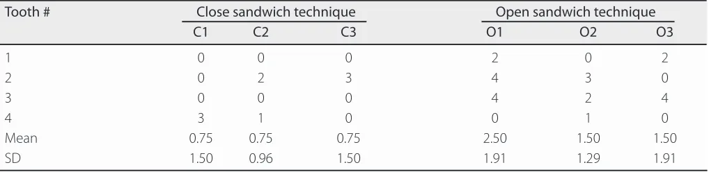
Distribution of the scores among the specimens by means and standard deviations are presented in Table 2. It can be noted that all score means of the close sandwich technique were less than one while all score means of the open sandwich technique were higher than one, with one sub-group (O1) higher than 2.
Frequencies and proportions of the scoring system according to the main groups and sub-groups are presented in Table 3. In the close sandwich technique group most scoring results were 0 (66.7%), followed by score 3 (16.7%), then score 1 and 2 (each at 8.3%) while score 4 was not observed.
[Epub ahead of print]
Table 1. Scoring system used for microleakage inspection.24Figure 2: Section under the microscope showing.
A: The dye penetration between GIC and gingival floor and absence of penetration between GIC and composite. B: The absence of dye penetration between GIC and gingival floor and between GIC and composite.
Score Description
0 No leakage.
1 Less than and up to one-half of the depth of the cavity preparation penetrated by the dye. 2 More than one-half of the depth of the cavity preparation penetrated by the dye but not
up to the junction of the axial and occlusal or gingival wall.
3 Dye penetration up to the junction of the axial and occlusal or gingival wall but not including the axial wall.
4 Dye penetration including the axial wall.
Figure 1: Flow chart of the techniques and types of resins used in the study.
Study groups
C Group
(Close sanwich technique)
O Group
(Open sanwich technique)
CharmfilTM CharmfilTM
O1 C1
4 teeth 4 teeth
ParaFillTM ParaFillTM
O2 C2
4 teeth 4 teeth
ProMedica® ProMedica®
O3 C3
4 teeth 4 teeth
[Epub ahead of print]
Score p-value
0 1 2 3 4
Main group Close sandwich Count 8 1 1 2 0 0.225
% 66.7% 8.3% 8.3% 16.7% 0.0%
Open sandwich Count 4 1 3 1 3
% 33.3% 8.3% 25.0% 8.3% 25.0%
Total Count 12 2 4 3 3
% 50.0% 8.3% 16.7% 12.5% 12.5%
Sub group C1 Count 3 0 0 1 0 0.587
% 75.0% 0.0% 0.0% 25.0% 0.0%
C2 Count 2 1 1 0 0
% 50.0% 25.0% 25.0% 0.0% 0.0%
C3 Count 3 0 0 1 0
% 75.0% 0.0% 0.0% 25.0% 0.0%
O1 Count 1 0 1 0 2
% 25.0% 0.0% 25.0% 0.0% 50.0%
O2 Count 1 1 1 1 0
% 25.0% 25.0% 25.0% 25.0% 0.0%
O3 Count 2 0 1 0 1
% 50.0% 0.0% 25.0% 0.0% 25.0%
Total Count 12 2 4 3 3
% 50.0% 8.3% 16.7% 12.5% 12.5%
n Mean Rank p-value
All groups C1 4 9.88 0.562
C2 4 10.75
C3 4 9.88
O1 4 17.25
O2 4 14.13
O3 4 13.13
Close sandwich C1 4 6.25 0.921
C2 4 7.00
C3 4 6.25
Open sandwich O1 4 7.88 0.627
O2 4 5.88
O3 4 5.75
Tooth # Close sandwich technique Open sandwich technique
C1 C2 C3 O1 O2 O3
1 0 0 0 2 0 2
2 0 2 3 4 3 0
3 0 0 0 4 2 4
4 3 1 0 0 1 0
Mean 0.75 0.75 0.75 2.50 1.50 1.50
SD 1.50 0.96 1.50 1.91 1.29 1.91
Table 2: Microfiltration scores, means, and SDs for the specimens.
Table 3: Frequency of the scores according to the application technique and types of composite resins.
[Epub ahead of print]
n Mean Rank Sum of Ranks p-value
Main group Close sandwich 12 10.17 122.00 0.083
Open sandwich 12 14.83 178.00
Sub group C1 4 4.25 17.00 0.741
C2 4 4.75 19.00
C1 4 4.50 18.00 1.000
C3 4 4.50 18.00
C2 4 4.75 19.00 0.741
C3 4 4.25 17.00
O1 4 5.25 21.00 0.378
O2 4 3.75 15.00
O1 4 5.13 20.50 0.445
O3 4 3.88 15.50
O2 4 4.63 18.50 0.882
O3 4 4.38 17.50
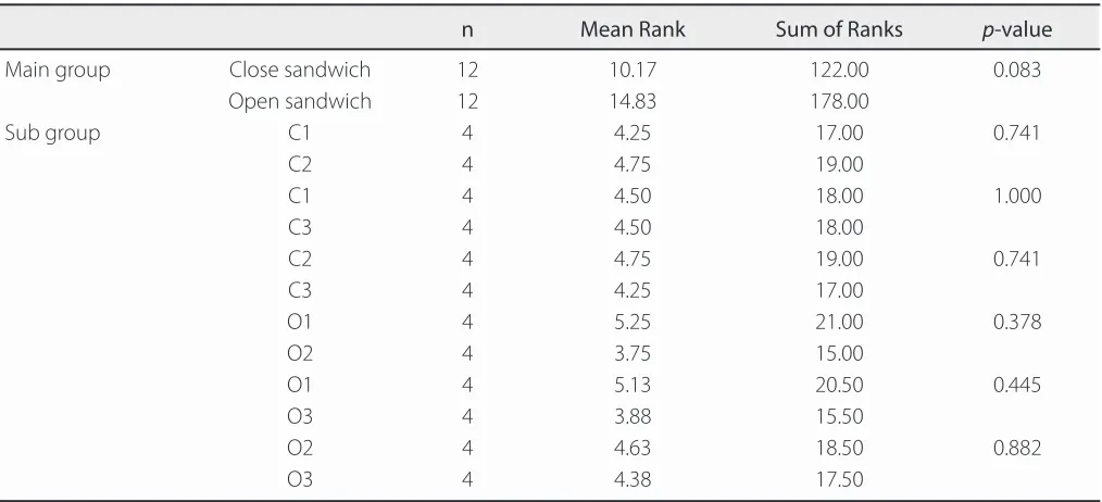
Table 5: Comparison of scores between main groups and between sub-groups of the application technique.
Chi-squared test for proportions in the main groups revealed no significant difference (p=0.225). Frequency according to sub-groups showed that score 4 was only recorded in open sandwich technique sub-groups particularly in sub-group O1 (2 cases) and sub-group O3 (1 case). Most frequent score for sub-groups was score 0 for C1 and C2 (3 cases each) while, scores 1 and 2 were not recorded for sub-groups C1 and C3. Similarly, scores 1 and 3 were not recorded for sub-groups O1 and O3. However, no statistically significant difference was found between sub-groups (p=0.587). Kruskal-Wallis for differences between means among all sub-groups revealed no significant differences with
p-value=0.562. Similarly, differences between means among sub-groups within each main group showed no significant differences p=0.921. (Table 4)
Mann-Whitney test for differences between means of two groups also revealed no significant differences (p> 0.05) for the differences between the two main groups (close and open sandwich techniques) and between each two sub-groups within each main group. (Table 5)
Microleakage inspection
The marginal microleakage could be seen under the microscope in the gingival floor and almost disappeared in junction area between GIC and composite as shown in Figure 2A. Absence of dye penetration in the gingival floor is shons in Figure 2B.
DISCUSSION.
It has been shown that the total-etch system significantly reduces microleakage compared with self-etch and three-step systems while self-etch-and-rinse adhesives remain the gold standard in terms of durability.6,21 The
present study revealed no significant differences between all specimens regarding marginal microleakage within the same technique using different composite resin materials, or between the two techniques. The result of no difference found between the different types of resins is in agreement with the study of Lotfi et al.,10 while,
the result of no difference between both techniques is consistent with the result of Sarfi et al.,2 in which the
difference between the oblique group and the vertical group was found to be statistically non-significant. In contrast, other studies5,6,16,22 found significant differences
between different types of composite resins. Also, some others13,18,19 found significant differences between the
two application techniques.
Marginal microleakage according to the results of the current study may be related to the internal stress that happens because of polymerization in light-cured composites resulting in adhesive failure. Moreover, the degradation in restoration adhesion can cause gap formation. Many previous studies11,17,23 have suggested
[Epub ahead of print]
hardening rate. Other factors such as the resin composite strength and its flow rate may improve the ability to decrease the internal stress even after the polymerization of composite. The volumetric shrinkage of the composite can cause the formation of negative pressure leading to internal deformation of the composite adherent to the cavity walls, thus increasing the possibility of marginal microleakage.18
Incremental application of the filling as small increments to fill the cavity leads to develop less stress in the composite on a large area of cavity walls, which is why this application technique is preferable.6,7,15 The
majority of microleakage studies reported greater dye tracer penetration in marginal dentin sites, as compared with those located in enamel. In this weak area, the open sandwich technique probably allows for a better seal with flowable resin composite, as minimal stress was created at the cervical margin.
In the case of cementum marginal microleakage in the open sandwich technique, as shown in the microscopic picture, a good marginal adaptation (condensation) of GIC is necessary to create a good penetration of the material inside the dentin tubules to achieve a successful long terms restoration.22
The evidence that the condensation of GIC plays the main role of achieving better adhesion to dentin tubules and decrease marginal microleakage was the presence of marginal microleakage in gingival floor and almost disappears in the junction area between GIC. Moreover, the low viscosity of GIC makes its soft and sticky and increases the possibility for slump and sticking to the application tool coming away from the dental structures at the time of GIC application on the gingival floor, and
thus it is difficult to condense.
On the other hand, when the composite is applied over the hard GIC restoration it is easier to achieve a good penetration of the bonding agent through the GIC base, allowing a good condensation of the composite layer resulting in a decrease in microleakage along this area.
Therefore, the viscosity of the first layer and the tendency to move away from dental tissues during application are the main causes of microleakage where the filling is not adhering from the beginning, unlike those that adhere from initially and then disintegrate from the dental tissues due to other factors. Although the current study has revealed evidence of no effect of the composite types and techniques used, it has some potential limitations. The small sample size warrants further studies with larger sample sizes to confirm these results.
CONCLUSION.
There is no statistically significant difference in marginal microleakage regardless of the composite type used or the application technique used to restore the cavity. The first increment of the material applied against the cavity floor is the primary factor to minimize the possibilities of marginal microleakage.
Conflict of interests: The author declares no conflict of
interest.
Ethics approval:None.
Authors’ contributions: The author contributed to the
manuscript.
Funding: Self-financed
Acknowledgements: None.
REFERENCES.
1. Elbishari H, Satterthwaite J, Silikas N. Effect of filler size and temperature on packing stress and viscosity of resin-composites. Int J Mol Sci. 2011;12(8):5330–8.
2. Sarfi S, Bali D, Grewal MS. Effect of different layering techniques on microleakage of nanofilled composite in class i restorations: An In Vitro study. JICDRO. 2017;9(1):8–11.
3. Velagapudi NJ, Reddy ER, Aduri R, Prasad MG, Sahana
S, Vaila A. Comparative Evaluation of Marginal Integrity and Microleakage in Nanoionomer and Low Shrinkage Posterior Composite Restorative Materials: An In Vitro Study. J Int Oral Health. 2016;8(2):261–6.
4. Ansari ZJ, Khalili H, Tork MAK, Siavashani MA.
Micro-shear Bond Strength of a Nanofiller Bonding agent with and
without Thermocycling in a Newly Invented Device. J Islam Dent Assoc Iran. 2013;25(3):222–7.
5. Jain A, Deepti D, Tavane PN, Singh A, Gupta P, Gupta
A. Evaluation of Microleakage of Recent Nano-hybrid Composites in Class V Restorations: An In Vitro Study. Int J Adv Health Sci. 2015;2(1):8–12.
6. Sarfi S, Sharma N, Garg ER, Bali D. Comparing
microleakage in Silorane based composite and nanofilled composite using different layering techniques in class I restorations : An in vitro study. Int Arch Integr Med. 2017;4(7):23–32.
[Epub ahead of print]
resin restorations. J Esthet Restor Dent. 2017;2(1):1–7.8. Moosavi H, Maleknejad F, Forghani M, Afshari E.
Evaluating Resin-Dentin Bond by Microtensile Bond Strength Test: Effects of Various Resin Composites and Placement Techniques. Open Dent J. 2015;9:409–13.
9. Spiller MS, Wright M, The Academy of Dental Learning
and OSHA Training. Dental Composites: A Comprehensive Review. 1st Ed. California: ADA CERP; 2017.
10. Lotfi N, Esmaeili B, Ahmadizenouz G, Bijani A, Khadem
H. Gingival microleakage in class II composite restorations using different flowable composites as liner: an in vitro evaluation. Caspian J Dent Res. 2015;4(1):10–6.
11. Fabianelli A, Sgarra A, Goracci C, Cantoro A, Pollington S, Ferrari M. Microleakage in class II restorations: open vs closed centripetal build-up technique. Oper Dent. 2010;35(3):308–13.
12. Pomohaci DD, Radu TM, Teodorovici P, Tanculescu O,
Andrian S. Preventing marginal microleakage in class II restoration using bioadhesive materials. J Romanian Med Dent. 2009;13(3):63–8.
13. Patel P, Shah M, Agrawal N, Desai P, Tailor K, Patel K. Comparative Evaluation of Microleakage of Class II Cavities Restored with Different Bulk Fill Composite Restorative Systems: An In Vitro Study. J Res Adv Dent. 2016;5(2):52–6.
14. Stansbury J, Bowman C. The Progress in Development of
Dental Restorative Materials. Material Matters. 2010;5(3):73.
15. Soares CJ, Faria-E-Silva AL, Rodrigues MP, Vilela
ABF, Pfeifer CS, Tantbirojn D, Versluis A. Polymerization shrinkage stress of composite resins and resin cements - What
do we need to know? Braz Oral Res. 2017;31(suppl 1):e62.
16. Somani R, Jaidka S, Arora S. Comparative evaluation of
microleakage of newer generation dentin bonding agents: An in vitro study. Indian J Dent Res. 2016;27(1):86–90.
17. Sidhu SK. Glass-ionomer cement restorative materials: a sticky subject? Aust Dent J. 2011;56(Suppl 1):23–30.
18. Kapoor N, Bahuguna N, Anand S. Influence of composite
insertion technique on gap formation. J Conserv Dent. 2016;19(1):77–81.
19. Roopa R, Anupriya B. Effect of four different placement techniques on marginal microleakage in class II restorations: An in vitro study. World J Dent. 2011;2:111–6.
20. Bona AD, Pinzetta C, Rosa V. Effect of acid etching
of glass ionomer cement surface on the microleakage of sandwich restorations. Appl Oral Sci. 2007;15(3):230–4.
21. Parolia A, Adhauliya n, de Moraes Porto ic, Mala k.
A comparative evaluation of microleakage around class V cavities restored with different tooth colored restorative materials. Oral Health Dent Manag. 2014;13(1):120–6.
22. Jia S, Chen D, Wang D, Bao X, Tian X. Comparing
marginal microleakage of three different dental materials in veneer restoration using a stereomicroscope: an in vitro study. BDJ Open. 2017;3:16010.
23. Arora V, Nikhil V, Sawani S, Arora P. The open sandwich technique with glass ionomer cement–a critical evaluation. Int J Innov Res Sci Eng Technol. 2013;2(8):3874–82.
24. Walsh EL, Hembree JH. Microleakage at the gingival