RESEARCH
Use of chitin and chitosan to produce
new chitooligosaccharides by chitinase
Chit42: enzymatic activity and structural basis
of protein specificity
Peter Elias Kidibule
1, Paloma Santos‑Moriano
2, Elena Jiménez‑Ortega
3, Mercedes Ramírez‑Escudero
3,
M. Carmen Limón
4, Miguel Remacha
1, Francisco José Plou
2, Julia Sanz‑Aparicio
3and María Fernández‑Lobato
1*Abstract
Background: Chitinases are ubiquitous enzymes that have gained a recent biotechnological attention due to their ability to transform biological waste from chitin into valued chito‑oligomers with wide agricultural, industrial or medi‑ cal applications. The biological activity of these molecules is related to their size and acetylation degree. Chitinase Chit42 from Trichoderma harzianum hydrolyses chitin oligomers with a minimal of three N‑acetyl‑d‑glucosamine (Glc‑ NAc) units. Gene chit42 was previously characterized, and according to its sequence, the encoded protein included in the structural Glycoside Hydrolase family GH18.
Results: Chit42 was expressed in Pichia pastoris using fed‑batch fermentation to about 3 g/L. Protein heterologously expressed showed similar biochemical properties to those expressed by the natural producer (42 kDa, optima pH 5.5–6.5 and 30–40 °C). In addition to hydrolyse colloidal chitin, this enzyme released reducing sugars from commercial chitosan of different sizes and acetylation degrees. Chit42 hydrolysed colloidal chitin at least 10‑times more efficiently (defined by the kcat/Km ratio) than any of the assayed chitosan. Production of partially acetylated chitooligosaccharides
was confirmed in reaction mixtures using HPAEC‑PAD chromatography and mass spectrometry. Masses correspond‑ ing to (d‑glucosamine)1–8‑GlcNAc were identified from the hydrolysis of different substrates. Crystals from Chit42 were grown and the 3D structure determined at 1.8 Å resolution, showing the expected folding described for other GH18 chitinases, and a characteristic groove shaped substrate‑binding site, able to accommodate at least six sugar units. Detailed structural analysis allows depicting the features of the Chit42 specificity, and explains the chemical nature of the partially acetylated molecules obtained from analysed substrates.
Conclusions: Chitinase Chit42 was expressed in a heterologous system to levels never before achieved. The enzyme produced small partially acetylated chitooligosaccharides, which have enormous biotechnological potential in medicine and food. Chit42 3D structure was characterized and analysed. Production and understanding of how the enzymes generating bioactive chito‑oligomers work is essential for their biotechnological application, and paves the way for future work to take advantage of chitinolytic activities.
Keywords: Chitinase, Trichoderma harzianum, Chit42 3D structure, Chitooligosaccharides, Partially acetylated chitooligosaccharides
© The Author(s) 2018. This article is distributed under the terms of the Creative Commons Attribution 4.0 International License (http://creat iveco mmons .org/licen ses/by/4.0/), which permits unrestricted use, distribution, and reproduction in any medium, provided you give appropriate credit to the original author(s) and the source, provide a link to the Creative Commons license, and indicate if changes were made. The Creative Commons Public Domain Dedication waiver (http://creat iveco mmons .org/ publi cdoma in/zero/1.0/) applies to the data made available in this article, unless otherwise stated.
Open Access
*Correspondence: [email protected]
1 Department of Molecular Biology, Centre for Molecular Biology Severo Ochoa (CSIC‑UAM), University Autonomous from Madrid, C/ Nicolás Cabrera, 1, Cantoblanco, 28049 Madrid, Spain
Background
Chitin, a linear polymer of β-1-4 linked N -acetyl-β-d-glucosamine (GlcNAc) units, gives strength to the exo-skeletons of insects, crustaceans and fungi cell walls, being the most widespread amino polysaccharide in nature. Deacetylation of chitin produces chitosan, poly-mer containing GlcNAc and d-glucosamine (GlcN) with the latter usually exceeding about 80% of the residues [1]. Chitin and chitosan have been used as functional materi-als in the fields of food, health or agriculture because of their biocompatibility, non-toxicity and availability from abundant and inexpensive biomass. Poor solubility at neutral pH values of both high molecular-weight biopol-ymers limits their potential use [2–4], a problem that could be overcome by using their derived oligomers and monomers. Indeed, the chitooligosaccharides (oligosac-charides derived from chitin or chitosan, COS) biologi-cal activity is well documented. They showed antioxidant, anti-inflammatory, antimicrobial, antiviral, antihyperten-sive, anti-tumoral and/or prebiotic properties [3, 5, 6]. The COS properties are strongly dependent on their size (defined by the degree of polymerization, DP) and charge (related to the degree of deacetylation, DD) [7–9]. How-ever, their use is quite limited due to its non-commercial availability.
COS can be produced by enzymatic conversions using chitinases (or chitosanases), chemical methods or by a combination of both strategies employing chitin or chi-tosan as starting material [10, 11]. Contrary to the chemi-cal hydrolysis that requires extreme reaction conditions of difficult control, the use of enzymes is a more envi-ronmentally friendly process that involves softer, spe-cific and controlled conditions [12]. Chitin lytic enzymes are extensively distributed Glycoside Hydrolases (GH), which cleave randomly at internal or terminal end β-1,4 glycosidic linkages of chitin generating COS, di-acetyl chitobiose ((GlcNAc)2) and/or GlcNAc units [13–16].
The biotechnological demand for these enzymes grows as the industrial-medical applicability of the products they generate increases. Therefore, to achieve efficient protocols for the production of both, chitinases and the products they generate, constitutes a challenge for the bioconversion of chitin waste.
Family GH18 (http://www.cazy.org) represents an ancient chitinase type found in all kingdoms of life, from lower organisms to humans, and possess a characteris-tic catalycharacteris-tic module consisting of a (β/α)8-TIM-barrel
structure [5, 17–19]. All chitinases described in yeast and fungi are included in the family GH18. They are not only involved in exogenous chitin decomposition but also in fungal cell wall degradation and morphogenesis where hydrolytic cleave of chitin is crucial for hyphal growth, septum formation and spore germination [20].
Some strains of the genus Trichoderma are used as powerful biocontrol agent against plant pathogens by the production of a wide variety of lytic enzymes, includ-ing several chitinases. The Trichoderma atroviride P1 strain produces several GH18 chitinases [21, 22]. Among them, the endochitinase Ech42 has been characterized at genomic and protein level and expressed in Escherichia coli [23], Pichia pastoris [24] and other T. harzianum strains [25]. The best heterologous protein level was obtained in P. pastoris, ~ 185 mg/L [24]. The role of some conserved residues in the substrate binding and cataly-sis of this protein has been enlightened using mutational analyses and three-dimensional structural models based on the crystal structure of the chitinase from the patho-genic fungus Coccidioides immitis [26, 27].
The chitinase Chit42 from T. harzianum (orthologous to Ech42) plays an important role in the fungus anti-phytopathogens activity [28–30]. This protein was able to hydrolyse chitin oligomers with a minimal DP of 3 units [28]. Gen chit42 has been previously characterized and encodes a protein of 423 amino acids including a puta-tive exportation signal of 34 residues [31]. Transformants of T. harzianum overexpressing ~ 20 mg/L of chitinase Chit42 had also been obtained [30].
In this work we have expressed the chitinase Chit42 from T. harzianum in P. pastoris to ~ 3 g/L, the best level obtained in a heterologous system for this protein. Enzymatic properties of the heterologous protein and its efficiency to produce COS from different chitinolytic materials were evaluated. In addition, crystallographic analysis of Chit42 has been performed to uncover the molecular basis explaining its observed COS-producing specificity.
Results and discussion
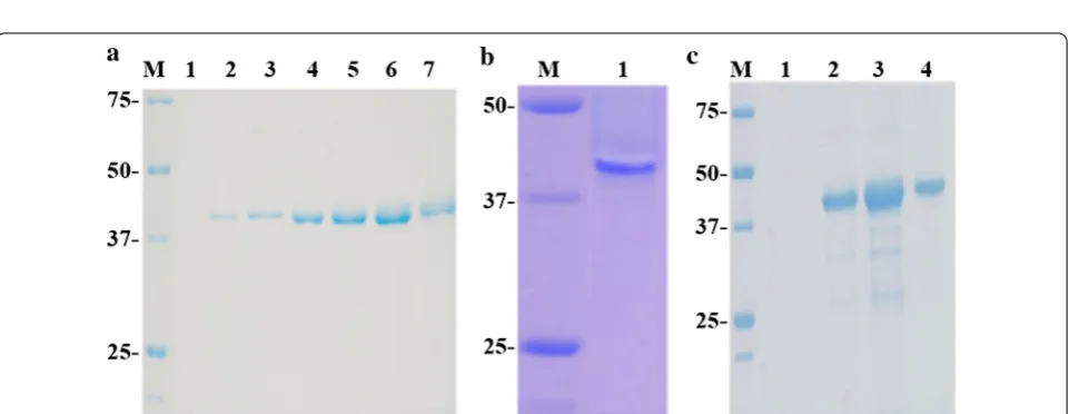
The highest chitinase activity, ~ 150 mU/mL culture, was detected in the extracellular medium of one of these transformants grown during 96 h in a metha-nol based medium (Fig. 1). As expected, this organ-ism secreted into the methanol based medium only a major protein of ~ 42 kDa showing chitinolytic activ-ity (Fig. 2a, b), the same molecular mass than the wild-type protein expressed in T. harzianum [28]. No extracellular protein was previously detected in the culture filtrates of control yeasts transformed with the empty vector pIB4 [32]. An extracellular protein con-centration of 29 μg/mL was quantified at the point of maximum protein expression, representing a specific chitinase activity of ~ 5.2 U/mg. Production of Chit42
was increased by ~ 100-times, to 2.9 mg/mL (15 U/mL; 5.2 U/mg), by growing the recombinant P. pastoris and inducing the protein expression in fed-batch fermenta-tion (Figs. 1b, 2c). As far as we know, this is the highest yield ever reported for the Chit42 from T. harzianum expressed in a heterologous system. Expression of fungi chitinases (orthologous to Chit42) in heterologous sys-tems has been previously analyzed with very different results. Thus, only 3 mg/L of chitinase Enc1 from T. harzianum T25-1 was obtained in S. cerevisiae [33] and less than 200 mg/L of chitinase Ech42 from T. atrovir-ide P1 [24] or about 6.2 g/L of chitinase Tachi1 from T. asperellum [34], both in P. pastoris.
Fig. 1 Activity profiles of cultures expressing Chit42. The P. pastoris transformant was grown in flask (a) and in fed‑batch fermenter (b) supplemented with methanol. OD600 (black circles), pH (empty cycles) and extracellular chitinase activity using colloidal chitin as substrate (blue circles) were measured at the indicated times at 35 °C. Each point of activity represents the average of three independent measurements and standard errors are indicated
Biochemical characteristics of Chit42 expressed in Pichia pastoris
Heterologous enzyme displayed maximum activity (> 85%) on colloidal chitin at pH 5.5–6.5 and 30–40 °C, and retained less than 40% activity at temperatures above 45 °C. In addition, when enzyme was incubated without substrate in the range of 25–60 °C for 10–90 min and then chitinase activity was assayed, protein maintained 100% activity at 25 °C, completely lost it after 10 min at 60 °C and retained 50% activity in the 43–50 °C range (Fig. 3). Within the conditions tested in this work, the enzyme expressed in P. pastoris appeared to be slightly more sensitive to temperature changes than protein pre-viously purified from T. harzianum for which maximum activity values were reported at 40–45 °C, and main-tained 50% of activity after 30 min at 50 °C [28]. How-ever, our data are closer to what was expected because, in general, the optimal temperature for the fungal chitinase activity varies between 20 and 40 °C [18].
Although less efficiently, chitinase Chit42 expressed in P. pastoris was also able to release reducing sugars from commercial chitosan of different sizes and acetylation degrees (Table 1).
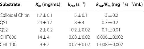
Enzyme kinetics with colloidal chitin and chitosan as substrates were examined (Table 2 and Additional file 1: Figure S1), and similar Km value for colloidal
chi-tin to that previously reported (~ 1 mg/mL) by using the enzyme expressed in T. harzianum [28] was obtained. However, only apparent kinetics parameters were deter-mined using any of the chitosan analysed because its low solubility did not allow a precise estimation of the Vmax,
and therefore of the Km values. Nevertheless, a priori the
enzyme showed a very different apparent catalytic effi-ciency (defined by the kcat/Km ratio) on the tested
sub-strates, and clearly hydrolysed colloidal chitin at least 10-times and 40-times more efficiently than chitosan including a DD in the range of ~ 77–80% and > 90%, respectively (Table 2).
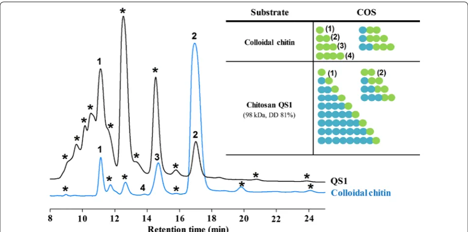
Products of the colloidal chitin and chitosan hydrolysis To evaluate the applicability of Chit42 in the COS produc-tion, the reaction products using both chitin and chitosan as substrate were analysed by a combination of HPAEC-PAD chromatography and mass spectrometry. With the aid of commercial standards, the fully acetylated series of COS (from 1 to 4 GlcNAc units) was identified when using colloidal chitin as substrate, being the disaccharide the most abundant product (Fig. 4 left, blue chromatogram). Curiously, and although the order of elution with PA-200 Fig. 3 Temperature, pH and thermostability dependence profiles. The effect of temperature (a) and pH (b) on the Chit42 chitinase activity was evaluated on colloidal chitin at pH 6 and 35 °C, respectively. c The chitinase was incubated for the indicated temperatures during the referred time periods (in min) prior to the addition of the substrate. Remaining activity was determined at 35 °C as described in the “Methods” section. Results represent the mean of three independent values. Standard errors are indicated
Table 1 Chit42 hydrolytic activity on the analysed substrates
100% activity: 0.2 U/mL
Data are the average of 3 independent experiments. Standard errors were indicated
n.d. not determined
a DD of initial chitin flakes
Substrate MW (kDa) DD (%) Activity (%)
Colloidal Chitin n.d. ≤ 5a 100 ± 6
QS1 98 81 29 ± 2
QS2 31 77 28 ± 2
CHIT600 600–00 > 90 2.3 ± 0.1
CHIT100 100–300 > 90 3.2 ± 0.2
Table 2 Catalytic constants on the analysed substrates
Apparent Km and kcat values were obtained using chitosan as substrate. Values
of kcat were calculated from Vmax considering a Chit42 protein molecular mass
of 42 kDa
Substrate Km (mg/mL) kcat (s−1) kcat/Km (mg−1/s−1/mL)
Colloidal Chitin 1.7 ± 0.1 5 ± 0.1 3 ± 0.2
QS1 24 ± 12 8 ± 4 0.3 ± 0.2
columns usually correlates with the increasing DP, the retention time of COS did not follow such order, probably due to the unusual eluting conditions (4 mM NaOH) and that the most acidic hydroxyl group (the 2-OH of glucose moieties) is substituted by NH2 or N-acetyl. The presence
of the commented acetylated oligosaccharides was con-firmed in the reaction mixture by mass spectrometry assay (Additional file 1: Figure S2 and Table S1). In addition, and most likely because chitin was not initially 100% acety-lated, masses corresponding to partially acetylated COS (paCOS) were also detected in the mixture.
Reactions using chitosan QS1 as substrate (Fig. 4, black chromatogram) yielded numerous signals by HPAEC-PAD, but only two products could be identified: GlcNAc and (GlcNAc)2 (peaks 1 and 2, respectively). The
remain-ing peaks did not elute at any known retention time for either fully acetylated or deacetylated available COS. Most likely they must be due to paCOS, as was suggested by the mass spectrometry assay in which masses corre-sponding to (GlcN)1–3-(GlcNAc)2 and (GlcN)1–8-GlcNAc
were detected (Additional file 1: Figure S2 and Table S2). Similar HPAEC-PAD results were obtained using chi-tosan QS2, CHIT100 and CHIT600 as substrates (Addi-tional file1: Figure S3).
In general, substrate-binding site of fungal chitinases is relatively long and accommodates at least five sugar units. The sugar-binding subsites are denominated as
− 3, − 2, − 1, + 1 and + 2, and the cleavage occurring between the − 1 and + 1 sugar. Chitinases from GH18 are retaining enzymes, which means that the β-anomeric configuration found in the substrate is retained in the product, showing an unusual substrate-assisted catalytic mechanism where the acid protonating the glycosidic bond (to be hydrolysed) is a conserved glutamate residue (included in the DXXDXDE sequence), and the nucleo-phile is the oxygen of the N-acetyl group (of GlcNAc) on the subsite − 1 sugar [20]. As described above, Chitinase Chit42 is able to hydrolyse chitin oligomers with a mini-mal size of 3 GlcNAc units [28] and the catalytic mecha-nism of chitinases included in the family GH18 requires a mandatory GlcNAc residue in the substrate − 1 posi-tion [20]. Thus, based on the enzyme specificity and mass spectrum analyses, it is feasible to think that the highest peak in the HPAEC-PAD chromatogram might well cor-respond to the tri-saccharide, showing the acetylated residue in the reducing end: (GlcN)2-GlcNAc (N-acetyl
chitotriose). Consequently, this will be very probably the main product obtained from any of the used chitosan.
Production of COS from different substrates
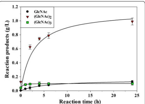
produced 1.2 g/L of fully acetylated molecules of which 0.13 g/L were GlcNAc, 0.99 g/L (GlcNAc)2 and 0.10 g/L
(GlcNAc)3 (Fig. 5). Standard errors for the
quantifica-tion of the acetylated COS were lower than 5%. Only small traces of (GlcNAc)4 were also detected that could
not be quantified (Fig. 4; Additional file 1: Figure S2 and Table S1). Thus, only 15% of the chitin suspension used as substrate was apparently transformed into these small size acetylated COS. Probability that not all the chitin molecules in the initial suspension are in soluble form or even that acetylation degree of colloidal chitin could be different from that of initial chitin flakes, could have contributed to the low COS yield. In fact, the reaction mixtures were quite turbid and all samples precipitated after the NaOH treatment, indicating that they contained polymers of high size that were not hydrolysed.
Concerning the reactions with chitosan as substrate, and because they are mostly deacetylated polymers, a priori, low yields in COS production could be expected. Large zones of substrate lacking GlcNAc residues that the enzyme could not be able to hydrolyse must be taken into account. In addition, as referred, paCOS could not be quantified due to the lack of commercial standards. Thus, only concentration of GlcNAc and (GlcNAc)2 could be
evaluated in reactions based on chitosan, with values of 0.25 and 0.14 g/L in the case of QS1, indicating that ~ 5% of the substrate was transformed into these two products. A peak with the same retention time as (GlcNAc)3 was
detected by HPAEC-PAD but the corresponding mass was not detected by mass spectrometry (Additional file 1: Figure S2 and Table S2). Because COS are very difficult to separate, it is not unusual to find two molecules with
the same retention time. Lack of (GlcNAc)3 in reactions
with chitosan can be explained by its high degree of dea-cetylation, which lowers the probability of finding three consecutive residues of GlcNAc in the chitosan chain. This peak is most likely due to a partially acetylated COS. Also, and in agreement to what was commented above, the major peak in the HPAEC-PAD chromatograms cor-responded most likely to the paCOS N-acetyl chitotriose. As referred before, biotechnological applications of COS include anticancer therapy, immune modulatory effect or antioxidant activity among many other [3, 5,
6]. The biological activity of these molecules is related to their DP and DD. Thus, antioxidant activity of paCOS exceeds those of the non-acetylated [9] and small size COS showed stronger antioxidant activity than the bigger ones [35]. In this context, the industrial market demand for COS with defined characteristics increases stead-ily. The enzymatic synthesis would clearly facilitate the production of homogenous batches of COS with defined properties in comparison to the less specific chitin chem-ical treatment, which requires large amounts of highly polluting chemical compounds such as HCl and NaOH [36]. All this gives an attractive biotechnological poten-tial to the chitinase Chit42 for the production of small paCOS.
Structural analysis of Chit42 specificity
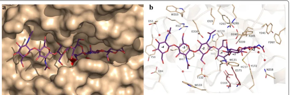
The structure of Chit42 was solved by molecular replace-ment at 1.8 Å resolution, showing the structural features previously described for other GH18 chitinases, i.e., a (β/α)8 TIM barrel fold with an additional α/β domain
inserted within the loop linking helix α8 to strand β9 of the barrel. This extra domain contributes to provide a groove-type shape to the active site (Fig. 6a). We have modeled a chitooligosaccharide within the active site channel by structural superimposition of the reported complex of this substrate with the Serratia marcescens ChiA-D313A mutant [37] onto the coordinates of Chit42 here presented. A detail of the proposed interaction with the oligosaccharide is shown in Fig. 6b. Although ChiA and Chit42 sequences are only 26% similar, the corre-sponding residues located at the active site are rather conserved. Therefore, the distorted conformation of the substrate at the cleavage point observed for ChiA fits well within the Chit42 active site groove. Thus, the complex model illustrates the main features involved in binding. First of all, the substrate is tightly recognized at subsites − 4 to + 2 by a net of hydrogen links to several Chit42 residues. Therefore, there are at least 6 substrate-bind-ing subsites in this protein. In particular, it seems that all the acetyl groups may be involved in hydrogen links with the expected exception of the sugar located at sub-site − 1. In general, each sugar makes two hydrogen links, Fig. 5 Evolution of the COS produced by Chit42 using colloidal chitin
one through the acetyl and the other through the O3/ O6 hydroxyls, but the sugar at subsite − 2 seems able to interact through all its free oxygen atoms.
The flexibility of the different subsites in allocating dea-cetylated COS within a Trichoderma chitinase has been discussed before [26, 27]. Taking into account these stud-ies, it was previously proposed that subsites − 2 to + 2 are more specific for GlcNAc binding, while distal subsites at the non-reducing end can accommodate modified sugars. However, and according to the analysis here reported, masses of the paCOS corresponding to (GlcN)1–3
-(GlcNAc)2 and also to (GlcN)1–8-GlcNAc, have been
identified from the hydrolysis of chitosan QS1 (Addi-tional file 1: Figure S2 and Table S2). Therefore, the acetyl moiety at subsites -2 must not be strictly required. Previ-ous mutagenesis experiments revealed that polar interac-tion of substrate with the Trichoderma chitinase residue Glu316 at subsites − 3 and − 2, is essential in binding [27]. According to our complex model, Glu316 is hydro-gen linked to O3 of the sugar at subsite − 2, but seems also able to link to a free amine group by a slight switch of its side-chain, therefore compensating the loss of the hydrogen bond to Trp378 and contributing to stabilized a deacetylated sugar at this subsite − 2. With respect to the sugar located at subsite − 3, the stacking interaction to Trp47 was previously proved to be critical in substrate binding, therefore making a broad specific subsite able to allocate modified sugars. Thus, a galactose unit, or fucose as a branch, were both accepted at subsite − 3 [26]. According to our model, Arg52 is also involved in allocat-ing the sugar at this subsite; therefore, its flexible chain provides additional plasticity to the sugar type occupying this binding subsite. Consequently, the structural analy-sis sheds light on the Chit42 specificity observed in our
work, and explains the chemical nature of the partially acetylated products obtained from chitosan.
A last interesting feature is the fact that the side-chain of Trp131, making subsite + 1, presents rather weak elec-tron density in our solved free enzyme crystal, revealing a marked disorder. This observation might be indicative of a process mechanism as proposed for the S. marcescens ChiA, where mobility of this aromatic residue may be essential to perform a productive sliding of the substrate. Alternatively, flexibility of this Trp131 might be required to facilitate distortion of the sugar occupying subsite − 1. In agreement with the disorder observed by us, two alternate conformations were previously found for this conserved Trp in the un-complexed crystals grown from the Aspergillus fumigatus ChiB1 [38]. Our work provides useful information on the functionality of Chit42, a chi-tinase of biotechnological interest for the production of pCOS, and could be useful to understand the enzymatic behaviour of other proteins included in the GH18 family.
Conclusion
paves the way for future work to take advantage of Chit42 activity.
Methods Chemicals
Chitin (from shrimp shells, practical grade coarse flakes; DD ≤ 5%), glycol chitosan, N-acetyl-glucosamine (Glc-NAc) and Biotin were from Sigma Aldrich (St. Louis, MO, USA). Colloidal chitin was obtained from chitin by the method of Jeuniaux [39]. Basically, 175 mL of 10 M HCl including 10 g of chitin was maintained 16 h at 4 °C and filtered through glass thick fibers into 1 L of ethanol. Chitin floccules were precipitated after 16 h at 4 °C, col-lected at 5000×g during 10 min and washed with distilled water. Then, 200 mL of 70 mM potassium phosphate pH 6 was added and colloidal chitin concentration was esti-mated by titrating the weight of solute contained in 1 mL of solution that was previously frozen at − 70 °C and lyophilized.
Glycol chitin was obtained from glycol chitosan as pre-viously reported [9]. Basically, 0.2 g of glycol chitosan was suspended in 50 mL methanol 50% (v/v) and 0.3 mL acetic anhydride. Two volumes of acetone were added and sample was centrifuged at 5000×g during 10 min. Precipitate was treated with 1 M sodium hydroxide, dialyzed against water, frozen at − 70 °C, and lyophi-lized. Chitosan CHIT100 and CHIT600 were from Acros Organics (Thermo Fischer Scientific Inc., Waltham, MA). Chitosan QS1 (from Paralomis granulosa) and QS2 (from Pandalus borealis) were from InFiQuS (Madrid, Spain). Chitosans (1 g) were dissolved in 90 mL of 0.1 M acetic acid and then 10 mL of 1 M sodium ace-tate pH 5.5 was added (1% (w/v) chitosan final concen-tration in 100 mM sodium acetate pH 5.5). Chitobiose ((GlcN)2), chitotriose ((GlcN)3), chitotetraose ((GlcN)4)
and N,N′,Nʺ-tri-N-acetyl-glucosamine ((GlcNAc)3) were
from Carbosynth Ltd. (Berkshire, UK). Yeast Nitro-gen base w/o amino acids (YNB) was from Difco (BD, Sparks, MD, USA). All other reagents were of the high-est purity grade.
Strains, growth and expression media
Pichia pastoris GS115 (his4-) (Invitrogen, Carlsbad, CA, USA) was used as expression host and was initially cul-tured at 30 °C and 250 rpm shaking in YED (1% yeast extract, 1% peptone, 2% glucose; all w/v). The yeast trans-formants were selected on MD medium (13.4 mg/mL YNB, 4 mg/mL biotin, 2% glucose; all w/v). Expression of the Chit42 protein was analysed on BMM after growing in BMG (both media same as MD but in 100 mM potas-sium phosphate pH 6.0 and 0.5% methanol or 1% glycerol as carbon source, respectively). BMG-F medium (same as BMG but 100 mM potassium phosphate pH 5.0 and
4% glycerol) was used for P. pastoris growth to high cell density. Growth was monitored spectrophotometrically at a wavelength of 600 nm (OD600). The Escherichia coli
DH5α strain was used as host for DNA manipulations using the standard techniques.
DNA amplification and cloning
flanking the site of insertion, with generate a 1527 bp amplification product. In the CHIT42-pIB4 construction, the 1902 bp of the gene Xd-INV was cleanly replaced by the last 1167 bp of chit42, which was fused to the MFα1 secretion signal sequence that includes the ATG initia-tion triplet. In addiinitia-tion, expression of Chit42 was under the control of AOX1p, which means that protein pro-duction can be strongly induced by methanol. Integ-rity of CHIT42-pIB4 construction was verified by DNA sequencing.
Pichia pastoris transformation and protein expression Plasmid CHIT42-pIB4 (6 μg) was linearized with Stu1 (into His4) and transformed into P. pastoris by electropo-ration according to the manual for protein expression in Pichia (Invitrogen, Carlsbad, CA, USA). Integration of gene chit42 in the transformants genome was con-firmed by PCR using the previously referred primers CHIT42F and CHIT42R. Transformants including the empty vector pIB4 were also obtained and used as con-trols. Expression of chitinase Chit42 in P. pastoris was analysed using BMM medium and heterologous activity was evaluated by measuring chitinase activity in culture filtrates. Initially, transformants carrying the construc-tion CHIT42-pIB4 were grown at 30 °C in 25 mL of BMG during 24 h, with shaking at 250 rpm, and then in 200 mL of BMM using 1 L flasks. Both, yeast growth (OD600) and the pH of the cultures were evaluated. Cells
were removed at 6000×g for 15 min. Extracellular frac-tion was concentrated and fracfrac-tionated (if required) trough 30,000 MWCO PES membranes by using a Viva-flow 50 system (Sartorius, Gottingen, Germany). About 68% of the chitinase activity was recovered. Yeast Pro-tein concentration was determined in a NanoDrop 1000 Spectrophotometer, V3.8 Thermo Fisher Scientific Inc (Wilmington, USA) at 280 nm using bovine serum albu-min as standard.
Fed‑batch fermentation
Recombinant P. pastoris expressing Chit42 was cultivated in 500 mL of BMG-F medium (three 1-L flasks contain-ing 166 mL of BMG-F each) durcontain-ing 24 h and then culti-vated to high cell density fed-batch fermentation using a 5-L bioreactor (Biostart BPluss Sartorius Ltd., Gottingen, Germany) containing 3.5 L of a batch medium including per 1 L: 40 g glycerol, 26.7 mL H3PO4 85%, 0.93 g CaSO4,
18.2 g K2SO4, 14.9 g MgSO4·7H2O, 4.13 g KOH, 2 mL
biotin (0.2 g/L) and 4.35 mL of PTM1 trace salts
(Invit-rogen, Carlsbad, CA, USA). Initial OD600 of the culture
was ~ 0.28 units. The fermentation parameters were maintained at 30 °C, 600 rpm agitation, 20% dissolved O2
and pH was controlled at 5.0 with NH4OH 28% (v/v)
dur-ing 24 h (~ 40 OD600). Then 100% methanol was added
continuously during 4 days at a rate of 20 μL/min/L of fermentation volume to induce the expression of pro-tein Chit42 (final OD600 ~ 290 units). Chitinase activity
and protein concentration of the fermenter culture were monitored throughout the process. Protein concen-tration was determined using NanoDrop at 280 nm as referred above.
Enzyme and kinetic analysis
Unless otherwise indicated, chitinase activity was deter-mined by detection of reducing sugars from colloidal chi-tin. Reactions were performed in 1.5 mL Eppendorf tubes by addition of 100 µL of the enzymatic solution (previ-ously diluted in 70 mM potassium phosphate pH 6, if required) to 400 µL of 1% (w/v) colloidal chitin and other substrates. Tubes were incubated at 900 rpm in a Thermo Shaker TS-100 (Boeco, Hamburg, Germany) during 30 min. A range of 25–60 °C was used in the tempera-ture dependence activity assay. Reactions were boiled for 10 min at 100 °C and one volume of 0.2 M NaOH (for precipitation of the remaining polysaccharides) was added. Polysaccharides were removed by centrifugation at 12000×g for 5 min. The quantification of reducing sugars in the supernatant was carried out using 3,5-dini-trosalicylic acid (DNS) method adapted to a 96-well microplate scale as described elsewhere [42]. A calibra-tion curve of d-glucosamine (0–3 mg/mL) was used. One unit of chitinase activity (U) was defined as that corre-sponding to the release of 1 μmol of reducing sugar per minute.
For estimation of chitinase activity at different pH values colloidal chitin was used in 70 mM potassium phosphate at the pH range: 5.5–8.0 and used as referred above. Unless otherwise indicated, activity was tested at 35 °C. The thermostability refers to the temperature required for 50% activity inactivation after maintaining the enzyme at 43–50 °C during 10–90 min, removing samples at regular intervals and estimating the residual chitinase activity. All the reactions were performed in triplicate. The Michaelis–Menten kinetic constants were determined using 0.1–15 mg/mL of analysed substrates and 35 °C. The plotting and analysis of the curves was carried out using SigmaPlot software (version 11.0), and the kinetic parameters were calculated fitting the initial rate values to the Michaelis–Menten equation. Standard errors were obtained by fitting the normalized equation as v = (kcat/Km)[S]/(1 + [S]/Km).
SDS‑PAGE and zymogram analyses
according to the standard Laemmli method [43]. Preci-sion Plus Protein Standards Unstained 10–250 kDa (Bio-Rad, CA, USA) were used as weight markers.
Chitinolytic activity was detected by zymogram analy-sis using basically the methodology developed by Zur et al. [44]. Proteins were separated on non-denaturing gels (PAGE 12% without SDS) containing 0.1% (w/v) gly-col chitin. Gels were run at 4 °C at 180 V. After electro-phoresis, gels were soaked in 100 mM sodium acetate pH 5.5 containing 1% (v/v) Triton X-100 and incubated with gentle agitation for 15 min at room temperature. Then, 100 mM sodium acetate pH 5.5 was added and gels were incubated during 1 h at 35 °C for the in gel-chitinolytic reaction. Finally, gels were washed with distilled water during 5 min, stained with 2.5 mg/mL Coomassie Bril-liant Blue R-250 (Bio-Rad, CA, USA) during 20 min, and then 20% (v/v) acetic acid was added. Chitinolytic activity was detected as a clear area (halo) against a dark purple background.
Characterization, quantification of COS by HPAEC‑PAD and mass spectrometry
Reactions were performed as described above using ~ 0.2 units of chitinase activity/mL of assay. Aliquots of 0.2 mL were withdrawn at different reaction times, mixed with 0.2 M NaOH and centrifuged as referred. The super-natant was diluted with 2.5 mM NaOH (final concentra-tion) and analysed by HPAEC-PAD as described before [45]. The chromatography equipment was a Dionex ICS3000 system (Dionex, Thermo Fischer Scientific Inc., Waltham, MA) consisting of an SP gradient pump, an electrochemical detector with a gold working electrode and a Ag/AgCl as reference electrode, and an auto sam-pler (model AS-HV). An anion-exchange 4 × 250 mm Carbo-Pack PA-200 column (Dionex) connected to a 4 × 50 mm CarboPac PA-200 guard column was used at 30 °C. The initial mobile phase was 4 mM NaOH at 0.3 mL/min for 30 min. Then, column was washed for 20 min at 0.5 mL/min with a solution containing 100 mM sodium acetate and 100 mM NaOH, and further equili-brated with 4 mM NaOH. Standards of fully deacetylated COS with DP ranging from 1 to 5 and fully acetylated COS with DP 1–4 were used to build the calibration curves for HPAEC-PAD analysis. Individual compounds were dissolved in NaOH (final concentration 10 mM) and serial dilutions were made from 0.12 to 0.005 g/L. Curves were adjusted to cubic or quadratic regressions using Chromeleon Software.
The molecular weight of COS was assessed by MALDI-MS using a mass spectrometer with Ultraflex III TOF/ TOF (Bruker, Billerica, MA, USA) and an NdYAG laser.
Registers were taken in positive reflector mode within the mass interval 40–5000 Da, with external calibration and with 20 mg/mL 2,5-dihydroxybenzoic in acetonitrile (3:7) (v/v) as matrix. Samples were mixed with the matrix in a 4:1 proportion and 0.5 µL were analysed.
Crystallization, data collection and crystal structure determination
Initial crystallization conditions for Chit42 (28 mg/ mL) were explored by high-throughput techniques with a NanoDrop robot (Innovadyne Technologies Inc.), using four different commercially screens: PACT and JCSG + Suites from Qiagen; and Index and SaltRx pack-ages from Hampton Research. Assays were carried out using the sitting-drop vapour-diffusion method in MRC 96 well crystallization plates (Molecular Dimensions). Elongated twinned needles grew from 20% polyethyl-ene glycol (PEG) 3000, 0.2 M zinc acetate, 0.1 M imida-zole pH 8, from JCSG crystallization screen. Conditions were further optimized by diluting the protein to half concentration (14 mg/mL) and including micro seeds in the drops, to final conditions containing 22% PEG 3K, 0.1 M zinc acetate, 0.1 M imidazole pH 8. For data col-lection, crystals were cryoprotected in mother liquor supplemented with 20% (v/v) ethylene glycol before being cooled in liquid nitrogen.
Diffraction data were collected at the ALBA synchro-tron station of Barcelona, Spain. Diffraction images were processed with XDS [46] and scaled using Aim-less from the CCP4 package [47] leading to space group P41212. The structure was solved by molecular
the RCSB Protein Data Bank under the accession code 6EPB. The active site contains acetate and Zn tightly bound at the active site, both ions being required for crystal growth. This fact impeded getting complexes by, either crystallization or soaking and, therefore, a com-plex was modelled as explained in the “Results and dis-cussion” section.
Abbreviations
COS: chitooligosaccharides; paCOS: partially acetylated COS; GlcN: d‑glucosa‑
mine; GlcNAc: N‑acetyl‑β‑d‑glucosamine; GH: glycoside hydrolase; DP: degree
of polymerization; DD: degree of deacetylation; PAGE: polyacrylamide gel electrophoresis.
Authors’ contributions
PEK: performed most of the experiments involving DNA, protein expression and enzymatic reactions as well as drafted part of the manuscript. PSM and FJP: performed chromatographic experiments, analysed data and drafted part of the manuscript. EJO and MRE performed the protein crystallization and crystallographic data analyses. JSA: contributed to experiment design, 3D protein structural characterization and final manuscript writing. CL contributed with some of the initial DNA construction and revised the whole manuscript; MR contributed in the data discussion and manuscript writing. MFL contributed to experiment design, discussions and suggestions during the work and wrote most of the manuscript. All authors read and approved the final manuscript.
Author details
1 Department of Molecular Biology, Centre for Molecular Biology Severo Ochoa (CSIC‑UAM), University Autonomous from Madrid, C/ Nicolás Cabrera, 1, Cantoblanco, 28049 Madrid, Spain. 2 Institute of Catalysis and Petrochemis‑ try, CSIC, C/ Marie Curie, 2, Cantoblanco, 28049 Madrid, Spain. 3 Department of Crystallography and Structural Biology, Institute of Physical Chemistry Roc‑ asolano (CSIC), C/ Serrano, 119, 28006 Madrid, Spain. 4 Department of Genetic, University of Sevilla, Avenida Reina Mercedes s/n, 41012 Seville, Spain.
Acknowledgements
We thank Mrs. Asunción Martín‑Redondo for technical support. We acknowl‑ edge support of the publication fee by the CSIC Open Access Publication Sup‑ port Initiative through its Unit of Information Resources for Research (URICI).
Competing interests
The authors declare that they have no competing interests.
Availability of data and materials
All data generated or analysed during this study are included in this manuscript.
Additional file
Additional file 1. Michaelis–Menten Kinetics and Lineweaver–Burk plots of Chit42 and the substrates analysed (Figure S1), mass spectrum data from the reaction mixtures obtained with colloidal chitin and chitosan QS1 as substrate (Figure S2, Tables S1 and S2) as well as the HPAEC‑ PAD chromatograms of reactions obtained using different chitosans as substrates (Figure S3). Figure S1. Michaelis–Menten Kinetics of chitinase Chit42 expressed in P. pastoris and the substrates: colloidal chitin (a1), chitosan QS1 (b1), chitosan QS2 (c1), chitosan CHIT600 (d1), and chitosan CHIT100 (e1) at the indicated concentrations in mg/mL. Lineweaver–Bruk plots (double reciprocal plots) for the referred substrates are shown in the panels of the right column (a2, b2, c2, d2 and e2). Vmax and Km values (from Sigma Plot version 11) are also shown. Figure S2. Mass spectrum of the reaction mixtures obtained with colloidal chitin (a) and chitosan QS1 (b) as substrate. Reaction conditions: substrates 0.8% (w/v), 35 ºC, 24 h. Molecular masses plus sodium were detected in positive mode. The peaks corresponding to identified COS, by the availability of the corresponding standard, are indicated. Figure S3. Chromatograms of reactions obtained with different chitosans as substrate. HPAEC‑PAD analysis of reactions based on the referred chitosans. Reaction conditions: 0.8% (w/v) of the chitosan indicated, 100 mM sodium acetate pH 5.5, 35 ºC, 24 h reaction. (1) GlcNAc; (2) (GlcNAc)2. The chromatogram of the indicated acetylated standards is also included. Other peaks could not be identified due to lack of the commercial corresponding standard.
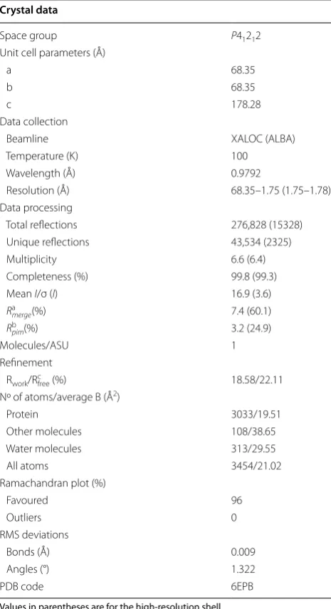
Table 3 Crystallographic data of Chit42
Values in parentheses are for the high-resolution shell
a R
merge= ∑hkl ∑i | Ii(hkl) − [I(hkl)]|/∑hkl ∑i Ii(hkl), where Ii(hkl) is the ith
measurement of reflection hkl and [I(hkl)] is the weighted mean of all measurements
b R
pim= ∑hkl [1/(N − 1)] 1/2 ∑i | Ii(hkl) − [I(hkl)]|/∑hkl ∑i Ii(hkl), where N is the
redundancy for the hkl reflection
c R
work/Rfree= ∑hkl | Fo − Fc |/∑hkl | Fo |, where Fc is the calculated and Fo is the
observed structure factor amplitude of reflection hkl for the working/free (5%) set
Crystal data
Space group P41212
Unit cell parameters (Å)
a 68.35
b 68.35
c 178.28
Data collection
Beamline XALOC (ALBA)
Temperature (K) 100
Wavelength (Å) 0.9792
Resolution (Å) 68.35–1.75 (1.75–1.78)
Data processing
Total reflections 276,828 (15328)
Unique reflections 43,534 (2325)
Multiplicity 6.6 (6.4)
Completeness (%) 99.8 (99.3)
Mean I/σ (I) 16.9 (3.6)
Rmergea (%) 7.4 (60.1)
Rpimb (%) 3.2 (24.9)
Molecules/ASU 1
Refinement
Rwork/Rcfree (%) 18.58/22.11
Nº of atoms/average B (Å2)
Protein 3033/19.51
Other molecules 108/38.65
Water molecules 313/29.55
All atoms 3454/21.02
Ramachandran plot (%)
Favoured 96
Outliers 0
RMS deviations
Bonds (Å) 0.009
Angles (°) 1.322
Consent for publication
Not applicable.
Ethics approval and consent to participate
Not applicable.
Funding
Work was supported by Spanish Ministry of Economy and Competitiveness: BIO2013‑48779‑C4‑1/‑2/‑4, BIO2016‑76601‑C3‑1/‑2/‑3, and by an institutional Grant from Fundación Ramón Areces to the Centro de Biología Molecular Severo Ochoa. Besides, it has received funding from the European Union’s Horizon 2020 research and innovation program [Blue Growth: Unlocking the potential of Seas and Oceans] under Grant Agreement No [634486; INMARE)]. PS‑M. was supported by a Spanish Ministry of Education University Personnel Training Plan (FPU) Grant.
Publisher’s Note
Springer Nature remains neutral with regard to jurisdictional claims in pub‑ lished maps and institutional affiliations.
Received: 21 December 2017 Accepted: 17 March 2018
References
1. Thadathil N, Velappan SP. Recent developments in chitosanase research and its biotechnological applications: a review. Food Chem. 2014;150:392–9. https ://doi.org/10.1016/j.foodc hem.2013.10.083. 2. Rinaudo M. Chitin and chitosan: properties and applications. Prog
Polym Sci. 2006;31(7):603–32. https ://doi.org/10.1016/j.progp olyms ci.2006.06.001.
3. Jung WJ, Park RD. Bioproduction of chitooligosaccharides: present and perspectives. Mar Drugs. 2014;12(11):5328–56. https ://doi.org/10.3390/ md121 15328 .
4. Khalil IR, Burns AT, Radecka I, Kowalczuk M, Khalaf T, Adamus G, Johnston B, Khechara MP. Bacterial‑derived polymer poly‑γ‑glutamic acid (γ‑PGA)‑ based micro/nanoparticles as a delivery system for antimicrobials and other biomedical applications. Int J Mol Sci. 2017;18(2):1–18. https ://doi. org/10.3390/ijms1 80203 13.
5. Rathore AS, Gupta RD. Chitinases from bacteria to human: properties, applications, and future perspectives. Enzyme Res. 2015. https ://doi. org/10.1155/2015/79190 7.
6. Hamed I, Özogul F, Regenstein JM. Industrial applications of crustacean by‑products (chitin, chitosan, and chitooligosaccharides): a review. Trends Food Sci Technol. 2016;48:40–50. https ://doi.org/10.1016/j. tifs.2015.11.007.
7. Xiong C, Wu H, Wei P, Pan M, Tuo Y, Kusakabe I, Du Y. Potent angiogenic inhibition effects of deacetylated chitohexaose separated from chitoo‑ ligosaccharides and its mechanism of action in vitro. Carbohydr Res. 2009;344:1975–83.
8. Wu H, Aam BB, Wang W, Norberg AL, Sorlie M, Eijsink VG. DuY. Inhibi‑ tion of angiogenesis by chitooligosaccharides with specific degrees of acetylation and polymerization. Carbohydr Polym. 2012;89:511–8. https :// doi.org/10.1016/j.carbp ol.2012.03.037.
9. Li Z, Cho S, Kwon IC, Janat‑Amsbury MM, Huh KM. Preparation and characterization of glycol chitin as a new thermogelling polymer for biomedical applications. Carbohydr Polym. 2013;92(2):2267–75. https :// doi.org/10.1016/j.carbp ol.2012.11.068.
10. Aam BB, Heggset EB, Norberg AL, Sorlie M, Varum KM, Eijsink VG. Production of chitooligosaccharides and their potential applications in medicine. Mar Drugs. 2010;8:1482–517. https ://doi.org/10.3390/md805 1482.
11. Sinha S, Chand S, Tripathi P. Enzymatic production of glucosamine and chitooligosaccharides using newly isolated exo‑β‑d‑glucosaminidase
having transglycosylation activity. 3. Biotech. 2016. https ://doi. org/10.1007/s1320 5‑015‑0330‑5.
12. Yang Y, Yu B. Recent advances in the synthesis of chitooligosaccharides and congeners. Tetrahedron. 2014;70:1023–46. https ://doi.org/10.1016/j. tet.2013.11.064.
13. Harman GE, Hayes CK, Lorito M, Broadway RM, Dipietro A, Peterbauer C, Tronsmo A. Chitinolytic enzymes of Trichoderma harzianum: purification of chitobiosidase and endochitinase. Mol Plant Pathology. 1993;83:313–8. 14. Sahai AS, Manocha MS. Chitinases of fungi and plants: their involvement
in morphogenesis and host‑parasite interaction. FEMS Microbiol Rev. 1993;11:317–38. https ://doi.org/10.1016/0168‑6445(93)90004 ‑S. 15. Schrempf H. Recognition and degradation of chitin by streptomy‑
cetes. Antonie Van Leeuwenhoek J Microb. 2001;79:285–9. https ://doi. org/10.1023/A:10120 58205 158.
16. Kasprzewska A. Plant chitinases‑regulation and function. Cell Mol Biol Lett. 2003;8:809–24.
17. Stoykov YM, Pavlov AI, Krastanov AI. Chitinase biotechnology: production, purification, and application. Eng Life Sci. 2015;15(1):30–8. https ://doi. org/10.1002/elsc.20140 0173.
18. Khan FI, Bisetty K, Singh S, Permaul K, Hassan MI. Chitinase from Thermo-myces lanuginosus SSBP and its biotechnological applications. Extremo‑ philes. 2015;19:1055–66. https ://doi.org/10.1007/s0079 2‑015‑0792‑8. 19. Kzhyshkowska J, Yin S, Liu T, Riabov V, Mitrofanova I. Role of chitinase‑
like proteins in cancer. Biol Chem. 2016;397(3):231–47. https ://doi. org/10.1515/hsz‑2015‑0269.
20. Hartl L, Zach S, Seidl‑Seiboth V. Fungal chitinases: diversity, mechanistic properties and biotechnological potential. Appl Microbiol Biotechnol. 2012;93(2):533–43. https ://doi.org/10.1007/s0025 3‑011‑3723‑3. 21. Kullnig C, Mach R, Lorito M, Kubicek C. Enzyme diffusion from
Trichoderma atrovidride (= T. harzianum P1) to Rhizoctonia solani is a prerequisite for triggering of Trichoderma ech42 gene expression before mycoparasitic contact. Appl Environ Microbiol. 2000;66(5):2232–4. https ://doi.org/10.1128/AEM.66.5.2232‑2234.2000.
22. Hoell IA, Klemsdal SS, Vaaje‑Kolstad G, Horn SJ, Eijsink VG. Overexpres‑ sion and characterization of a novel chitinase from Trichoderma atroviride strain P1. Biochim Biophys Acta. 2005;1748(2):180–90. https ://doi. org/10.1016/j.bbapa p.2005.01.002.
23. Boer H, Simolin H, Cottaz S, Soderlund H, Koivula A. Heterologous expression and site‑directed mutagenesis studies of two Trichoderma harzianum chitinases, Chit33 and Chit42, in Escherichia coli. Protein Expr Purif. 2007;51(2):216–26. https ://doi.org/10.1016/j.pep.2006.07.020. 24. Pérez‑Martínez AS, De León‑Rodríguez A, Harris LJ, Herrera‑Estrella A,
Barba de la Rosa AP. Overexpression, purification and characterization of the Trichoderma atroviride endochitinase, Ech42, in Pichia pastoris. Protein Expr Purif. 2007;55(1):183–8. https ://doi.org/10.1016/j.pep.2007. 25. Kowsari M, Motallebi M, Zamani M. Protein engineering of chit42 towards
improvement of chitinase and antifungal activities. Curr Microbiol. 2014;68:495–502. https ://doi.org/10.1007/s0028 4‑013‑0494‑3. 26. Boer H, Munck N, Natunen J, Wohlfahrt G, Soderlund H, Renkonen O,
Koivula A. Differential recognition of animal type β‑4‑galactosylated and α‑3‑fucosylated chito‑oligosaccharides by two family 18 chitinases from Trichoderma harzianum. Glycobiology. 2004;14(12):1303–13. https ://doi. org/10.1093/glyco b/cwh12 1.
27. Lienemann M, Boer H, Paananen A, Cottaz S, Koivula A. Toward under‑ standing of carbohydrate binding and substrate specificity of a glycosyl hydrolase 18 family (GH‑18) chitinase from Trichoderma harzianum. Gly‑ cobiology. 2009;19(10):1116–26. https ://doi.org/10.1093/glyco b/cwp10 2. 28. De la Cruz J, Hidalgo‑Gallego A, Lora JM, Benítez T, Pintor‑Toro JA, Llobell
A. Isolation and characterization of three chitinases from Trichoderma harzianum. Eur J Biochem. 1992;206:859–67.
29. Benítez T, Rincón MA, Limón MC, Codón CA. Biocontrol mechanisms of Trichoderma strains. Int Microbiol. 2004;7:249–60.
30. Limón MC, Chacón MR, Mejías R, Delgado‑Jarama J, Rincón AM, Codón AC, Benítez T. Increased antifungal and chitinase specific activities of Trichoderma harzianum CECT 2413 by addition of a cellulose binding domain. Appl Microbiol Biotechnol. 2004;64(5):675–85. https ://doi. org/10.1007/s0025 3‑003‑1538‑6.
31. García I, Lora JM, de la Cruz J, Benítez T, Llobell A, Pintor‑Toro JA. Cloning and characterization of a chitinase (CHIT42) cDNA from the mycoparasitic fungus Trichoderma harzianum. Curr Genet. 1994;27(1):83–9. https ://doi. org/10.1007/BF003 26583 .
• We accept pre-submission inquiries
• Our selector tool helps you to find the most relevant journal • We provide round the clock customer support
• Convenient online submission • Thorough peer review
• Inclusion in PubMed and all major indexing services • Maximum visibility for your research
Submit your manuscript at www.biomedcentral.com/submit
Submit your next manuscript to BioMed Central
and we will help you at every step:
33. Draborg H, Christgau S, Halkier T, Rasmussen G, Dalboge H, Kauppinen S. Secretion of an enzymatically active Trichoderma harzianum endochi‑ tinase by Saccharomyces cerevisiae. Curr Genet. 1996;29:404–9. 34. Tang W, Li Y, Liu L, Zhang J, Xian H. Characterization and production
optimization of a chitinase (Tachi1) from Trichoderma asperellum in recombinant Pichia pastoris expression system. Wei Sheng Wu Xue Bao. 2012;52(3):345–52.
35. Chen A‑S, Taguchi T, Sakai K, Kikuchi K, Wang M‑W, Miwa I. Antioxidant activities of chitobiose and chitotriose. Biol Pharm Bull. 2003;26(9):1326– 30. https ://doi.org/10.1248/bpb.26.1326.
36. Ravi Kumar MN. A review of chitin and chitosan applications. React Funct Polym. 2000;46(1):1–27. https ://doi.org/10.1016/S1381 ‑5148(00)00038 ‑9. 37. Papanikolau Y, Prag G, Tavlas G, Vorgias CE, Oppenheim AB, Petratos K.
High resolution structural analyses of mutant Chitinase A complexes with substrates provide new insight into the mechanism of catalysis. Biochem‑ istry. 2001;40(38):11338–43. https ://doi.org/10.1021/bi010 505h. 38. Rao FV, Houston DR, Boot RG, Aerts JM, Hodkinson M, Adams DJ, Shiomi
K, Omura S, van Aalten DM. Specificity and affinity of natural product cyclopentapeptide inhibitors against A. fumigatus, human, and bacterial chitinases. Chem Biol. 2005;12(1):65–76. https ://doi.org/10.1016/j.chemb iol.2004.10.013.
39. Jeuniaux C. Chitinases. Methods Enzymol. 1966;8(C):644–50. https ://doi. org/10.1016/0076‑6879(66)08117 ‑5.
40. Sears IB, O’Connor J, Rossanese OW, Glick BS. A versatile set of vectors for constitutive and regulated gene expression in Pichia pastoris. Yeast. 1998;14(8):783–90.
41. Van den Ent F, Löwe J. RF cloning: a restriction‑free method for inserting target genes into plasmids. J Biochem Biophys Methods. 2006;67(1):67– 74. https ://doi.org/10.1016/j.jbbm.2005.12.008.
42. Álvaro‑Benito M, de Abreu M, Fernández‑Arrojo L, Plou FJ, Jiménez‑ Barbero J, Ballesteros A, Polaina J, Fernández‑Lobato M. Characteriza‑ tion of a β‑fructofuranosidase from Schwanniomyces occidentalis with
transfructosylating activity yielding the prebiotic 6‑kestose. J Biotechnol. 2007;132(1):75–81. https ://doi.org/10.1016/j.jbiot ec.2007.07.939. 43. Laemmli UK. Cleavage of structural proteins during the assembly of the
head of bacteriophage T4. Nature. 1970;227:680–5.
44. Żur I, Gołębiowska G, Dubas E, Golemiec E, Matušíková I, Libantová J, Moravčíková J. β‑1,3‑Glucanase and chitinase activities in winter triticales during cold hardening and subsequent infection by Microdochium nivale. Biologia. 2013;68(2):241–8. https ://doi.org/10.2478/s1175 6‑013‑0001‑0.
45. Santos‑Moriano P, Fernandez‑Arrojo L, Mengibar M, Belmonte‑Reche E, Peñalver P, Acosta FN, Ballesteros AO, Morales JC, Kidibule P, Fernández‑ Lobato M, Plou FJ. Enzymatic production of fully deacetylated chi‑ tooligosaccharides and their neuroprotective and anti‑inflammatory properties. Biocatal Biotransformation. 2017;35(1):1–11. https ://doi. org/10.1080/10242 422.2017.12952 31.
46. Kabsch W. XDS. Acta Crystallogr D Biol Crystallogr. 2010;66(2):125–32.
https ://doi.org/10.1107/S0907 44490 90473 37.
47. Winn MD, Ballard CC, Cowtan KD, Dodson EJ, Emsley P, Evans PR, et al. Overview of the CCP4 suite and current developments. Acta Crystallogr D Biol Crystallogr. 2011;67(4):235–42. https ://doi.org/10.1107/S0907 44491 00457 49.
48. Vagin A, Teplyakov A. Molecular replacement with MOLREP. Acta Crystal‑ logr D Biol Crystallogr. 2010;66(1):22–5. https ://doi.org/10.1107/S0907 44490 90425 89.
49. Murshudov GN, Vagin AA, Dodson EJ. Refinement of macromolecular structures by the maximum‑likelihood method. Acta Crystallogr D Biol Crystallogr. 1997;53(3):240–55. https ://doi.org/10.1107/S0907 44499 60122 55.
50. Emsley P, Cowtan K. Coot: model‑building tools for molecular graphics. Acta Crystallogr D Biol Crystallogr. 2004;60(12–1):2126–32.