R E S E A R C H
Open Access
Automatic artefact removal in a self-paced
hybrid brain- computer interface system
Xinyi Yong
1*, Mehrdad Fatourechi
1, Rabab K Ward
1and Gary E Birch
1,2Abstract
Background: A novel artefact removal algorithm is proposed for a self-paced hybrid brain-computer interface (BCI) system. This hybrid system combines a self-paced BCI with an eye-tracker to operate a virtual keyboard. To select a letter, the user must gaze at the target for at least a specific period of time (dwell time) and then activate the BCI by performing a mental task. Unfortunately, electroencephalogram (EEG) signals are often contaminated with artefacts. Artefacts change the quality of EEG signals and subsequently degrade the BCI’s performance.
Methods: To remove artefacts in EEG signals, the proposed algorithm uses the stationary wavelet transform combined with a new adaptive thresholding mechanism. To evaluate the performance of the proposed algorithm and other artefact handling/removal methods, semi-simulated EEG signals (i.e., real EEG signals mixed with simulated artefacts) and real EEG signals obtained from seven participants are used. For real EEG signals, the hybrid BCI system’s performance is evaluated in an online-like manner, i.e., using the continuous data from the last session as in a real-time environment.
Results: With semi-simulated EEG signals, we show that the proposed algorithm achieves lower signal distortion in both time and frequency domains. With real EEG signals, we demonstrate that for dwell time of 0.0s, the number of false-positives/minute is 2 and the true positive rate (TPR) achieved by the proposed algorithm is 44.7%, which is more than 15.0% higher compared to other state-of-the-art artefact handling methods. As dwell time increases to 1.0s, the TPR increases to 73.1%.
Conclusions: The proposed artefact removal algorithm greatly improves the BCI’s performance. It also has the following advantages: a) it does not require additional electrooculogram/electromyogram channels, long data segments or a large number of EEG channels, b) it allows real-time processing, and c) it reduces signal distortion.
Background
A brain-computer interface (BCI) system allows humans to use their brain signals (such as EEG) to control vari-ous devices such as a virtual keyboard [1-3], a functional electrical stimulator [4], an orthosis [5], amongst others. BCIs can be operated in a synchronized mode or an asyn-chronous (self-paced) mode [6]. In a synchronized BCI system, the periods when a user can control the system are determined by the system itself. The system usually sends an external cue to the user and the user must then issue a control command within a window of opportu-nity provided by the system. This limits the use of a
*Correspondence: [email protected]
1Department of Electrical and Computer Engineering, University of British Columbia, 2356 Main Mall, Vancouver, V6T1Z4 Canada
Full list of author information is available at the end of the article
synchronized BCI system in practical applications. A self-paced BCI system, on the other hand, allows users to control the system whenever they desire. Hence, the users have a more natural and flexible means for controlling an object [6].
Designing a self-paced BCI system with high perfor-mance is associated with two major challenges. They are:
1. identifying the user’s intentional control (IC) state reliably [IC periods are periods when the user intends to issue control] and
2. reducing the number of false activations (false positives during the no control (NC) periods). [NC periods are periods when the user does not intend to activate the system such as when he/she is obtaining
information from the computer screen, thinking about a problem, talking, resting, etc].
NC periods are usually much longer compared to IC periods. As a high number of false positives can result in user frustration, it is especially important to design a sys-tem that generates a very low (ideally zero) number of false positives.
It is not easy and straightforward to apply existing pure (i.e., non-hybrid) self-paced BCI systems to operate a practical system such as a virtual keyboard. The reason is that these systems can only recognize a limited num-ber of mental tasks as unique IC commands (mostly one or two). This number is much smaller than the number of letters used in spelling applications. Furthermore, most self-paced BCI systems generate a large number of false positives per minute on average, which is not suitable for most practical applications.
To overcome the above problems, in [7] we have pro-posed a hybrid system that combines a self-paced BCI with an eye-tracker to operate a virtual keyboard. Our proposed hybrid BCI system also successfully overcomes the ‘Midas Touch’ problem, which is a major prob-lem experienced by conventional eye-gaze interfaces, and results in a significantly smaller false positives generated per minute [7]. The ‘Midas Touch’ problem is the diffi-culty of determining whether or not the user is intending to select a certain object as the user might be gazing at the object for reasons other than to enter it [8].
As the hybrid BCI system relies on eye movements to control the cursor, it is no surprise that the EEG signals in the system are more contaminated with ocular artefacts compared to EEG signals in a pure BCI system. Also as in other BCI systems, EEG signals are also contaminated with artefacts caused by muscle activities, power line interference, and electrode movements [9]. These arte-facts can affect the performance of the system in several ways. In particular, they can:
1. significantly reduce the amount of data available for designing the system;
2. result in false positives during the NC periods and 3. decrease the true positive rate of the system.
Although some studies have clearly shown that artefacts affect the performance of pure self-paced BCI systems [10,11], little attention has been paid to handle artefacts so far.
In this paper, to minimize the effects of artefacts and improve the performance of our hybrid BCI, we propose a new artefact removal algorithm. The proposed artefact removal algorithm is integrated with our artefact detec-tion algorithm proposed in [12]. Both algorithms use the stationary wavelet transform (SWT). The wavelet coef-ficients obtained from the artefact detection algorithm
are thresholded by applying a new adaptive thresholding procedure that we propose to remove artefacts in EEG sig-nals. Its advantages over state-of-the-art artefact removal algorithms are:
1. it can be fully automated;
2. it uses an adaptive mechanism to reduce signal distortion;
3. it is computationally inexpensive and allows real-time processing; and
4. it does not require additional electrooculogram (EOG) or electromyogram (EMG) channels, long data segments or a large number of EEG channels.
We compare the performance of different algorithms using real EEG signals and semi-simulated EEG signals (i.e., real EEG signals mixed with simulated artefacts). With semi-simulated EEG signals, we show that the pro-posed algorithm achieves lower signal distortion in both time and frequency domains. Next, using real EEG sig-nals, we fully investigate and compare the performance of the hybrid BCI system in the following situations: 1) when artefacts are ignored (i.e., the original data are used); 2) when EEG segments with artefacts are rejected (i.e., the output of the system is blocked in the presence of artefacts and the system becomes unavailable); and 3) when auto-matic artefact removal algorithms such as the proposed algorithm and Blind Source Separation (BSS) algorithms are employed. We show that for dwell time of 0.0s (i.e., the user can activate the system any time right after he/she gazes at a letter/word), the true positive rate (TPR) achieved using the proposed artefact removal algorithm is 44.7% with 2 false positives generated per minute. This TPR value is 33.6% and 20.1% higher than those achieved when artefacts are rejected and ignored respectively. We also show that our proposed method outperforms BSS by at least 16.2%.
In the following subsections, we briefly review our self-paced hybrid BCI system, current artefact handling methods in the literature as well as the state-of-the-art of artefact removal algorithms.
The structure of the self-paced hybrid BCI system
A hybrid BCI is defined as a system that combines a BCI with another system (such as another BCI or an eye-tracker) [13]. In this section, the overall structure of the hybrid self-paced BCI system proposed in our earlier work is presented [7]. This system combines a BCI and an eye-tracker to operate a virtual keyboard.
Hybrid BCI System
Self−Paced BCI
Figure 1Our Hybrid BCI System.The hybrid BCI system proposed in [7].
to have large selection boxes, and a word prediction functionality. The eye-tracker acts as thepointingdevice and the user’s eye gaze controls the cursor movement. The use of eye gaze is natural and fast because people often look at the object of interest before controlling it [8]. The BCI, on the other hand, acts as the clicking device. Its inputs are the continuous EEG signals recorded from the user’s scalp and the output is a binary control signal (i.e., it is either ‘0’ or ‘1’).
To make a selection (i.e., aclickoperation), a user has to gaze at the target for at least a specific period of time (called the dwell time) and then activate the self-paced BCI with a mental task (which is an attempted hand exten-sion), as demonstrated in Figure 1. When changes in the EEG signals due to an attempted hand extension move-ment are detected by the signal processing unit in the BCI, a click command (an intentional control or IC) is initiated. Note that an attempted hand extension results in an imagined movement by users with movement dis-abilities who cannot move their hands. For able-bodied individuals, it leads to an actual hand movement [15]. Evidence from the literature shows that the patterns aris-ing from attempted movements are very similar to those of real movements [16,17]. This evidence allows the use of real movements in our study. However, note that the attempted hand movement can be replaced by any other mental task.
Our previous study showed that increasing the dwell time (Tdwell) reduces the number of false positives [7].
This is because our design restricts the BCI’s operation to the periods during which the user’s point of gaze is within a region on the monitor that can be clicked on and the user gazes at that region for at least Tdwell seconds. For the rest of the time, the BCI is put in
the so-called ‘sleep’ mode, i.e., it does not process the input EEG signals nor generates any output. By using this arrangement, we can greatly reduce the number of false positives during the NC periods, as demonstrated in [7].
The above system has one main drawback. When the users are looking at different locations of the virtual key-board to make a selection, the amount of eye movement activity is significant. Therefore, EEG signals are more fre-quently contaminated with ocular artefacts compared to pure (non-hybrid) BCI systems. Hence, it is important to design an algorithm that can efficiently handle artefacts in this hybrid system.
Artefact handling methods
A review of methods for handling EOG and EMG arte-facts in BCIs shows that more than half of the 250 BCI papers studied did not report as to whether or not they had considered or handled EOG and/or EMG artefacts [9]. For those who did, three methods were generally employed:
1. Ignore: ignoring the presence of artefacts;
2. Reject: automatic rejection of artefact-contaminated EEG segments; and
3. Remove: automatic removal of artefacts.
Rejecting contaminated EEG segments (Reject) is com-mon in BCI literature. However, this approach has two major disadvantages:
1. In the training phase, it can significantly reduce the amount of available data for training the classifier; 2. In the testing phase, it forces the BCI system into a
non-responsive state for a significant portion of the time. This subsequently reduces the information transfer rate of the system.
Due to these shortcomings,Rejectneeds to be replaced by methods that do not discard any data during artefact-contaminated periods.
Unless the signal processing algorithms employed to process EEG signals are robust to the presence of artefacts, ignoring the artefacts in EEG signals (Ignore) is usually not an efficient approach either. This is due to the fact that artefacts affect the different frequency bands in EEG signals and therefore impact the performance of a self-paced BCI system. For example, a study conducted by Bashashatiet al.[10] shows that the performance of the proposed self-paced BCI system deteriorates, when the data with ocular artefacts are included in the analysis. Based on the results obtained from eight participants, the amount of decrease in the true positive rate (TPR) value varied from 2.3% to 15.1% (with an average of 6.8%), when the time-normalized false positive rate (TNFPR) was set to 9 FPs/min. In another study, Fatourechi et al. [11] combined the use of features extracted from three neuro-logical phenomena: movement-related potentials (MRPs), and the power of mu and beta rhythms to design a self-paced BCI system that is robust in the presence of arte-facts. Using a five-fold nested cross validation, the average TPR and TNFPR achieved were 56.2% and 0.5 FPs/min for non-contaminated data and 51.8% and 2 FPs/min for artefact-contaminated data. The deterioration in some individuals was much greater, e.g., a drop of 13.2% and an increase of 0.5 FPs/min in the TPR and TNFPR, respec-tively, were observed in one person. The results of the above studies show that current state-of-the-art pattern recognition algorithms employed in self-paced BCI sys-tems cannot efficiently handle artefacts. As a result, other solutions need to be explored.
A better alternative solution to handle artefacts in a self-paced BCI system is to apply automatic artefact removal algorithms to EEG segments contaminated with artefacts (Remove). Although removing artefacts is not straightfor-ward and increases the complexity of the BCI system, the major advantage is that the BCI system becomes available for user’s control at all timesincluding those with arte-facts happen. Besides, the performance of the system may be improved if the artefact removal algorithm removes the artefacts effectively without distorting the EEG signals.
In the rest of this section, we provide a brief review on artefact removal algorithms (for a more detailed review, please see [9]).
Regression analysis is widely used to remove ocular artefacts from EEG signals [18-21]. It assumes that the observed EEG signals are a linear superposition of EEG and EOG components [18]. The proportion of any EOG component that is present in the EEG signal is estimated and then removed using the least squares criterion. This method has the disadvantage of requiring the recording of source signals from the EOG channels to remove ocular artefacts. For the case of muscle artefacts, it is not straight-forward to identify the source signals as these sources can originate from different muscle groups [21]. For this reason, different reference channels from multiple muscle groups are required. This in turn can greatly increase the complexity of the algorithm.
Another popular approach for artefact removal is blind source separation (BSS) [22-25], including Independent Component Analysis (ICA) algorithms [20,26-28]. These algorithms estimate the underlying sources from EEG sig-nals recorded from electrodes. The sources related to artefacts are removed to obtain denoised EEG signals. As an example, Hung et al.automated the identification of EEG activities of interest using several manually identi-fied movement-related spatial maps and used the cleaned signals in the classification of motor imagery EEG sig-nals [26]. Halderet al. proposed the use of the AMUSE (Algorithm for Multiple Unknown Source Extraction) and ICA Infomax algorithms to isolate artefacts from 3-second EEG segments. A combination of support vec-tor machines was used to classify the isolated artefacts extracted using the proposed BSS and ICA algorithms [22]. While BSS/ICA algorithms are widely used in the literature for removing artefacts, a study conducted by Wallstrom et al. [20] showed that these algorithms may overestimate the spectrum of artefacts and thus cause spectral distortion in EEG signals. Moreover, such meth-ods require multi-channel data and long data epochs to produce reliable results [29].
In this study, we have explored the use of SWT in removing various types of artefacts in EEG signals. The main reason is that it is computationally inexpen-sive and no additional EOG/EMG channels and long data segments are required. To overcome the problem encountered when using the SURE threshold selection procedure, we have proposed a new adaptive thresholding mechanism.
In the next section, we first describe the experimental procedure and the type of EEG data used in this study. Next, the artefact detection algorithm and our proposed artefact removal algorithm are discussed. Finally, the met-rics used to evaluate the performance of the artefact removal algorithm is presented.
Methods
Experimental procedure
Data description
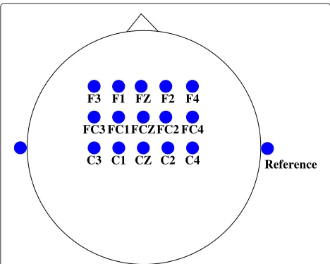
The experiments [7] were approved by the UBC Behav-ioral Research Ethics Board. We recruited seven able-bodied individuals, who did not wear glasses for this study. Their age ranges from 26 to 31. Participants gave an informed consent before participating in the experiment. Each individual was seated comfortably approximately 75 cm in front of a computer monitor and wore a 64-channel electrode cap. EEG signals were recorded from 15 elec-trodes placed over the motor cortex area of the brain as shown in Figure 2. Electrooculogram (EOG) signals were recorded by two pairs of electrodes placed around both eyes. Facial muscle activities were recorded by four pairs of electromyogram (EMG) surface electrodes placed sym-metrically on two related facial muscles from each side of the face: zygomaticus major and corrugator supercilii. All electrodes were referenced to the linked right and left
C3 CZ C4
FC4
C1 C2
FC3FC1FCZFC2 F2 F4 F1 FZ
F3
Reference
Figure 2Electrode Montage.The EEG channels used in our system.
earlobes. All signals were amplified and sampled at 128 Hz using a L64 Sagura EEG amplifier system [35].
For eye-tracking, we used a Mirametrix S1 system [36]. This eye-tracker employed a single high-resolution cam-era to estimate the point of gaze. The eye-gaze informa-tion such as the x and y coordinates of the fixainforma-tion point, the pupils’ center x and y coordinates amongst other information were recorded during the experiments.
Experimental protocol
Each experiment for each participant lasted for approx-imately 2.5 hours. At the beginning of each experiment, the eye-tracker was calibrated. Next, the participants were given approximately ten minutes to practise a text entry task with the eye-tracker and the Dynamic Keyboard so that they became more comfortable with using the system. The participants were then requested to rest for two min-utes. The data recorded during this resting period were later used to determine the thresholds for the artefact detection algorithm [12].
Next, the participants were asked to type a sentence dis-played by the graphical user interface (GUI), at their own speed. Once a user finished typing one sentence, a new sentence appeared and replaced the old one. This proce-dure was repeated until the end of the ten-minute session. The sentences were randomly selected from the ‘Phrase Set’ provided by MacKenzie and Soukoreff [37], which consisted of 500 phrases, with lengths varying from 16 to 43 characters. Each experiment consisted of three to five sessions.
areas, false positives are blocked and they do not result in any false selection. Hence, the total TNFPR would be lower.
Throughout the experiment, a participant could ask for a break whenever needed. Furthermore, whenever a participant felt that the eye-tracker was becoming more difficult to control, we recalibrated the eye-tracker.
Generating semi-simulated EEG signals
The EEG data collected from the experiments described above were used to evaluate the performance of the hybrid BCI system when various algorithms were used for arte-fact removal. As the exact percentage of artearte-facts in EEG signals is not clear, it is difficult to measure the effec-tiveness of different methods in terms of the amount of artefacts removed. For this reason, we have generated semi-simulated EEG signals so that the amount of arte-facts and signals removed by various artefact removal algorithms can be quantified. The semi-simulated EEG signals were constructed by adding simulated artefacts to real EEG data acquired from the experiments. As the clean EEG signals, the artefacts and their mixing process are now known, evaluating the performance of different artefact removal algorithms becomes easier.
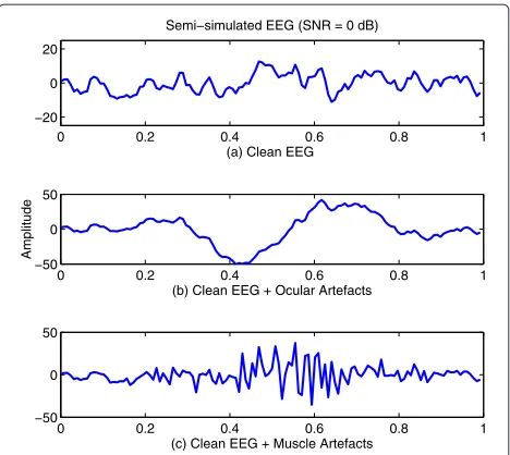
For each of the 15 EEG channels, to generate a 1-second semi-simulated EEG signal, a 1-second clean EEG seg-ment from each channel was mixed with artefacts. Two different types of artefacts were simulated: eye-blinks and muscle artefacts. Eye-blinks were simulated by band-pass filtering a random noise from 1 to 3 Hz. The filter was obtained using a finite impulse response (FIR) filter based on Kaiser’s window [39]. Muscle artefacts were simulated by band-pass filtering a random noise from 20 to 60 Hz using an FIR filter based on the Kaiser’s window [39]. The level of artefact contamination for each EEG channel was estimated from real EEG signals. Then, the amplitudes of the simulated artefacts were adjusted such that the semi-simulated signals have a signal-to-noise ratio (SNR) of 0 dB for the EEG channel that has the largest arte-fact contamination level. Figure 3 shows two examples of semi-simulated EEG signals with ocular and muscle artefacts added respectively.
To simulate real-life scenarios where EEG segments are contaminated with artefacts at different locations, each simulated artefact was shifted and mixed with each clean EEG signal to generate different semi-simulated EEG signals.
Automatic artefact detection
Our BCI system is composed of four main modules (see Figure 4):
1. an artefact detection module; 2. an artefact removal module;
3. a feature extraction module; and 4. a feature classification module.
This system employs Ne = 15 monopolar EEG
chan-nels. It continuously segments the EEG signals using a 1-second sliding window, with 87.5% overlap. Therefore, eight EEG segments are obtained each second. The arte-fact detection algorithm is first applied to each EEG seg-ment, before that segment is processed by the artefact removal, feature extraction and feature classification mod-ules. In the remaining part of this section, the artefact detection algorithm [12] is briefly discussed.
The automatic artefact detection algorithm is based on the stationary wavelet transform (SWT) in [12]. It only employs EEG signals acquired from the premotor and sen-sorimotor cortex areas of the brain. This allows us to bypass the use of additional EOG and EMG signals, as well as frontal and temporal EEG electrodes in our artefact detection module. The algorithm also has a low compu-tational complexity because it uses a simple thresholding method for artefact detection. Furthermore, to minimize human intervention, the thresholds used in the algorithm are obtained automatically using the EEG data collected at the beginning of each experiment as the user is requested to rest and have minimal movement [12].
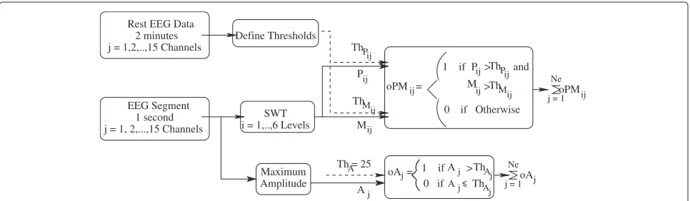
The artefact detection algorithm uses the maximum amplitude of EEG signals and the SWT coefficients to detect artefacts (see Figure 5).
In Figure 5,Ajis the maximum amplitude of an EEG
seg-ment in channelj. In addition,PijandMijare the power
0 0.2 0.4 0.6 0.8 1
−20 0 20
Semi−simulated EEG (SNR = 0 dB)
(a) Clean EEG
0 0.2 0.4 0.6 0.8 1
−50 0 50
(b) Clean EEG + Ocular Artefacts
Amplitude
0 0.2 0.4 0.6 0.8 1
−50 0 50
(c) Clean EEG + Muscle Artefacts
Control Commands Feature Extractor Feature Translator
Amplitude Thresholding
Stationary Wavelet Transform Inverse SWT
Thresholding SWT coefficients
EEG Signals (15 Channels)
Denoised EEG Signals
Automatic Artefact Detection Artefact Removal
Figure 4Structure of the Proposed Self-Paced BCI System.The structure of the proposed self-paced BCI system.
and the maximum amplitude of theithlevel wavelet coeffi-cients for the EEG channeljrespectively as defined below:
Pij=
1 N
N
t=1
a2i,j,t (1)
Mij=maxt=1:N|ai,j,t| (2)
whereai,j,tis thetthsample of theithlevel wavelet
coeffi-cients obtained for the EEG channeljandNis the number of coefficients available.
As shown in Figure 5,Pij,Mi, andAjfor each EEG
seg-ment in channeljare computed and each of these features is compared with one of the three thresholds (ThPij,ThMij
and ThAj). The thresholds for these features are
deter-mined using the reference EEG signals collected when the participants were requested to rest (please see [12] for more details).
As different wavelet coefficient levels correspond to dif-ferent frequency bands, the algorithm could be used to identify two major types of artefacts: (a) low frequency artefacts (e.g., ocular, electrode movement and head movement artefacts), and (b) higher frequency artefacts
(e.g., facial muscle and electrode movement artefacts). The low frequency artefacts are declared present if:
• the features of the last level of the detail coefficients and the approximation coefficients in at leastNCh EEG channels exceed their thresholds; or
• any of the EEG channels has a valueAjthat exceeds 25μV
Also, the high frequency artefacts are declared to be present if the higher frequency features (Pij,Mijfori=1,
2, and 3) in at leastNChEEG channels exceed the values of
their corresponding thresholds.
Here, NCh denotes the number of EEG channels that
are observed to havePij andMij values exceeding their
corresponding thresholds. This parameter affects the sen-sitivity (the percentage of correctly detected segments with artefacts) and the specificity (the percentage of cor-rectly identified artefact-free segments). The choice of NCh = 0 is too stringent. Although it results in a high
sensitivity value, the specificity value is often too low. In our study, we have experimentally found thatNCh = 5
(i.e., one third of the electrodes) provides a reasonable
ij Rest EEG Data
2 minutes j = 1,2,..,15 Channels
1 second EEG Segment
j = 1, 2,...,15 Channels
Maximum Amplitude
Pij ij ThP
Mij Mij
Aj = 25
ThA Th
Aj oPM =ij
j = 1oPM Ne
ij Pij
Ne
j = 1oA j Define Thresholds
SWT i = 1,..,6 Levels
Th
oA =j
0 if Aj
Aj
1 if > Th
Aj and ThM
0 if Otherwise Th
Pij 1 if >
M ij>
specificity and sensitivity values. It is clear that there is a trade-off between the sensitivity and the specificity val-ues. For our application, a high sensitivity value (i.e., a high artefact detection rate) is more desirable because arte-facts can affect the performance of the system. Those EEG segments that are falsely declared as contaminated with artefacts would not be rejected or discarded and therefore no data loss would result.
In this paper, we have integrated this artefact detection algorithm with our proposed artefact removal algorithms to denoise EEG signals. If artefacts in an EEG segment are declared as present by the artefact detection algorithm, the artefact removal algorithm is then applied to remove them, as explained in the next section.
Artefact removal algorithm
We propose to remove the artefacts using the station-ary wavelet transform (SWT) with an adaptive thresh-olding mechanism. As shown in Figure 4, the wavelet coefficients generated by the artefact detection module are used in our artefact removal algorithm to denoise the EEG signals. The denoised signals are obtained by performing an inverse SWT on the thresholded wavelet coefficients. The performance of the proposed algorithm is compared with those of other artefact removal algo-rithms such as blind sources separation (BSS) algoalgo-rithms. The details of these algorithms and the performance evaluation criteria used are provided in the following subsections.
Background
The discrete wavelet transform (DWT) is not transla-tion invariant. Small shifts in a signal can cause large changes in the wavelet coefficients of the signal and large variations in the distribution of energy in the different wavelet scales [30]. Besides, due to the lack of the transla-tion invariance property, denoising with DWT sometimes introduces artefacts (small ripples) in the signal near dis-continuities that are created by thresholding the wavelet coefficients [40]. A solution to the translation invariance problems is the use of a translation invariant estimation such as SWT [30].
SWT is translation invariant because there is no down-sampling of data involved in the algorithm that decom-poses a signal [30]. Instead, the wavelet filters are dilated at each decomposition level of the transform [30]. To remove the noise from a signal using SWT, three steps need to be performed [40]:
1. Transform the signal into the wavelet domain; 2. Apply a thresholding function to the resulting
wavelet coefficients; and
3. Transform the modified wavelet coefficients back to the original domain to obtain the denoised signal.
Therefore, when applying SWT for artefact removal, two important issues need to be taken into consideration: 1) the thresholding function used to attenuate the wavelet coefficients; and 2) the estimation procedure for obtaining the optimal threshold. These issues are discussed next.
Thresholding function
The thresholding function is used to remove or reduce a selected number of wavelet coefficients so as to remove artefacts from a signal. Depending on the application and the assumptions made, the large wavelet coefficients are related to either the signal of interest or to the artefacts. In our application, we assume that the artefacts that obscure the EEG signals introduce large wavelet coefficients in the wavelet domain. Hence, the wavelet coefficients (that are larger than a particular thresholdT) correspond to noisy samples and the wavelet coefficients smaller thanT cor-respond to the signal of interest. Of course, the amount of the attenuation of these coefficients depends on the thresholding function employed.
The two most widely used thresholding functions are the hard thresholding (Eq. 3) and the soft thresholding functions (Eq. 4) [40]. The hard thresholding function has a discontinuity. This discontinuity results in a bigger vari-ance in the estimated signal (i.e., the output estimate is sensitive to small changes in the input data) [41]. The soft thresholding function on the other hand results in a bigger bias (and hence larger errors) in the estimated signal [41]. To overcome the drawbacks of both the hard and the soft thresholding, the non-negative garrote shrinkage function (Eq. 5) was proposed in [41]. This function is continuous, less sensitive to small changes in the data and has a smaller bias.
δHard(x)=
0 |x| ≤T
x |x|>T (3)
δSoft(x)=
⎧ ⎨ ⎩
0 |x| ≤T
x−T x>T x+T x<T
(4)
δ+Garrote(x)=
0 |x| ≤T
x−T2/x |x|>T (5)
Another shrinkage function called the Smooth Sigmoid-Based Shrinkage (SBSS) function has been proposed by Attoet al.[42]. This function is defined as:
δSBSS(x)=
0 |x| ≤T
sgn(x)(|x|−T)
1+e−τ (|x|−λ) |x|>T
(6)
amplitudes;λis the threshold height (λ > T). Finally,τ is the attenuation we want to impose on data with ampli-tudes in the interval ]T,λ[ and ]−λ,T[. Please see [42] for more details about the SBSS shrinkage function. The advantages of this shrinkage function are:
1. It is smooth and it introduces small variability among coefficients with close values. Thus, it induces less error when reconstructing the signals;
2. It can control the degree of attenuation imposed on wavelet coefficients: high attenuation on the small coefficients and weak attenuation on the large coefficients.
In this paper, we investigate the different threshold-ing functions. Among these functions, the non-negative garrote thresholding function and the SBSS shrinkage function have not been explored in the BCI literature to remove artefacts from EEG signals and will be investigated for the first time in our paper.
Threshold value selection
The thresholds selected for wavelet denoising, Ti, are
important as they decide the degree of attenuation imposed on both artefacts and signals. Over-estimating the thresholds results in the under-estimation of artefacts and thus, the artefacts are not completely removed from the signal of interest. On the other hand, under-estimating the thresholds results in the over-estimation of artefacts and thus, the signal of interest is over-corrected.
Two possible approaches to estimating the thresholds include: 1) estimating the thresholds based on some ref-erence signals [31] (denoted by SWT-REF) and 2) using the so-called universal threshold proposed by [40] (Eq.7), which is denoted by SWT-UNV.
T0i =σi √
2lnN (7)
whereT0i is the universal threshold estimated for theith decomposition level wavelet coefficientsai:,σiis the
esti-mated noise variance forai:, andNis the number of data samples. For this formula,σi=MADN(ai:)where MADN is the normalized version of the median absolute deviation defined below:
MADN(x)= 1
cmedian(|x−median(x)|) (8)
wherec=0.6745, as this value results in an estimate that is unbiased when the data is normally distributed [43].
Both approaches provide fixed thresholds, which are not necessarily optimal. For instance, the universal thresh-old tends to be bigger than necessary and over-smooths the signal [41]. For our application, this implies that this threshold value fails to effectively remove artefacts.
To adaptively find the optimal thresholds, Donoho and Johnstone proposed a threshold selection procedure based on the Stein’s unbiased risk estimate (SURE) for soft-thresholding [44]. This procedure is not valid for hard thresholding because the hard thresholding function is not continuous and therefore it does not have bounded weak derivative (in Stein’s sense) [41].
When applying SWT with soft thresholding and using the SURE procedure (denoted by SWT-SURE) to remove artefacts in EEG signals, we have observed that the esti-mated thresholds tend to be lower than the optimal thresholds. That means the thresholds do not only remove the artefacts, but they also remove some parts of the sig-nals as well. The evidence to support our observation will be presented in the Results section.
To overcome the problems encountered in the exist-ing threshold selection procedures discussed above, we propose an adaptive thresholding algorithm, which is explained next.
Proposed adaptive SWT Denoising Algorithm - ASWTD
SWT with hard thresholding [31] and soft thresholding [33,34] have been applied in the literature to remove noise in EEG signals. These studies, however, have only focussed on ocular artefact removal. Hence, only the wavelet coef-ficients that correspond to lower frequency bands (i.e., up to 16 Hz) are thresholded. To the best of our knowledge, SWT has not been used to remove other artefacts such as muscle and electrode artefacts.
Our proposed algorithm, which is denoted by Adaptive SWT-based Denoising (ASWTD), is different from the above studies in two main aspects:
1. It uses a newadaptive thresholding procedure that minimizes the effects of artefacts, while preserving the features of the signal of interest and preventing the signal from being over-corrected.
2. To remove thevarious EEG artefacts in a self-paced BCI system, ASWTD thresholds the wavelet coefficients at all the decomposition levels.
We also investigate four different thresholding func-tions (i.e., the hard, soft, non-negative garrote and SBSS thresholding functions), when the proposed procedure is employed.
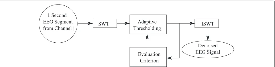
Figure 6 depicts the basic idea of the ASWTD algo-rithm. The thresholds are data-driven and adaptively updated. The adaptive thresholding procedure requires a performance-based criterion to decide how the thresholds should be adjusted with respect to the requirements of our application. These requirements include reducing the presence of artefacts and preserving the features of EEG signals in a computationally efficient manner.
Adaptive Thresholding
SWT ISWT
Evaluation Criterion
Denoised EEG Signal from Channel j
EEG Segment 1 Second
Figure 6The Proposed SWT-Based Artefact Removal Algorithm.The basic structure of the proposed SWT-based artefact removal algorithm.
wavelet coefficients related to denoised EEG signals (see Eq. 1). ˚Pijprovides the frequency information of the signal,
as the different wavelet decomposition levels correspond to the different frequency bands. If ˚Pij > ThPij(the same
threshold value used in the artefact detection module), this means that the artefacts are still present in the signal. The threshold values of the thresholding function for each decomposition leveliand EEG channeljare then modified as follows:
Tij=Tij−μTij (9)
where μ is the learning rate of the adaptive algorithm (0 < μ < 1). The larger theμvalue, the faster the algo-rithm is in finding the optimal threshold. However, ifμ is too large, it might result in over-estimating the artefact components and subsequently the signal distortion. We use the two values 0.1 and 0.5 forμin this study. The value that results in a higher performance in the algorithm (i.e., a larger true positive rate and a larger time-normalized false positive rate in validation EEG data and less distor-tion in the semi-simulated EEG data) is selected. For the hard thresholding, the non-negative garrote and the SBSS functions, 0.1 is used. For the soft thresholding function, 0.5 is used.
As shown in Figure 4, ASWTD is integrated with the artefact detection module. In the artefact detection mod-ule, each of the 1-second EEG segments collected from 15 EEG channels is decomposed into five levels using SWT. As SWT is only translation invariant under circular con-volution [30], any discontinuities at the borders can create large wavelet coefficients at those locations. To reduce this boundary effect, each 1-second EEG segment is extended symmetrically on the right before the `a trous algorithm is applied. As most of the artefacts that contaminate the EEG signals are ocular artefacts, the wavelet function employed is Coiflet 3 because it resembles the shape of eye-blink artefacts [31]. Whenever artefacts are detected by the artefact detection module, ASWTD is applied to the wavelet coefficientsaij:to remove them.
A summary of ASWTD is as follows:
1. Define the initial level-dependent threshold for each wavelet decomposition level using the universal threshold specified in Eq. 7.
2. Threshold the wavelet coefficients. The modified wavelet coefficientsa¯ij:correspond to artefacts. The
wavelet coefficients that correspond to the EEG signalsa˚ij:are obtained by finding the difference
betweenaij:anda¯ij:(i.e.,a˚ij:=aij:− ¯aij:).
3. Find the power ofa˚ij:(P˚ij) as defined in Eq. 1 and compare it to the thresholdThPij. WhileP˚ij>ThPij, the threshold value is modified according to Eq. 9. 4. Apply the inverse SWT to the final coefficient values
˚
aij:to reconstruct the denoised EEG signals.
Performance evaluation
It is difficult to evaluate the performance of artefact removal algorithms because a good estimate of the clean EEG activity is usually unavailable. For this reason, some studies do not quantify the performance of their proposed artefact removal algorithms. Instead, they use qualitative visual comparison, i.e., contaminated EEG signals and the corrected or denoised EEG signals are plotted and qualita-tively compared [23,24,31,45]. Unfortunately, such quali-tative measures are subjective. Some researchers therefore have attempted to quantify the performance by using criteria such as the ratio between the spectral density functions of the corrected and the raw EEG signals [46] and expert scoring [18].
the performance of the different artefact removing algo-rithms. The performance metrics used include the signal distortion:
1. in the time domain by using the mean square error (MSE); and
2. in the frequency domain by using the spectral distortionPSDddefined as:
PSDd=
40
f=1PSDest(f)2
40
f=1PSDclean(f)2
(10)
wherePSDclean(f)andPSDest(f)are the spectral values atf Hz for the known clean EEG signal and the denoised EEG signal obtained using an artefact removal algorithm, respectively. The ideal value of PSDdis 1, i.e.,PSDest =PSDclean. Values of PSDd <1indicate that the algorithm over-corrects the semi-simulated EEG signals. On the other hand, if PSDd >1, the artefacts are not completely removed from the semi-simulated EEG signals or some distortion is possibly introduced by the algorithm.
Besides using semi-simulated EEG signals, we also eval-uate the performance of the different artefact algorithms when applied torealEEG data. The performance of the system was evaluated using the true positive rate (TPR) and the time-normalized false positive rate (TNFPR) of the hybrid BCI system. TPR is the percentage of IC com-mands that are correctly detected by the system. False positive rate (FPR) is the percentage of false positives gen-erated by the system during NC periods. However, FPR is NOT a good performance metric to summarize the detec-tion performance over NC periods [7]. This is because dif-ferent self-paced BCI systems may have different number of output decisions per second. Therefore, even though two systems may have the same FPR, the number ofFPs per unit of timemight be substantially different if their output rates are different. For example, consider systems A and B, where both A and B have an FPR of 1%. Sys-tem A produces 8 decisions every second and therefore it is expected to generate approximately 4.8 FPs per minute. On the other hand, System B, which produces 16 decisions
every second is expected to generate approximately 9.6 FPs per minute (i.e., twice the number of FPs generated by System A). As a result, it is more meaningful to compare the performance of different systems during NC periods using atime-normalized measure of FPs as proposed in [11], and defined as follows:
TNFPR= FPR
100 ×output rate×60 (FPs/min) (11)
To be consistent with our previous studies, a TP was declared as present when the BCI system was activated at least once in a window from 0.5s before to 1.0s after a hand switch activation [15]. Any EEG segment obtained outside the TP window was labeled as an NC trial. There-fore, any activation that occurred outside the TP window was considered as an FP. The BCI system generated 8 deci-sions every second. As a result, an FPR of 0.42% results in TNFPR = 0.0042×8×60=2 FPs/min (see Eq. 11).
Feature extraction and classification algorithms
After processing the EEG signals by the artefact detection and removal modules, the feature extraction and classifi-cation modules are applied next. The structure of these modules is shown in Figure 7 and their details are dis-cussed in our previous work [7]. A brief description of their structure is as follows: First, thirty combinations of bipolar EEG signals are generated by calculating the difference between adjacent monopolar channels: Cz-C1, Cz-C2, Cz-C3, Cz-C4, C1-C2, C1-C4, C1-C3, C3, C2-C4, C3-C2-C4, FCz-Cz, FC1-C1, FC2-C2, FC3-C3, FC4-C2-C4, Fz-FCz, F1-FC1, F2-FC2, F3-FC3, F4-FC4, FC1, FCz-FC2, FCz-FC3, FCz-FC4, FC1-FCz-FC2, FC1-FC4, FC1-FC3, FC2-FC3, FC2-FC4, FC3-FC4. Then, the power spectral density of each bipolar signal is computed by applying Fast Fourier Transform (FFT) [a window size of one second was used]. The frequency components from 1 to 35 Hz are used because they correspond to the movement related potentials as well as the mu and beta rhythms. This results in a total of 35×30 = 1050 features for each windowed EEG segment.
Next, the stepwise Linear Discriminant Analysis (step-wise LDA) [48] selects the features that best discriminate between the IC and NC classes. In this study, the number
State ’1’ or ’0’ PSD (FFT) Stepwise LDA
Classifier
LDA Moving
Average Debounce
Feature Translator Feature Extractor
EEG
’0’ ’1’
Commands Control
of features selected by stepwise LDA is subject-specific and varies from 80 to 140. Finally, Linear Discriminant Analysis (LDA) [48] is applied as a classifier [7,49]. For every participant, the EEG data collected from all ses-sions he/she completed (ns sessions) are divided into
three parts:
1. training data: the EEG data obtained from session 1 to ns−1, except for the last minute of the sessionns−1; 2. cross-validation data: the last minute of the EEG data
obtained from sessionns−1;
3. testing data: all the EEG data obtained from the last (nths ) session.
The stepwise LDA and LDA classifier are trained using the training data. The value for the parameter μin our proposed artefact removal algorithm is chosen using the cross-validation data. For testing the LDA classifier, all EEG segments of the last session were tested continu-ously in an online-like manner (i.e. as is done in an online experiment).
During testing, the LDA classifies EEG features every 0.125 seconds as a state ‘0’ (NC) or a state ‘1’ (IC). As shown in Figure 7, a moving average filter (with the length of 2 samples) and a debounce block are also employed to further improve the detection performance [11,49,50]. Debouncing the BCI output is similar to the debouncing of a physical switch. After an activation is detected by the LDA (i.e., a change from a state ‘0’ to a state ‘1’), the LDA output is set to a state ‘1’ for one sample. The nextTdb
samples, however, are forced to be the NC state ‘0’, where Tdbis the debounce period in samples. Similar to our
pre-vious study [7], a debounce component with a Tdb of 8 decision samples is used here as well.
Results
The performance of our proposed ASWTD is compared to those of SWT-REF, SWT-UNV, SWT-SURE, and three different blind source separation (BSS) algorithms (imple-mented from ICALAB toolbox [51]):
1. SOBI (Second Order Blind Identification) [24,25], 2. ERICA (Equivariant Robust ICA - based on
Cumulants) [52] and
3. AMUSE (Algorithm for Multiple Unknown Source Extraction) [22,25].
To be consistent with the way the EEG signals were segmented in our hybrid BCI system, the EEG signals were continuously segmented using a one-second mov-ing window (N = 128 samples), with an 87.5% overlap, before any BSS algorithm is applied. The mean values were removed from the 15-channel EEG segments and then the data were pre-whitened with a prewhitening matrix [53] to remove any correlations in the data. The
BSS algorithms are then applied to the prewhitened EEG segments to estimate the source components of the EEG signals. The detected artefact components were removed and the denoised EEG signals were reconstructed.
We identified the artefact components automatically, based on the statistical and spectral characteristics of the source components (s) [39]. If one of the conditions stated below was satisfied, then artefacts were declared as present in the component:
1. Amplitude thresholding: artefacts were declared as present, if|st|>Ths, wherestis the amplitude of the tthsample ofs. The threshold Thswas defined using the robust version of the ‘three sigma rule’ [43]: Ths=median(so)+3MADN(so), wheresoare the amplitudes of the estimated source components of the clean reference EEG signals collected when the participants were resting.
2. Kurtosis thresholding: artefacts were declared as present if|k|>Thk, wherek is the kurtosis of a source component and Thkis the threshold. Before the kurtosis of each component was computed, all one-second source components were normalized to the zero mean and a unitary standard deviation [52]. The threshold Thkwas defined as:
Thk =median(ko)+2MADN(ko), wherekois the kurtosis of the normalized source components of the clean reference EEG signals. The ‘three sigma rule’ was not used in this case because we found that this particular threshold failed to detect some artefact components. Therefore, a smaller threshold value was used.
3. Spectral ratio thresholding: when high frequency artefacts were detected in the EEG signals, the artefact components were identified using a thresholding method based on the relative power spectral values,Pratio, as defined in Eq. 12. This parameter quantifies the ratio of the spectral values of the high frequency components (21 - 40 Hz) to the spectral values of the low frequency components (5 -10 Hz).
Pratio=
40
i=21Pi
10
i=5Pi
(12)
wherePiis the power spectral of a source component at the frequencyi (Hz). Artefacts were declared as present in a source component, ifPratio >Thpr. The value of Thprwas determined using the robust version of the ‘three sigma rule’:
1 2 3 4 5 6 7 8 9 10 0
50 100 150 200 250 300 350
Methods
MSE
a)
MSE (SNR = 0dB) Ocular Artefacts Muscle Artefacts1 2 3 4 5 6 7 8 9 10 0
1 2 3 4 5 6 7 8 9 10
Methods
Spectral Distortion
b)
Spectral Distortion (SNR = 0dB) Ocular ArtefactsMuscle Artefacts 1: SWT−REF (Soft)
2: SWT−UNV (Soft) 3: SWT−SURE (Soft) 4: ASWTD−Hard 5: ASWTD−Soft 6: ASWTD−Garrote 7: ASWTD−SBSS 8: SOBI
9: ERICA 10: AMUSE Ideal Value = 0.0
1: SWT−REF (Soft) 2: SWT−UNV (Soft) 3: SWT−SURE (Soft) 4: ASWTD−Hard 5: ASWTD−Soft 6: ASWTD−Garrote 7: ASWTD−SBSS 8: SOBI
9: ERICA 10: AMUSE Ideal Value = 1.0
Figure 8The MSE and Spectral Distortion Obtained from Different Artefact Removal Algorithms.The (a) MSE and (b) spectral distortion obtained from the different artefact removal algorithms when semi-simulated EEG signals are used.
To compare the performance of different artefact removal algorithms, we use different criteria depending on whether the data are semi-simulated or real EEG sig-nals, as summarized below:
1. Semi-simulated EEG: MSE and Spectral Distortion; 2. Real EEG: Qualitative Evaluation;
3. Real EEG: TPR and TNFPR of the Hybrid BCI System; 4. Real EEG: Inter-Trial Variability and Processing
Time.
The results are now presented.
MSE/Spectral Distortion/Qualitative Evaluation
Figure 8 presents the MSE and spectral distortion (PSDd)
for different artefact removal algorithms when semi-simulated EEG signals (with ocular and muscle artefacts added) are used. As seen in the figure, both SWT-REF and SWT-UNV have large MSE andPSDdvalues. In
par-ticular, the PSDd values are much larger than the ideal
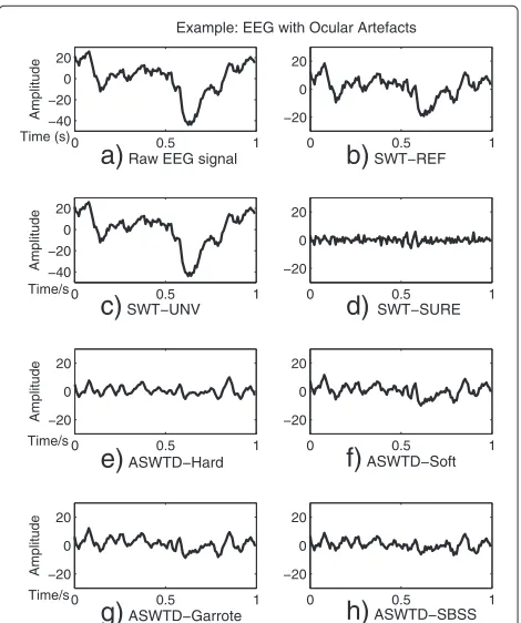
value of 1. Figure 9 shows an example when SWT-REF and SWT-UNV are applied to a real EEG signal con-taminated with ocular artefacts. The artefacts are not effectively removed when the two approaches mentioned above are used (Figure 9(b) and Figure 9(c)). The rea-son is that the estimated threshold values are bigger than the optimal thresholds and hence, the wavelet coeffi-cients corresponding to the artefacts are not completely removed.
We also observe from Figure 8 that SWT-SURE has very smallPSDdvalues (PSDd<<1). For EEG signals
contam-inated with ocular artefacts, only the wavelet coefficients
0 0.5 1
−40 −20 0 20
a)
Raw EEG signalAmplitude
Example: EEG with Ocular Artefacts
0 0.5 1
−20 0 20
d)
SWT−SURE0 0.5 1
−20 0 20
b)
SWT−REFTime/s 0 0.5 1
−40 −20 0 20
c)
SWT−UNVTime (s)
Amplitude
0 0.5 1
−20 0 20
e)
ASWTD−HardAmplitude
0 0.5 1
−20 0 20
f)
ASWTD−Soft0 0.5 1
−20 0 20
g)
ASWTD−GarroteAmplitude
Time/s 0 0.5 1
−20 0 20
h)
ASWTD−SBSSTime/s
that correspond to the lower frequency bands (i.e., up to 16 Hz) are thresholded [33]. For EEG signals contami-nated with muscle artefacts, the wavelet coefficients from all decomposition levels are thresholded as the artefacts affect the EEG signals in all frequency bands. Hence, a greater over-correction (a smallerPSDdvalue) is observed
in the case of muscle artefacts. Figure 9(d) shows the denoised EEG signal obtained using SWT-SURE, when applied to the real EEG signal mentioned above. We note that the amplitude of the denoised signal is relatively small due to the over-correction.
As shown in Figure 8, the proposed ASWTD achieves smaller distortion: 1) the MSE values are smaller than other artefact removal algorithms and closer to the ideal value of 0, and 2) the spectral distortion valuesPSDdare
close to the ideal value of 1. Among all the threshold-ing functions, the non-negative garrote function has the best performance. The BSS algorithms, on the other hand, have larger MSE values compared to our ASWTD. The PSDd values for the case of ocular artefacts are larger
than 1, as the artefacts are not completely removed and some signal distortion may have been introduced by the algorithms. For the case of muscle artefacts, the BSS algo-rithms are not as efficient in isolating artefacts from the EEG signals, as compared to the case of ocular artefacts. Thus, more source components are identified as contam-inated with muscle artefacts and these components are unfortunately removed [22]. This may have resulted in an over-estimation of artefacts (and larger distortion in the estimated signals). Hence,PSDdvalues of less than one are
observed.
Figure 9 (e) - (h) presents the denoised signals obtained when ASWTD (with various thresholding functions) are used to remove artefacts in the real EEG signal Figure 9(a). Based on visual inspection, the artefacts are effectively removed by ASWTD. For the SBSS function, less informa-tion from the small coefficients is removed from the EEG signals and more information from the large coefficients (corresponding to artefacts) has been removed. Hence, the denoised signal obtained shows slightly more details (and therefore is less smooth) compared to the rest.
Examples of applying SWT-SURE, ASWTD and BSS algorithms to real EEG signals are shown in Figure 10 and Figure 11. The raw EEG segments are contaminated with an eye-blink and fEMG artefacts respectively. As shown in Figure 10, SOBI, AMUSE and ERICA remove the artefacts to a certain extent. In Figure 11(d), however, SOBI fails to completely remove the artefacts. For AMUSE, ERICA and SWT-SURE, the EEG signals are over-corrected and the distortion is observed in the denoised signals. On the other hand, ASWTD with the non-negative garrote thresholding function gives the best results. It has smaller signal distortion as well as a smaller variance between the two estimated denoised signals.
0 0.5 1
−100 −50 0
a)
Raw EEG signalAmplitude
0 0.5 1
−20 0 20
b)
ASWTD−Garrote0 0.5 1
−20 0 20
c)
SWT−SUREAmplitude
0 0.5 1
−20 0 20
d)
SOBITime/s
0 0.5 1
−20 0 20
e)
ERICAAmplitude
Time/s 0 0.5 1
−20 0 20
f)
AMUSETime/s
Figure 10Denoised EEG Signals Obtained Different Artefact Removal Algorithms (Ocular Artefacts).The real EEG signal contaminated with ocular artefacts is shown ina). The denoised EEG signals obtained using the different artefact removal algorithms are shown inb) -f):b) ASWTD-Garrote;c) SWT-SURE;d) SOBI;e) ERICA; andf) AMUSE.
TPR/TNFPR of the hybrid BCI
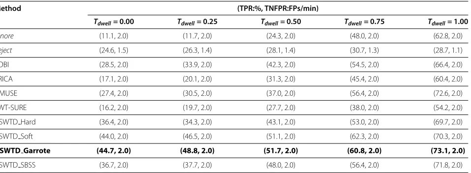
Table 1 compares the average performance achieved by the hybrid BCI system for seven individuals, when dif-ferent artefact handling methods and dwell times (Tdwell)
are used (note that a dwell time of 0.0s implies that the user can select a target immediately once he gazes at it).
0 0.5 1
−50 0 50
a)
Raw EEG signalAmplitude
Time/s 0 0.5 1
−20 0 20
Example: EEG with Muscle Artefacts
b)
ASWTD−Garrote0 0.5 1
−20 0 20
c)
SWT−SUREAmplitude
Time/s 0 0.5 1
−20 0 20
d)
SOBI0 0.5 1
−20 0 20
e)
ERICAAmplitude
Time/s 0 0.5 1
−20 0 20
f)
AMUSETable 1 Comparing the Performance of Different Artefact Handling Methods
Method (TPR:%, TNFPR:FPs/min)
Tdwell= 0.00 Tdwell= 0.25 Tdwell= 0.50 Tdwell= 0.75 Tdwell= 1.00
Ignore (11.1, 2.0) (11.7, 2.0) (24.3, 2.0) (48.0, 2.0) (62.8, 2.0)
Reject (24.6, 1.5) (26.3, 1.4) (28.1, 1.4) (30.7, 1.3) (28.7, 1.1)
SOBI (28.5, 2.0) (33.9, 2.0) (42.3, 2.0) (54.5, 2.0) (66.4, 2.0)
ERICA (17.1, 2.0) (20.1, 2.0) (31.3, 2.0) (45.4, 2.0) (60.4, 2.0)
AMUSE (27.4, 2.0) (30.5, 2.0) (37.0, 2.0) (56.4, 2.0) (72.6, 2.0)
SWT-SURE (16.2, 2.0) (19.7, 2.0) (27.7, 2.0) (38.0, 2.0) (54.2, 2.0)
ASWTD Hard (36.4, 2.0) (34.3, 2.0) (43.1, 2.0) (53.0, 2.0) (69.7, 2.0)
ASWTD Soft (44.0, 2.0) (46.5, 2.0) (51.1, 2.0) (62.3, 2.0) (70.3, 2.0)
ASWTD Garrote (44.7, 2.0) (48.8, 2.0) (51.7, 2.0) (60.8, 2.0) (73.1, 2.0)
ASWTD SBSS (36.7, 2.0) (37.7, 2.0) (48.0, 2.0) (56.4, 2.0) (71.8, 2.0)
Comparing the performance of the hybrid BCI system with the different artefact handling methods and dwell times.
All the real EEG segments obtained from the last session are included in the analysis (including those contaminated with artefacts). We consider the following three artefact handling methods (explained in Section 2):
1. Ignore: No artefact handling is employed; 2. Reject: Contaminated EEG segments are rejected; 3. Remove: An artefact removal algorithm (ASWTD,
SWT-SURE, SOBI, ERICA or AMUSE) that denoises contaminated EEG segments is applied.
For ASWTD, different thresholding functions are used:
1. ASWTD Hard: ASWTD + hard thresholding 2. ASWTD Soft: ASWTD + soft thresholding 3. ASWTD Garrote: ASWTD + non-negative garrote
thresholding
4. ASWTD SBSS: ASWTD + SBSS thresholding
A two-way Analysis of Variance (ANOVA) [54] was carried out to examine the statistical significance of the results. ANOVA showed that the mean performances of the hybrid BCI system with different artefact handling methods and different dwell times were significantly dif-ferent at a significance level of 0.01.
As shown in Table 1, the hybrid BCI system withIgnore has an average TPR = 11.1% and TNFPR = 2.0 FPs/min, when the dwell time is 0.0s. As the dwell time increases to 0.5s, and finally to 1.0s, the TPR increases to 34.3% and then to 62.8% (for the same TNFPR).
WhenRejectis used, many EEG segments are rejected and blocked by the system due to the presence of artefacts. The explanation is as follows. The EEG data recorded from seven participants during the last session contained an average of 88± 19 IC trials and 2595±698 NC tri-als (IC tritri-als = the number of attempted hand extension
executed; NC trials = the number of 1-second EEG seg-ments obtained outside the TP window, as defined ear-lier). Approximately 48.4±38.8% of IC and 90.2±11.4% of NC trials were contaminated with artefacts. Reject-ing these trials means that these data are discarded and not presented as inputs to the system. Therefore, when-ever artefacts are detected, the availability of the BCI for control is significantly reduced. This may lead to generating many false negatives (i.e., missed true activa-tions) because many IC trials are blocked due to arte-facts. Hence, both the TPR and TNFPR values are small and the results are not significantly different for various dwell times.
On the other hand, Remove allows the users to have more control over the BCI system, as the system is oper-ational even in the presence of artefacts. Besides, this approach reduces the effects of artefacts and achieves a better performance when compared toIgnoreandReject. This performance improvement is especially significant, when the value ofTdwellis small. For example, when dwell
time is 0.0s, the TPR achieved using ASWTD Garrote is 44.7% , which is more than 20% of those of Ignore andReject. As the dwell time increases, the performance difference between the methods decreases. The reason is that increasing the dwell time reduces the availability of the system to only those periods for which a selec-tion might happen. Thus, the system is put in the so-called ‘inactive’ mode more frequently and the effects of artefacts on the system’s performance are significantly reduced.
ASWTD using different thresholding functions also outperforms SWT-SURE and other BSS algorithms. Among all the thresholding functions, the non-negative garrote thresholding achieves the best performance, i.e.,
Table 2 Comparing the Performance ofIgnoreand ASWTD Garrote
Method (TPR:%, TNFPR:FPs/min)
Tdwell= 0.00 Tdwell= 0.25 Tdwell= 0.50 Tdwell= 0.75 Tdwell= 1.00
Ignore (11.1, 2.0) (11.7, 2.0) (24.3, 2.0) (48.0, 2.0) (62.8, 2.0)
ASWTD Garrote (22.2, 2.0) (23.7, 2.0) (35.4, 2.0) (52.5, 2.0) (66.6, 2.0)
Comparing the performance of the hybrid BCI system usingIgnoreand ASWTD Garrote when the classifier is trained using only clean EEG trials.
increases steadily to 73.1% when the dwell time increases to 1.0s.
In Table 1, the performance of ASWTD is obtained from the BCI classifier trained using both clean and denoised EEG trials. We also investigate the performance of ASWTD Garrote, when the BCI classifier is trained using only clean EEG trials (denoted by BCIclean). The results are presented in Table 2. Note that the TPR val-ues obtained in Table 2 are lower than those in Table 1 because a smaller number of EEG trials are available to trainBCIcleandue to artefact contamination. WhenIgnore
is used and the dwell time is 0.0s, TPR = 11.1% and TNFPR = 2.0 FPs/min are obtained. ASWTD Garrote, on the other hand, removes artefacts in contaminated EEG trials and successfully improves the TPR values from 11.1% to 22.2% (at the same TNFPR). The contribution to the improvement comes entirely from those EEG tri-als with artefacts, because the proposed algorithm does not operate on clean EEG trials (i.e., the performance from of both artefact handling methods remain the same when only clean EEG trials are evaluated). The results in Table 2 suggest that when artefacts are ignored, the artefacts results in a change in the quality of the EEG signals and therefore affect the performance ofBCIclean.
ASWTD Garrote successfully minimizes the effects of artefacts and improves the classifier’s performance. When a larger number of trials are used in training the classi-fier (Table 1), ASWTD Garrote achieves even higher TPR values (at the same TNFPR).
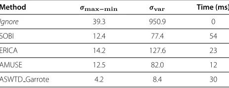
Inter-Trial Variability/Processing Time
When an artefact removal algorithm shows a large trial-by-trial variability in the amplitudes of the denoised sig-nals, this might suggest that the algorithm is not efficient in removing artefacts. Possible causes of such a large inter-trial variability could be that:
1. the algorithm does not completely remove artefacts or
2. the algorithm sometimes removes the artefacts efficiently, but sometimes over-corrects the EEG signals or does not completely remove the artefacts.
Here, we quantify the inter-trial variability in the ampli-tudes of the denoised EEG signals (estimated using
various artefact removal algorithms) when applied to real EEG signals by finding the standard deviation of:
1. the variance of each estimated denoised EEG signals (σvar)
2. the difference between the maximum and minimum value of each denoised EEG signals (σmax−min)
The results are presented in Table 3. Evidently, theσvar
and σmax−min are large when the artefacts are ignored
because of the large differences between the amplitudes of clean and contaminated EEG signals. ASWTD, how-ever, has a significantly smallerσvarandσmax−min values. The BSS algorithms have largerσvarandσmax−minvalues because the denoised EEG signals estimated by these algo-rithms are less consistent. For example, in Figure 10(d), SOBI successfully removes the ocular artefacts, whereas in Figure 11(d), SOBI fails to completely remove the muscle artefacts. This results in a larger inter-trial variability.
Besides inter-trial variability, we also examine another performance metric that needs to be taken into consid-eration for online implementation: the processing time required to run the artefact algorithms (see the last column of Table 3). In this study, all algorithms were run in Matlab 2009b environment. For SWT, the Rice Wavelet Toolbox from RICE University was used [55]. The processor used was an 2.93 GHz Intel (R) Core i7 870. As shown in Table 3, all algorithms require no more than 60 ms to process a 1-second EEG segment with 15 channels, indicating their suitability for online applications.
Table 3 Inter-Trial Variability and Processing Time
Method σmax−min σvar Time (ms)
Ignore 39.3 950.9 0
SOBI 12.4 77.4 54
ERICA 14.2 127.6 23
AMUSE 12.5 82.0 12
ASWTD Garrote 4.2 8.4 30
Discussion
This paper proposes a fully automated algorithm to remove artefacts from EEG signals and subsequently improve the performance of our hybrid BCI system. Specifically:
1. we propose an adaptive thresholding method based on SWT to remove various artefacts in EEG signals. It is shown that the proposed method (ASWTD) greatly improves the performance of the hybrid BCI system and reduces signal distortion and
2. we investigate the effects of using different thresholding functions in the performance of ASWTD.
In the following subsections, more details about the above claims are provided.
Comparison of different artefact handling methods We have investigated and compared the performance of our hybrid BCI system, when different artefact handling methods are used to denoise the real EEG data. The performance is evaluated using a pseudo-online testing paradigm, where all real EEG data (both clean and contaminated) are included in the testing. Such testing provides us with a better understand-ing of the system’s performance in a real-world online application, where artefacts are present in the EEG signals.
We need to emphasize the importance of the system having a low TNFPR. A low TNFPR ensures that the sys-tem does not cause too much frustration for users. This is because users are in an NC state for most of the time when using the system. Also, it is usually easier to deal with a missed IC command than with a false activation (i.e., an FP). For example, in a text-writing application, a false positive results in selecting the wrong letter/word. Con-sequently, the user has to initiate additional commands to de-selectthe wrong letter/word and then select the cor-rect desired letter/word. On the other hand, in the case of a missed IC, the user only has to issue the IC command again. Therefore, it is important to lower the TNFPR as much as possible.
Table 1 and Table 2 show that artefacts can affect the BCI system’s performance. If artefacts are ignored (Ignore), the system has a low TPR value, especially when the dwell time is small. The rejection of contaminated EEG segments (Reject), on the other hand, reduces the amount of time for which the hybrid BCI system is avail-able for control. In addition, this approach rejects IC trials contaminated with artefacts, which results in lower TPR values (Table 1). The drawbacks ofIgnoreandReject sig-nify the need to minimize the effects of artefacts by apply-ing artefact removal algorithms. As shown in Table 1,
Removegreatly improves the performance of the hybrid BCI system.
Our study demonstrated that the proposed artefact removal algorithm ASWTD can improve the hybrid BCI system’s performance in two ways:
1. ASWTD Garrote reduces the effects of artefacts and improves the performance of the hybrid BCI system. This is when the BCI classifier is trained with clean EEG trials only (see Table 2);
2. ASWTD Garrote increases the number of clean EEG trials available for training the BCI classifier. Both the clean and denoised EEG trials are used to train the classifier. This further increases the detection performance of the hybrid BCI system (see Table 1).
ASWTD also has another advantage: a smaller dwell time can be used when the algorithm is incorporated into the hybrid BCI system. Thus, the user does not have to gaze at the target for too long to make a selection. For example, ASWTD Garrote achieves a TPR of 48.8% at a TNFPR of 2 FPs/min when the dwell time is 0.25s. This performance is as good as the one achieved byIgnorebut when the dwell time is 0.75s (TPR = 48.0%, TNFPR = 2 FPs/min).
Comparison of different artefact removal algorithms Our results shows that ASWTD outperforms SWT-SURE, SOBI, ERICA and AMUSE. More specifically, it achieves:
1. lower MSE values and less spectral distortion when semi-simulated EEG signals with ocular and muscle artefacts are used (see Figure 8);
2. larger TPR values when real EEG signals are used (see Table 1); and
3. smaller inter-trial variability in the amplitudes of the denoised EEG signals when real EEG signals are used (see Table 3).
As the proposed artefact removal algorithm introduces less distortion in EEG signals, false artefact detection (i.e., the artefact detector falsely detects artefacts in a clean EEG signal) may not pose too much of a problem. An example is shown in Figure 12, where a clean EEG segment is processed using ASWTD Garrote, SWT-SURE, SOBI, ERICA and AMUSE. The denoised signal obtained using ASWTD has less distortion while the other algorithms over-correct the EEG signal. Also, when semi-simulated EEG signals are used (see Figure 8), ASWTD also achieves smaller MSE and PSDd values (which are closer to the
ideal values).
SWT-SURE does not perform as well because the esti-mated thresholds often lead to the over-estimation of arte-facts and hence it removes some EEG features (PSDd <<
![Figure 1 Our Hybrid BCI System. The hybrid BCI system proposed in [7].](https://thumb-us.123doks.com/thumbv2/123dok_us/9066863.1899158/3.595.59.542.87.283/figure-hybrid-bci-hybrid-bci-proposed.webp)
![Figure 7 Structure of the Feature Extraction and Classification Blocks of the BCI. The structure of the feature extraction and classificationalgorithms of the self-paced BCI system [7].](https://thumb-us.123doks.com/thumbv2/123dok_us/9066863.1899158/11.595.58.541.632.704/figure-structure-feature-extraction-classication-structure-extraction-classicationalgorithms.webp)