Journal Homepage: vrf.iranjournals.ir
Effects of nano-selenium on mRNA expression of markers for spermatogonial
stem cells in the testis of broiler breeder males
Seyed Sattar Jalali1, Alireza Talebi1*, Manoochehr Allymehr1, Ali Soleimanzadeh2, Mazdak Razi3
1 Department of Poultry Health and Diseases, Faculty of Veterinary Medicine, Urmia University, Urmia, Iran; 2 Department of Theriogenology, Faculty of Veterinary Medicine, Urmia University, Urmia, Iran; 3 Department of Basic Sciences, Faculty of Veterinary Medicine, Urmia University, Urmia, Iran.
Article Info Abstract
Article history:
Received: 03 June 2018 Accepted: 24 July 2018 Available online: 15 June 2019
Fertility is one of the most important parameters in breeder farms and cockerels play an outstanding role in the fertility of eggs in broiler breeder farms. Todays, supplementation of chicken diet with additives such as organic selenium is used to increase fertility. The aim of this study was to evaluate the effects of different levels of nano-selenium (Nano-Se) on the expression of molecular markers of spermatogonial stem cells (SSCs) in the testis of broiler breeder males. A total of 30 roosters of 40 weeks of age were randomly divided into five groups. Groups were as follows: 1) control (normal diet) group, 2) diet supplemented with 0.30 mg kg-1 sodium selenite, 3) diet supplemented with 0.15 mg kg-1 Nano-Se, 4) diet supplemented with 0.30 mg kg-1 Nano-Se, and 5) diet supplemented with 0.60 mg kg-1 Nano-Se. At the end of the experimental period (5th week), birds were autopsied and samples from testis of all birds were collected. The testis samples were used to examine the β1-integrin (CD29), thy-1 (CD90) and NANOG mRNA expression by real-time PCR. The results showed that testis of the groups fed with the diets supplemented with 0.60 mg kg-1 and 0.15 mg kg-1 of Nano-Se had the highest and lowest mRNA expression of SSCs markers, respectively. In conclusion, the present study indicated that Nano-Se had advantages over sodium selenite. Diet supplemented with 0.60 mg kg-1 of Nano-Se may contribute to optimal fertility via increasing the mRNA expression of SSCs markers of roosters’ testis and could be used to delay the reduction of fertility caused by aging in broiler breeder males.
© 2019 Urmia University. All rights reserved.
Key words:
Broiler breeder males Molecular markers Nano-selenium
Spermatogonial stem cells
راثآ لولس یلوکلوم یاهرکرام نایب رب موینلس ونان سورخ هضیب یداینب یاه یتشوگ ردام هلگ یاه هدیکچ هفطن مخت یراد غرم یم بوسحم ردام غرم یاهدحاو رد مهم یلیخ یاهروتکاف زا یکی اه سورخ و دوش هتسجرب شقن اه مخت یروراب رد ار یا غرم یم افیا اه هزورما .دننک هب نم ظ سورخ یروراب شیازفا رو -لمکم زا اه یم هدافتسا موینلس تابیکرت لیبق زا یندوزفا یاه لوکلوم یاهرکرام نایب رب موینلس ونان لمکم فلتخم حوطس تارثا یبایزرا هعلاطم نیا زا فده ،تارذ ونان تابیکرت نساحم هب تیانع اب .دوش ی لولس ( ینوگوتامرپسا یداینب یاه SSCs خ هضیب ) سور عومجم رد .دوب یتشوگردام هلگ یاه 03 نس اب یتشوگردام هلگ سورخ هعطق 03 هب یفداصت تروصب ،هتفه جنپ هورگ .دندش میسقت هورگ :لماش اه 1 ) ،)لامرن هریج( لرتنک 2 یواح هریج ) 03 / 3 یلیم ،مرگولیک رد میدس تینلس مرگ 0 یواح هریج ) 11 / 3 یلیم ،مرگولیک رد موینلسونان مرگ 0 ج ) یواح هری 03 / 3 یلیم ،مرگولیک رد موینلسونان مرگ 1 یواح هریج ) 03 / 3 یلیم .دندوب مرگولیک رد موینلسونان مرگ نایاپ رد ( شیامزآ هرود مجنپ هتفه ) ییاشگدبلاک ، و هدنرپ مامت هضیب زا یرادرب هنومن هنومن .دیدرگ ماجنا اه ب هضیب یاه ا شور شنکاو جنز ی هر ا ی لپ ی زارم نامز رد یعقاو نایب رظن زا mRNA لولس یلوکلوم یاهرکرام ینوگوتامرپسا یداینب یاه β1-integrin (CD29) و thy-1 (CD90) و NANOG نیرتمک و نیرتشیب هک داد ناشن جیاتن .دنتفرگ رارق شیامزآ دروم نایب نازیم mRNA لولس یلوکلوم یاهرکرام ( ینوگوتامرپسا یداینب یاه SSCs هب ) هورگ رد بیترت اه هریج اب هدش هیذغت ی یواح یاه 03 / 3 و 11 / 3 یلیم مرگولیک رد موینلسونان مرگ دوب . جیاتن رگنایب قیقحت نیا فرصم و هتشاد تیحجرا موینلس تینلس هب تبسن موینلسونان لمکم هک تسا نآ 03 / 3 یلیم نایب شیازفا اب هریج مرگولیک رد موینلس ونان مرگ mRNA لولس یلوکلوم یاهرکرام یاه ینوگوتامرپسا یداینب ( SSCs یم ) هب دناوت دوبهب سورخ یروراب کمک اه و دیامن سورخ نس هب هتسباو یروراب شهاک ت هب ار یتشوگردام غرم یاه أ .دزادنایب ریخ :یدیلک یاه هژاو یتشوگ ردام هلگ سورخ ، لولس موینلس ونان ،یلوکلوم یاهرکرام ،هضیب یداینب یاه *Correspondence:
Alireza Talebi. DVM, MVM, PhD
Department of Poultry Health and Diseases, Faculty of Veterinary Medicine, Urmia University, Urmia, Iran E-mail: [email protected]
Forum
Introduction
The final product of breeder farms is one-day-old chicks. The eggs hatchability is mostly influenced by the fertility of cockerels. Nowadays, various feed additives including organic selenium are used in the diet of broiler breeder males in order to increase fertility. Recent studies indicated that nano-particles of selenium may have some advantages over organic selenium during spermatogenesis. Four types of spermatogonial cells such as spermatogonia dark type A (Ad), pale type A (Ap1, Ap2), and type B, have been described in birds and the type Ad spermatogonia have been considered as the spermatogonial stem cells (SSCs).1-3 The SSCs are the early precursor for spermatozoa and are responsible for the continuation of spermato-genesis in adult males' testis. These cells have the capability to undergo self-renewal division as well as produce daughter cells destined for differentiation into spermatozoa and transferring genetic information from an individual to the offsprings.4-6 During a routine spermato-genesis process, the SSCs infrequently self-renew, but they can divide frequently in response to harmful conditions such as damage due to chemical compounds.7 Therefore, the performance of SSCs during spermatogenesis influences both the number of spermatogenic cells and semen parameters including sperm concentration.8 Similar to the other tissue-specific stem cells, SSCs are rare in chicken. The proportions of SSCs to testes’ germ cells in young chickens and adult males are approximately 0.40% and 0.03% respectively.9,10
The SSCs reside within a specialized micro-environment called ‘niche’ which regulates the behavior of the stem cells and its differentiating progeny. The SSCs performance (self-renewal and differentiation) is tightly regulated by a combination of intrinsic gene expression within the SSC and the extrinsic gene signals from the niche. Communication between the niche and SSCs is crucial to maintain proper spermatogenesis7,11,12
Aging leads to an increase in reactive oxygen (ROS) and when the generation of ROS in a system exceeds, the occurrence of oxidative stress may cause a reduction in antioxidant enzymes in spermatogonial stem cells.13-19 During oxidative stress, free radical production is increased causing damages to different organs/cells. In order to decrease harmful effects of free radicals on reproductive organs and subsequently to prevent the reduction of males' fertility, dietary supplementation of an antioxidant such as selenium compounds is necessary.20,21 Selenium is a major structural component of many antioxidant enzymes and has an important role in a variety of biological processes including antioxidant defense system, fertility, immune function, and muscle metabolism.22-31 Selenium protects spermatozoa against oxidative damage during spermato-genesis and in fact, the optimal level of Se is required for normal development of male reproductive tissue.8,32-35
Nowadays, nanotechnology proposed several new effective forms of dietary supplementation components due to their low toxicity and high bioavailability. Among those, nano-selenium (Nano-Se) particles, because of their excellent characteristics including great surface area, effective surface activity, lots of surface active centers, high catalytic efficiency, strong absorbing ability and low toxicity have many advantages in comparison to sodium selenite.36 The purpose of this study was to determine the most optimal dose of supplemental Nano-Se on the mRNA expression of molecular markers of SSCs in the testis of broiler breeder males.
Materials and Methods
Ethics of experimentation. All experimental procedures were carried out according to the standard animal experimentation protocols of the Veterinary Ethics Committee of Faculty of Veterinary Medicine, Urmia University (IR-UU-AEC-266/DP3).
Experimental design. Thirty broiler breeder males (Arbor Acres Plus strain) at the age of 40 weeks were used in this study. The birds were randomly divided into five groups (six birds per group) and housed in the pens of identical size in a deep litter system with wood shaving floor. Each group had three replicates (two birds per pen). The first week of the experiment was designed as an adaptation period and during this period birds of all the groups were fed with standard basal diet according to the catalog of Arbor Acres Plus.37 After adaptation period, group 1 (control group) was fed with basal diet, group 2 was fed with basal diet supplemented with 0.30 mg kg-1 sodium selenite, groups 3, 4 and 5 were fed with basal diet supplemented with 0.15 mg kg-1, 0.30 mg kg-1, 0.60 mg kg-1 of Nano-Se, respectively. All birds fed with the described diets for four weeks after the adaptation period.38
Selenium. Sodium selenite (Merck, Darmstadt, Germany) and Nano-Se (American Elements, Los Angeles, USA) with the size of 10-45 nm and purity of 99.99% were purchased.
Sample collections. At the end of the 5th week, birds of all groups were humanely euthanatized by cervical dislocation.39 Then testes were collected, rinsed with normal saline, frozen immediately in liquid nitrogen and stored at – 80 ˚C to investigate the mRNA expression levels of SSCs markers β1integrin (CD29), thy-1 (CD90) and NANOG) in testicular tissue with Real-Time PCR.8
(5’ TCAGCCTCACCAGACAACA 3’) and antisence (5’ GGACGCACTTCTCCACTTT 3’);8 GAPDH (housekeeping gene) specific primers amplifying a fragment of 293 bp in size: Sence (5’ GCCCAGAACATCATCCCA 3) and antisence (5’ CCAGCACACGCATCAAAG 3’);8 NANOG specific primers amplifying a fragment of 195 bp in size: Sence (5’ CTCCAGCAGCAGACCTCTCCTTG 3’) and antisence (5’ CCTTCCTTGTCCCACTCTCACCTT 3’).40
RNA extraction. The total RNA of testis samples were extracted using Cinna Pure-RNA kit according to the manufacturer’s instructions (CinnaGen, Tehran, Iran). The concentration and purity of the total RNA was determined by spectrophotometer (ND-2000; Thermo Fisher Scientific, Waltham, USA). Total RNA was stored at – 70 ˚C until cDNA synthesis.
cDNA synthesis. First strand cDNA was synthesized from 10 μg of total RNA using random hexamers primers,
dNTP Mix, M-MLV Reverse Transcriptase (Vivantis Technologies, Selangor, Malaysia), 10X Buffer M-MuLV according to the manufacturer’s instructions. The reaction incubated at 42 ˚C for 60 min and terminated by incubating at 85 ˚C for 5 min followed by cooling at 4 ˚C. Synthesized cDNA was stored at – 20 ˚C until processed.
PCR assay. The PCR thermal program for β1-integrin (CD29), thy-1 (CD90), NANOG and GADPH consisted of 95 ˚C for 10 sec, followed by 40 cycles of 95 ˚C for 10 sec, and 60 ˚C for 30 sec.
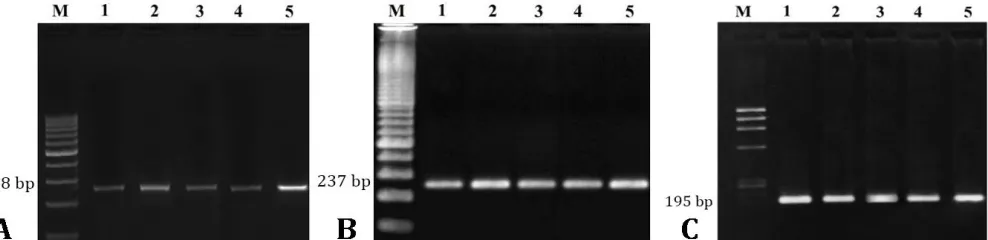
Electrophoresis. Agarose gel electrophoresis was performed with the PCR products to verify the primers specificity (Fig. 1).
Real-Time PCR assay. The mRNA expression levels of SSCs markers (β1-integrin (CD29), thy-1 (CD90), NANOG and GADPH were quantified by the Real-Time RT-PCR using SinaSyber Blue HF- qPCR Mix (CinnaGen) and was performed on a Step One™ Real-Time PCR System (Applied Biosystems, Carlsbad, USA) in 25.00 μL reaction volume. PCR reactions were comprised of 12.50 μL of SinaSyber Blue HF- qPCR Mix, 0.25 μL of each primer (2.00 μM), 2.00 μL of cDNA, 9.50 μL of nuclease-free water. PCR thermal condition was 95 ˚C for 10 sec, followed by 40 cycles of 95 ˚C for 10 sec, and 60 ˚C for 30 sec.
We used a standard curve from cDNA reaction mixture to determine PCR efficiencies. The efficiencies of PCR were observed between 90 and 110 percent. The values for R2 for all curves were between 0.997 to 0.999. For quantification of the product of PCR, we used the amounts of CT and expressed the relative expression level of the target gene as 2-ΔCT. We measured ΔCT after subtracting CT of housekeeping gene (GAPDH), from CT of target gene.8
Statistical analysis. The results were analyzed using SPSS s (version 23.0; IBM Corp, Chicago, USA) employing one-way ANOVA. The means of different treatments were compared with Tukey post-hoc test. Data are expressed as the mean ± standard error of mean (mean ± SE). The differences were considered to be significant at p ≤ 0.05.
Results
The results of mRNA expressions of the SSCs markers in the experimental groups are expressed as fold-increase in comparison to the reference gene are shown in Figures 2 to 4. As shown in Figure 2, the mRNA expressions of the β1-integrin (CD29) gene of the groups 1-5 (control, 0.30 mg kg-1 sodium selenite, 0.15 mg kg-1 Nano-Se, 0.30 mg kg-1 Nano-Se, 0.60 mg kg-1 Nano-Se) were 1.63 ± 0.17, 1.72 ± 0.21, 1.69 ± 0.19, 1.80 ± 0.21 and 2.15 ± 0.17, respectively. The diet containing different level of Nano-Se had an increasing impact on mRNA expression of β1-integrin and the group fed with 0.60 mg kg-1 Nano-Se supplemented diet had the highest mRNA expressions of the β1-integrin (CD29).
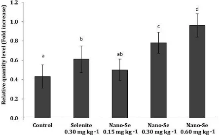
In regards to thy-1 (CD90) gene, the mRNA expressions of the groups 1-5 were 1.04 ± 0.21, 1.14 ± 0.24, 1.07 ± 0.14, 1.21 ± 0.17 and 1.33 ± 0.13, respectively (Fig. 3) and the highest mRNA expressions was seen in the group fed with diet containing 0.6 mg kg-1 Nano-Se. As shown in Figure 4, the mRNA expressions of the NANOG gene of the groups 1-5 were 0.43 ± 0.12, 0.61 ± 0.14, 0.50 ± 0.11, 0.78 ± 0.11 and 0.96 ± 0.12, respectively and the group fed with 0.6 mg kg-1 Nano-Se supplement diet had the highest level of NANOG gene expression.
Overall, the lowest level of the mRNA expression of all three genes was in the group fed with diet containing 0.15 mg kg-1 Nano-Se (Figs. 2 to 4). Primer specificity and cDNA synthesis were verified (Fig. 1).
Fig. 2. Level of mRNA expressions of β1-integrin (CD29) in the treatment groups.
Different letters indicate significant differences between the groups (p < 0.05).
Fig. 3. Level of mRNA expressions of thy-1 (CD90) in the treatment groups.
Different letters indicate significant differences between the groups (p < 0.05).
Fig. 4. Level of mRNA expressions of NANOG genes in the treatment groups.
Different letters indicate significant differences between the groups (p < 0.05).
Discussion
Selenium deficiency significantly decreased the population of SSCs and causing low fertility.8,32-34,41 Therefore, selenium in both inorganic (selenite, selenate) and organic (selenomethionine, selenocysteine and methylselenocysteine) forms is used as a feed supplementation to improve immune responses and fertility in the poultry industry.42 Recent studies have demonstrated that excess and deficiency of selenium supplementation can stimulate oxidative stress, which probably leads to the reduction of SSCs population, therefore many studies have been designed to determine the optimal dose of selenium in poultry.8,32,43,44 In recent years, concurrent nano-particle science progresses, therefore Nano-Se has been paid attention as a novel prospect for nutritional supplementation.45 Thus limited studies are available on Nano-Se, especially in the poultry. As Nano-Se has some advantages (lower toxicity and higher bioavailability) in comparison to inorganic forms' of selenium, therefore, investigating the effective dose of Nano-Se on poultry performances is of great interest to researchers. During this study, the effects of different doses of supplemental Nano-Se on the expression of SSCs markers in the testis of broiler breeder males were investigated by using SSCs surface antigens including β1-integrin (CD29), thy-1 (CD90) and NANOG that known as the molecular markers.
As shown in Figures 2-4, the mRNA expressions of SSCs markers were elevated by the supplementation of the diet with both sodium selenite and Nano-Se when compared to the control group. The result of real-time RT-PCR indicated that similar to inorganic selenium, Nano-Se dietary supplementation could influence the mRNA expression of SSCs specific markers in testis of broiler breeder males. The results of the present study were in agreement with the previous reports which indicated that supplementation of selenium compounds increases SSCs markers.8
The results obtained in the present study clearly showed that supplementation of the poultry diet with Nano-Se was more effective than sodium selenite. The dose of 0.60 mg kg-1 of Nano-Se could be used in the diet of roosters in order to improve their performances and in particular their fertility without any side effects. Regarding the mechanism of action, Selenium as an essential trace element plays an important role in antioxidation46,47 and protects various systems against damage caused by free radicals and oxidative stress.48 The physiological functions of Selenium are considered to be mediated through selenoproteins.49,50 Unlike the previous studies using inorganic selenium which showed that higher levels of sodium selenite reduces the number of SSCs markers,8 the results of the present study revealed that the increasing levels of Nano-Se in the diet did not cause a detrimental effect on the population of SSCs markers (Figs. 2-4). These differences could be attributed to the differences in selenium sources.
In conclusion, the supplementation of the diet with 0.60 mg kg-1 of Nano-Se can be used in roosters feeding to improve their performance in particular when fertility is declining due to aging in broiler breeder males from 40 weeks of age onward.
Acknowledgments
This study was supported by Vice-Chancellor for Research of Urmia University.
Conflicts of interest
The authors notify that they have no conflicts of interest.
References
1. Dym M. Spermatogonial stem cells of the testis. Proc Natl Acad Sci USA 1994; 91(24): 11278-11289.
2. Lin M, Jones RC, Blackshaw AW. The cycle of the seminiferous epithelium in the Japanese quail (Coturnix coturnix japonica) and estimation of its duration. J Reprod Fertil 1990; 88 (2): 481-490. 3. Lin M, Jones RC. Renewal and proliferation of
spermatogonia during spermatogenesis in the Japanese quail, Coturnix coturnix japonica. Cell Tissue Res 1992; 267(3): 591-601.
4. de Rooij DG. Proliferation and differentiation of spermatogonial stem cells. Reproduction 2001; 121(3): 347-354.
5. Aponte PM, Soda T, Teerds KJ, et al. Propagation of bovine spermatogonial stem cells in vitro. Reproduction 2008; 136(5): 543-557.
6. Phillips BT, Gassei K, Orwig KE. Spermatogonial stem cell regulation and spermatogenesis. Phil Trans R Soc Lond Bio Sci 2010; 365(1546): 1663-1678.
7. Singh SR, Burnicka-Turek O, Chauhan C, et al. Spermatogonial stem cells, infertility and testicular cancer. J Cell Mol Med 2011; 15(3): 468-483.
8. Shi L, Zhao H, Ren Y, et al. Effects of different levels of dietary selenium on the proliferation of spermatogonial stem cells and antioxidant status in testis of roosters. Anim Reprod Sci 2014; 149(3-4): 266-272.
9. Han JY, Lee HC, Park TS. Germline-competent stem cell in avian species and its application. Asian J Androl 2015; 17(3): 421-426.
10. Jung JG, Lee YM, Park TS, et al. Identification, culture, and characterization of germline stem cell-like cells in chicken testes. Biol Reprod 2007; 76(1): 173-182. 11. Dadoune JP. New insights into male gametogenesis:
what about the spermatogonial stem cell niche. Folia Histochem Cytobiol 2007; 45(3): 141-147.
12. Oatley JM, Brinster RL. Regulation of spermatogonial stem cell self-renewal in mammals. Annu Rev Cell Dev Biol 2008; 24(1): 263-286.
13. Harman D. Aging: A theory based on free radical and radiation chemistry. J Gerontol 1956; 11(3): 298-300. 14. Schoneich C. Reactive oxygen species and biological
aging: a mechanistic approach. Exp Gerontol 1999; 34(1): 19-34.
15. Ryu BY, Orwig KE, Oatley JM, et al. Effects of aging and niche microenvironment on spermatogonial stem cell self-renewal. Stem Cells 2006; 24(6): 1505-1511. 16. Zhang X, Ebata KT, Robaire B, et al. Aging of male
germ line stem cells in mice. Biol Reprod 2006; 74(1): 119-124.
17. Agarwal A, Makker K, Sharma R. Clinical relevance of oxidative stress in male factor infertility: an update. Am J Reprod Immunol 2008; 59(1): 2-11.
18. Gharagozloo P, Aitken RJ. The role of sperm oxidative stress in male infertility and the significance of oral antioxidant therapy. Hum Reprod 2011; 26(7): 1628-1640.
19. Schmidt JA, Abramowitz LK, Kubota H, et al. In vivo and in vitro aging is detrimental to mouse spermatogonial stem cell function. Biol Reprod 2011; 84(4): 698-706 20. Surai PF. Natural antioxidants in poultry nutrition:
New developments. World Poultry Science Association. In Proceedings: 16th European Symposium on Poultry Nutrition. Strasbourg, France 2007; 669-676.
21. Boostani A, Sadeghi AA, Mousavi SN, et al. The effects of organic, inorganic, and nano-selenium on blood attributes in broiler chickens exposed to oxidative stress. Acta Sci Vet 2015; 43: 1264.
22. Brenneisen P, Steinbrenner H, Sies H. Selenium, oxidative stress, and health aspects. Mol Aspects Med 2005; 26(4-5): 256-267.
24. Foresta C, Flohe L, Garolla A, et al. Male fertility is linked to the selenoprotein phospholipid hydro-peroxide glutathione peroxidase. Biol Reprod 2002; 67(3): 967-971.
25. Kaur P, Bansal MP. Effect of selenium-induced oxidative stress on the cell kinetics in testis and reproductive ability of male mice. Nutrition 2005; 21(3): 351-357.
26. Ebeid TA. Organic selenium enhances the antioxidative status and quality of cockerel semen under high ambient temperature. Br Poult Sci 2009; 50(5): 641-647. 27. Brennan KM, Pierce JL, Cantor AH, et al. Source of
selenium supplementation influences testis selenium content and gene expression profiles in Single Comb White Leghorn roosters. Biol Trace Elem Res 2012; 145(3): 330-337.
28. McKenzie RC, Rafferty TS, Beckett GJ. Selenium: An essential element for immune function. Immunol Today 1998; 19(8): 342-345.
29. Arthur JR, McKenzie RC, Beckett GJ. Selenium in the immune system. J Nutr 2003; 133(5): 1457-1459. 30. Cantor AH, Moorhead PD, Musser MA. Comparative
effects of sodium selenite and selenomethionine upon nutritional muscular dystrophy, selenium-dependent glutathione peroxidase, and tissue selenium concentrations of turkey poults. Poult Sci 1982; 61(3): 478-484.
31. Zhang J, Li J, Zhang Z, et al. Ubiquitous expression of selenoprotein N transcripts in chicken tissues and early developmental expression pattern in skeletal muscles. Biol Trace Elem Res 2012;146(2): 187-191. 32. Kaushal N, Bansal MP. Dietary selenium
variation-induced oxidative stress modulates CDC2/cyclin B1 expression and apoptosis of germ cells in mice testis. J Nutr Biochem 2007; 18(8): 553-564.
33. Kaushal N, Bansal MP. Selenium variation induced oxidative stress regulates p53 dependent germ cell apoptosis: plausible involvement of HSP70-2. Eur J Nutr 2009; 48(4): 221-227.
34. Ranawat P, Bansal, MP. Apoptosis induced by modulation in selenium status involves p38 MAPK and ROS: Implications in spermatogenesis. Mol Cell Biochem 2009; 330(1–2): 83-95.
35. Song R, Yao X, Shi L, et al. Effects of dietary selenium on apoptosis of germ cells in testis during spermatogenesis in roosters. Theriogenology 2015; 84(4): 583-588.
36. Bhattacharjee A, Basu A, Sen T, et al. Nano-Se as a novel candidate in the management of oxidative stress related disorders and cancer. Nucleus 2017; 60(2): 137-145.
37. Aviagen website. Arbor Acres plus parent stock nutritional specification. Available at: http://eu.aviagen. com/assets/Tech_Center/AA_Breeder_ParentStock//A APlus-S-PS-NS-2016-EN.pdf. Accessed: Nov 02, 2016. 38. Surai PF, Fisinin VI. Selenium in poultry breeder
nutrition: An update. Anim Feed Sci Tech 2014; 191: 1-15.
39. Collett SR. Principles of disease prevention, diagnosis, and control introduction. In: Swayne DE (Ed). Diseases of Poultry. 13th ed. Ames, USA: Wiley Blackwell 2013; 4-39.
40. Sisakhtnezhad S, Bahrami AR, Matin MM, et al. The molecular signature and spermatogenesis potential of newborn chicken spermatogonial stem cells in vitro. In Vitro Cell Dev Biol Anim 2015; 51(4): 415-425. 41. Erkekoglu P, Zeybek ND, Giray B, et al. The effects of di
(2-ethylhexyl) phthalate exposure and selenium nutrition on sertoli cell vimentin structure and germ-cell apoptosis in rat testis. Arch Environ Contam Toxicol 2012; 62(3), 539-547.
42. Garousi F. The toxicity of different selenium forms and compounds – Review. Acta Agraria Debreceniensis 2015; 64, 33-38.
43. Zhang JL, Zhang ZW, Shan AS, et al. Effects of dietary selenium deficiency or excess on gene expression of selenoprotein N in chicken muscle tissues. Biol Trace Elem Res 2014; 157(3): 234-241.
44. Khalid A, Khudhair N, He H, et al. Effects of dietary selenium supplementation on seminiferous tubules and SelW, GPx4, LHCGR, and ACE expression in chicken testis. Biol Trace Elem Res 2016; 173(1): 202-209. 45. Skalickova S, Milosavljevic V, Cihalova K, et al. Selenium
nanoparticles as a nutritional supplement. Nutrition 2017; 33, 83-90.
46. Yao HD, Wu Q, Zhang ZW, et al. Selenoprotein W serves as an antioxidant in chicken myoblasts. Biochim Biophys Acta 2013; 1830(4): 3112-3120.
47. Liu C, Fu J, Liu C, et al. The role of nitric oxide and autophagy in liver injuries induced by selenium deficiency in chickens. RSC Adv 2015; 5(62): 50549-50556.
48. Rayman MP. The importance of selenium to human health. Lancet 2000; 356(9225): 233-241.
49. Yuan D, Zheng L, Guo XY, et al. Regulation of selenoprotein P concentration and expression by different sources of selenium in broiler breeders and their offspring. Poult Sci 2013; 92(9): 2375-2380. 50. Liu CP, Fu J, Lin SL, et al. Effects of dietary selenium