Cite as
Kempiński R, Łukawska A, Krzyżanowski F, Ślósarz D, Poniewierka E. Clinical outcomes of non-alcoholic fatty liver disease: Polish-case control study. Adv Clin Exp Med. 2019;28(12):1615–1620. doi:10.17219/acem/106173
DOI
10.17219/acem/106173
Copyright
© 2019 by Wroclaw Medical University This is an article distributed under the terms of the Creative Commons Attribution Non-Commercial License (http://creativecommons.org/licenses/by-nc-nd/4.0/)
Address for correspondence
Radosław Kempiński
E-mail: [email protected]
Funding sources
None declared
Conflict of interest
None declared
Received on February 28, 2019 Reviewed on March 15, 2019 Accepted on April 4, 2019
Published online on April 23, 2019
Abstract
Background. Non-alcoholic fatty liver disease (NAFLD) is becoming the most common cause of chronic liver disease worldwide, affecting up to 30% of population. Non-alcoholic fatty liver disease can lead to non-alco-holic steatohepatitis (NASH), fibrosis, cirrhosis, and hepatocellular carcinoma. Age, obesity, insulin resistance, type 2 diabetes, and dyslipidemia are important risk factors for developing hepatic steatosis. Concomitant diseases, especially cardiovascular, are discussed as important causes of death in NAFLD patients. Objectives. The objective of this study was to conduct a retrospective comparison of the frequency of con-comitant diseases in NAFLD patients and controls, especially metabolic syndrome and cardiovascular disease (CVD).
Material and methods. A total of 1,058 (558 NAFLD patients and 500 controls). Diagnosis of NAFLD was established with ultrasound examination in the absence of other causes of fatty liver. The control group included patients with no history of liver disease, normal liver image in ultrasound examination and normal liver laboratory tests.
Results. Overweight and/or obesity were diagnosed in 80.8% of patients in the study group and 40.8% in the controls (p < 0.001). Metabolic syndrome was present in 48.7% patients in the study group compared with 14.4% controls, (p < 0.001). In the study group, we found higher prevalence of hypertension (56.1% vs 37%; p < 0.001), type 2 diabetes mellitus (24.4% vs 8.6%; p < 0.001), decreased concentration of serum HDL (35.1% vs 19.5%; p < 0.001), elevated serum triglycerides (36.5% vs 15.4%; p < 0.001). Cardiovascular disease was found in 13.6% of individuals in the study group and in 15% controls (NS, p = 0.32). The most frequent concomitant gastrointestinal disease present in the study group was gastroesophageal reflux disease (GERD) (31.9% vs 22.8%; p < 0.001) followed by colonic diverticulosis (23.7% vs 15.8%; p < 0.005). Conclusions. Metabolic syndrome with its components is more common in NAFLD patients compared to matched controls. Additionally, NAFLD patients are more often affected by GERD and colonic diverticulosis but not by CVD.
Key words: metabolic syndrome, cardiovascular disease, concomitant diseases, non-alcoholic fatty liver disease
Clinical outcomes of non-alcoholic fatty liver disease:
Polish-case control study
Radosław Kempiński
1,A–F, Agata Łukawska
2,B,D–F, Filip Krzyżanowski
2,B,D–F,
Dominika Ślósarz
2,B,D–F, Elżbieta Poniewierka
1,A,D–F1 Department of Gastroenterology and Hepatology, Wroclaw Medical University, Poland 2 Gastroenterology Student Organization, Wroclaw Medical University, Poland
A – research concept and design; B – collection and/or assembly of data; C – data analysis and interpretation; D – writing the article; E – critical revision of the article; F – final approval of the article
Introduction
Non-alcoholic fatty liver disease (NAFLD) recently has become the predominant cause of chronic liver disease in many parts of the world.1 The spectrum of the disease
ranges from simple liver steatosis through non-alcoholic steatohepatitis (NASH) with possible fibrosis leading to cir-rhosis. Furthermore, hepatocellular carcinoma is the com-plication of liver cirrhosis in patients with NAFLD. In the last decade, NAFLD was revealed to be multisystem disease, affecting also extra-hepatic organs.2 Non-alcoholic
fatty liver disease is diagnosed in approx. 30–40% of men and 15–20% of women.3 In certain subgroups, like type 2
diabetes mellitus (T2DM) individuals, it occurs even in up to 70% of this group of patients.4 Increasing incidence rates
of NAFLD are related to the growing prevalence of obesity that is associated with a wide range of complications, in-cluding metabolic syndrome (MetS). Non-alcoholic fatty liver disease is strongly connected with metabolic syn-drome and its components. Many cross-sectional studies have demonstrated that NAFLD is significantly associated
with MetS.5–8 Recent data showed that NAFLD increased
the overall mortality by 57%, mainly from liver-related and cardiovascular disease (CVD) causes, and the risk of T2DM was increased approx. twofold.9 Cardiovascular
disease is the most common cause of death globally, with an estimated 17.9 million people dying from CVD in 2016. Emerging evidence shows that NAFLD is also connected to other chronic diseases, such as endocrinopathies (e.g., hypothyroidism, polycystic ovary syndrome, hypogonad-ism), colorectal cancer, sleep apnea, osteoporosis, and
pso-riasis.10 Recent meta-analysis reported that NAFLD was
associated with a twofold increased risk of chronic kidney disease.11 As colonic diverticulosis shares the same
path-ways with NAFLD (obesity, hypertension, dyslipidemia)12
and its risk is higher in patients with visceral fat
accumu-lation.13 The possible connection between NAFLD and
colonic diverticulosis is additionally investigated.
Liver biopsy is still a gold standard for the diagnosis of NAFLD and its stage (simple steatosis, steatohepatitis, fibrosis, cirrhosis). It is an invasive procedure, requiring most often hospital admission. Therefore, non-invasive methods were recently implemented, especially to iden-tify the patients with liver fibrosis. Abdominal ultra-sound is a simple method to diagnose liver steatosis, but, unfortunately, like computed tomography or magnetic resonance techniques, it is not accurate in defining fi-brosis in the liver. Transient elastography estimates liver tissue stiffness in ultrasound method and is a novel im-aging technique to evaluate patients with liver fibrosis. Serum cytokeratin-18 is a promising and accurate non-invasive marker of non-alcoholic steatohepatitis (NASH). The staging of liver fibrosis with simple serum marker panels is still a challenge, the most accurate are FIB-4 test and NAFLD fibrosis score.14 A novel method
of fibro-sis detection in NAFLD is 13C-methacetin breath test.15
The objective of our study was a retrospective evalua-tion of patients with NAFLD and a comparison of the fre-quency of concomitant diseases in NAFLD patients and controls, especially metabolic syndrome with its compo-nents and CVD.
Material and methods
Study group
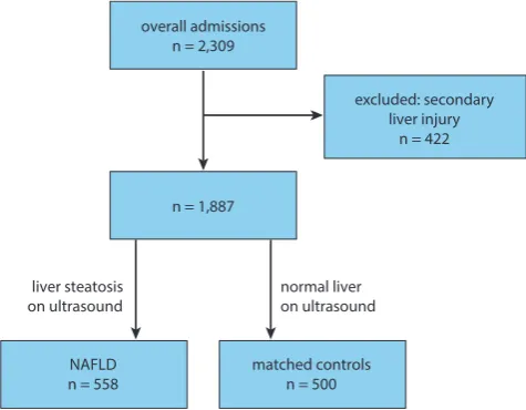
A total of 2,309 consecutive hospital charts of patient hospitalized in the Department and Clinic of Gastroen-terology and Hepatology of Wroclaw Medical University between 2017–2018 were meticulously searched. Four hun-dred twenty-two individuals were excluded due to second-ary causes of liver injury: viral hepatitis, autoimmune hepa-titis, hemochromatosis, Wilson’s disease, α-1 antitrypsin deficiency, drug-induced hepatic injury, cholestatic liver disease, and alcohol consumption higher than 30 mg/day for men and 20 mg/day for women.
One thousand eighty-seven patients were left for analy-sis. The study NAFLD group consisted of 558 consecutive patients with liver steatosis. Five hundred individuals with no history of liver disease, normal liver laboratory tests and normal image of liver on ultrasound examination were matched for age and sex with the study group and served as controls. Flowchart for patient inclusion was shown in Fig. 1.
Diagnosis of type 2 diabetes mellitus (T2DM) was de-fined as a registered diagnosis in patient charts, a non-fasting glucose value ≥180 mg/dL or a a non-fasting glucose value ≥126 mg/dL, or having treatment for diabetes. Hy-pertension was diagnosed when there was a registered diagnosis in the patient’s chart, a resting blood pres-sure ≥140/90 mm Hg or if the patient had any anti-hy-pertensive drug prescription. Dyslipidemia was defined
Fig. 1. Flowchart for patient inclusion overall admissions
n = 2,309
excluded: secondary liver injury
n = 422
NAFLD
n = 558 matched controlsn = 500 normal liver on ultrasound liver steatosis
on ultrasound
when the patient had a fasting triglyceride concentration value ≥150 mg/dL or HDL-cholesterol value <40 mg/dL (male), <50 mg/dL (female). Body mass index (BMI) was calculated as (weight [kg]/height [m]2). The diagnosis
of metabolic syndrome was established according to Adult
Treatment Panel III criteria.16 Cardiovascular disease
was defined as one of the following: coronary heart dis-ease, cerebrovascular disdis-ease, peripheral arterial disdis-ease, rheumatic heart disease, congenital heart disease, deep vein thrombosis, or pulmonary embolism. The diagnosis of colonic diverticulosis was based on an endoscopy and/ or radiological examination.
Laboratory parameters extracted from patients’ charts were blood morphology, AST, ALT, HDL-cholesterol, tri-glycerides, and glucose. Other parameters were incomplete and not included in the study.
The diagnosis of liver steatosis was defined in ultra-sound examination. Patients were examined in the supine and left lateral decubitus position under fasting condi-tions. A gastroenterologist with over 10 years of abdomi-nal ultrasound experience evaluated the echogenicity of the liver and the right kidney. Increased hepatorenal echogenicity, bright hepatic echoes, and vascular blur-ring of portal or hepatic vein were classified as exclusive features of NAFLD.
Statistical analysis
Continuous, normally distributed variables were sum-marized as mean ± standard deviation (SD). Student’s t-test was performed to compare the means in groups with nor-mally distributed data. In groups with non-normal distri-bution, the Mann–Whitney U-test was used. To compare mean prevalence differences between groups, the χ² test (categorical variables) was performed. Statistical analysis was performed using STATISTICA v. 13.3 software (Stat-Soft Inc., Tulsa, USA).
Ethical considerations
The study protocol was approved by local ethics com-mittee in accordance with the Helsinki Declaration.
Results
Five hundred fifty-eight consecutive patients with liver steatosis and 500 controls were enrolled into the study. According to the absence of other causes of liver ste-atosis, all the patients in the study group were classified as NAFLD. Twenty-four patients (4.3%) in this group were diagnosed with liver cirrhosis. The mean age of the patients was 58.1 years in NAFLD group and 57.5 years in the con-trols, respectively. Individuals in the 7th decade of life were
most often represented. A total of 50.4% of the patients in the study group were male. The mean age and the sex distribution in both groups did not differ statistically sig-nificantly. We tried to estimate the prevalence of NAFLD in all the patients admitted to the gastroenterology depart-ment: 558 cases out of 2,309 overall admissions = 24.2%. Selected clinical data of patients in NAFLD patients and controls is shown in Table 1.
The mean BMI was significantly higher in the study group compared to controls (29.2 vs 24.4, p < 0.001). Pa-tients with NAFLD had significantly higher serum TG and lower HDL concentration.
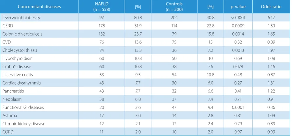
The prevalence of metabolic syndrome and its com-ponents in NAFLD patients and controls were estimated (Table 2, number of patients in the brackets in cases where the missing data made it impossible to complete the calcu-lation). Not surprisingly, MetS with components (hyper-tension, T2DM, dyslipidemia) was strongly correlated with NAFLD. The concomitant diseases in NAFLD and con-trols were evaluated. We managed to estimate the preva-lence of consecutive diseases with odd ratios ( Table 3). The concomitant diseases connected with NAFLD were: overweight/obesity, gastroesophageal reflux disease (GERD), colonic diverticulosis and cholecystolithiasis. Functional gastrointestinal diseases were less common in NAFLD than in the controls. The prevalence of other diseases was not significantly different in the study group and controls.
We chose FIB-4 scoring system to estimate the pos-sibility of coexisting fibrosis.17 In 348 patients (62.4%),
the FIB-4 score was lower than 1.45, suggesting no ad-vanced fibrosis present. These patients will not require
Table 1. Selected clinical data of NAFLD patients and controls
Clinical data NAFLD Controls p-value
Age [years]a 58.1 ±14.1 57.5 ±16.2 0.73
Sex (female/male) 277/281 252/248 0.81
BMI [kg/m2]a 29.2 ±5.5 24.4 ±3.9 <0.001
ALT [U/L]a 33.5 ±32.5 26.2 ±16.5 <0.001
AST [U/L]a 31.8 ±24.0 22.5 ±16.8 <0.001
Glucose [mg/dL]a 108.5 ±30.6 103.6 ±32.2 0.35
TG [mg/dL]a 147.1 ±86.8 108.4 ±60.5 <0.001
HDL (male: >40 mg/dL, female: >50 mg/dL)a 50.1 ±14.4 56.6 ±16.7 <0.001
ALT – alanine transaminase; AST – aspartate transaminase; HDL – high-density lipoprotein; BMI – body mass index; TG – triglycerides; a mean, standard
further diagnostic procedure. Fifteen patients with FIB-4 score above 3.25 are likely to have advanced liver fibrosis. One hundred ninety-one (34.2%) patients with interme-diate score 1.45–3.25 require additional diagnostic pro-cedures (Fig. 2). These patients were sent for ambulatory transient elastography.
Discussion
Non-alcoholic fatty liver disease, a chronic condi-tion of the liver related to hepatic steatosis, was recently recognized as the most common chronic liver disease. Increasing prevalence rates of the risk factors for NAFLD are the following: obesity, diabetes and metabolic syn-drome will most probably result into further increasing incidence rates of NAFLD all over the world.
The mean age of the patients in the study group was 58.1. Non-alcoholic fatty liver disease was found slightly more often in men than in women (not statistically significant difference in our study group). This is consistent with the re-sults of the epidemiological studies performed in the USA. The frequency of hepatic steatosis varies significantly with ethnicity (45% in Hispanics; 33% in Whites; 24% in Blacks).3
Our study group was monoethnical – 100% individuals were White. We can only estimate the prevalence of hepatic ste-atosis in all the patients admitted to the gastroenterology department as approx. 25% (558 out of 2,309).
Non-alcoholic fatty liver disease afflicts overweight and obese people and presents the coexistence with Fig. 2. FIB-4 score in patients with NAFLD
Table 3. Concomitant diseases in NAFLD patients and controls
Concomitant diseases (n = 558)NAFLD [%] (n = 500)Controls [%] p-value Odds ratio
Overweight/obesity 451 80.8 204 40.8 <0.0001 6.12
GERD 178 31.9 114 22.8 0.0009 1.59
Colonic diverticulosis 132 23.7 79 15.8 0.0014 1.65
CVD 76 13.6 75 15 0.32 0.89
Cholecystolithiasis 74 13.3 36 7.2 0.0013 1.97
Hypothyroidism 60 10.8 50 10 0.69 1.08
Crohn’s disease 60 10.8 38 7.6 0.078 1.46
Ulcerative colitis 53 9.5 54 10.8 0.48 0.87
Cardiac dysrhythmia 43 7.7 30 6.0 0.27 1.31
Pancreatitis 43 7.7 32 6.6 0.41 1.22
Neoplasm 38 6.8 37 7.4 0.71 0.91
Functional GI diseases 20 3.6 47 9.4 0.0001 0.36
Asthma 17 3.0 14 2.8 0.81 1.09
Chronic kidney disease 12 2.1 12 2.4 0.79 0.89
COPD 11 2.0 10 2.0 0.97 0.99
COPD – chronic obstructive pulmonary disease; CVD – cardiovascular disease; GERD – gastroesophageal reflux disease; GI – gastrointestinal.
<1.45 1.45–3.25
FIB-4 value
number of patient
s
>3.25 348
191
15 0
50 100 150 200 250 300 350 400
Table 2. The prevalence of metabolic syndrome and its components in NAFLD patients and controls
Components of metabolic syndrome NAFLD [%] Controls [%] p-value
Metabolic syndrome 268 (n = 550) 48.7 72 (n = 487) 14.4 <0.001
Hypertension 313 56.1 185 37 <0.001
Type 2 diabetes mellitus 136 24.4 43 8.6 <0.001
Hyperlipidemia (hypertriglyceridemia) 195 (n = 534) 36.5 75 (n = 487) 15.4 <0.001
Hyperlipidemia (decreased HDL) 183 (n = 522) 35.1 91 (n = 467) 19.5 <0.001
the MetS-associated disorders, like T2DM, hypertension and dyslipidemia.18 These comorbidities have a negative
impact on the natural course of NAFLD.19 In fact,
progres-sion to fibrosis in NAFLD is highly influenced by the pres-ence of T2DM and obesity.20,21 In a very recent review,
authors have mapped shared gene/protein interaction
networks and performed gene-disease analysis.18 Shared
mechanisms among NAFLD and the MetS diseases were revealed and provided evidence that NAFLD and espe-cially NASH, requires taking multi-target approaches, rather than focusing on single mechanisms of disease. Indeed, in our study we have confirmed strong connec-tions between either MetS itself or its components and NAFLD. The prevalence of MetS was 48.7%, overweight/ obesity was 80.8%, atrial hypertension 56.1%, T2DM 24.4%, hypertriglyceridemia 36.5%, and decreased HDL 35.1%, respectively. The difference in all the cases was significantly different from the corresponding rates in the control group.
Unexpectedly, the frequency of CVD was almost the same in the study group compared to controls (13.6% vs 15%). As patients with NAFLD have features of MetS, they also have important clinical implications for the de-velopment of future CVD. Many studies have addressed this issue before, finding the prevalence of CVD signifi-cantly higher in NAFLD patients.22 Recently, a vast
Eu-ropean study has been performed in Sweden23 and its
authors claim that it is the largest ever study of biopsy-proven NAFLD. Over 600 patients were followed up during a mean of 20 years. The authors found that death rates due to cardiovascular reasons did not differ statistically in NAFLD group compared to controls. The most common gastrointestinal disease in NAFLD group was GERD. It was statistically more common compared to controls. Large co-hort study (over 34,000 participants) showed that NAFLD is not independently associated with the risk of the devel-opment of reflux esophagitis after adjusting for BMI and other metabolic factors.24 The authors suggest that reflux
esophagitis is primarily the consequence of increased BMI commonly associated with NAFLD. Probably for the same reason, cholecystolithiasis appeared more often in NAFLD patients, as both diseases share the same main risk factor: overweight and obesity. Moreover, this association is more strongly seen in females than in males.25
In the literature there is lacking evidence on connection between NAFLD and colonic diverticulosis. Only 1 case-control study claimed that diverticulosis in the elderly (>65 years) was a negative predictor of liver steatosis.26 Our
findings in the younger group were different. In NAFLD patients, the prevalence of diverticulosis was much higher compared to controls.
We have evaluated the FIB-4 score that is simple and easy to calculate and to estimate the risk of fibrosis in the liver. In our study group, most cases were classified as not having fibrosis, whereas only 1/3 of patients required additional examinations (for instance transient elastography).
Conclusions
Overweight and obesity as well as metabolic syndrome with its components: hypertension, type 2 diabetes mel-litus, and dyslipidemia are more common in NAFLD pa-tients compared to matched controls. No significant dif-ference between the study group and controls was found in the frequency of CVD. Additionally, NAFLD patients are more often affected by GERD, colonic diverticulosis and cholecystolithiasis.
ORCID iDs
Radosław Kempiński https://orcid.org/0000-0002-6030-2700 Agata Łukawska https://orcid.org/0000-0002-3766-3073 Filip Krzyżanowski https://orcid.org/0000-0002-7571-7925
Dominika Ślósarz https://orcid.org/0000-0002-8159-1660 Elżbieta Poniewierka https://orcid.org/0000-0002-2074-976X
References
1. Masuoka HC, Chalasani N. Nonalcoholic fatty liver disease: An emerg-ing threat to obese and diabetic individuals. Ann N Y Acad Sci. 2013; 1281:106–122.
2. Armstrong MJ, Adams LA, Canbay A, Syn WK. Extrahepatic compli-cations of nonalcoholic fatty liver disease. Hepatology. 2014;59(3): 1174–1197.
3. Browning JD, Szczepaniak LS, Dobbins R, Nuremberg P, Horton JD, Cohen JC. Prevalence of hepatic steatosis in an urban population in the United States: Impact of ethnicity. Hepatology. 2004;40(6):1387–1395. 4. Blachier M, Leleu H, Peck-Radosavljevic M, Valla DC, Roudot- -Thoraval F. The burden of liver disease in Europe: A review of avail-able epidemiological data. J Hepatol. 2013;58(3):593–608. 5. Hamaguchi M, Kojima T, Itoh Y, et al. The severity
of ultrasonograph-ic findings in nonalcoholof ultrasonograph-ic fatty liver disease reflects the metabolof ultrasonograph-ic syndrome and visceral fat accumulation. Am J Gastroenterol. 2007; 102(12):2708–2715.
6. Musso G, Gambino R, Bo S, et al. Should nonalcoholic fatty liver dis-ease be included in the definition of metabolic syndrome? A cross-sectional comparison with Adult Treatment Panel III criteria in non-obese nondiabetic subjects. Diabetes Care. 2008;31(3):562–568. 7. Kwon YM, Oh SW, Hwang SS, Lee C, Kwon H, Chung GE. Association
of nonalcoholic fatty liver disease with components of metabolic syndrome according to body mass index in Korean adults. Am
J Gas-troenterol. 2012;107(12):1852–1858.
8. Speliotes EK, Massaro JM, Hoffmann U, et al. Fatty liver is associat-ed with dyslipidemia and dysglycemia independent of visceral fat: The Framingham heart study. Hepatology. 2010;51(6):1979–1987. 9. Musso G, Gambino R, Cassader M, Pagano G. Meta-analysis:
Natu-ral history of non-alcoholic fatty liver disease (NAFLD) and diagnos-tic accuracy of noninvasive tests for liver disease severity. Ann Med. 2011;43(8):617–649.
10. Musso G, Cassader M, Olivetti C, Rosina F, Carbone G, Gambino R. Association of obstructive sleep apnoea with the presence and sever-ity of non-alcoholic fatty liver disease. A systematic review and meta-analysis. Obes Rev. 2013;14(5):417–431.
11. Musso G, Gambino R, Tabibian JH, et al. Association of non-alcoholic fatty liver disease with chronic kidney disease: A systematic review and meta-analysis. PLoS Med. 2014;11(7):e1001680.
12. Kopylov U, Ben-Horin S, Lahat A, Segev S, Avidan B, Carter D. Obesity, metabolic syndrome and the risk of development of colonic diver-ticulosis. Digestion. 2012;86(3):201–205.
13. Nagata N, Sakamoto K, Arai T, et al. Visceral abdominal obesity mea-sured by computed tomography is associated with increased risk of colonic diverticulosis. J Clin Gastroenterol. 2015;49(10):816–822. 14. Festi D, Schiumerini R, Marzi L, et al. Review article: The diagnosis
alcoholic fatty liver disease – availability and accuracy of non-invasive methods. Aliment Pharmacol Ther. 2013;37(4):392–400. 15. Kempiński R, Neubauer K, Wieczorek S, Dudkowiak R, Jasińska M,
16. Executive Summary of The Third Report of The National Cholester-ol Education Program (NCEP) Expert Panel on Detection, Evaluation And Treatment of High Blood Cholesterol In Adults (Adult Treatment Panel III). JAMA. 2001;285(19):2486–2497.
17. Sterling RK, Lissen E, Clumeck N, et al. Development of a simple non-invasive index to predict significant fibrosis in patients with HIV/HCV coinfection. Hepatology. 2006;43(6):1317–1325.
18. Sookoian S, Pirola CJ. Review article: Shared disease mechanisms between non-alcoholic fatty liver disease and metabolic syndrome – translating knowledge from systems biology to the bedside.
Aliment Pharmacol Ther. 2019;49(5):516–527.
19. Rinella ME. Nonalcoholic fatty liver disease: A systematic review.
JAMA. 2015;313(22):2263–2273.
20. Friedman SL, Neuschwander-Tetri BA, Rinella M, Sanyal AJ. Mecha-nisms of NAFLD development and therapeutic strategies. Nat Med. 2018;24(7):908–922.
21. Noureddin M, Rinella ME. Nonalcoholic fatty liver disease, diabe-tes, obesity, and hepatocellular carcinoma. Clin Liver Dis. 2015;19(2): 361–379.
22. Targher G, Byrne CD, Lonardo A, Zoppini G, Barbui C. Non-alcoholic fatty liver disease and risk of incident cardiovascular disease: A meta-analysis. J Hepatol. 2016;65(3):589–600.
23. Hagström H, Nasr P, Ekstedt M, et al. Fibrosis stage but not NASH predicts mortality and time to development of severe liver disease in biopsy-proven NAFLD. J Hepatol. 2017;67(6):1265–1273.
24. Min YW, Kim Y, Gwak GY, et al. Non-alcoholic fatty liver disease and the development of reflux esophagitis: A cohort study.
J Gastroen-terol Hepatol. 2018;33(5):1053–1058. doi:10.1111/jgh.14042
25. Jia L, Haiyan L, Chengqi Z, et al. Non-alcoholic fatty liver disease asso-ciated with gallstones in females rather than males: A longitudinal cohort study in Chinese urban population. BMC Gastroenterol. 2014; 14:213.