Production, quality control and biological evaluation of
153Sm-TTHMP as a possible bone palliation agent
Zohreh Naseri1, Amir R. Jalilian2, Ali Nemati Kharat1, Ali Bahrami-Samani2, Mohammad Ghannadi-Maragheh2
1
Inorganic Chemistry Department, Faculty of Sciences, Tehran University, Tehran, Iran
2
Radiopharmaceutical Research and Development Lab,
Nuclear Science and Technology Research Institute, Atomic Energy Organization of Iran, Tehran, Iran
(Received 2 October 2011, Revised 10 November 2011, Accepted 18 November 2011)
ABSTRACT
Introduction: Various bone palliative therapeutic agents have been developed and widely used for bone
metastasis such as 153Sm-EDTMP. In this study, production, quality control and biodistribution studies of a newly developed therapeutic compound have been presented followed by imaging studies in wild-type rodents.
Methods: 153Sm-TTHMP was prepared starting from 153Sm-SmCl3, prepared by neutron activation of an
enriched 152Sm sample (purity >98%), and in-house synthesized TTHMP in 1h at 25C followed by stability tests, partition coefficient determination and biodistribution studies of in wild-type rodents using scarification and SPECT imaging.
Results: The radiolabled Sm complex was prepared in high radiochemical purity (>99%, ITLC) and specific
activity of 278 GBq/mmol and demonstrated significant stability at 4, 25 and 37C (in presence of human serum). Initial biodistribution data showed significant bone accumulation of the tracer in 48h.
Conclusion:153Sm-TTHMP can be a potential candidate for bone pain palliation therapy in skeletal metastases,
although further biological studies in other mammals is still needed.
Keywords: Sm-153, TTHMP, Radiopharmaceutical, Therapy, Biodistribution, Imaging
Iran J Nucl Med 2011;19(2):60-68
Corresponding author: Dr Amir R Jalilian, Radiopharmaceutical Research & Development Laboratory
(RRDL), Nuclear Science and Technology Research Institute (NSTRI), AEOI, Tehran, Iran. E-mail: [email protected]
O
r
ig
in
a
l A
r
ti
c
Ir
a
n
J
N
u
c
l
M
e
d
2
0
1
1
,
V
o
l
1
9
,
N
o
2
(
S
e
ri
a
l
N
o
3
6
)
61
INTRODUCTIONBone metastases are common in the course of various tumors such as prostate, breast, and lung carcinoma and they often entail an occurrence of progressive pain (1). Bone metastases occur in many patients with solid malignant tumors (2). Approximately 50% of patients with breast carcinoma and 80% of patients with prostate carcinoma develop metastatic bone disease and nearly half of them experience bone pain (3). In these patients who have progressive disease despite treatment, a systemic bone-avid radiopharmaceutical for treatment of widespread bony metastases has potential benefit (4). Radionuclide therapy using 32P,
89
Sr, 90Y, 153Sm and 186Re has been proposed as an alternative modality for management of bone pain (5).
Samarium-153 has favorable radiation characteristics, medium-energy beta particle emissions (Emax = 810 keV) which is
desirable for treatment, medium-energy gamma photon (103 keV) which is suitable for imaging, and short half-life (46.3 h). This radionuclide is the most widely used pain palliation radiopharmaceutical in the United States in form of EDTMP complex (Lexidronam) (6).
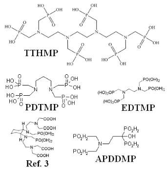
However, the search for the development of new ligands with higher stability, better pharmacokinetics and lower unwanted tissue uptakes (liver and GI) is still ongoing. Various complexes including new cyclic mixed phosphonate/carbonate ligands (7), alkyl phosphonates (PDTMP) (8) and hydroxyl-containing phosphonates (APDDMP) (9) have been developed and evaluated while none of these 153 Sm-complexes demonstrated better performance compared to Lexidronam (Figure 1).
In continuation of developing bone pain palliation agents for the use in the country as well as developing better quality compounds (10, 11), in this work, we report the preparation, quality control and
biodistribution of a new Sm-153 complex of recently synthesized ligand (12), Triethylene Tetramine Hexa (Methylene Phosphonic acid) (153Sm-TTHMP) developed for ultimate bone pain palliation therapy (Figure 1).
Figure 1. Structures for some phosphonates ligands
used in 153Sm labeling.
METHODS
Ir
a
n
J
N
u
c
l
M
e
d
2
0
1
1
,
V
o
l
1
9
,
N
o
2
(
S
e
ri
a
l
N
o
3
6
)
62
Production and quality control of153
SmCl3 solution
Samarium-153 was produced by neutron irradiation of 1 mg of enriched 152Sm2O3
(152Sm, 98.7% from ISOTEC Inc.) according to reported procedures (13) in the Tehran Research Reactor at a thermal neutron flux of 5×10¹³ n.cm-2.s-1 for 2 days. Specific activity of the produced 153Sm was 345 mCi/mg. The irradiated target was dissolved in 100 µl of 1.0 M HCl, to prepare
153
SmCl3 and diluted to the appropriate
volume with ultra pure water, to produce a stock solution. The mixture was filtered through a 0.22 µm biological filter and sent for use in the radiolableing step. The radionuclidic purity of the solution was tested for the presence of other radionuclides using beta spectroscopy as well as HPGe spectroscopy for the detection of various interfering beta and gamma emitting radionuclides. The radiochemical purity of the 153SmCl3 was checked using 2
solvent systems for ITLC (A: 10mM DTPA pH.4 and B: ammonium acetate 10%: methanol (1:1)) and HPLC.
Synthesis of triethylene tetramine
hexa(methylenephosphonic Acid)
(TTHMP)
The experimental procedure for the synthesis of TTHMP ligand was according to other bis-phosphonates as reported (14). Briefly, a quantity of 0.48 g (3.3 mmoles) of triethylenetetramine was dissolved in 0.75 ml of concentrated HC1 and a concentrated aqueous solution of 1.62 g (20 mmoles) of phosphorous acid. The resulting solution was heated to reflux temperature and 3.2 ml of 37% aqueous formaldehyde solution (40 mmoles) was added dropwise in the course of 1 h to the refluxing solution and refluxing was continued for another 1h. The result of reaction is an ethanol precipitated of a slightly yellow product from the concentrated reaction solution [m.p. 90-92ºC, 1H-NMR (D2O, δ ppm): 3.02-3.25(m,
12 H, >N-CH2CH2-N<), 3.37-3.47(m, 12 H,
-NCH2-PO3H2)].
Radiolabeling of TTHMP with 153SmCl3
A stock solution of TTHMP was prepared by dissolution in 1 N NaOH and diluted to the appropriate volume with ultra pure water by dissolving 250 mg of TTHMP in 1.5 ml NaOH (2N) and 3.5 ml distilled H2O, pH.
12. Then 0.3 ml of this solution was added to 200µl 153SmCl3 (5.7 mCi) (S.A. 345
mci/mg) and pH adjusted to 7 using phosphate buffer. The reaction mixtures were incubated with stirring at room temperature for 1h. Various parameters such as ligand concentration, pH of the reaction mixture, incubation time, reaction temperature were optimized to achieve maximum complexation yield. Sterility, apyrogenicity and toxicity were ascertained by routine methods. The radiolabeling yield of the ligand was determined with paper chromatography using Whatman No. 2 paper by sampling 5µl of the reaction mixture on the paper strip followed by developing in NH4OH:MeOH:H2O
(2:20:40) mixture.
Stability studies
The stability of the complex stored at room temperature (22ºC ambient), fridge (4ºC) and presence of freshly-prepared human serum (at 37ºC) was studied at different intervals of time by determining the radiochemical purity of the complex by
paper chromatography in
NH4OH:MeOH:H2O (2:20:40) system.
Ir
a
n
J
N
u
c
l
M
e
d
2
0
1
1
,
V
o
l
1
9
,
N
o
2
(
S
e
ri
a
l
N
o
3
6
)
63
sampled and counted in HPGe detector for20 seconds.
Biodistribution of 153Sm cation and 153
Sm-TTHMP in wild-type rats
To determine its biodistribution, 153 Sm-TTHMP was administered to normal rats. For comparison, free Sm3+ cation buffer solution was also administered. Briefly, 200μl of final 153Sm-TTHMP solution with 0.7 mCi radioactivity was injected intravenously to rats through their tail vein. The animals were sacrificed at the exact time intervals (2, 4, 24, 48 hours), and specific activity of different organs was calculated as percentage of injected dose per gram using HPGe detector (%ID/g).
Single photon emission computed
tomography (SPECT) imaging of 153
Sm-TTHMP in wild-type rats
For imaging studies, 153Sm-TTHMP solution (7.4 MBq, 200 L) was injected intravenously to rats through their tail veins followed by propofol-xylazine mixture injection for anaesthetization. The images were acquired after administration of the radiopharmaceutical by a single-head SPECT system (Siemens, Germany) based on 103 keV peak (15% energy window). The rat-to-septa distance was 12 cm.
RESULTS AND DISCUSSION
Ligand synthesis
TTHMP ligand was synthesized and the structure was determined using H NMR, C NMR, P NMR and IR methods which was equivalent to other commercial authentic samples of bis-phosphonates used in radiopharmacy, according to the conventional method (Figure 2).
Ir
a
n
J
N
u
c
l
M
e
d
2
0
1
1
,
V
o
l
1
9
,
N
o
2
(
S
e
ri
a
l
N
o
3
6
)
64
Radionuclide productionThe radionuclide was prepared in a research reactor according to regular methods with a specific activity of 350 mCi/mg for radiolabeling use. The radioisotope was dissolved in acidic media as a starting sample and was further diluted and evaporated for obtaining the desired pH and volume followed by sterile filtering. Gamma-ray spectrum revealed the presence of 154Eu (<4.7×10⁻⁵% of 153Sm) and 155Eu (<2.4×10⁻⁵% of 153Sm) at the end of irradiation.
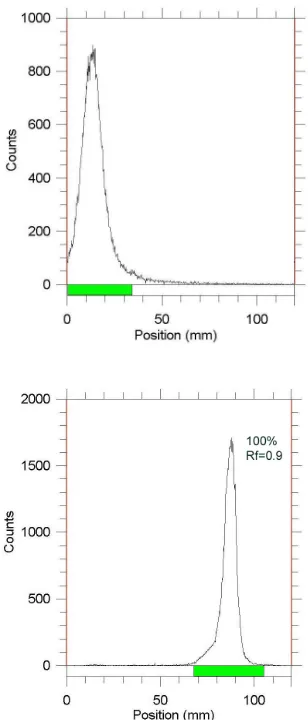
Figure 3. ITLC chromatograms of 153Sm-SmCl3
(above) and 153Sm- TTHMP (below) solutions on Whatman 2 MM paper using NH4OH: MeOH: H2O (0.2:2:4).
Radiochemical impurities in the 153Sm sample used in the radiolabeling step were checked by two systems. As stationary phase for paper chromatography system, Whatman 2 MM paper was used. In %10 ammonium acetate:methanol, the free samarium cation in 153Sm3+ form remains at the origin (Rf = 0.0) and while other Sm-153
species migrate to higher Rfs (0.8). Another
eluent for Sm3+ detection was 10 mM DTPA aquoes solution at pH.3 (Rf = 0.8).
Labeling optimization studies
In order to obtain maximum complexation yields, several experiments were carried out by varying different reaction parameters such as ligand concentration, pH, reaction time and temperature. Ligand concentration was varied between a wide ranges starting from 10 to 50mg/ml for TTHMP. It was observed that at room temperature 99% complexation was achieved with 15mg/ml of TTHMP. The best ITLC mobile phase was considered Whatman 2 MM paper using NH4OH: MeOH: H2O (0.2:2:4) as shown in
Figure 3.
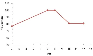
Variation of complexation yields with respect to TTHMP concentration is shown in Figure 4. The effect of variation of pH on complexation yield at room temperature was also studied by varying the pH of the reaction mixture from 2 to 12 using 1M HCl or 2M NaOH solution. Maximum yield of 100% was observed at pH 7-8 for complex. The effect of pH on the complexation yield for 153Sm-TTHMP complex is shown in
Figure 5.
Ir
a
n
J
N
u
c
l
M
e
d
2
0
1
1
,
V
o
l
1
9
,
N
o
2
(
S
e
ri
a
l
N
o
3
6
)
65
Figure 4. Effect of ligand concentration on
complexation yield of 153Sm-TTHMP.
Figure 5. Effect of variation of pH on complexation
yield of 153Sm-TTHMP at room temperature.
Stability
The stability of the 153Sm-TTHMP complex prepared under optimized reaction conditions was studied and observed that the complex showed excellent stability even when stored at room temperature. The complex remained stable to the extent of 96% up to 72h, whereas stability this compound was shown 90% for 48h in refrigerator.
In vitro stability against human serum This was carried out by incubating 0.1 ml of the radioactive complex and 0.3 ml human serum at 37ºC and at pH= 7 for 4 h. After 4
h, activity was measured by RTLC in NH4OH: MeOH:H2O (2:20:40) system. The
result of RTLC was shown that this complex is stable (95%) in this condition.
Determination of water/lipid solubility A mixture of 100 μl of 2-Octanol and 100 μl of radiolabeled Samarium complex at 37°C was vortexed for 2 hours and left for 30 minutes in room temperature. Then 5μl of the octanol and aqueous phases were sampled and counted in HPGe detector for 20 seconds. P was calculated from the ratio showing very high water solubility of the complex leading to very low unwanted liver and gastrointestinal tract uptake of the complex and also high excretion through kidneys.
Biodistribution of 153Sm cation and 153
Sm-TTHMP in wild-type rats
The animals were sacrificed by CO2
Ir
a
n
J
N
u
c
l
M
e
d
2
0
1
1
,
V
o
l
1
9
,
N
o
2
(
S
e
ri
a
l
N
o
3
6
)
66
Figure 6. Biodistribution of 153Sm-free in different organs of normal rat.
Ir
a
n
J
N
u
c
l
M
e
d
2
0
1
1
,
V
o
l
1
9
,
N
o
2
(
S
e
ri
a
l
N
o
3
6
)
67
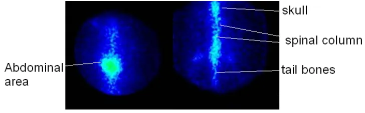
Figure 8. SPECT images of 153Sm-TTHMP (right) and 153Sm (left) 24 h post injection in wild-type rats.
The kidney plays an important role in 153Sm cation excretion especially after 24 h.
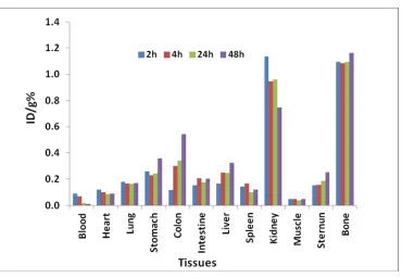
A volume (200 μl) of final 153Sm-TTHMP solution with 0.7 mCi radioactivity was injected intravenously to rats through their tail vein. The animals were sacrificed at the exact time intervals (2, 4, 24, 48 hours), and specific activity of different organs was calculated as percentage of injected dose per gram using HPGe detector (Figure 7). As shown in Fig 7. The major radioactivity is accumulated in bones as expected for bone-avid radiopharmaceuticals, also due to the presence of anionic properties of the complex and relatively small size of the molecules, the complex is also excreted through the kidneys. Due to liver uptake a significant GI uptake is observed.
The cation radioactivity is almost reaching a maximum after 48 h while the complex is rapidly removed from the circulation into the bones as expected for a bone pain palliation such as 153Sm-EDTMP (3-4%). Also a significant difference is observed for the liver accumulation among two species, free cation, like many other lanthanides, accumulates 1-4% in the liver while the complex is less than %0.5 at all time intervals as expected for a suitable bone agent. The spleen uptake is low for 153 Sm-TTHMP while a minor cation uptake is observed in this tissue as observe in other radio metals.
The bone uptake among the two species is not significantly different in 24h while after 48 hours the bone uptake increases in case of 153Sm-TTHMP and the cation uptake. As shown in other works (12), the Lu-177 uptake of the complex. Compared to that work, the liver uptake of the Sm-complex is less that than the Lu-complex, leading to more safety aspects of the complex due to unwanted irradiation to the surrounding tissues. On the other hand comparison of the complex with the well known Sm-153 EDTMP complex demonstrates the high specific activity of Sm-153 EDTMP in bones and less unwanted uptake in the other organs. That makes the Sm complexes of the discussion superior to the Lu-177 analogs. For the imaging studies of the radiopharmaceutical a wild type rats was used showing a distinct skeletal uptake in all time intervals (Figure 8).
CONCLUSION
Ir
a
n
J
N
u
c
l
M
e
d
2
0
1
1
,
V
o
l
1
9
,
N
o
2
(
S
e
ri
a
l
N
o
3
6
)
68
and 37C (in presence of human serum).The final preparation was administered to wild-type rats and biodistribution of the radiopharmaceutical was checked 4-48 hours later showing major accumulation of the drug in the bone tissues. Preliminary imaging studies also demonstrate bone uptake in rats after 24h. 153Sm-TTHMP can be a potential candidate for bone pain palliation therapy in skeletal metastases, although further biological studies in other mammals is still needed.
Acknowledgements
The authors wish to thank Mr M. Mirfalah for conducting animal studies.
REFERENCES
1. Serafini AN. Therapy of metastatic bone pain. J Nucl Med. 2001;42(6):895-906. 2. Bayouth JE, Macey DJ, Kasi LP, Fossella
FV. Dosimetry and toxicity of samarium-153-EDTMP administered for bone pain due to skeletal metastases. J Nucl Med. 1994;35(1):63-9.
3. Campa JA 3rd, Payne R. The management of intractable bone pain: a clinician's perspective. Semin Nucl Med. 1992;22(1):3-10.
4. Eary JF, Collins C, Stabin M, Vernon C, Petersdorf S, Baker M et al. Samarium-153-EDTMP biodistribution and dosimetry estimation. J Nucl Med. 1993;34(7):1031-6.
5. Holmes RA. [153Sm]EDTMP: a potential therapy for bone cancer pain. Semin Nucl Med. 1992;22(1):41-5.
6. Pandit-Taskar N, Batraki M, Divgi CR. Radiopharmaceutical therapy for palliation of bone pain from osseous metastases. J Nucl Med. 2004;45(8):1358-65.
7. Ouadi A, Loussouarn A, Morandeau L, Remaud P, Faivre-Chauvet A, Webb J et al. Influence of trans-1,2-diaminocyclohexane structure and mixed carboxylic/phosphonic group combinations on samarium-153 chelation capacity and stability. Eur J Med Chem. 2004;39(5):467-72.
8. Majali MA, Mathakar AR, Shimpi HH, Banerjee S, Samuel G. Studies on the preparation and stability of samarium-153 propylene diamine tetramethylene phosphonate (PDTMP) complex as a bone seeker. Appl Radiat Isot. 2000;53(6):987-91.
9. Zeevaart JR, Jarvis NV, Louw WK, Jackson GE. Metal-ion speciation in blood plasma incorporating the tetraphosphonate, N,N- dimethylenephosphonate-1-hydroxy-4-aminopropilydenediphosphonate
(APDDMP), in therapeutic radiopharmaceuticals. J Inorg Biochem. 2001;83(1):57-65.
10. Bahrami-Samani A, Ghannadi-Maragheh M, Jalilian AR, Meftahi M, Shirvani-Arani S, Moradkhani S. Production, quality control and biological evaluation of 153Sm-EDTMP in wild-type rodents. Iran J Nucl Med 2009;17(2):12-19. 11. Bahrami-Samani A, Bagheri R, Jalilian
AR, Shirvani-Arani S, Ghannadi-Maragheh M, Shamsaee M. Production, quality control and pharmacokinetic studies of Ho-EDTMP for therapeutic applications. Sci Pharm. 2010;78(3):423-33.
12. Chakraborty S, Das T, Unni PR, Sarma HD, Samuel G, Banerjee S et al. 177Lu labelled polyaminophosphonates as potential agents for bone pain palliation. Nucl Med Commun. 2002;23(1):67-74. 13. Manual for reactor produced
radioisotopes. IAEA-TECDOC-1340, IAEA, Vienna, 2003. p. 71.