Cholesterol
Levels in Rats
GRIMALDO CARVALHO
Department
of
Surgical Pathology,
Medical College
of
Virginia,
Richmond 23219
Introduction
It is well known that in rats
estrogen and testosterone influence the cholesterol concentration in blood and tissues, and that blood cholesterol levels are higher in
young adult females than in males of the same age. At birth there is no difference in the blood
cho-lesterol levels between the sexes,
but differences begin to appear at approximately 21 days of age and
gradually increase with age. Experiments by Fillios et al. in 1958, in which blood cholesterol levels were measured during the
several phases of the rat menstrual cycle, verified the existence of higher cholesterol concentrations during the preovulatory and ovula-tory phases, as identified by vaginal
smears, when estrogenic activity is higher. The two phases coincide with the time of greatest activity of
the ovarian follicles. When this
activity increases, the blood cho-lesterol concentration rises. The
authors also observed that injec-tion of testosterone in females with ovarian activity decreased the blood cholesterol levels. Therefore, they believed the biosynthesis of endogenous cholesterol in rats to be
stimulated by estrogen and de-pressed by testosterone.
Studying two groups of rats that received a high-cholesterol and a low-cholesterol diet, respectively, Morris and Chaikoff observed in 1959 that testicular cholesterol was of endogenous origin. In liver,
small intestine and adrenals, this
endogenous origin of cholesterol
was not completely suppressed,
even in those animals that received
prolonged rations of a high-cho-lesterol diet.
In previous work with rabbits on high-cholesterol and on high-cho-lesterol plus triparanol diets, I
observed that no offspring were produced, even though the animals
were not carefully separated by sex. Consequently, it seemed advis-able, as the main purpose, to re-examine the possibility that a diet high in cholesterol, with or with-out triparanol, might affect repro-duction. In this study rats were
used instead of rabbits. Since tri-paranol inhibits the conversion of
desmosterol to cholesterol, it
seemed advisable to study the effect
of triparanol on blood and tissue cholesterol itself. It also seemed of interest to study the distribution of
cholesterol in various tissues. Materials and Methods
Three groups of young adult
Wistar white rats, averaging 154 gm in weight, were divided into the
following groups.
Group A: 26 rats-13 males and 13 females. They received a daily diet of 18 gm rat chow containing 0.27 gm of pure cholesterol plus 0.01 gm of an inhibitor of cho-lesterol synthesis ( triparanol) in
addition to the normal daily
sup-plement of vitamins, mineral salts,
etc., which was totally eaten. Group B: Same number of ani-mals as in the previous group, with
FERTILITY AND ELEVATED CHOLESTEROL
gm rat chow containing 0.27 gm of pure cholesterol plus vitamins, mineral salts, etc.
Group C: 16 rats-eight males and eight females. They received a daily normal diet of 18 gm stand-ard rat chow and served as the con-trol group.
The animals were kept in in-dividual cages and received their rations in two stages at 12-hour intervals. The whole amount given was eaten.
Before the experiment started, two animals from both sexes were sacrificed, and the cholesterol and desmosterol concentrations were determined in both blood and tis-sues. These animals were on ordi-nary diet and were later included in Group C as control animals.
After one month on the diet, four animals from each group were killed and cholesterol and desmos-terol levels were determined in their blood and tissues.
Desmosterol was determined in-directly by measuring the color de-veloped with Liebermann-Burchard reagent at 400µ, and 620µ, accord-ing to the procedure of Abell et al., ( 19 52). It is possible that other
TABLE 1
non-cholesterol sterols besides mosterol are included in the des-mosterol values.
After two months, we placed the remaining animals in pairs within their own groups and observed them for three months.
Results and Discussion
There was weight gain in the three groups, the average weight in Groups A and B being 160 gm; in Group C, 158 gm.
Table 1 shows that the blood cholesterol of animals on high cho-lesterol diet (Group B) increased, while 'the desmosterol level de-creased. In the animals on high cholesterol plus triparanol diet (Group A), the cholesterol level was higher than in the control group but lower than in Group B, and the desmosterol level in Group A was higher than in the other groups.
In the rats from Group B, the cholesterol level in the heart was higher than in the other two groups that showed similar values for both cholesterol and desmo-sterol levels. However, the desmo-sterol levels in Group A and in
Cholesterol, Desmosterol, and Total Steroid Concentration of Serum and Tissues."'
Group A
I
Group BHigh Cholesterol Diet
+
Triparanol High Cholesterol Diet' I
Organ Cholesterol I Desmosterol Total Cholesterol Desmosterol Total mg/ml serum mg/ml serum Steroids mg/ml serum mg/ml serum Steroid
mg/gm wet mg/gm wet mg/ml serum mg/gm wet mg/gm wet mg/ml serum tissue tissue mg/gm wet tissue tissue mg/gm wet
tissue tissue
Serum 0.62 ± 1.0
I
0.13±0.7I
7.5 ± 0.8 0.76 ± 0.91 0.04 ± 0.51 0.8±0.7 Heart 9.9 ± 0.9I
2.5 ± 0.9I
12.4 ± 0.9 15.7± 1.oJ 0.03 ± 0.31 15.7 ± 0.6 Aorta 20 ± 0.8I
1.8±0.7
I
21.8±0.7 55.9 ± 1.21 0
I
55.9 ± 1.2
Spleen 47.1 ± 1.1 1.7± 0.9 48.8 ± 1.0 26.6 ±0.8 2.2 ± 0.8 28.8 ± 0.8
Adrenals 113 ± 1.0
I
6.5 ± 1.0 119.5 ± 1.0 182.6 ± 1.31 0 1182.6 ± 1.3 Testicles 11 ± 0.9I
6.9 ± 1.0 17.9 ± 0.9 22± 0.81 1 7 ± 0.9I
23.7 ± 0.8I
I
I
!
II
I
Group C were higher than in Group B. Similar changes were seen in the aorta, but the values were higher.
In the spleen, the high cho-lesterol diet was associated with increased endogenous cholesterol. The adrenals were very sensitive to the diets. With the high cho-lesterol diet, the synthesis of cho-lesterol seemed to be completely abolished, because desmosterol was absent. With the cholesterol plus triparanol diet, desmosterol was again present in higher levels than in Group C.
An interesting result was ob-served in the testicles, where the highest level of endogenous cho-lesterol was found. In the control group, for example, a 93 mg/10 gm level of desmosterol and a 140 mg/10 gm level of cholesterol were observed, indicating high choles-terol biosynthesis activity. This was the highest desmosterol level found in tissues. In Group A, the levels of both cholesterol and desmosterol decreased in comparison to Group C. In Group B, the desmosterol level dropped and the cholesterol level rose.
Group C Normal Diet
I
Cholesterol Desmosterol Total
mg/ml serum mg/ml serum Steroids mg/gm wet mg/gm wet mg/ml serum
tissue tissue mg/gm wet
serum 0.45 ± 0.9 t 0.1 ± 0.5 0.55±0.7
9.6± 0.91 2.6± 0.1 [ 12.2 ± 0.8 15.5 ± 1.1 2.2± 0.7 17.7±0.9
33 ± 1.2 0.'2 ± 0.4 33.2 ± 0.8 74.2±1.1 1.2 ± 0.5 75.4 ± 0.8
14 ± 0.9
I
9.3 ± 0.9I
23.3 ± 0.9I
I
• 10 gm from each of these organs were used in homogenized solution. The figures given are average values ±standard errors of the means. Standard
deviation in the method used was 1.03 mg/lOgrn.
The puzzling results observed in
the testicles led to histological
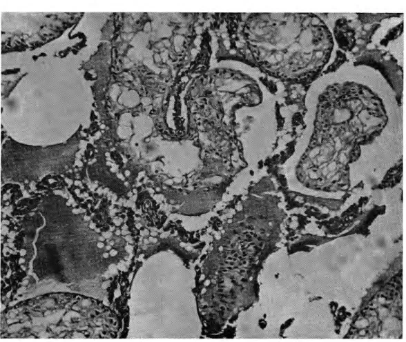
sec-tions of these organs being made from animals of the three groups (Fig. 1, 2, 3). At the same time,
the remaining animals were bred
within their own groups and
ob-served for three months.
The control group, with four
males and four females, had
nor-mal offspring and were considered
to be 100% fertile.
Eleven pairs from Group A
mated normally but produced fewer
offspring than Group C. They
were 63 % fertile, and their babies
had little or no possibility of sur
-vival, as they showed a high
in-cidence of malformation such as
phocomelia and sirenomelia. Four
of them had almost complete
or-ganic agenesia. The abdominal
cavity of one of these babies ap-peared empty (Fig. 4). In the chest cavity, there was only the heart and
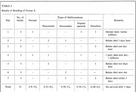
TABLE 2
Results of Breeding of Group A
No. of
Pair babies Normal
1 5 l
2 3
-3 2
-4 3 1
5 3
-6 2
-7 3
-Total I
21 2/9.53
rudiments of lungs. The posterior
portion of the body was not formed,
and the lower limbs and the tail
were missing. Seven pairs had 21
babies. Nineteen of these babies
had malformations, as shown in
Table 2, and all 21 died before three days of life. One of the
moth-ers from this group died of uterine
hemorrhage immediately after
de-livery of five dead malformed
babies. Three of them had organic
agenesia.
The rats from Group B were
100% sterile. In view of the com
-plete sterility found in the pairs
from this group, there was
con-siderable interest in determining
whether the male, the female, or
both were at fault. Continuing to
submit all the animals to the same
diets, they were cross-mated as
follows:
a) Males from Group B were
coupled with control females for
Types of Malformations
Organic
Phocomelia Sirenomelia agenesia
- - 3
2 1
--
--1 - 1
2 -
-- 2
--
1-5/23.83 4/19.13 4/19.13
two months and continued sterile.
b) Control males were coupled
with females from Group B for
the same time and had normal off-spring.
c) Males from Group B were
mated with females from Group A
and continued sterile.
d) Males from Group A were
mated with females from Group B.
Sixty-seven per cent of the matings
resulted in offspring with a 20%
lower incidence of malformations
than in pairs from Group B.
Naturally, it was thought that
only the males from Group B
be-came sterile with the high choles-terol diet. Attempts were then made
to determine the type of lesion and
its location.
The diets in the study groups
were discontinued, and all the
ani-mals received the same normal
meals as in Group C. After one
month on this diet, all the groups
Remarks
Dwarfism
1 Mother died; babies
stillborn
- Babies died 3 days later
2 Babies died one day
later
- l baby died next day;
1 stillborn
l Babies died two days
later
- Babies died next day
2 Babies died within 3
days
were again cross-mated, as before. All the animals then had normal offspring, except the males from Group B that remained sterile.
Histological sections from testi-cles of animals from Groups A and B were performed during the special diets and three months after these diets were discontinued. Com-parative studies with testicles of rats from the control group were also done. The following results were obtained.
Group A: There were some changes in the testicular tissue while on the diet. These changes resembled fat degeneration but
were not severe. There was no
atrophy, and apparently spermato-genesis was not seriously affected. Only lower numbers of sperm were seen. When the diet was dis-continued, these changes disap-peared (Fig. 3) ..
Group B: There were serious
and irreversible lesions, which def-initely blocked spermatogenesis. These lesions were fat degeneration
and complete atrophy of testicular
tissue; they suggested somewhat the changes seen in inanition (Fig. 2).
However, the rats were well
nourished, with a good supply of
vitamins and mineral salts in their diets.
It should be emphasized that the sterility of males from Group B did not have any relation to im-potence, since there was no differ-ence between the sexual activity of these males and the males from other groups.
Summary and Conclusions
Several conclusions were drawn from these experiments.
1. Desmosterol levels were higher
in the testicles of animals on a
normal diet, and lower in animals
that became sterile on a high
cho-lesterol diet. This suggested that
desmosterol, in addition to being
a precursor of cholesterol, could
also be a direct precursor of a male hormone (or hormones) which controls or influences
spermato-FERTILITY AND ELEVATED CHOLESTEROL
Fig. I-Normal testicular tissue, with normal spermatogenesis from animals
of the control group. X 400 (Trichrome staining technique).
Fig. 2-Severe degeneration of the testicular tissue, with spermatogenesis
genesis. However, this remains hypothetical.
2. The sterility induced in males on a high cholesterol diet could not be reversed by the discontinuation of the diet.
3. The cholesterol
concentra-tions in the adrenals increased to
the highest levels found in the two
studied groups, and desmosterol
was absent in Group B. No
ana-tomical or histological changes
were seen.
4. Cholesterol and desmosterol levels in serum of animals from the
three groups are compatible with
the hypothesis that triparanol
in-hibits the synthesis of cholesterol
in its last stage.
It is my hypothesis that the higher cholesterol and lower
des-mosterol levels in the heart and
aorta of animals from Group B
probably mean that the
endo-genous cholesterol was replaced by
the cholesterol from the diet. The fat degeneration and
com-plete atrophy of testicular tissue in
animals from Group B were
prob-ably due to the poor nutrition of
the testicular tissue provoked by diet.
References
ABELL, L. L., B. B. LEVY, B. B.
BRODIE AND F. E. KENDALL. A
simplified method for the
estima-tion of total cholesterol in serum
and demonstration of its specificity.
J. Biol. Chem. 195: 357-366, 1952.
BECKER, M. C. Clinical experience
with triparanol. J. New Jersey Med. Soc. 59: 59-64, 1962.
CONNOR, W. E. Dietary cholesterol
and the pathogenesis of
athero-sclerosis. Geriatrics 16: ~07-415, 1961.
DORFMANN, R. I. Metabolisme de la testosterone. ( Colloque sur la
fonc-tion endocrine du testicule, juillet
9-11, 1957.) Presse Med. 65: 1891, 1957.
DOWNIE, V. J. Death of a testicle.
Lancet I: 370-371, 1959.
Fig. 3-Slight degeneration of the testicular tissue, with oligospermatogenesis,
as seen in animals from Group A (high cholesterol plus triparanol diet).
Fig. 4-Newborn rat with organic agenesia. Notice the presence of the heart
and rudiments of lungs as well as the absence of the inferior portion of the
FILLIOS, L. C., R. KAPLAN, R. S. MARTIN AND F. J. STARE. Some as-pects of the gonadal regulation of
cholesterol metabolism. Am. J.
Physiol. 193: 47-51, 1958. FORBES, A. P. La deuxieme hormone
testiculaire. ( Colloque sur la fonc-tion endocrine du testicule, juil!et
9-11, 1957.) Presse Med. 65:
1891, 1957.
GILBERT-DREYFUS. Utilisation
prati-que des gonadotrophines et des androgenes dans le traitement des insuffisances testiculaires. ( Colloque sur la fonction endocrine du
testi-cule, juillet 9-11, 1957.) Presse Med. 65: 1892, 1957.
GILBERT-DREYFUS, J. SEBAOUN AND J. BELAISCH. Les facteurs hormonaux de la spermatogenese chez l'homme sur la fonction spermatogenetique du testicule humain. Presse Med.
67: 743, 1959.
GOLDSMITH, G. A. Highlights on the cholesterol-fats, diets and athero
-sclerosis problem. JAMA 176: 783-790, 1959.
Gouyoou, M. G. Characteres
histo-logiques de la spermatogenese nor-male; les spermatozoides normaux.
Presse Med. 67: 743, 1959. GOYETTE, E. M. AND J. C. ELDER.
The use of triparanol to lower
serum cholesterol. Am. Heart J.
60: 536-538, 1960.
HARRISON, M. R. G. Facteurs vascu-laires dans la physiologie normale
de la spermatogenese. Presse Med.
67: 743, 1959. .
Hms IN'T VELD, M. L. G. Influence de la fonction endocrinienne du testicule sur !'excretion urinaire des 17-cetosteroldes neutres. ( Colloque sur la fonction endocrine du
testi-cule, juillet 9-11, 1957.) Presse Med. 65: 1891, 1957.
JAYLE, M. F., R. SHOLLER, G.
GARONNE AND F. MOREL. Action
des gonadotrophines chorioniques sur !'excretion des 17-cetosteroldes, des phenolsterolds, des cortocoldes et du pregnandiol chez l'homme.
( Colloque sur la fonction endo-crine du testicule, juillet 9-11,
1957.) Presse Med. 65: 1891, 1957.
KEUTEL, M. H. J. Etude des proteines
du plasma seminal humain. Presse Med. 67: 744, 1959.
KRAWCZUK, M. A. Etude
compara-tive sur le niveau des
17-cetos-teroldes et des oestrogenes, et
!'image presentee par la glande
inter-FERTILITY AND ELEVATED CHOLESTEROL
stitielle et Jes vesicules seminales
d'hommes steriles. Presse Med. 67:
745, 1959.
MELBY, J. C., M. ST. CYR AND S. L.
DALE. Reduction of adrenal steroid
production by an inhibitor of cho-lesterol biosynthesis. New Engl. J.
Med. 264: 583-587, 1961.
MITCHELL, J. R. A. AND S. M. BRIERS. Effect of cholesterol, cholesterol esters, and neutral fats on fibrinol-ysis. Lancet 2: 435-436, 1959.
MORRIS, M. D. AND I. L. CHAIKOFF. The origin of cholesterol in liver, small intestine, adrenal gland, and testis of the rat: dietary versus
endogenous contributions. J. Biol. Chem. 234: 1095-1097, 1959. ROMANI, J. D. Valeur des
explora-tions hormonales dans !es troubles
de la spermatogenese. Presse Med.
67: 744, 1959.
RYAN, K. J. AND 0. W. SMITH. Bio-genesis of estrogens by the human ovary. III. Conversion of cho
-lesterol-4-C14 to estrone. J. Biol. Chem. 236: 2204-2206, 1961. STAMLER, J., R. PICK AND L. N.
KATZ. Effects of dietary proteins, methionine and vitamins on plasma
lipids and atherogenesis in cho-lesterol-fed cockerels. Circulation Res. 6: 442-446, 1958.
- - - : Effects of dietary protein and carbohydrate level on
cholestero-lemia and atherogenesis in cockerels
on a high-fat, high-cholesterol mash. Circulation Res. 6: 447-451,
1958.
VAGUE, J., G. FAVIER AND C. DELVOY.
Participation des hormones aux
al-terations du comportement sexuel
chez l'homme. (Colloque sur la
fonction endocrine du testicule, juil
-let 9-11, 1957.) Presse Med. 65: 1891, 1957.
VERNE, M. J. Morphologie, histologie et histo-chimie du testicule endo-crinien chez les mammiferes adultes. (Colloque sur la fonction endo-crine du testicule, juillet 9-11,