Page i
BONE GROWTH DURING PUBERTY AND THE EFFECTS OF EXERCISE AND CALCIUM ON
BONE MASS ACCRUAL
A DOCTORAL THESIS
SANDRA lULIANO
2001
\j[)ER. THESIS 612.75 lUL 30001007574298 luliano, Sandra
Page a
Dedication
No thesis can be completed without the support and encouragement of all those around you. This thesis is dedicated to everybody that has played a part in its creation, from editing through to moral support.
Acknowledgments
The following are acknowledged in no particular order for their contribution to this thesis. Royal Childrens Hospital
• Kay Gibbons for her nutritional expertise in the development of the calcium supplementation component of the intervention.
• Robyn and Sue for cooking the foods used in the program. Murray Gouldbum Co-op
• Dr. Wayne Sanderson and Michael Phillips for providing the milk minerals and the T-shirts for all the participants.
University of Saskatchewan
• Professor Don Bailey and Dr. Bob Mirwald for allowing me to work with them on their pediatric bone mineral accraal study and exposing me to the most significant learning experience of my life.
• The staff of the College of Physical Education for making me feel so welcome during my time in Canada.
• Susan Whiting for collaborating on the dietary calcium intake paper. Milgate primary school
• The principal Ms Christine Lister for her foresight in seeing the value in the program and promoting its implementation at the school.
• The staff at Milgate Primary School who so willingly embraced the program and incorporated it into the school.
• All the girls who participated in the study - the hugest THANKS, and I hope they all enjoyed it. Research team
• Dr Geraldine Naughton for believing in my vision when I first approached her with the research idea.
• Professor John Carlson for adopting me so willingly, and providing me with his honest and respected feedback.
• Dr. Shona Bass for her direction and attention to detail to ensure the project was of the highest standard.
• Leanne Saxon for being the other half of the project team, and without whom the project would not have succeeded.
• Deb Oliver who was pivotal in the mnning of this program and without her enthusiasm and wonderful rapport with the staff and students the program would not have been as successful as it was.
Supporters
• My husband Scott for his wonderful patience as I toiled over the thesis. • Family and friends for living through the thesis with me.
Page Hi
Awards
• luliano-Bums, S. - Roger Melick Young Investigator Award - Winner (2000)
Australia and New Zealand Bone & Mineral Society 10''' Annual Scientific Meeting, Hamilton Island, QLD, Australia, Nov 4 - 7 .
(Chapter Six) Publications
• luliano-Bums, S., Whiting, S.J., Faulkner, R.A., Bailey, D.A. (1999)
Levels, Sources and Seasonality of Dietary Calcium Intake in Children and Adolescents enrolled in the University of Saskatchewan Pediatric Bone Mineral Accmal Study.
Nutrition Research 19:1471-1483. (Chapter Four)
• luliano-Bums, S., Mirwald, R.L., Bailey, D.A. (2001)
The timing and magnitude of peak height velocity and peak tissue velocities for early, average and late maturing boys and girls.
Amer. J. Human Biol 13(1): 1-8. (Chapter Five)
Presentations
• Whiting, S.J., luUano-Bums, S., Faulkner, R.A., Bailey, D.A. (1999)
Levels, Sources and Seasonality of Dietary Calcium Intake in Children and Adolescents enrolled in the University of Saskatchewan Pediatric Bone Mineral Accmal Study (Poster).
Canadian Federation of Biological Societies, Winnipeg, Manatoba, Canada, June 2-5 (Chapter Four)
• luliano-Bums, S., Mirwald, R.L., Bailey, D.A. (1999)
The timing and magnitude of peak height velocity and peak tissue velocities for early, average and late maturing boys and girls.
Austtalia and New Zealand Bone & Mineral Society 9"^ Annual Scientific Meeting, Cairns, QLD, Australia, June 30 - July 2.
(Chapter Five)
• luliano-Bums, S., Saxon, L., Oliver, D., Naughton, G., Bass, S. (2000)
Weight bearing exercise and increased dietary calcium intake have a synergistic effect on bone mass accrual at the femur in pre- and early-pubescent girls.
Austtalia and New Zealand Bone & Mineral Society 10* Annual Scientific Meeting, Hamilton Island, QLD, Austtalia, Nov 4 - 7.
(Chapter Six)
• Saxon, L., luliano-Bums, S., Naughton, G., Bass, S. (2000)
The osteogenic response to different mechanical loading regimes in pre and early pubertal girls. Australia and New Zealand Bone & Mineral Society 10* Annual Scientific Meeting, Hamilton Island, QLD, Austtalia, Nov 4-7. (Poster)
(Chapter Six)
• Saxon, L., luUano-Bums, S., Naughton, G., Bass, S. (2000)
Exercise and increased calcium intake: A synergistic effect at the loaded sites in pre and early pubertal girls.
Special pre-American Society Bone & Mineral Research Meeting on Pediatric Bone. MacMaster University, Hamilton, Canada, October.
Page iv
• Saxon, L., luliano-Bums, S., Naughton, G., Bass, S. (2000)
Exercise and increased calcium intake: A synergistic effect at the loaded sites in pre and early pubertal girls.
American Society Bone & Mineral Research Meeting, Toronto, Canada, October.
Page V
TABLE OF CONTENT
Tide Dedication
Acknowledgments
Awards, Publications and Presentations Table of Content
List of Tables List of Figures List of Appendices ABSTRACT
11
ii iii V
ix xi xii
1
CHAPTER ONE - hitroduction 1.1 Introduction
1.2 Rationale 1.3 Purpose 1.4 Limitations 1.5 Delimitations 1.6 Definitions of Terms
3 5 5 7 8 10
CHAPTER TWO - Literature review 2.1 Overview of bone growth
2.2 Normal bone development in growth 2.3 Linear growth of the human skeleton 2.4 Dimensional changes to bone with growth
2.5 Bone mineral accrual and loss during the lifespan 2.6 Peak bone mass
2.7 The role of calcium in the body 2.8 Regulation of plasma calcium levels 2.9 Overview of calcium metabolism 2.10 Calcium balance
2.11 Calcium absorption 2.12 Calcium excretion
2.13 Calcium intake recommendations
2.13.1 Trends in calcium intake in children of developed countries 2.14 The effect of calcium on bone remodelling
2.15 Calcium intake and adult bone mass
Page vi
2.16 Calcium intake and bone mass in children 41 2.17 The effects of calcium supplementation on bone mineral accmal in children 44
2.17.1 The effect of calcium supplementation on bone mass accrual at 50 different stages of maturity
2.17.2 Does daily dietary calcium intake influence the osteogenic response 52 of bone to calcium supplementation?
2.17.3 The effect of calcium supplementation withdrawal on bone mineral 53 accmal: The long term benefits
2.18 The effects of physical activity on bone mass 55 2.19 The mechanisms of the osteogenic response to mechanical loading 56
2.20 The osteogenic response of adult bone to exercise 57 2.21 The osteogenic response of immature bone to exercise 59
2.21.1 The effect of exercise on measures of bone mass in children 60 2.21.2 The effect of unilateral loading on the growing skeleton 62 2.21.3 The effect of exercise intervention on the growing skeleton 65 2.22 The long-term benefits of childhood physical activity on bone mass 68
2.23 The combined effects of exercise and calcium on bone mass 72
CHAPTER THREE - Methodology 3.1 Methods
3.2 Recruitment
3.3 Allocation to study groups 3.4 Pubertal staging
3.5 Body composition and bone mineral content 3.5.1 Regional analysis of total body scan 3.5.2 Total body scan
3.5.3 Femoral neck scan 3.5.4 Lumbar spine scan 3.6 Anthropometry
3.7 Physical activity 3.8 Dietary intake
3.9 Calcium supplementation 3.10 Exercise intervention 3.11 Ethical approval 3.12 Power calculations 3.13 Statistical analysis
Page vii
CHAPTER FOUR Level, Sources and
- Study one
Seasonality of dietary Calcium Intake in Children and Adolescents Enrolled in the University of Saskatchewan Pediatric Bone Mineral Accrual Study
4.1 4.2 4.3 4.4 4.5 Abstract Introduction Methods 4.3.1 4.3.2 4.3.3 4.3.4 4.3.5 4.3.6 4.3.7 4.3.8 Results 4.4.1 4.4.2 4.4.3 Discussion Recmitment Dietary intake Exclusion criteria Nutrient analysis Food grouping Seasonal categories Maturity categories Statistical analysis
Total dietary calcium intake Calcium sources
Calcium sources and seasonal variations
88 89 90 90 91 92 92 92 94 94 94 95 95 95 96 96
CHAPTER FIVE - Study Two
The Timing and Magnitude of Peak Height Velocity and Peak Tissue Velocities for Early, Average and Late Maturing Boys and Girls
5.1 5.2 5.3 5.4 5.5 Abstract Introduction Methods 5.3.1 5.3.2 5.3.3 5.3.4 5.3.5 5.3.6 Results 5.4.1 5.4.2 5.4.3 Discussion Recmitment
Body composition and bone mineral content Anthropometry
Inclusion criteria Maturity categorisation Statistical analysis
Comparison of males and females Maturity comparisons - males Maturity comparisons - females
Page via
5.5.1 Comparisons of peak height and peak tissue velocities between
males and females 116 5.5.2 Maturity comparisons 117
CHAPTER SIX - Study Three
The Effect of Weight Bearing Exercise and Calcium Supplementation on Bone Mass Accmal in Pre-and Early-Pubertal Girls
6.1 Abstract 122 6.2 Introduction 123 6.3 Methods 123
6.3.1 Statistical analysis 124
6.4 Results 124 6.4.1 Group characteristics and compliance 124
6.4.2 Maturation 124 6.4.3 Exercise intervention 126
6.4.4 Anthropometry and body composition 130
6.4.5 Dietary intake 130 6.4.6 Calcium supplementation and food compliance 131
6.4.7 Baseline bone mass 132 6.4.8 Th effect of 8.5 months of high- or low-impact exercise on
bone mass accmal 132 6.4.9 The effect of 8.5 months of calcium supplementation on
bone mass accmal 135 6.4.10 The combined effect of calcium and exercise on bone
mass accmal 138
6.5 Discussion 138 6.6 Conclusion 142
CHAPTER SEVEN - Conclusion and Recommendations
7.1 Conclusion 144 7.2 Summary of Recommendation 146
REFERENCES 147
Page ix
List of Tables
Number Title Page
2.1 Peak height velocity data from previous growth studies 16 2.2 Dietary calcium intakes of children derived from studies from various
countries around the world 33 2.3 Effects of calcium supplementation on bone loss in postmenopausal women 40
2.4 The effect of calcium supplementation on measures of bone mass is children
and adolescents 47 2.5 Correlation between the relative side to side differences in BMD and training
variables for players of different maturity status 64 2.6 Summary of previous studies investigating the relationship between calcium,
exercise and bone mass 74 3.1 Data collection time lines for the calcium/exercise intervention program 80
4.1 Data Collection Times and Subjects in the University of Saskatchewan
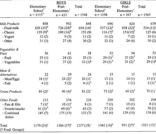
Pediatric Bone Mineral Accmal Study 91 4.2 Total dietary calcium intake from food in mg per day and contribution
of food groups to total calcium intake by sex and social-maturational groups 99 4.3 Percentage contribution of food groups to total dietary calcium intake by sex
and social-maturational groups 100 4.4 Seasonal variations in total dietary calcium intake from food in mg per day
and contribution of food groups to total calcium intake in males by
social-maturational groups 101 4.5 Seasonal variations in total dietary calcium intake from food in mg per day
and contribution of food groups to total calcium intake in females by
social-maturational groups 102 5.1 Sex comparison of mean values for the timing and magnitude of peak
height and tissue velocities 111 5.2 Correlations between age and magnitude of peak height and peak tissue
velocities for males 113 5.3 Timing of peak height and tissue velocities for early, average and late
maturing males and females 113 5.4 Magnitude of peak height and tissue velocities for early, average and late
maturing males and females 114 5.5 Tissue values at time of PHV, for early, average and late maturing males
Page X
5.6 Correlations between age and magnitude of peak height and peak tissue
velocities for females 115 6.1 Group characteristics for pre- and early pubescent girls involved in 10 months
of calcium supplementation and either high impact or low impact exercise 125 6.2 Sequential comparison of the number and types of impacts for the high impact
and low impact exercise groups 126 6.3 Body composition, anthropometry and bone mass values of pre- and
early-pubescent girls before and after in 10 months of calcium supplementation
and either high impact or low impact exercise 127 6.4 Group characteristics of pre- and early-pubescent girls before and after
10 months of calcium supplementation or a placebo equivalent 128 6.5 Anthropometry, bone mass and body composition values of pre- and
early-pubescent girls before and after 10 months of calcium supplementation
or a placebo equivalent 128 6.6 Group characteristics of pre- and early-pubescent girls before and after
10 months of either high impact or low impact exercise 129 6.7 Anthropometry, bone mass and body composition values of pre- and
early-pubescent girls before and after 10 months of either high impact or
low impact exercise 129 6.8 Dietary intake values for pre- and early-pubescent girls before, during and
after 10 months of calcium supplementation and either high impact or low
impact exercise 133 6.9 Dietary intake values for pre- and early-pubescent girls before, during and
after 10 months of calcium supplementation or a placebo equivalent 133 6.10 Dietary intake values for pre- and early-pubescent girls before, during and
Page xi
List of Figures
Figure Title Page
2.1 The dimensions of bone 15 2.2 Endosteal resorption and apposition during the growth period 17
2.3 The relationship between hormonal and physiological responses to changes
in plasma calcium levels 23 2.4 Calcium metabolism in the human body 24
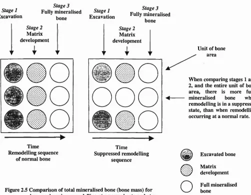
2.5 Comparison of total mineralised bone (bone mass) for bone undergoing
remodelling at a normal rate and at a suppressed rate 37 2.6 Side-to-side difference in BMC of the upper arm of players relative to the
age they commenced training 63 3.1 Total calcium supplementation intervention period demonstrating the timing
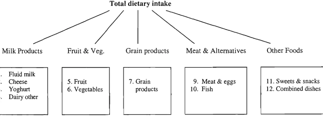
of the introduction of the three calcium supplementation methods 84 4.1 Categorisation of foods from the Canadian Food Composition Tables into
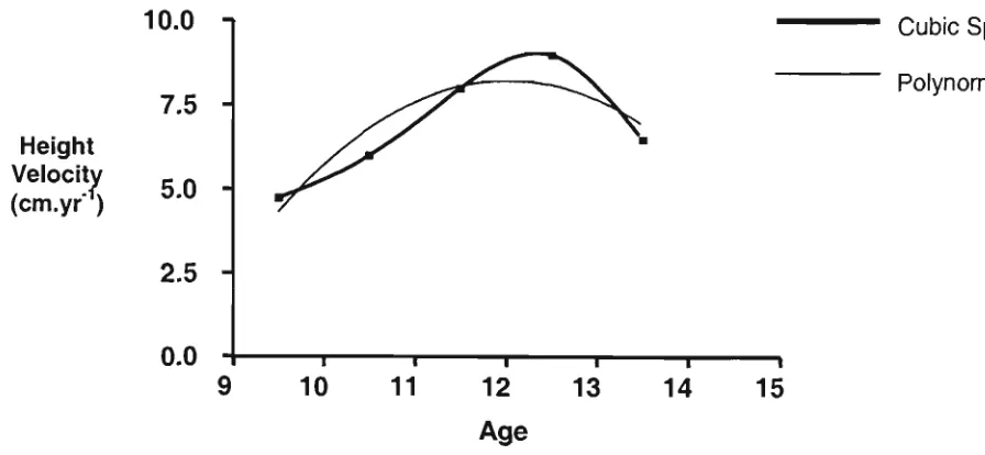
12 groups 93 5.1 Comparison of PHV values using two different curve fitting procedures 103
5.2 Breakdown of male and female participants into early-, average- and
late-maturity groups based on the age of PHV 108 5.3 Sequence of PHV and peak tissue velocities for males and females 110
5.4 Absolute values for height, body mass, lean mass, BMC and fat mass for
males and females at the age of PHV 112 6.1 Seasonal changes in dietary calcium intake in pre- and early-pubescent girls 131
6.2 The main effect of calcium and exercise over 10 months in pre- and
early-pubescent girls after adjusting for baseline bone mass and change in bone length 134 6.3 Group comparisons in percent bone mass accmal in pre- and early-pubescent
girls following 10 months of calcium and exercise intervention after adjusting
for baseline bone mass and change in limb length 136 6.4 Group comparisons in bone mass accrual in pre- and early-pubescent
girls following 10 months of calcium and exercise intervention after adjusting
Page xii
List of Appendices
Appendix Title
3.1 Recipes for foods used in the calcium / exercise intervention trial 165
4.1 Seasonal variations in Vitamin D intake from food in iu per day (SD) and
contribution of food groups to Vitamin D intake in elementary school females 168
6.1 Group characteristics, anthropometry, bone mass, body composition and dietary intake values for Asian and Caucasian pre- and early-pubescent girls at baseline and after 10 months of calcium supplementation (or placebo) and either high
Abstract 1 Abstract
Chapter One 2
Sub-1.1 1.2 1.3 1.4 1.5 1.6
•headings
Introduction Purpose Rationale Limitations Delimitations
Definitions of Terms
Chapter One
Chapter One 3
1.1 Introduction
Osteoporosis is a disease characterised by "low bone mass and microarchitectural deterioration of bone tissue, leading to enhanced bone fragility and a consequent increase in fracture risk" fWHO, 1994]. The disease has recently come to the fore as a major medical, social and health concern. Exuberant costs are involved in the treatment of those afflicted with osteoporosis. In 1992 it was
*
estimated that in Australia the annual cost of treating osteoporotic fractures in the 60+ age group was $779 million Australian^Commonwealth Dept. Health & Family Services, 1997]. Fractures may not only cause permanent disabilities they may also be fatal, with one in four sufferers of hip fractures dying within a year of the fracture [Commonwealth Dept. Health & Family Services, 1997]. The problem is projected to escalate in the future as the Australian population ages [Wark, 1996]. Preventative strategies aimed at promoting bone mineral accrual during growth or reducing bone loss in later life may be salient in attenuating this potentially 'epidemic' disease. Deciding on the most effective time during the lifespan to implement preventative strategies may determine the success of the strategies.
Although osteoporotic fractures manifest in old age, osteoporosis may have its origins in youth [Bonjour, 1994]. Bone mass and subsequent fracture risk in the elderly results from the amount of bone accmed by skeletal maturity, and the rate of bone loss in later life. Johnston and Slemenda (1995) suggest that low bone mineral density (BMD) may resuh from one or both of these factors. Low peak bone mass (PBM) or the failure to maximise bone mass accrual during growth is considered by some investigators to be the most important determinant of bone mass and fracture risk in old age [Bonjour, 1994]. Entering adulthood with maximal bone mass may mean that for a given rate of bone loss in later life, more time would be needed before bone mass would be reduced to the point where skeletal integrity may be compromised and fractures may ensue.
Chapter One 4
Once adult stature has been attained the ability to significantly enhance bone mass through modifications to diet and exercise appear limited. Calcium or exercise intervention studies involving adults have reported no effect or small improvements in BMD [Dawson-Hughes et al., 1990; Kelly, 1998a]. In contrast calcium [Bonjour et al., 1997; Johnston et al., 1992] and exercise [Bradney et al., 1998; Morris et al., 1997] intervention trials involving children have demonstrated positive effects on measures of bone mass accmal. Evidence from bilateral, cross sectional and retrospective studies in children further support the claim that childhood may be a more opportune time than adulthood, to influence bone mass [Bass et al., 1998; Haaspasalo et al., 1994; Kannus et al., 1995: Ruiz et al., 1995]. Haaspasalo et al. (1994) reported greater side-to-side differences between the playing and non-playing arms of elite female tennis players when compared to non-players. More importantly, the side-to-side difference was greater in players who commenced playing before menarche compared to those who commenced after menarche. Kannus et al. (1995) further investigated the side-to-side difference in BMC of the humems of elite squash and tennis players and reported a greater side-to-side difference in girls who commenced playing > 5 years before menarche (20-23%) compared to those who commenced playing > 15 years after menarche (9-10%). The findings reported by Haaspasalo et al. (1994) and Kannus et al. (1995) however, may be challenged as differences in loads experienced at differing stages of pubertal development were not quantified.
Independently, calcium and exercise intervention studies have reported positive effects on bone mass accmal. The existing evidence however, indicates that dietary calcium and exercise may act upon different constituents of bone and influence different aspects of the growth process. Calcium is reported to influence the remodelling process, by slowing bone turnover [Johnston et al., 1992], while exercise is believed to exert a greater influence on the modelling process [Bailey, 1995]. These observations raise the question as to the interactive effect of exercise and calcium on bone mass accmal. Limited data exist describing the combined effects of exercise and calcium on measures of bone mass in adults [Prince et al., 1995]. To date no data are available describing the combined effects of calcium and exercise on bone mass accmal during growth. For example, it is not known if the combined effects of calcium and exercise are synergistic or cumulative. Furthermore, there is a need to determine if exercise when combined with calcium would provide a greater osteogenic benefit to growing bone than the independent effects of exercise or calcium alone.
Chapter One 5
The timing of skeletal maturity demonsttates large variance both within and between the sexes. The effect of these temporal differences on the magnitude of bone and tissue accmal has not been investigated using longitudinal analysis. Calcium and exercise are two important and modifiable lifestyle factors that may enhance bone mass accmal. Existing dietary intake data indicates that dietary calcium intake, especially in females may not be meeting recommended levels. It is not known what dietary habits are contributing to this failure to meet recommended calcium intake levels. Based on available research evidence, modifying childhood calcium intakes and exercise levels may be more beneficial to bone mass than similar strategies in adults. The rationale for this study is threefold. Firstiy, there is a need to determine if calcium recommendations are being met, and if not which sex or maturity group are at the greatest risk and why. Information of this type may assist future educational or promotional campaigns aimed at ensuring an adequate calcium intake in our younger populations. Secondly, details of the differences in bone mass accrual between early, average and late maturing children may provide insight into the most appropriate means to meet skeletal needs during this period of rapid skeletal development. Finally, there is a need to understand the effects of calcium and exercise on bone growth, and to determine the independent and combined effects calcium and exercise may have on bone mineral accmal. The minimal amount, and type of exercise, or the level of calcium intake that may exert a positive effect on bone mineral accmal however, has not been clearly defined. There is a need, therefore to identify the nature and quantity of exercise and calcium that may provide an osteogenic benefit to growing bone.
1.3 Purpose
The purpose of this thesis was to measure calcium intakes in children both seasonally and at different maturity levels, to investigate bone mass accmal in children of differing age of maturity, and to determine the effects of increased calcium intake and high impact weight-bearing activity on bone mass accmal in pre- and early-pubertal girls. The specific objectives were:
1. To determine and compare total dietary calcium intake and sources of dietary calcium for younger and older boys and girls and to determine if differences are apparent between sexes, maturational groups and seasons.
It was hypothesised that calcium intake would decrease with age in girls but not boys and that milk intake would decrease in the colder month.
Chapter One 6
It was hypothesised that late maturing children would have lower peak height and tissue velocities than early nmtures due to growth occurring over a longer period of time.
3. To determine whether 8.5 months of calcium supplementation using calcium-enriched foods resulted in greater increases in the bone mineral content of pre- and early-pubertal girls when compared with pre- and early-pubertal girls not receiving supplementation.
// was hypothesised that calcium supplementation would result in enhanced bone mass accrual.
4. To determine whether twenty minutes of high impact weight-bearing activity, three times a week for 8.5 months resulted in greater gains in the bone mineral content of pre- and early-pubertal girls when compared with girls who participated in a similar program involving low impact activities.
// was hypothesised that high impact exercise would result in enhanced bone mass accrual at weight bearing sites.
5. To determine whether the combined effect of calcium supplementation and high impact weight-bearing activity on the bone mineral content of pre- and early-pubertal girls was greater than the individual effects of calcium supplementation or high impact weight-bearing activity.
It was hypothesised that a combined effect would be evident at weight bearing sites.
Study one consisted of 226 participants aged 8 to 19 years involved in a six year longitudinal study examining skeletal development and lifestyle factors that may influence bone mass accmal. Dietary intake was determined by serial 24-hour recalls with each participant providing up to four recalls per year. Three thousand one hundred and thirteen diet records were assessed.
Study two consisted of a group of 60 male and 53 female adolescents measured annually over a six-year period using standard measuring procedures and dual-energy x-ray absorptiometry (DXA). Peak height and peak tissue velocities were determined using a cubic spline curve fitting procedure of the individual data.
Chapter One 7
assigned to either the high- or low-impact exercise group, and to receive either the calcium-enriched or non-enriched foods.
1.4 Limitations
The recognised limitations associated with studies one and two were:
1. The group of 8 to 14 year old boys and girls who initially enrolled in the study were not representative of children of this age in the total population.
A representative sample in a study of this nature would demand a nation-wide random sample of children. The logistics of these demands mean that it would not have been feasible to use a representative sample of children.
2. The normal calcium intake of the participants was monitored at intervals throughout the study period.
For practical purposes, calcium intake was determined using 24-hour recall up to 4 times a year. Due to the large number of participants and the multiple number of data collection times it was impractical to use weight food records. However, it is acknowledged that weighed food records are the gold standard by which to record nutrient intake.
The recognised limitations associated with study three were:
1. The group of 7 to 10 year old girls who participated in this study was not representative of 7 to 10 year old girls in the total population.
A representative sample in a study of this nature would demand wide scale co-operation from a random sample of schools. Furthermore, it would intmde on a large number of school curriculum policies and programs, as the intervention would have to have been incorporated into the physical education curriculum. The logistics of these demands mean that it would not have been feasible to use a representative sample of girls.
2. The environmental conditions in which the program was undertaken could not be controlled.
Chapter One 8
nature of the impact and the subsequent osteogenic responses of the participants to the variable impacts.
3. The motivational levels of the participants may have varied during the program.
The amount of motivation may have influenced the amount of effort by the participants. This may in tum, have influenced the amount of impact incurred by participants.
4. The normal activity levels and calcium intakes of participants outside of the program were monitored but not controlled.
High levels of physical activity or calcium may have masked the potential influence of the program. In addition, participants may have adopted lifestyle changes during the intervention period. These changes may have altered their response to the intervention.
5. Participants were of Caucasian and Asian decent.
Although racial differences in bone mass have been reported, adjustments were made to baseline data during the analysis to minimise the potential for discrepancies associated with racial differences in response to the intervention.
1.5 Delimitations
The recognised delimitations associated with this study were:
1. Females only were included.
The existence of sex-specific differences in the response to calcium and exercise are unknown. A single sex group was used therefore, to eliminate the potential for sex-related differences.
2. All girls were pre- or early-pubertal.
Hormonal changes associated with advanced puberty may obscure changes as a result of the intervention. Seven to ten year old girls were selected as this helped to ensure that the participants were pre- or early-pubertal at the commencement of the intervention period, and remained pre- or early-pubertal for the duration of the intervention period.
3. Participating girls were delimited to girls of similar socio-economic backgrounds.
Chapter One 9
4. Participating girls were randomly allocated to the study groups.
This type of allocation was made to reduce the potential for selection bias, as girls more suited to exercise may have self selected to be in the high impact group.
5. A calcium supplementation level of 450 mg.day'^ was administered.
This level was to increase calcium intake levels to be above or near the recommended levels for children of this age.
6. No one methodology describes the impact of exercise on human bone in vivo.
The activity levels of the girls within the confines of the program therefore, were monitored using the following techniques. The number of impacts during the program was determined by video surveillance. The average landing forces and peak magnitude of impacts of various movements performed during the sessions were determined using a pedar mobile system on a subset of girls. The amounts and type of activity that may have been effectual on bone, therefore could be estimated.
7. The duration of the intervention period was delimited to 8.5 months.
This duration was selected as it could be undertaken and completed within one school year. The duration however, was long enough to allow a potential osteogenic response to be observed.
8. Baseline dietary calcium intake and physical activity levels were determined before, during and after the intervention period.
The determination of dietary calcium intake was delimited to a 3-day food record. The determination of physical activity was delimited to the use of a pediatric specific questionnaire, which was validated with video surveillance. These methodologies were employed to determine if differences between the groups were apparent in baseline measures, which may have confounded the effects of the intervention.
9. Changes in bone mineral measurements and body composition were delimited to detection via DXA.
Chapter One 10
10. Calcium supplementation was delimited to calcium-enrichment of food products.
Calcium supplementation may be in mineral form or of food sources, however, only supplementation via the enrichment of food sources has demonstrated residual benefits to measures of bone following the cessation of supplementation.
11. Blood and urinary measures were not taken
Due to the age group of the participants in the study, the inclusion of these measures would have severely reduced the number of girls who volunteered.
1.6 Definition of Terms
The following terms were defined with respect to their usage in this thesis
BMC Bone mineral content
The amount of bone mineral contained within the skeletal envelope. Expressed in grams (g).
BMAD Bone mineral apparent density
Bone mass divided by the volume of the region scanned. Expressed as grams per centimetre cubed (g/cm^). BMD Bone mineral density
Bone mass divided by the projected area of the area scanned. Expressed in grams per centimette squared (g/cm).
DPA Dual photon absorptiometry
Method of measuring bone mass and mineral density using low dose radiation. DXA Dual energy x-ray absorptiometry
Method of measuring bone mass and mineral density using low dose radiation. ECF Extra cellular fluid
Fluids located outside of the body's cells. GH Growth hormone
Hormone released from the pituitary gland that is the primary mediator of linear growth.
IGF-1 Insulin like growth factor
Secreted in response to GH, IGF-1 stimulates the differentiation and proliferation of many different cells including the cells (chondrocytes) within the growth plates of long bones.
HI High impact
Chapter One 11
LM Lean Mass
Total mass - (fat mass + bone mineral content).
MESm Minimal effective strain for controlling modelling drifts Strain magnitude at which bone modelling is initiated. MESr Minimal effective strains for controlling remodelling units
Strain magnitude below that which bone remodelling predominates and bone loss occurs.
MU Mechanical usage
Sttain threshold at which bone modelling and remodelling are initiated. PBM Peak hone mass
Maximal bone mass achieved by the time of skeletal maturity. PBMCV Peak bone mineral content velocity
Maximal velocity of bone mineral content accmal. PFV Peak fat velocity
Maximal velocity of fat mass accmal. PHV Peak height velocity
Maximal velocity of height gain, which is used as a marker of maturity. PLV Peak lean velocity
Maximal velocity of lean mass accmal. PMV Peak mass velocity
Maximal velocity of body mass accmal. PTH Parathyroid hormone
Hormone released from the parathyroid gland, which is involved in calcium homeostasis.
SPA Single photon absorptiometry
Chapter Two 12
Chapter Two
Literature Review
Sub-headings
2.1 Overview of bone growth
2.2 Normal bone development in growth 2.3 Linear growth of the human skeleton 2.4 Dimensional changes to bone with growth
2.5 Bone mineral accraal and loss during the lifespan 2.6 Peak bone mass
2.7 The role of calcium in the body 2.8 Regulation of plasma calcium levels 2.9 Overview of calcium metabolism 2.10 Calcium balance
2.11 Calcium absorption 2.12 Calcium excretion
2.13 Calcium intake recommendations
2.13.1 Trends in calcium intake in children of developed countries 2.14 The effect of calcium on bone remodelling
2.15 Calcium intake and adult bone mass 2.16 Calcium intake and bone mass in children
2.17 The effects of calcium supplementation on bone mineral accmal in children
2.17.1 The effect of calcium supplementation on bone mass accmal at different stages of maturity
2.17.2 Does daily dietary calcium intake influence the osteogenic response of bone to calcium supplementation?
2.17.3 The effect of calcium supplementation withdrawal on bone mineral accmal: The long term benefits
2.18 The effects of physical activity on bone mass
2.19 The mechanisms of the osteogenic response to mechanical loading 2.20 The osteogenic response of adult bone to exercise
2.21 The osteogenic response of immature bone to exercise
Chapter Two 13
2.1 Overview of bone growth
Childhood and adolescence are time periods characterised by the rapid growth and development of the skeletal system. Growth occurs in length, in width and in the mineral content contained within the skeletal envelope. To optimise the development of bone it is important to identify and promote factors that may have a positive effect on bone mass accrual. Two lifestyle factors that can be easily modified to enhance bone mass accmal are dietary calcium intake and exercise.
Adolescence is a time of accelerated skeletal growth therefore calcium needs may be heightened during this time. In spite of the greater calcium need during puberty, calcium intake levels of many young people do not meet recommended intake levels [English, 1989]. Furthermore, calcium intakes in females are reported to decrease during puberty, often well below recommended intake levels [Albertson et al., 1997]. The effect of these apparent inadequacies in calcium intake on skeletal integrity is yet to be determined.
Animal- and human-based studies have shown that mechanical loads applied to bone during growth results in greater bone mass accmal. The promotion of appropriate weight bearing exercise during childhood and adolescence may assist in the optimisation of bone mass accmal. If the enhanced bone mass accrued is maintained beyond the growth years, then the benefits of exercise undertaken during childhood and adolescence may be continued into later life. Exercise during childhood and adolescence may help reduce the risk of osteoporotic fractures in the elderly by promoting the accumulation of a greater reserve of bone, which can be drawn from to accommodate age-related bone loss.
Calcium supplementation and high impact weight bearing activity are both reported to have a positive effect on bone mass accraal [Johnston et al., 1992; Morris et al., 1997]. The amount of calcium or level and type of physical activity that elicits a positive osteogenic response is yet to be determined. In addition there is a paucity of information regarding the combined effect of calcium and exercise.
Chapter Two 14
relationship between exercise and bone mass, and more specifically the effects of school-based exercise interventions on bone mass accraal in children. The literature review concludes with a discussion of the combined effects of calcium and exercise.
2.2 Normal bone development in growth
Longitudinal growth and skeletal consolidation are dynamic processes that commence in utero and conclude some time in the third decade [Matkovic, 1991]. During this time the skeleton will approximately triple in length, and accrae over 40 fold its mitial calcium content, to contain approximately 1200 g of calcium by adulthood [Heaney, 1991].
The processes that bring about growth and expansion of the human skeleton are very detailed, involving the complex interaction of genetics, hormones and environmental factors. The importance of each of these factors is acknowledged however, for the purpose of this section, the discussion will be on the dimensional and mineral content changes of endochondral bone (that which replaces an existing cartilage model) from the postnatal period to skeletal consolidation.
The skeleton reaches its adult state through the processes of growth, modelling and remodelling. All three process can occur simultaneously throughout the skeleton however, growth and modelling predominate during the growth phase [Bailey et al., 1996]. Growth is the process of skeletal enlargement and elongation. Modelling enables the shape of the bone to be altered and the mass of the bone to increase, as new bone is added without the prior resorption (removal) of existing bone. The process of remodelling involves the replacement of fatigue damaged bone, with the existing bone being removed as new bone is added [Mundy, 1999]. The growth of bone therefore occurs in length, in thickness and in mineral content [Bailey et al., 1996].
2.3 Linear growth of the human skeleton
The lengthening of long bones occurs at the epiphysis, the band of cartilage that separates the diaphysis (shaft of the bone) from the epiphysis (end of the bone). The cells within the metaphysis multiply, and elongate the cartilage band. The cartilage closest to the diaphysis is then replaced, initially by woven bone, then by lamellar bone, with the later being layered and stmcturally sttonger [Solomon et al., 1990].
Chapter Two 15
been reported in yearly rates of height gains. Hagg and Taranger (1991) reported growth rates of 25.6 cm.yr"' and 25.1 cm.yr'' for male and female Swedish infants, respectively. Tanner et al. (1966) reported growth rates of 5.7 cm.yr"' and 5.8 cm.yr"' for British male and female children ages 5-10 years, respectively. Sexual dimorphism in height gains occurs during the pubertal period [Matkovic
1992].
Sex differences are apparent in the timing and magnitude of peak height velocity (PHV), which is the acceleration of linear growth which coincides with puberty [Bailey, 1997]. The earlier age of PHV reported for females is consistent with the earlier onset of maturity in females compared to males [Malina & Bouchard, 1991]. The magnitude of PHV however, is greater in males than females. The timing and magnitude of PHV from previous growth studies are summarised in Table 2.1. Calculated from the data presented in Table 2.1 PHV occurs approximately 1.9 years earlier in females than males. The magnitude of PHV is approximately 1.3 cm.yr"' greater in males compared to females.
The majority of adult stature is achieved before the completion of puberty. Bailey (1997) determined that 90% of adult height in males and 92% of adult height in females was achieved by the age of PHV. Following sexual maturity, the rate of height gain slows considerably. Kroger et al. (1993) reported gains in height of less than 0.2 cm.yr'' after menarche in 37 females. Overall height results from lengthening of the legs and the trank. During childhood, linear growth results from a proportionally greater gain in leg length compared to tmnk length, while pubertal growth is characterised by an acceleration in the rate of gain in tmnk length, and a slowing in the rate of growth of the legs [Bass et al., 1999].
2.4 Dimensional changes to bone with growth
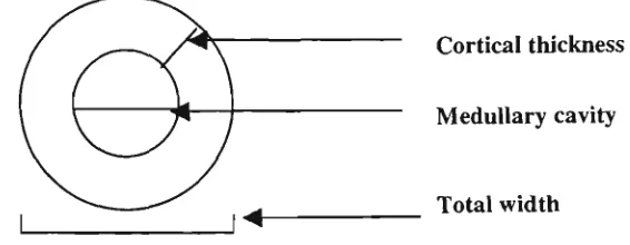
Dimensional changes to bone result from differing rates of apposition and resorption from the inner (endosteal) and outer (periosteal) surface of bone. These differences result in changes to the total width of the bone, the width of the medullary cavity and to the thickness of the cortical wall (Figure 2.1). Sex-related differences are evident in relation to the dimensional changes to bone.
Cortical thickness
Medullary cavity
Total width
\^
vO > X •a S 03 VI <0
s
PL, >ft f j
C/3 •"5 3 & (/] 3 O 3 > .SP "5 JC 03 O H f2 s «3 E tL| :2. 1 -M 0 \ o6 * +1 o OS 1.4 * +1 0 0 ON SO OS r^i t>-1. 2 +1 >n ON 1.2 * -i-i ON 00 ,8± 1 r-o 1±1 . i > CN 3±1 . 00 8. 6 6. 3 0 0 d +1 u a 4^ >. o m c CO 13 m (^ o >. T3 S CO O O a u > 3 d +1 0 0 +1 >, T3 3
• 4 - 1
00 13 c 3 Lon g ^ ca ( / ] *>. ^ >^ X ) 3 1/3 "rt di n 3 ongi t hJ -G o
•c 3
N OS d +1 ON c 2 u -G <u c X J
I
6 o T3 S 05 0 0 <N ON ON ^^ «. —^ CQ . - Ja> c D C 3 V OQ ON ON .. ' rt - 4 - ^
U
> H
u c
Chapter Two 17
Total bone width increases rapidly within the first year of life and again during the pubertal growth spurt, with moderate gains in bone width occurring during childhood. Tanner et al. (1981) reported gains of approximately 1 mm.yr'' in tibia width in pre-pubertal children. The accelerated gain in width which corresponds with the adolescent growth spurt occurs earlier in females than males however, the magnitude of the gains in bone width are greater in males than females [Malina & Bouchard, 1991]. Tanner et al. (1981) reported 13% greater bone width at the tibia and humems in males compared to females after puberty. Overall, males have greater bone width due to; a later pubertal spurt (longer childhood growth period), a greater magnitude of gain during puberty, and the overall growth period of bone width expansion lasting longer in males compared to females [Gam,
1970].
Changes on the endosteal (inner) surface influence the size of the medullary cavity. Gam (1970) indicated that the endosteal surface of bone move from a phase of resorption, to a phase of apposition, then back to resorption again. Gam (1970) reported that resorption occurred at the endosteal surface rapidly during infancy, then more slowly until approximately aged 14 years in females and approximately age 18 years in males. Resorption commenced again at approximately age 40 in both males and females. The rate of resorption peaks at approximately age 45 in females. The findings by Gam (1970) are supported in part by work from others. Johnston and Watt (1969) reported a 16% reduction in the width of the medullary cavity of the second metacarpal in post-menarcheal ghls compared to pre-menarcheal girls. Song et al (1994) reported a decrease in medullary diameter with increasing chronological and skeletal age in girls aged 12-18 years. Apposition of bone on the endosteal surface occurs at a greater rate in females compared to males, resulting in a smaller medullary cavity in girls compared to boys [Gam, 1970]. Tanner reported a 13% greater medullary width of the tibia in males than females after puberty. Sex-related differences are depicted in Figure 2.2. Following the period of apposition, resorption occurs at the endosteal surface during adulthood and older age in both males and females [Gam, 1970]. Ruff and Hayes (1988) reported no sex-related difference in the increase in medullary area of the tibia and femur in adults based on cross-sectional data.
Infancy & childhood Post-pubescent ^ ^^^ p i
endosteal resorption endosteal apposition / \ / , . ,, ' ^^ results in a smaller
medullary cavity than males
Male apposition occurs at a slower rate resulting in a larger medullary cavity than females
Chapter Two 18
The differences in the timing and rates of resorption and apposition of bone on the inner and outer surface of bone will influence the thickness of the cortex. Cortical wall tiiickness as described by Gam (1970) generally increases from infancy to adulthood, with males generally demonsttating a greater cortical thickness compared to females. Tanner et al. (1981) reported similar gains in cortical thickness of the tibia in boys and giris during childhood. Gryfe and Exton-Smith (1971) reported that boys on average had a greater cortical area at the second metacarpal compared to girls. Sexual dimorphism in cortical area or thickness occurs during the pubertal period. Gryfe and Exton-Smith (1971) observed that the spurt in growth in cortical area of the metacarpal occurred 2 years earlier in females than males (at approximately 10 years of age). Gam (1970) noted that the increase in cortical thickness in females during puberty results from apposition of bone on both the endosteal (inner) surface and periosteal (outer) surface of bone. This observation is supported by the findings of Song et al. (1994). The authors reported an increase in cortical area and thickness of the second metacarpal with skeletal age in adolescent females. Cortical area increased form 36.4 mm^ to 50.8 mm^ between the ages of 12 to 18 years. Concurrently there was a significant decrease in medullary area and an increase in total area with skeletal age in this group of girls. Although during early adolescence the cortical thickness is greater in females than males, the period of apposition is longer in males compared to females [Gam, 1970], resulting in greater cortical thickness in males compared to females, by late adolescence. Tanner et al (1981) reported a 13% greater cortical area in males compared to females by the end of puberty.
Bouxsein et al. (1994) reported greater cortical area and thickness at the distal radius and ulna in younger women (n = 21, 20-30 years of age) compared to older women (n = 22, 64-84 years of age), indicating a decrease in cortical area with age in women. Ruff and Hayes (1988) described earlier made a similar observation. They observed that following adjustment for bone length, cortical area of the tibia decreased with age in women (aged 20 - 90 years), however remained relatively constant with age in similarly aged men. The authors suggest that the compensatory apposition of bone on the periosteal surface in males offsets the loss of bone at the endosteal surface (significant increase in medullary area) thus maintaining the cortical area and intum the stractural integrity of the bone.
Chapter Two 19
2.5 Bone mineral accrual and loss during the lifespan
Bone mineral is progressively accmed in bone up to the age of peak bone mass, which is reported to occur between the ages of 15 to 30 years [Matkovic et al., 1994; Ott, 1990; Theintz et al., 1992]. Following the attainment of peak bone mass, and a period of maintenance of bone mass during adulthood, calcium is progressively lost from bone [Seeman, 1997]. Although all individuals accme and lose bone mineral content during their lifetime, the rates of accrual and loss are not constant throughout the lifespan, and vary depending on age and sex.
At birth the human body contains approximately 25 grams of calcium, which increases to approximately 1000-1200 mg by adulthood [Heaney, 1991]. The greatest rates of BMC accraal occur during early infancy [del Rio et al., 1994; Southard et al., 1991] and the pubertal grovvth spurt [Bailey, 1997], with lesser rates of accraal reported during childhood [Zanchetta et al., 1995]. Del Rio et al. (1994) used BMD as a measure of bone mineral content and size. Based on data collected for gains in lumbar spine BMD and BMC, an almost four fold increase in annual gain in BMD was observed in 2 year old children compared to 6-7 year olds (0.14 g.cm^ v 0.03 g.cm^).
Mean rates of total body BMC accraal in children (calculated from cross sectional data) have been estimated at between 100-150 g.yr''. Zanchetta et al. (1995) reported annual rates of total body BMC accraal of 104 g.yr'' and 105 g.yr'* for Argentine boys and girls aged 5 to 10 years, respectively. Molgaard et al. (1997) reported annual total body BMC accraal rates of 123 g.yr' and 121 g.yr' for 7 to 10 year old Danish boys and girls, respectively. Nelson et al. (1997) reported slightly higher rates of total body BMC accrual at 145 g.yr'' and 154 g.yr"' for 9-10 year old American boys and girls, respectively.
Based on cross sectional analysis it appears that prior to puberty, little sex-related differences occur in rates of bone mineral accraal. Nelson et al. (1997) described above noted no difference in total body BMC accraal between the 158 males and 130 females in thefr study. Bonjour et al. (1991) also reported no sex-related differences in rates of gains in BMD at the femoral neck, lumbar spine and femoral shaft between boys and girls aged 9-13 years. Sex-related differences in the timing and rate of bone mineral accraal appear during puberty.
Chapter Two 20
ages of 12-14 years in females and 14-16.5 years in males. Limited data are available that describe peak total body bone mineral accraal velocities during puberty. Bailey (1997) used longitudinal analysis to determine peak rates of total body bone mineral content (BMC) accraal in a cohort of over 200 Canadian children aged 8-14 years. Peak rates of total body BMC accraal were estimated at 320 g.yr' for males and 230 g.yr"' for females. The absolute amount of BMC accraed during the pubertal period is greater for males than females [Bailey, 1997]. Bailey (1997) calculated that the total body BMC accraed during the two years before and two years after the age of PHV (over a four year period) was 1072 grams for males and 772 grams for females, which represented 36% of adult BMC.
Age related bone loss is greater in females than males. Perry et al. (1996) noted that approximately 230g grams of calcium is lost from the adult female skeleton from the time of peak bone mass through to old age, while males lose about lOOg [Seeman, 1997]. As the female skeleton is generally smaller than the male skeleton, this absolute loss of bone mineral will result in a greater proportional loss of total bone mass.
From the growth data presented it is apparent that males tend to demonsttate overall greater bone mass than females. The sex-related difference results from greater height in males, due to a longer growth period, and greater magnitude of height gain at peak. Males also have greater bone width than females, which is also reflected in more bone mineral content. Males therefore, posses a larger and geometrically sttonger bone than females. The greater bone mass achieved by males may be a conttibuting factor to their greater resistance to osteoporosis in later life when compared to females. Maximising peak bone mass may be a means of preventing low bone mass in later life.
2.6 Peak bone mass
Chapter Two 21
Matkovic et al. (1994) investigated the timing of PBM in 265 pre-menopausal Caucasian females aged 8-50 years using a cross sectional method of assessment. Using regression models, it was reported that most of the BMC at the lumbar spine, proximal femur, radius and for total body was accraed by late adolescence. The inflection in BMC accraal with age occurred between the ages of 17- 22 years depending on the site being assessed. It was noted that beyond the age of 18 years no differences were found between the younger and older pre-menopausal women for measures of bone mass at weight bearing sites.
Haaspasalo et al. (1996) evaluated lumbar spine, femoral neck, trochanter and distal radius BMC in 330 pre-menopausal females aged 7 - 4 7 years. Peak BMC values occurred at 21 years of age for lumbar spine, 19 years of age for the trochanter and femoral neck and 18 years of age for the radius. Peak values for BMD occurred between the ages of 18 - 21 years.
Chapter Two 22
2.7 The role of calcium in the body
Calcium is a major inorganic element present in all living cells. At birth the human body contains approximately 25 grams of calcium. This amount increases to approximately 1000 - 1200 mg by adulthood [Heaney, 1991]. Ninety-nine percent of the body's calcium is found in bone (skeleton and teeth). The remaining 1% is found in extra cellular fluid, plasma, and in cell membranes [Kanis, 1994].
Calcium is an essential element, and performs both regulatory and stractural roles in the body. In its regulatory role, calcium is necessary for blood clotting, muscle contractions, nerve transmission, hormonal function, and membrane transport [Gibson, 1990]. Calcium also has an important role in bone stmcture. Calcium is a major mineral component of bone representing approximately one third of the mineral in bone. Calcium is combined with phosphate to form the mineral hydroxyapatite, which is deposited into the collagen (protein) matrix that makes up bone tissue. The mineralisation of bone tissue contributes to its strength and hardness [Broadus, 1999]. The skeleton also serves as a reserve for this nutrient. Bone is the major tissue involved in the regulation of plasma calcium levels.
2.8 Regulation of plasma calcium levels
Chapter Two 23
xl Plasma calcium
t
i Ca release from bone t Urinary excretion i Intestinal absorption
t P T H secretion
T Ca release from bone 4 Urinary excretion T Intestinal absorption
T Calcitonin secretion
T Plasma calcium
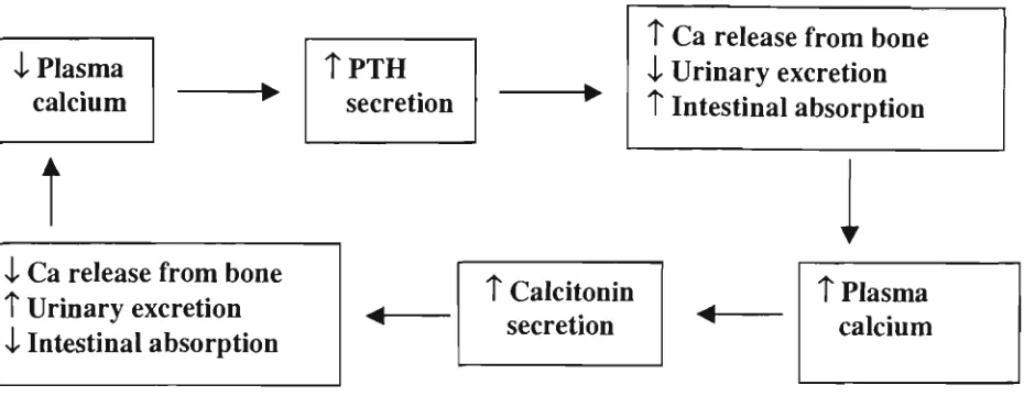
Figure 2.3. The relationship between hormonal and physiological responses to changes in plasma calcium levels.
2.9 Overview of calcium metabolism
Chapter Two 24
Intestinal absorption 200 mg.day*
Extra cellular
fluid /
Dermal losses 15 mg.day''
Bone 700 mg.day"*
Urinary losses 100 - 350 mg.day"*
Calcium intake 1000 mg.day'*
Faecal losses 800 mg.day'*
Figure 2.4: Calcium metabolism in the human body [Passmore & Eastwood, 1986].
2.10 Calcium balance
Calcium balance refers to the amount of calcium needed to maintain adult bone mass, or in the case of children, the amount needed to allow for skeletal development [Matkovic, 1991]. Adulthood is characterised by a zero calcium balance, as calcium enters and leaves the bone at equal rates. The equilibrium of skeletal calcium occurs via the process of remodelling. This process involves the coupling of the actions of osteoclasts (which remove bone) and osteoblasts (which replace bone) at discrete locations on the skeleton. The mechanisms that result in the coupling of these two contrasting actions are not clearly understood [Mundy, 1999]. The end result is maintenance of skeletal calcium. Calcium balance during childhood and adolescence is generally positive as new bone is added. Bone modelling occurs during childhood and adolescence and is responsible for the growth and re-shaping of bone. Unlike remodelling, during modelhng bone formation is not immediately preceded by resorption [Baron, 1999]. The end result is the accraal of skeletal calcium. While researchers agree that a positive balance is necessary for skeletal growth, the degree of positive balance needed to maximise bone mineral accraal and achieve peak bone mass remains unknown [Matkovic, 1991].
The determination of calcium balance during adulthood is based on an assumed relationship between calcium intake and excretion using the following equation [Matkovic et al., 1990]:
^ a Balance — ^ a intake ~ v*^a feecal "r '-^a urinary/
Chapter Two 25
Standard rate of calcium absorption to establish the amount of dietary calcium needed to maintain zero balance.
These balance studies used to determine recommended calcium levels rely on the existence of a relationship between calcium intake and excretion. The rates of absorption and urinary excretion in the metabolism of calcium however, may be influenced by other dietary and regulatory factors, and vary independenUy of each other. For example calcium absorption varies with maturity [Weaver et al., 1995], and is influenced by the dietary fibre content of the diet [Heaney & Weaver, 1989; Levenson & Bockman, 1994]. Calcium excretion may be influenced by the protein content of the diet [O' Brien et al., 1996]. These factors challenge the accuracy of calcium requirements determined from balance studies.
Calcium balance has been linked to calcium intake [Matkovic et al. 1990; Matkovic, 1991] however, the relationship is not necessarily maintained for high calcium consumers, which led to the concept that calcium may be a threshold nutrient. Matkovic (1991) reviewed 487 balance studies spanning the period of skeletal growth to peak bone mass (0 - 30 years). From this analysis, Matkovic (1991) reported a positive correlation between calcium intake and calcium balance (r = 0.589, p < 0.001). The relationship was evident in all groups representing different stages of development (infancy, childhood, adolescence and adulthood). A further review of 34 published balance studies by Matkovic and Heaney (1992) reported a positive correlation between calcium intake and balance for children and adolescents aged 9-17 years. The intake of participants ranged from 225 to 988 mg of calcium a day (n = 33, r = .803, p < 0.01). No relationship was found for those participants with very high calcium intakes (1730 - 2721 mg.day"').
Chapter Two 26
Furthermore, the relationship between calcium intake and balance is likely to be confounded by variations in absorption and excretion during the metabolism of calcium.
2.11 Calcium absorption
Calcium absorption occurs via two mechanisms: active transport in the duodenum and passive diffusion throughout the small intestine [Breslau, 1996]. During active transport calcium is moved through the epithelial cells bound to the carrier protein calbindin. This process is Vitamin D dependent and is down regulated when calcium intake is sufficient, and up regulated when calcium intake is low [Bronner & Pansu, 1999]. Passive diffusion is not regulated [Breslau, 1996], and is the principle mechanism of calcium absorption when calcium intake is adequate or high [Bronner & Pansu, 1999].
Calcium absorption is reported to increase during times of high need such as rapid skeletal growth (infancy and adolescence), pregnancy, and lactation [Passmore & Eastwood, 1986]. Matkovic (1991) compared calcium absorption rates for participants with calcium intakes within a normal range (683 to 1297 mg.day''). The analysis involved studies including infants ( 0 - 1 year, n = 88), children ( 2 - 8 years, n = 82), adolescents (9 - 17 years, n = 117) and young adults ( 1 8 - 3 0 years, n = 159). Higher rates of calcium absorption were reported for infants (36% - 44%) and adolescents (40% - 41%), when compared with children (24% - 27%) and adults (20% - 24%).
Further studies have reported similar findings to Matkovic (1991). Weaver et al. (1995) compared the net calcium absorption from a controlled mixed diet, between 14 adolescent girls (13.1 ±0.9 years) and 11 young adult females (22.2 ± 4.2 years) involved in a three-week balance study. A greater rate of calcium absorption was reported for the adolescent girls compared to the adult women (32.3% versus 20.6%, p < 0.05). Abrams and Stuff (1994) compared calcium absorption rates from self-selected diets in 51 females aged between 4.9 years to 16.7 years, who represented three stages of maturity. A greater rate of calcium absorption (34.4%) was reported in early-pubescent females (n = 13), when compared with pre-pubescent (absorption = 27.7%, n = 21, p < 0.05) and late-pubescent females (absorption = 25.9%, n = 17, p < 0.01). Thus in girls, the skeletal expansion that occurs during early puberty (eg. PHV) may be accommodated by greater rates of calcium absorption, which have been reported during this stage of development compared to the pre- and post-pubertal periods [Abrams et al., 2000]. These observed changes in rates of calcium absorption may serve to compensate for the reduction in calcium intake in females during adolescence
Chapter Two 27
The percentage of dietary calcium absorbed (fractional calcium absorption) was reported to increase with decreased calcium intake in healthy young girls, adult women, osteoporotic women, and girls with a family history of osteoporosis [Heaney et al., 1990; O'Brien et al., 1996; O'Brien et al., 1998]. Heaney et al. (1990) determined the fractional calcium absorption in 24 healthy women aged 29 to 45 years. Each woman consumed three of five prescribed calcium loads (15 mg, 36 mg, 86 mg, 205 mg, 500 mg) in a random sequence. The fractional absorption of calcium was inversely correlated with the logarithm of the load (p < 0.001). At the lowest calcium load, fractional absorption averaged 64.0%. In contrast, fractional calcium absorption averaged 28.6% at the highest calcium load.
O'Brien et al. (1996) found similar results to those reported above. These investigators evaluated calcium absorption efficiency in 11 girls (mean age 11.9 ± 2.4 years) after 10 days on a low calcium diet (7.05 ± 2.03 mmol.d'') and 10 days on a high calcium diet (35.30 ± 2.28 mmol.d"'). Fractional calcium absorption was greater for the low calcium diet compared to the high calcium diet (58.2% ± 8.7% and 26.0% ± 6.8%, respectively, p < 0.0001). These data indicate that fractional calcium absorption appears to increase with decreased intake in females. However, if the calcium intakes are very low, in spite of the greater rate of absorption, the amount of available calcium may still be compromised.
Net calcium absorption is reported to increase with calcium intake [Matkovic et al., 1990]. For example, if 15% of lOOOmg of dietary calcium is absorbed then the net Ca absorbed is 150mg. However, if 30% of 300 mg of dietary calcium is absorbed then the net Ca absorbed is 90 mg. This is supported by work presented in the literature. Heaney et al. (1990) reported that the net amount of calcium absorbed based on the calcium loads and fractional absorption rates were 10 mg, 102 mg and 143 mg, respectively for calcium loads of 15 mg, 300 mg and 500 mg. In addition, O'Brien et al. (1996) demonstrated that the actual calcium absorbed was greater for the young girls when they consumed the high calcium diet when compared with the low calcium diet (8.98 ± 2.65 mmol.day"' and 4.04 ± 1.00 mmol.day'', respectively, p < 0.001).
Chapter Two 28
chronic or long-term reductions in calcium intake, where alternative adaptations may occur [Heaney, 1991].
Calcium absorption occurs within the context of a total diet. Constituents of other foods in the diet may influence the absorption of calcium. For example, calcium forms insoluble complexes with phytates in wholegrain cereal products, oxalates contained in green leafy vegetables, nuts and tea, and the uronic acid component of dietary fibre [Breslau, 1996]. The insoluble complexes reduces the solubility (and bioavailability) and subsequent absorption of calcium [Heaney & Weaver, 1989; Levenson & Bockman,
1994]. In addition, fatty acids, especially saturated fatty acids form insoluble soaps, which also hinder calcium absorption. Excessive phosphate and an alkaline environment may also decrease calcium absorption [Breslau, 1996]. Alternatively, some amino acids, lactose and an acidic environment in the intestine increase the bio-availability of calcium thus promoting absorption. Adequate vitamin D is also necessary for the absorption of calcium [Levenson & Bockman, 1994].
Dairy foods are often promoted as the most readily absorbable food source of calcium [Peacock, 1991]. Recent reviews of calcium absorption studies presented below however, indicate that calcium from other sources may be absorbed as efficiently as calcium from milk and milk products. Calcium absorption rates calculated from several studies indicate that calcium absorption from milk averages 33%, which compared less favourably with mean absorption rates of 37% for calcium chloride and 35% - 36% for calcium citrate maleate [Heaney & Weaver, 1989; Levenson & Bockman, 1994; Miller et al., 1988]. Calcium absorption rates less than those reported for milk were found for calcium oxalate (10% - 11%) [Heaney & Weaver, 1989], calcium carbonate (26% - 28%) [Miller et al., 1988], tricalcium phosphate (25%) and calcium cittate (22%) [Levenson & Bockman, 1994]. Similar rates of absorption to milk were reported for calcium lactate (32%) and calcium acetate (32%) [Levenson & Bockman, 1994].
Chapter Two 29
These data presented indicate that calcium absorption is greater during infancy and adolescents, compared to childhood and young adulthood. Also within the period of puberty in females, absorption appears to be greater during early puberty, compared to the rates of absorption observed during the pre- and post-pubertal periods. At a high calcium intake more absolute calcium is absorbed than at low intakes however, the proportion of calcium that is absorbed is less (smaller percentage). The form of the calcium, and other components contained in the diet may also influence absorption rates. In addition to calcium intake and absorption potentially influencing calcium balance, calcium losses may also influence calcium balance.
2.12 Calcium excretion
Calcium balance is determined from calcium intake and calcium excretion. As previously outlined, calcium losses may occur from dermal, faecal and urinary avenues. Dermal losses were estimated at approximately 15 mg.day'' however, losses from active children have been estimated in excess of 60 mg.day'' [Charles et al., 1983]. Faecal calcium losses are not regulated. These losses appear to be a function of intake and are reported to increase with increasing calcium intakes in children and young adults [Matkovic, 1991]. The principle means of regulating calcium excretion is via urinary losses. While earlier studies assumed a relationship between urinary calcium excretion and dietary calcium intake, other dietary constituents may influence this relationship.
Urinary calcium was reported to decrease with short-term reductions in calcium intake in 11 girls aged 11.6 ± 2.4 years who were fed a low calcium diet for 10 days [O'Brien et al., 1996]. Matkovic et al. (1995) however, reported that urinary calcium was not related to intake in 370 girls aged 8-13 years when consuming their usual diet. Matkovic et al. (1990) earlier reported that urinary calcium losses remained unchanged in spite of variations in calcium intake (270 mg.day'' - 1242 mg.day'') in 31 fourteen-year-old girls involved in a calcium supplementation trial. Jackman et al. (1997) reported that only 6% of the variance in urinary calcium excretion of 35 post-menarcheal girls could be accounted for by calcium intake.

![Figure 2.4: Calcium metabolism in the human body [Passmore & Eastwood, 1986].](https://thumb-us.123doks.com/thumbv2/123dok_us/7965635.1321185/37.580.22.505.99.329/figure-calcium-metabolism-human-body-passmore-eastwood.webp)