ABSTRACT
KUMAR, ARUN. Process Development for Expansion of Human Mesenchymal Stem Cells (hMSCs) using Three Dimensional (3D) Constructs in Novel Perfusion Bioreactors. (Under the direction of Dr. Binil Starly).
Process Development for Expansion of Human Mesenchymal Stem Cells (hMSCs) using Three Dimensional (3D) Constructs in Novel Perfusion Bioreactors.
by Arun Kumar
A dissertation submitted to the Graduate Faculty of North Carolina State University
In partial fulfillment of the requirements for the degree of
Doctor of Philosophy
Industrial Engineering
Raleigh, North Carolina 2017
APPROVED BY:
--- Dr. Binil Starly
Chair of Advisory Committee
--- Dr. Paul Cohen
--- Dr. Rohan Shirwaiker
DEDICATION
BIOGRAPHY
ACKNOWLEDGMENTS
First of all, I would like to acknowledge the guidance and support of my academic advisor Dr. Binil Starly. He mentored me with a systematic and realistic approach during my entire PhD journey. He is a pragmatic personality. Without his support and guidance, it was not possible for me to get where I am today.
I would also like to extend my gratitude for Dr. Susan Bernacki. Except teaching tissue engineering techniques, she was pivotal in explaining various techniques used in BMECC (Biomedical Engineering Culture Core) during the course of this research. I also appreciate the actions of Mr. Wing Lau of 3D Biotek for providing me critical industry exposure and helping me conduct this research.
Additionally, I would like to thank my colleagues Lokesh Karthik Narayanan, Atin Angrish together with other DIME lab members for supporting my research and conducting valuable discussions. I would also like to acknowledge the support and motivation provided by my friend Kimberly S Harms at every step. Moreover, the Dum Dum group holds a special place for getting me through the tough times during 2013-14.
TABLE OF CONTENTS
LIST OF TABLES ... viii
LIST OF FIGURES ... ix
Chapter 1 Manufacturing of Regenerative Medicine Products ... 1
1.1 Introduction ... 1
1.2 Growing demand of Stem Cells ... 2
1.2.1 Autologous Therapy ... 4
1.2.2 Allogeneic Therapy ... 5
1.2.3 Manufacturing Process Flow of Cells/Tissues/Organ Products ... 6
1.3.1 2-D Culture & Limitations ... 8
1.3.2 Exploiting the Third Dimension ... 10
1.3.3 Bioreactors ... 10
1.3.4 Knowledge Gap ... 11
1.4 Research Goal and Objectives ... 15
1.5 Results & Contributions ... 18
1.6 Dissertation Outline... 19
Chapter 2 Literature Review ... 20
2.1 Overview of 3D Culture methods for cell expansion... 20
2.1.1 Microcarriers (solid and porous) ... 20
2.1.2 Micro-encapsulation ... 23
2.1.3 Porous 3D scaffolds and fibers ... 25
2.1.4 Cell Spheroids ... 26
2.2 Current State of Art in Stem Cell Expansion ... 27
2.2.1 Cell Stack / Cell Factory ... 29
2.2.2 Specific types of Bioreactors ... 30
Chapter 3 Materials and Methods ... 46
3.1 Alginate Source and Sterilization ... 46
3.2 Cell Micro-encapsulation in Alginate Microspheroids using Electrostatic Deposition... 47
3.3 Characterization of Alginate Microspheroids ... 49
3.4 De-crosslinking of Alginate Microbead: Cell Harvesting ... 50
3.5 Batch and PBS Mini-Bioreactor cultures ... 51
3.6 PS Scaffold Fabrication & Characterization ... 52
3.7 Plasma Treatment of Poly-Styrene Scaffolds ... 53
3.8 Cell Harvesting Protocol for Poly-Styrene scaffolds ... 54
3.9 hMSCs & MG-63 Cell Culture ... 55
3.10 AlamarBlue® Cell Proliferation Assay ... 56
3.11 LIVE/DEAD® Cell Viability Assay ... 57
3.12 Lactate Concentration Assay ... 58
3.13 Glucose Concentration Assay ... 58
3.14 Osteogenic Differentiation ... 59
3.15 Alizarin Red S Staining ... 59
3.16 Adipogenic Differentiation ... 60
3.17 Oil Red O Staining ... 61
Chapter 4 Microencapsulation as a Vehicle for Cell Expansion ... 62
4.1 Introduction ... 62
4.2 Material and Methods ... 65
4.3 Results ... 66
4.4 Discussion ... 71
Chapter 5 Micro-Encapsulated hMSCs Cultured in Rotating Vertical Wheel Bioreactor ... 74
5.1 Introduction ... 74
5.2 Material and Methods ... 78
5.3 Results and Discussion... 78
Chapter 6 hMSCs Expansion using 3D Printed Scaffolds in Static Culture... 87
6.1 Introduction ... 87
6.2 Materials and Methods ... 88
6.3 Results and Discussion... 92
6.4 Conclusion and Future Work ... 100
Chapter 7 Perfusion bioreactor with Packed 3D Printed Poly-Styrene Scaffolds ... 102
7.1 Introduction ... 102
7.2 Material and Methods ... 103
7.3 Results and Discussion... 107
7.4 Conclusions ... 115
Chapter 8 Design, Development and Evaluation of a Tubeless Perfusion Bioreactor for Expansion of human Mesenchymal Stem Cells... 116
8.1 Introduction ... 116
8.2 Material and Methods ... 119
8.3 Results ... 126
8.4 Conclusion and Discussion ... 143
Chapter 9 Overall Summary, Research Contributions and Future Work ... 147
9.1 Overall Summary and Research Contributions ... 147
LIST OF TABLES
Table 1: Table summarizing the advantages and limitations of various bioreactors... 28
Table 2: Factors and levels for seeding time DoE. ... 90
Table 3: Fold Expansion and Percent viability after harvesting ... 96
Table 4: Bioreactor run results (n=3) with RPM=20. ... 113
Table 5: Variables used to set up screen boundary condition. ... 138
LIST OF FIGURES
Figure 1-1: Manufacturing Process Flow for Cell based Autologous Regenerative Medicine
Therapy [34] ... 4
Figure 1-2: Manufacturing Process Flow for Cell based Allogeneic Regenerative Medicine Therapy ... 5
Figure 1-3: Two-Dimensional culture platforms. A) Cell culture Flasks B) Cell Stack/Factory ... 8
Figure 2-1: Micro-carriers ... 21
Figure 2-2: Cell Microencapsulation ... 25
Figure 2-3: Wave Bioreactor with stem cells grown on microspheres. ... 30
Figure 2-4: Stirred Tank Bioreactor... 32
Figure 2-5: Hollow Fiber Bioreactor ... 35
Figure 2-6: Rotating Wall Vessel Bioreactor (RWVB) ... 37
Figure 2-7: Packed Bed bioreactor setup. ... 39
Figure 2-8: Roller Bottle Bioreactor ... 40
Figure 3-1: Nisco Cell Encapsulator Unit... 47
Figure 3-2: Alginate Cell Microencapsulation Process ... 49
Figure 3-3: De-crosslinking Alginate Microbead ... 50
Figure 3-4: Polystyrene scaffold characterization. ... 52
Figure 4-1: Microencapsulated MG-63 model bone cells ... 66
Figure 4-2: Non-invasive characterization of Microbeads ... 67
Figure 4-3: Distribution of Microbead Diameter [148]... 68
Figure 4-4: Proliferation as percent AlamarBlue reduction ... 69
Figure 4-5: Qualitative assessment of viability using LIVE/DEAD assay ... 70
Figure 5-1: Non-invasive characterization of microbeads. ... 78
Figure 5-2: Cell growth and lactate concentration in batch culture [140]. ... 79
Figure 5-4: Comparison of lactate concentration from model and experiment in batch culture
[140]. ... 82
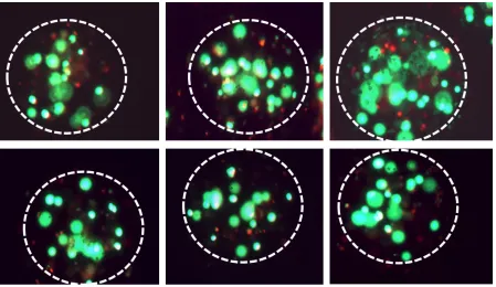
Figure 5-5: Live/Dead viability assay fluorescence images. ... 83
Figure 5-6: Lactate concentration (ngm/uL) in bioreactor. ... 84
Figure 5-7: Glucose content (mg) in bioreactor... 85
Figure 6-1: Flow diagram of evaluating 3D PS scaffolds for cell expansion in static culture ... 91
Figure 6-2: Pareto Plot for seeding time DoE. ... 92
Figure 6-3: Scaled estimates for seeding time DoE. ... 93
Figure 6-4: Cell Seeding Efficiency on Poly-Styrene Scaffolds ... 94
Figure 6-5: Cell proliferation as percent alamarBlue reduction. ... 95
Figure 6-6: LIVE/DEAD assay for Non-Treated Scaffolds. ... 97
Figure 6-7: LIVE/DEAD assay for Treated Scaffolds. ... 98
Figure 6-8: LIVE/DEAD assay for Control well at different magnification levels. ... 98
Figure 6-9: Alizarin Red S and Oil Red O staining images for treatment group (Scale=500um) ... 99
Figure 6-10: Alizarin Red S and Oil Red O staining images for control group (Scale=500um) ... 100
Figure 7-1: A petri-dish size actual scaffold picture with alternate strand lay details. Cross-section detail of printed strands. Each layer is offset to ensure maximum seeding efficiency. ... 103
Figure 7-2: Seeding and Perfusion culture of hMSCs on Polystyrene Scaffolds ... 105
Figure 7-3: Harvesting of hMSCs after expansion. ... 106
Figure 7-4: Polystyrene scaffolds characterization. ... 108
Figure 7-5: Mass Flow Rate vs Velocity graph for perfusion culture. ... 109
Figure 7-6: Inlet Velocity vs Pressure Gradient for a single PS scaffold. ... 109
Figure 7-7: Average Inlet Velocity vs Average Wall Shear Stress on cells growing on scaffold surfaces. ... 110
Figure 7-9: Flow through polystyrene scaffolds. ... 111
Figure 7-10: A) Bioreactor with 3 PS Scaffolds B) Flowlines through modeled porous media ... 112
Figure 7-11: Alizarin Red S and Oil Red O staining images ... 114
Figure 8-1: Bioreactor Assembly with 3 scaffolds and associated parts. ... 121
Figure 8-2: Actual prototype of novel tubeless bioreactor with assembly. ... 121
Figure 8-3: Effective surface area available for cell expansion marked as BLUE. ... 125
Figure 8-4: RPM vs Average Surface Velocity ... 127
Figure 8-5: 37 Point estimates of the surface velocity of fluid on the scaffold strands ... 128
Figure 8-6: Variation in surface velocity with position of scaffolds. ... 128
Figure 8-7: Average Velocity vs Average Wall Shear Stress (WSS) ... 129
Figure 8-8: Average WSS was determined by sampling 40 points on the surface of the scaffold ... 130
Figure 8-9: Percentage area available for the cells to proliferate on the scaffold at 75RPM. ... 131
Figure 8-10: A) Bioreactor with 3 porous scaffolds B) Sectional view of flow trajectories at 75 RPM with scale. ... 132
Figure 8-11: 6-well size model with 10 scaffolds. ... 133
Figure 8-12: Average Surface Velocity for each scaffold for 50 RPM impeller speed. ... 134
Figure 8-13: Average Wall Shear Stress for each scaffold for 50 RPM impeller speed. ... 134
Figure 8-14: 6-well size model with 3 scaffolds. ... 135
Figure 8-15: Average Surface velocity for each scaffold for 50 RPM impeller speed. ... 135
Figure 8-16: Average Wall Shear Stress for each scaffold for 50 RPM impeller speed. ... 136
Figure 8-17: 2D geometry representing media inlets (A, a, b), Oxygen Concentration Boundary B, Outflow Boundary C, Scaffolds D and Internal Fixture Boundary E... 138
Figure 8-18: "Finer" physics controlled mesh with No Flux boundaries (Blue color). ... 139
Figure 8-19: Flow Velocity distribution within the fluid domain. ... 139
Figure 8-20: Oxygen Concentration surface plot. ... 140
Chapter 1
Manufacturing of Regenerative Medicine Products
1.1 Introduction
Regenerative Medicine (RM) aims at enhancing the ‘self-healing’ of damaged organs, tissues or cells by using the body’s natural healing mechanism coupled with foreign biological materials/tissues. These biological materials/tissues are aimed at rebuilding/replacing damaged tissue with the aid of Tissue Engineering (TE). Thus, Tissue Engineering pertains to the practice of integrating biomaterials, cells and suitable biochemical factors to produce functional tissues/organs. Even though regenerative medicine and tissue engineering differ slightly in their definitions, these terms are used interchangeably in practice and referred to as TERM (Tissue Engineering and Regenerative Medicine). TERM have been used for a plethora of therapeutic applications including bone [1–5] and cardiac regeneration [6–11]. Advancements in these fields have also shown potential ability to regenerate damaged/lost limbs [12,13], next-gen tools for drug development [14–17] and as advanced models to understand disease pathology [18–23]. With the advancement in TERM technologies during the past couple of decades, FDA has approved several regenerative products categorized under Biologics, Cell-based medical devices and Biopharmaceuticals [24].
as raising livestock accounts for a considerable portion of greenhouse gases responsible for global warming. Therefore, TERM offers an alternative to meet therapeutic as well as non-therapeutics needs. Still, a lot of research and development continue to be done to overcome the challenges associated with realizing the full potential of this field. This research addresses one such challenge, i.e. a key component of engineered tissue – the Cells. Critical to the function of engineered tissues, whether it be for non-therapeutic or therapeutic applications, are the ‘Living Cells’ that are required in the hundreds of millions of quantities required for
critical function.
1.2Growing demand of Stem Cells
Cellular biomanufacturing technologies are a critical link to the successful application of in vitro tissue models, organ-on-chip devices and disease models. An emerging technology that is related to the fabrication of such living engineered tissue are through what is known as ‘Biofabrication’ processes. These processes, both biopatterning and bioprinting, when scaled
up to commercial level production processes, will require the initial raw cellular material stock to be produced in large quantities. This cellular stock material cannot be produced by labor intensive processes as is currently done in research laboratories. In commercial production, cellular stock materials must be initially produced in large viable numbers and then distributed to individual biomanufacturing processes that require them. Preparing this key stock material is quintessential for any biofabrication process since a successfully biofabricated construct will depend on the quality of cell types contained in them.
1.2.1 Autologous Therapy
Figure 1-1: Manufacturing Process Flow for Cell based Autologous Regenerative Medicine Therapy [34]
Autologous therapies are initiated with the extraction of cells from a patient, expanded in large quantities and later administered to the same patient as shown in Figure 1-1. These cells obtained from the patient must be expanded 100–200 times its initial amount to have the desired clinical delivery yields. For a single patient, quantities in excess of 300million are needed for a single therapeutic application in cardiac regeneration when using mesenchymal stem cells. This is partly due to low efficiency in current delivery methods, when a majority of the therapeutic cells are lost in the blood stream when administered to the patient [35]. The second reason is the sheer number of cells needed to treat a critical size defect such as in bone, liver or cartilage regeneration. Encouraging results have been reported in bone regeneration towards DDD (Disk Degeneration Disease) [36], Gingivostomatitis [37], psoriasis vulgaris and psoriatic arthritis [38] using Mesenchymal Stem Cells (MSCs). The goal would be to rapidly ‘scale out’ operations by using a single facility to process cells from multiple patients
simultaneously. This process will need to be automated and most likely cannot be performed by humans to remain economically viable. However, current methods employed are extremely labor intensive and will lead to very expensive treatment options for patients.
Tissue Acquisition Isolation of Cell Types of Interest Cell Expansion Cell Harvest Volume
1.2.2 Allogeneic Therapy
While autologous therapies aim at regenerating/treating the same patient from which the cells are derived, allogeneic therapy provides a more generic solution. These therapies are paving the pathway towards “off the shelf” stem cell treatments for mass population. The
manufacturing process flow of such therapy is shown in Figure 1-2. Like Autologous therapy, the process starts with the acquisition of tissue of interest from the donor. Cells of interest are then isolated from the tissue for further processing in an expansion device usually bioreactors. Once the cell population reaches desired quantities, the cells are harvested and concentrated for packaging. These therapeutic products need to be cryopreserved for long term storage and transportation. Intensive testing and regulatory bodies are involved in the release of such products which could considerably increase the time to market. Once successfully released, these products are shipped to suitable surgical/medical facilities for administration on patients.
Figure 1-2: Manufacturing Process Flow for Cell based Allogeneic Regenerative Medicine Therapy
prevented Glucocorticoid-Induced Osteoporosis [39]. A second allogeneic Hematopoietic Cell Transplantation (HCT) was shown to be efficacious in patients who have previously experienced a graft failure or relapse [40]. After undergoing allogeneic therapy couple of patients were reported to have developed graft-versus-host disease after subsequent radiation therapy [41].
1.2.3 Manufacturing Process Flow of Cells/Tissues/Organ Products
Tissue Engineering and Regenerative Medicine manufacturing process flow initiates with the acquisition of tissue from a healthy donor. The donor is carefully chosen and the acquired tissue dictates most of the upstream processes. Once the tissue is successfully acquired, it is subjected to sequence of operations to isolate the cells of interest. These cells could range from a few thousand to millions in quantity. For a successful allogeneic therapy, cells in the order of 1010 to 1012 would be required. Extensive cell manufacturing and culture should be carried out in order to scale-up the initial quantity of cells isolated from the donor to these large numbers. This phase of the upstream process is known as the Cell Expansion phase. During the course of cell expansion, cells are initially plated in 2D culture plates/flasks for expansion to millions in quantity. Hereafter, the scale-up to billions in quantity is often carried out in bioreactors suited for cellular expansion. These bioreactors are discussed in detail in the latter part of the document.
enzyme can cleave essential proteins and damage the self-renewal capacity of the cells leading to senescence. Therefore, cell harvesting protocols can be extremely challenging and tailor-made for specific cell types. This cellular harvest is then processed by centrifugation to concentrate the yield in smaller volumes ready for packaging. Final reduced volume is then cryopreserved and stored until the product is ready to be shipped and passes various regulatory requirements. Extensive testing and clinical trials are a major component of this downstream processing of the cellular product. Phase I/II/III trials are conducted to ensure the efficacy and safety of the product before it can be shipped and made available to the final patient. After the testing is successful and product is deemed safe for final use, it is shipped and available for end use. For most of the allogeneic therapy the above manufacturing cycle is applicable while autologous therapy might bypass the packaging, cryopreservation and logistics.
This research work addresses the ‘Cell Expansion’ and ‘Cell Harvest’ phase of the
of cells needed for both therapeutic and non-therapeutic applications. This research will address the development of one such technology path – to provide an alternate method to grow cells to production level quantities. The next section elaborates on these details and form the core motivation of the work performed.
1.3 Research Motivation
1.3.1 2-D Culture & Limitations
The most expensive and time consuming step in the process flow (as shown in Figure 1-2) is the cell expansion phase. In laboratory and early clinical trials, this expansion step is typically carried out in tissue culture flasks or stacked culture flasks. Several review papers
A)
Inlet Outlet
B)
have highlighted the deficiencies of flat plate based expansion of cells in RM [31,42,43]. The 2D surfaces are best utilized in research laboratories, pre-clinical and Phase I trials. With flat plate culture technology, it is laborious and expensive to expand cells in the hundreds of millions to a few billion quantities. High quality manufacturing floor space to expand cells will come at a premium and may not be financially sustainable for cell manufacturers. Use of bioreactors for cell expansion is critical to the production of cells. In an allogeneic setting, most therapeutic applications will require cells on the order of 1010–1012 cells produced for effective therapy in a single cell expansion system. Flat-plate based static culture system cannot be scaled up to meet such demands. Industrial scale bioreactors designed for cell expansion offer key advantages in providing the right environment and reproducibility needed to achieve stable stem cell proliferation while maintaining sterility and potency characteristics.
there is a need to create and maintain conducive working conditions to help ensure uniform product quality.
1.3.2 Exploiting the Third Dimension
For these reasons, cell expansion in any RM approach, is critically important to the translation of RM therapies to commercial operations. Moving away from flat plate culture, three-dimensional culture of adherent cells provides an exciting new approach to expand cells and when combined with bioreactors, provides a possible solution to culturing massive quantities of cellular raw material. There is a lot of potential for the use of 3D scaffolds and constructs as matrices upon which anchorage dependent cell expansion can be carried out. Expansion of therapeutic cells on 3D surfaces through the use of microbeads, microgels and scaffolds will be discussed further. Followed by explanation of how anchorage dependent 3D culture is enabled in bioreactor based cell expansion. Each bioreactor type is described by its working principle, summary of published work and will focus on work related to the use of bioreactors in stem cell manufacturing. However, it is important to note that cell expansion processes does not impact stem cells alone, but affects other anchorage dependent progenitor and mature cell types.
1.3.3 Bioreactors
proliferation. These 3D surfaces include microencapsulation, microcarriers, scaffolds, nanotubes/nanofibers, hollow fibers etc. Chapter 2 provides a detailed explanation of these 3D attachment and encapsulation surfaces. Choosing the right attachment surface can significantly impact the scale-up potential and quality of the final yield from the bioreactor.
Bioreactors such as Wave, Stirred Tank, Hollow Fiber, Rotating Wall, Packed Bed and Roller Bottle are amongst most widely used bioreactors for adherent cells. Even though these bioreactors provide a solution to three-dimensional expansion of adherent cells, they still face several limitations. Due to large volume of cellular content inside the bioreactor and the size of the culture volume, nutrients and metabolites gradients are often present in such bioreactors limiting the growth profile of the therapeutic cells. Moreover, retrieval of cells after expansion is another avenue requiring deft skills and rigorous protocols. Finally, shear stress induced due to a moving fluid or rotating shaft/impellers may cause excessive shear stress resulting in a non-uniform cellular yield. Apart from these common challenges across most current bioreactor, there are several other limitations linked to specific type of bioreactors which will be discussed in the Literature review portion of this document.
1.3.4 Knowledge Gap
precisely defining the micro-environment of cells. This would ensure defined pores and surfaces for attachment as well as spaces for mass transfer and media circulation. Therefore, non-uniformly defined 3D surfaces like non-woven textiles, suspended nanotubes, porous microparticles etc. can lead to variable yields and limit process consistency. Moreover, even if they produce a conducive surface to grow, methods are still lacking on how they can be efficiently retrieved back from the 3D surface. A digitally defined scaffold can overcome these limitations and provide the necessary surface and pores for expansion and dynamic culture. Another challenge with the existing bioreactors is the black-box nature of these devices. In-situ sensors providing pH, dissolved oxygen, metabolites and glucose concentration can change the nature of these bioreactors. Providing more uniform culture conditions and reducing the gradients of nutrients with time. This can in turn lead to better and uniform yields with improved knowledge about the cellular product being manufactured.
A bioreactor containing a stacked array of digitally defined scaffold and produced by additive manufacturing methods with precisely defined micro-environment is proposed in this dissertation. This bioreactor would be designed to be a completely closed system with no external moving parts other than the ones in the bioreactor chamber itself. Computational Fluid Dynamics is conducted to calculate optimal flow characteristics and associated wall shear stress. Being a closed system with limited involvement of pumps and tubes, this system would be capable of reproducibly expanding cells while maintaining a sterile environment.
1) Cell Seeding on Scaffolds: Poly-Styrene scaffolds with highly uniform characteristics are being considered for the 3D surface to be used inside this bioreactor. Apart from increasing the culture surface available for the attachment, these scaffolds also provide a well-defined micro-environment for cells. But, cellular expansion can only happen once these scaffolds are successfully seeded before introduction into the bioreactor. The desired cell seeding density and initial attachment duration must be determined before expansion within any class of bioreactors.
2) Proliferation: Once successfully seeded onto the scaffolds, it must be ensured whether the surface and environment is conducive for the cells to proliferate during the expansion phase. Qualitative assessment of this phenomenon can be useful in determining the duration to confluency and stopping criterion. Statically cultured scaffolds can provide qualitative proliferation data leading to successful assessment of proliferation rate and confluency. This work will study the use of various bioreactor types suitable for cellular expansion.
4) Viability: The final yield has to be evaluated for accessing viability of the cells. Several factors can impact viability ranging from seeding, proliferation and harvesting. If not properly seeded, the cells can dislodge once the media is circulated through the scaffold. Moreover, during proliferation, a high shear stress induced by the flow could result in cell death and compromise viability. Harvesting can also significantly affect cellular viability due to prolonged exposure to undesirable enzymes which can damage surface cellular proteins and cellular membrane. There is limited research work conducted to study how much viability loss is present during the critical phases of cellular expansion with 3D printed scaffolds.
5) Undifferentiated Stem Cells (Stemness): Expansion of stem cells for TE and RM applications is based on the premise of the yield being undifferentiated. Later, the cells are differentiated towards lineages required for any particular therapy. In order to access the undifferentiated state of the stem cells harvested, the cells can be exposed to differentiation media for sufficient time interval. After that duration, the cells can be stained with differentiation assay dyes to confirm deposition of differentiation material. If the cells exposed to differentiation media show stains of differentiation w.r.t. control (cells with just the growth medium) then it can be reasonably be concluded that the harvested cells have preserved stemness characteristics.
At the end, a new bioreactor design is developed to help address some of the current technology limitations in existing bioreactor systems.
1.4 Research Goal and Objectives
As discussed above, there is a critical need for large quantities of stem cells as a raw material for tissue engineering and regenerative medicine. This research is focused on studying the process of large scale expansion of adherent cells particularly human Mesenchymal Stem Cells (hMSCs) using 3D constructs and bioreactor technologies. Parameters such as seeding efficiency, proliferation, harvesting, viability and stemness (undifferentiated stem cells) are being reported for successful evaluation of techniques wherever possible. This work is categorized under the following three objectives. Results obtained have been reported for all the objectives in this dissertation document with discussion of research contributions and future work.
utilized the microgels produced to expand the culture of hMSCs within the bioreactor. Microbeads were sampled out of the reactor and evaluated for cellular yield. We have also assessed how glucose and lactate varies as the cells proliferate within the hydrogels. Harvesting protocols along with methods to determine yield and viability have been developed as a part of this objective.
Objective 2: To identify dynamic culture conditions that affect the efficiency of the expansion of hMSC cells in a perfusion bioreactor system containing packed 3D printed porous polystyrene scaffolds.
Objective 3: To design, develop and test a closed loop perfusion bioreactor system for the expansion of hMSC on 3D printed poly-styrene scaffolds.
The perfusion bioreactor system evaluated in objective 2 has its limitations in that it is not a completely closed system. It includes a series of tubes going in and out of the incubator that severely limit it scalability and increases the chances of contamination. Moreover, a single inlet port for circulating media inside the bioreactor provides poor mixing and high wall shear stress concentration in certain portions of the scaffolds. In order to improve upon these limitations and make the bioreactor a closed system, a novel bioreactor is designed. This bioreactor would still include 3D printed Poly-Styrene scaffolds as the cell attachment surface. This design employs no tubes or mechanical peristaltic pumps for the circulation of media. Through the use of a magnetically activated stirrer, we have achieved a continuous closed loop circulation of media with adequate oxygenation within the media. This system is designed to sit inside an incubator for optimal culture conditions to be maintained. We have examined the fluid dynamics within the reactor and developed a multi-physics model to assess oxygen gradients within the bioreactor.
cells is important. Fold expansion as compared to the initial seeding density is considered a metric towards quantity of the cells. Similarly, the ability of these hMSCs to differentiate into different lineages dictates the undifferentiated quality of the product. These metrics are of paramount importance when looking at large scale expansion of stem cells for RM and tissue engineering applications.
1.5 Results & Contributions
three-dimensional space for cellular expansion, scaffolds are limited with the surface area of strands available for cell attachment and growth. However, harvesting of cells can be significantly easier with 3d printed scaffolds as opposed to microencapsulation. With these sets of experiments and modeling, new product development related to cell expansion can be successfully accomplished. In a nutshell, this dissertation provides feasibility of using different 3D constructs inside perfusion bioreactor and their interactions.
1.6 Dissertation Outline
Chapter 2 Literature Review
2.1 Overview of 3D Culture methods for cell expansion
For cell expansion at production scale quantities, 3D matrices have been studied to explore the use of the third-dimension to provide an active surface for cells to attach and proliferate. Among the main advantages of this method are: (1) a much higher surface to volume ratio, thereby concentrating the growth of cells within a confined volume space; (2) for adherent dependent cells, the surface properties can be modulated to provide a conducive surface for attachment and proliferation. Many of the therapeutic cell types of interest are adherent based and need a surface to attach; and, (3) three-dimensional surfaces expands the possibilities of providing a microenvironment that can regulate cell behavior and function through engineered biomaterials and cell niches, inherently seen in in-vivo physiology. The following three methods are currently utilized by the community for adherent cell based expansion for clinical trials and commercial production.
2.1.1 Microcarriers (solid and porous)
(1) solid spherical or disc shaped particles, where in cells are solely attached to the surface of the bead (Figure 2-1 (A)), and
(2) porous beads prepared from cellulose or gelatin which allow cells to infiltrate and colonize into the microbead (Figure 2-1 (B)).
Figure 2-1: Micro-carriers
A) Cells grown on a solid microcarrier; B) Cells grown on a porous microcarrier. Surface area provided by the spheres helps improve cell yield within a given bioreactor volume [34].
Solid microcarriers (Cytodex made from dextran and SoloHill made from polystyrene) have been used for mesenchymal, induced pluripotent and embryonic stem cell expansion [44,45]. Extensively used in stirred tank type bioreactors, the microcarriers are suspended within the bioreactor while providing the surface for adhesion and proliferation of anchorage dependent cells. Harvesting of cells can be a challenge since enzymes used to remove cells from the microcarriers can destroy adhesion proteins which may negatively affect cells for therapeutic applications. If hydrodynamic forces in the suspension culture are not controlled, the microcarriers can strike each other causing damage to the attached cells on the exterior surface. Commercially available porous carriers (CultiSpher™, Cytopore™ and Cytoline™)
culture based bioreactors. While successfully used in viral vaccine and recombinant protein production, their use in cell therapy might be a challenge due to the difficulty of safely harvesting cells from within the porous microcarriers.
environmental changes. From a cellular biomanufacturing process, this requires us to develop smarter control strategies for the robust culture of stem cells.
2.1.2 Micro-encapsulation
A B C
Figure 2-2: Cell Microencapsulation
A) Cells encapsulated within a hydrogel. B) Two or more cell types within a hydrogel for an in-vivo like environment. Safely harvest therapeutic cells while preserving viability and potency can present a challenge in a production based environment; (C) Cell adhering to
one-another to form spheroid like bodies, also known as cell aggregates [34].
2.1.3 Porous 3D scaffolds and fibers
fibronectin have been used to expand the growth of bone marrow (BM) derived mesenchymal stromal cells (MSCs) [60]. However, very limited studies have been conducted utilizing this method for cell expansion and harvesting. Most papers utilize this method in the production of recombinant proteins, growth factors, exosomes or viral vectors—all products produced from immobilized cells within the matrices. In such applications, the need for harvesting cells does not arise and hence fibers provide a contained reactor for growth of the desired cell types.
2.1.4 Cell Spheroids
2.2 Current State of Art in Stem Cell Expansion
Table 1: Table summarizing the advantages and limitations of various bioreactors.
Type of Bioreactor Advantages Limitations
Wave Bioreactor - Scale-up
- Ease of automation
- Operation in different batch
modes
-Scale up to >100L is
challenging.
-Much larger floor space needed.
Stirred Tank - Ease of design
- Scale-up achievable
- Operation in different batch
modes
- Online and in situ monitoring
including media and cells.
-Satisfying the oxygen demand
of large volume, high-density cell cultures.
-Excessive shear stress caused by
the impeller
Hollow Fiber Bioreactor - Increased surface to volume
ratio with highest culture density.
- Low shear environments.
- In vivo setting as fibers mimic blood vessels.
-In-situ monitoring is
challenging.
-Scale-up has not been
demonstrated.
-Gradient of nutrients and
metabolites can be detrimental.
-Retrieval of cells is challenging.
Rotating Wall Vessel Bioreactor - Better nutrient supply and
metabolic waste removal
- Extremely low shear and
turbulence
- No internal moving parts and
mechanical agitation.
- No gradients of shear stress
-Limited in their capacity to
produce relevant number of cells for clinical application
-Scale-up is not possible due to
rotary motion.
Packed-Bed Bioreactor - Large surface area present for
cell attachment and proliferation
- Do not require frequent
passaging thereby savings generated from costs related to culture media.
-Concentration gradients,
particularly in large vessels.
-Packing material can limit cell
harvesting.
-In-situ monitoring possible but
only measure media.
Wave Bioreactor - Scale-up
- Ease of automation
- Operation in different batch
modes
-Scale up to >100L is
challenging.
-Much larger floor space needed.
Roller Bottle Bioreactor - Both adherent and suspension
based cells can be cultured
- Low shear stress
- Simple in design
- Low capital investment required
-Labor intensive
-Large physical space required
-Limited surface area (bottle
surfaces)
-High media cost due to
2.2.1 Cell Stack / Cell Factory
A cell stack consists of multiple two-dimensional (2D) plates assembled on top of each other with media inlet/outlet ports on the top (Figure 1-3 (B)). Each plate provides 2D area equivalent to almost 9 T-75 culture flasks. This enables almost 10 mi cells to be obtained from each layer. Cell Stack (CS) is classified according to the number of layers of 2D surfaces mounted. One (CS-1), two (CS-2), four (CS-4), ten (CS-10) and forty (CS-40) are the most commonly used configurations. Cell stack have been used for large scale expansion of pre-isolated bone marrow mesenchymal stem cells using CS-10 [66]. Similarly, CS-5 (five layer cell stack) was reported for expansion of dental pulp stromal cells [67]. Clinical-scale production with 31.6 +/- 14.5 fold expansion was reported for granulocyte and post-progenitor cells using cell stack [68].
2.2.2 Specific types of Bioreactors
2.2.2.1 Wave bioreactor
A wave bioreactor utilizes the wave motion of culture medium generated by a rocking platform to provide good cell bead suspension. This rocking motion also destroys any nutrient, pH or oxygen gradients present with low to negligible shear stress to stem cells in disposable culture
Figure 2-3: Wave Bioreactor with stem cells grown on microspheres.
Peristaltic pumps feed fresh medium with a separate bag for harvesting microspheres for further downstream processing [34].
demonstrated for culture volumes upto 500 L [71]. Eibl et al describes the various modes this bioreactor has been operated and analyzed via batch, fed-batch, repeated fed-batch, continuous and continuous perfusion mode [72]. Some of the commercially available wave-agitation bioreactors are BioWave®, Wave Bioreactor™, BIOSTAT® CultiBagRM, AppliFlex,
Tsunami® Bioreactor, CELL-tainer®, wave and undertow bioreactor [72]. Wave agitation bioreactors have been demonstrated to achievemore than10times expansion of neutrophils from hematopoietic progenitor cells using a 10 L unit [71]. Likewise, a 10 L Wave® Bioreactor was employed for GMP manufacturing of biologically active Xcellerated T Cells™ [73].
Microcarriers have been previously used in this bioreactor for cultivating Madin Darby Canine Kidney cells [74,75]. Expansion of adherent embryonic feline lung fibroblasts on microcarriers was also carried out using this bioreactor [76]. However, this bioreactor still needs to be explored for expansion of stem cells, particularly since they have not demonstrated to be used in high volume production.
2.2.2.2 Stirred Tank Bioreactor
operation in different batch modes along with culturing the cells on various 3D platforms including the ability to integrate online monitoring probes and control of culture variables [82,83]. Recently, an average 24-fold expansion per 6 day passage of hPSCs cultured on treated microcarriers was reported with 85% retrieval efficiency along with maintenance of cell characteristics [84].
Figure 2-4: Stirred Tank Bioreactor
Stirred Tank Bioreactor with marine impeller type blades stirring the medium containing cell attached on microcarriers. Suite of dissolved oxygen sensors, temperature probes,
Stirred tank bioreactors have been used for microcarrier based expansion of ESCs in a serum-free culture medium with maintenance of pluripotency [79]. Also, microcarrier based expansion of BM MSC and adipose tissue-derived stem/stromal cell was established using a 1 L stirred-tank bioreactor to (1.1 ± 0.1) x 108 and (4.5 ± 0.2) x 107 cells respectively within 7 days of culture [81]. Similarly, stirred tank bioreactor was used to expand mES cells on microcarriers using serum-free medium yielding an 85 ± 15 fold expansion over 11 days. Moreover, these cells retained their pluripotency markers and differentiation potential [82]. Another study reveals the generation of 2 x 10⁸ hiPSCs/100 mL using microcarriers with maintenance of cell characteristics [85]. Likewise, directed differentiation to hepatocyte as well as undifferentiated expansion of hESCs on microcarriers to 1 x 10⁶ cells/mL within 2 days was successfully reported [86]. A 5 L stirred tank bioreactor was used for microcarrier based expansion of hMSCs to more than 6-fold over 12 days of culture with maintenance of cell surface markers after harvesting [87]. Similar application to 14 days microcarrier based culture of MSCs (derived from BM and adipose tissue) in xeno-free medium yielded (2.0 ± 0.2) x 105 and (1.4 ± 0.5) x 105 cells/mL respectively [88]. Carmelo et al describes a potential protocol for expansion of MSCs derived from different human sources with different micro-carriers using this bioreactor [89].
employed to maintain required oxygen concentration [90]. But sparging of air within the medium can induce undesired shear forces on the cells cultured in the reactor. Excessive shear stress caused by the impeller is yet another problem with increased agitation rates and mixing resulting in cell death. Finally, lack of expertise on harvesting myriad of cells from beads after differentiation or during passaging [83] limits the projected benefits.
Vertical Wheel Bioreactor
2.2.2.3 Hollow Fiber Bioreactor
Figure 2-5: Hollow Fiber Bioreactor
Hollow Fiber Bioreactor with cells grown between the spaces of the packed hollow fibers. Culture medium flowing through the IC space delivers nutrients while waste material is carried out through the exit port of the system. Fiber membranes have designed pore sizes
to retain cells allowing only relatively smaller molecules such as nutrients to move back and forth across the membrane [34].
flow operation is known as intra-capillary inoculation with extra-capillary perfusion [93]. HF bioreactors provide an increased surface to volume ratio for cell attachment and growth as compared to static cultures but present challenges in monitoring and scale-up [42]. The fibers mimic blood vessels in coordinating nutrient supply and rejection of waste [99] while oxygen exchange is managed by diffusion between intra-capillary and extra-capillary spaces. Multi-cellular tissue culture or cultures with scaffolds can limit the oxygen exchange and distribution of culture medium inhibiting cell proliferation and growth [100]. In addition, heterogeneous distribution of cells with gradient of nutrients and metabolites has been previously found that limit the use of these bioreactors in cell therapy applications [99]. Techniques to overcome these limitations include use of oxygen carriers [100], increasing flow rates and rotating the HF reactor in timed cycles to reduce oxygen gradients. Some of the commercial sources for HF bioreactor include FiberCell Systems (FiberCell Systems Inc., Frederick, MD, USA) [95]; NxStage (Lawrence, MA, USA) [94]; Quantum® Cell Expansion System (Terumo BCT) [96– 98]; and Stemcell Systems GmbH, Berlin, Germany [101].
2.2.2.4 Rotating Wall Vessel Bioreactor
Figure 2-6: Rotating Wall Vessel Bioreactor (RWVB)
RWVB schematic with any of the three 3D culture types rotating about the horizontal axis as shown [34].
Rotating wall vessel bioreactor (RWVB) was developed by NASA’s (National
A comparison of HARV and STLV was presented by Goodwin and co-researchers to conclude superiority of HARV over STLV for culturing T-24, a human bladder transitional epithelial cell line, on Cytodex-3 microcarriers [105].
2.2.2.5 Packed-bed bioreactor
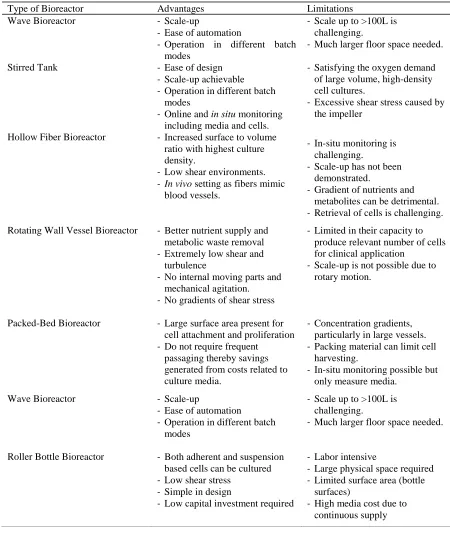
Figure 2-7: Packed Bed bioreactor setup.
Controller station (not shown), which typically contains the thermal mass flow controller with gas connections to control oxygen, air and carbon-dioxide and sensor probe
interconnects [34].
after 7 days of culture [117]. Expanded cells maintained their potential for differentiation and were found positive for many common markers to MSC. Recently, a version of the packed bed, termed as the fibrous bed bioreactor was used to expand mES cells for 30 days in two passages, 15 days in each passage. A 60–77 fold expansion was recorded in each passage while maintaining the pluripotency and differentiation markers for mES cells [118]. Due to large surface area present for cell attachment and proliferation, these bioreactors do not require frequent passaging, thereby savings generated from costs related to culture media. Even though fixed bed bioreactors provide large surface for cell attachment, they suffer from concentration gradients, particularly at large cellular yields which could alter cellular behavior, particularly with stem cell expansion.
2.2.2.6 Roller Bottle Bioreactor
Figure 2-8: Roller Bottle Bioreactor
Roller bottles rotated continuously to allow culture media to flow over the surface attached cells.
improve oxygen transfer and maintain a homogeneous environment inside the roller bottle. Since the cells are continuously washed by media, it helps remove waste materials and metabolites [119]. Rolling motion for mass transfer and mixing produces extremely low shear stress as compared to stirred bioreactors [120]. Fresh medium is continuously circulated in the chamber and product/samples can be harvested during operation for necessary monitoring of culture.
2.3 Chapter Summary
Industrial scale bioreactor technology approaches discussed in this document have been derived from cell expansion used in the production of bio-therapeutics such as recombinant proteins and viral vector production. In expansion for cell therapy applications, these bioreactors exploit the rapid expansion capacity of cells, particularly embryonic stem cells and progenitor cells. Cell stack factories are being utilized by the industries for producing enough cells for Phase II/III clinical trials. Unlike biopharma production, distinct processes must be developed for the expansion and safe retrieval of adherent cells. The following engineering challenges still need to be addressed before commercial cell expansion utilizing 3D culture can gain widespread acceptance.
packing material currently utilized as 3D surfaces for cell culture. Mass transfer and retrieval of cells from an open pore architecture scaffold will be considerably easier. In addition, biopatterning processes can create customized microenvironment best suited to the growth of cell types of interest. We foresee biofabrication processes playing a key role in producing the next generation of 3D culture surfaces specifically meant for adherent cell expansion in bioreactors.
(2) The microenvironment: The bioreactor microenvironment has clear advantages in terms of automated cell expansion. However, our understanding of the microenvironment within the bioreactor and how it affects cellular behavior is still lacking. Large nutrient gradients within bioreactors can significantly alter cellular behavior and function, particularly with the expansion culture of mesenchymal stem cells and iPSC cells. Efficient removal of adherent cells from surfaces without significant alteration of the cell-surface proteins is needed. This will also improve harvesting efficiency with minimal effect on the therapeutic cells. Currently, use of large amounts of trypsin or other chelating agents to detach cells from microcarriers or hydrogels affect cell membrane proteins and also leads to lower cellular yield.
through which we can improve the surface to volume ratio will improve cellular yield/volume ratio, leading to a reduced need for frequent media changes. Manipulation and optimizing the cell microenvironment can be an added method to minimize culture media use [127]. Novel biomaterials such as nanofibrous gelatin substrates have proven to stabilize cell phenotype, particularly with the hPSCs [128]. Others have shown that switches in chemical environment within a hydrogel system can induce differentiation without the need for specialized media [129]. Similarly, by binding hydroxyapatite peptides to scaffolds, promising research has shown to induce differentiation of MSCs to an osteogenic line [130,131]. More work must be done to identify microenvironment factors that best suits the expansion of therapeutic cells. Limiting the amounts of media changes and number of passages performed can be simple steps taken to lower COGs associated with culture medium.
small molecules as real-time sensors can be adapted to be included within bioreactors [135]. These sensors can provide the needed process data to perform adequate process monitoring. Due to the lack of real-time, in situ sensors, FDA led activities such as process analytical technology is hard to implement in cellular biomanufacturing. The ability to measure changes to the product is critical to ensuring product consistency and quality.
Chapter 3
Materials and Methods
During the course of this entire dissertation research, several methods were employed for culturing adherent cells on 3D platforms. Collections of new and established methods were employed for testing and evaluation of cell seeding, proliferation, harvesting, viability and assessing stemness characteristics. New methods developed include sterilization of Low-G variety alginate salt, encapsulation technique for obtaining ~400 um microbeads, scaffold characterization, non-invasive characterization of microbeads after encapsulating MG-63/hMSCs and harvesting procedure for 3D printed polystyrene scaffolds. Moreover, established methods like biological proliferation assays, LIVE/DEAD, cell culture etc. were used for evaluation of growth and viability qualitatively. We also developed protocols for culturing microgels in rotating wheel bioreactors. Following are the collection of all the materials and methods utilized for conducting this research. Rather than repeating the methods multiple times, this chapter will explain all common protocols utilized. Specific methods will be described in their individual section in the dissertation.
3.1 Alginate Source and Sterilization
required concentration. The cell-alginate concoction was then loaded into a sterile syringe for use in the microspheroid generation unit.
3.2 Cell Micro-encapsulation in Alginate Microspheroids using Electrostatic Deposition
Figure 3-1: Nisco Cell Encapsulator Unit
(A) Syringe pump (B) Cell-alginate concoction (C) Peristaltic pump (D) Nozzle (E) Positive electrode pole (F) Calcium chloride solution (G) Peristaltic pump control (H) Agitator
The cells (hMSCs/MG- 63) were encapsulated in alginate microspheroids using an electrostatic encapsulator (Nisco Var V1, Nisco Engineering AG, Zurich, Switzerland). A static electric field (10 kV) was applied between grounded nozzle tip (Ø 350 µm) of a syringe pump and a charged electrode plate resulting in droplet generation. The syringe pump delivered the cell-alginate concoction to the nozzle at a flow rate of 5 ml/hr to match the force exerted by the electric field. Calcium chloride (CaCl2) solution (2% w/v) was placed at spraying distance (4cm) below the nozzle (Figure 3-1). This is where the sodium ions in alginate get replaced with calcium ions in the dish and the bead gets crosslinked. The 10 kV voltage applied to the electrode pole with one end submerged in the CaCl2 solution created an electric field which pulled the droplets off the end of the nozzle and into CaCl2, leading to the formation of calcium-alginate microspheroids. The process was continued until the entire solution in the syringe was emptied.
Figure 3-2: Alginate Cell Microencapsulation Process 3.3 Characterization of Alginate Microspheroids
3.4 De-crosslinking of Alginate Microbead: Cell Harvesting
Figure 3-3: De-crosslinking Alginate Microbead
3.5 Batch and PBS Mini-Bioreactor cultures
Figure 3: PBS Mini Bioreactor
(A) LED light switch (B) Zoomed-out view of floating hMSCs inside Alginate Microspheroids (C) Magnetic Drive rotor for efficient mixing (D) RPM adjustment knob (E) RPM Display
screen [141]
Vertical Wheel Bioreactor (PBS Biotech, Camarillo, CA) employs bidirectional mixing through specially designed peripheral paddles and vanes magnetically [34]. This bioreactor provides uniform low-shear mixing and is ideally suited for shear sensitive cell culture. PBS mini-bioreactor culture was set up simultaneously with the batch culture. Encapsulated hMSCs were perfusion cultured in a 500ml PBS mini Vertical Wheel bioreactor with 280ml working volume for 21 days. Samples were withdrawn after every 48 hours (2 days) for first 6 days and thereafter 96 hours (4 days). Sample pH was recorded before freezing them at -20 ºC, the first sample was treated as reference. Samples were analyzed for Lactate concentration once 4 samples were available. A visual confirmation on the integrity of the alginate microspheroids was confirmed during every sample extraction. The bioreactor was operated at 25rpm plugged into a power backup source to provide uninhibited power supply and placed within the incubator at all times except during sample extraction.
3.6 PS Scaffold Fabrication & Characterization
Figure 3-4: Polystyrene scaffold characterization.
Scaffolds fabricated on a 3D Printer (3D Biotek, LLC, NJ) with Poly-Styrene (PS) pellets as the raw material were used wherever required. Scaffolds were printed in a 0/90 deg strand lay down pattern to develop a 4 layered 8in x 8in initial scaffold. These scaffolds were then sliced into requisite shapes and sizes for use in cell culture applications. After obtaining these scaffolds from 3D Biotek, scaffolds were subjected to measurements of its weight, strand width, and channel width between adjacent strands. Scaffolds were weighed using a digital precision balance (BP 211 D, Sartorius, GmbH). Strand and channel width were measured using a digital microscope (100X magnification, KH- 7700, Hirox, Hackensack, NJ), with two random measurements in each of the four quadrants per scaffold. All metrology data have been reported as mean ± standard deviation of these recorded readings. The scaffold porosity was calculated using the relative density method (Eqs. (1) and (2))
VP = VT – (ms/ρ) (1)
Porosity = (VP/VT) x 100 (2)
where VP is the volume of pores (mm3), VT is the total scaffold volume (mm3), ms is the scaffold mass (g), and ρ is the PS density (1.02 x 10⁻³ g/mm3).
3.7 Plasma Treatment of Poly-Styrene Scaffolds
min using PDC-32G plasma cleaner (Harrick Plasma, Ithaca, New York, USA). This fresh treatment improved the surface hydrophilicity of the scaffold and led to rapid spreading of media-cell concoction on the scaffold.
3.8 Cell Harvesting Protocol for Poly-Styrene scaffolds
Three-dimensionally cultured human mesenchymal stem cells on Poly-Styrene scaffolds can be statically harvested using the following protocol:
i) Prepare the scaffolds in a multi-well plate according to their size in a sterile culture hood. If cultured statically then the scaffolds would already be present in this setting.
ii) Collect the media in each well into a separate conical tube.
iii) Wash the scaffold 2x with 0.5ml HBSS (Sigma Aldrich, St. Louis, MO) and collect the solution in the same tube.
iv) Expose each scaffold with 250uL TryplE (ThermoFisher Scientific, Grand Island, NY) for a 12-well plate and proportional amounts for other sizes.
v) Use an orbital shaker and incubate the plate on the shaker for 15mins. vi) Remove the plate from the incubator and bring to sterile culture hood. vii) Gently pipette the solution in the wells to dislodge the cells.
viii) Collect the solution from the respective wells into corresponding tubes. ix) Observe the scaffold under microscope for residual attached cells.
xi) If cells are observed to be still rounded and present onto the scaffold prepare 4% solution of Liberase (Sigma Aldrich, St. Louis, MO) in TryplE (ThermoFisher Scientific, Grand Island, NY) and incubate with shaking for 15 mins.
xii) Collect the concoction into the tube and wash with HBSS 2x followed by collection in the same tubes.
xiii) Observe under microscope for all the cells removed successfully, if not repeat xi) & xii) once more.
xiv) Securely screw the cap onto the conical tubes and centrifuge at 100g for 8 mins. xv) Aspirate out the supernatant and re-suspend the cells fresh pre-warmed media by
gently pipetting.
xvi) Sample 50ul of this homogeneous cell suspension in another snap-cap tube. Add 50uL of Trypan Blue Exclusion Assay (ThermoFisher Scietific, Grand Island, NY) dye to this tube and mix gently.
xvii) Micro-pipet 10uL of this concoction onto each side of a Hemocytometer.
xviii) Count the live (transparent) and dead cells (stained blue) under a microscope in each grid of the hemocytometer.
3.9 hMSCs & MG-63 Cell Culture
Human Mesenchymal Stem Cells (hMSC) obtained from adipose tissue (hMSC’s
donated by Dr. Elizabeth Loboa’s Laboratory, North Carolina State University) were grown in MesenPRO RS™ Medium (Basal Medium, ThermoFisher Scientific, Grand Island, NY) with MesenPRO RS™ Growth Supplement (ThermoFisher Scientific, Grand Island, NY) and 1%
culture flasks at 37ºC (5% CO2). The cells were passaged when they reached 80% confluency. Trypan blue viability assay (Life Technologies, Grand Island, NY) with a hemocytometer was used to count the number of viable cells during cell passaging and at the end of cell culturing.
MG-63 cells (ATCC® CRL-1427) were grown in Eagle’s minimum essential medium (EMEM 30-2003, ATCC, Manassas, VA) with 10% heat-inactivated fetal bovine serum in 75 cm2 cell culture flasks at 37 C (5% CO2). The cells were passaged when they reached 90% confluency. Trypan blue viability assay (Life Technologies, Grand Island, NY) was used to count the number of viable cells during cell passaging and at the end of cell culturing.
3.10 AlamarBlue® Cell Proliferation Assay
observation. Moreover, per hour reduction rates can also be reported. After 24 hours from each reading, wells were again replenished with fresh 10% alamarBlue® solution. A post-hoc nonparametric statistical analysis was performed to determine if there were significant changes in alamarBlue® reduction rate during the experimental interval. The daily alamarBlue® reduction rates were compared to one another using the Kruskal–Wallis test followed by Mann–Whitney U tests with a significance level α = 0.05 (SAS® 9.4, Cary, NC) to test significant difference between observations.
3.11 LIVE/DEAD® Cell Viability Assay
3.12 Lactate Concentration Assay
Frozen samples were analyzed for Lactate Concentration using Lactate Assay Kit (Sigma Aldrich, St. Louis, MO). All the samples were thawed in a water bath (37 ºC) and then diluted 20 times using Lactate assay buffer to obtain 50µL sample in wells of a 96-well plate (Costar®, Corning, NY). Thereafter, 50µL of Master reaction mix was added to each well and mixed thoroughly before incubating the reaction for 30mins (37 ºC, 5% CO2). Due to sensitivity of the reaction to light, the plate was covered with an aluminum foil during incubation and transfers. After the appropriate incubation interval (5-45mins), absorbance readings were obtained at 570 nm using a Microplate reader (Tecan Group Ltd., Männedorf, Switzerland) and MAGELLANTM data analysis software (Tecan Group Ltd.)
3.13 Glucose Concentration Assay
Frozen samples were analyzed for Glucose Concentration using Glucose (GO) Assay Kit (Sigma Aldrich, St. Louis, MO). All the samples were thawed in a water bath (37 ºC) and then diluted 20 times using deionized water to approximately 20-80 ug glucose/ml. Glucose concentration from a single standard was used to calculate the glucose concentration in the samples. Reagent blank with 1ml of deionized (DI) water and Standard with 0.95ml DI water & 0.05ml Glucose Standard were used for comparing and standardizing the results. Test tubes with Reagent Blank, Standard and 1ml of samples were prepared and 2ml of Assay Reagent was added to each tube at time zero. An interval of 30 seconds was maintained between each subsequent tube and the tubes were placed in water bath (37 C) for 30 mins for the reaction to
Hereafter, the absorbance readings were obtained at 540 nm using a cuvette reader (Tecan Group Ltd., Männedorf, Switzerland) and MAGELLANTM data analysis software (Tecan Group Ltd.).
3.14 Osteogenic Differentiation
Osteogenic differentiation media (StemPro Osteogenesis Differentiation Kit, ThermoFisher Scientific, Grand Island, NY) was used for accomplishing osteogenic differentiation of harvested cells. The harvested cells were re-plated on the multi-well plates and cultured for 4-6 days with media change at 3-4 day interval. After the cells were almost 70% confluent, medium was aspirated and the wells were gently washed with DPBS. Pre-warmed TryplETM (ThermoFisher Scientific, Grand Island, NY) was then pipetted onto the monolayer and the multi-well plate was incubated for 5 minutes. The detached cells were gently pipetted to create single cell suspension with added pre-warmed MesenPro RSTM medium (ThermoFisher Scientific, Grand Island, NY) and counted using a hemocytometer. Cells at a density of 5000 cells/cm2 were then inoculated and cultured in multiple wells of a 12-well plate with MesenPro RSTM medium for 3 days. After 3days, the MesenPro RSTM medium was replaced by osteogenic differentiation medium and the cells were cultured for 14days with media changes every 3-4 days.
3.15 Alizarin Red S Staining
washing, the cells were fixed with 10% Formaldehyde solution for 10min at room temperature. Formaldehyde was then aspirated and the wells were washed with DPBS twice for removing any residual formaldehyde. A 2% (w/v) solution of Alazarin Red S dye was created using DI water and pH was adjusted to 4.1-4.3. A quantity of 1ml of this solution was added to the wells for at room temperature. After 15 minutes, the staining solution was aspirated and the wells were washed with DI water twice before observing under Leica DM5500B Microscope.
3.16 Adipogenic Differentiation
3.17 Oil Red O Staining
Chapter 4
Microencapsulation as a Vehicle for Cell Expansion
4.1 Introduction
Stem cells including induced Pluripotent Stem cells (iPSC), mesenchymal stem cells (MSC), embryonic stem cells (ESC) have promising applications in regenerative medicine, drug screening and disease modeling [12,13,29]. Stem cells also show promise for non-therapeutic applications such as cell-based sensors for chemical/biological threat monitoring, manufactured ‘meat’ for food consumption and engineered leather for industrial/consumer
goods [14–17,19,21,23,142,143]. Yet to realize these benefits, cell based products must be manufactured consistently, must be cost-effective and must adhere to strict cGMP standards, beginning from raw material stage to the final end product [144,145]. Unlike conventional manufacturing, a living product is being mass produced at industrial level scales. Advanced cell and tissue bioprocessing systems are needed to deliver sufficient numbers of cells in compliance with regulatory protocols to maintain purity, phenotype and therapeutic cell potency.


![Figure 5-2: Cell growth and lactate concentration in batch culture [140].](https://thumb-us.123doks.com/thumbv2/123dok_us/1655776.1207605/93.612.102.527.199.508/figure-cell-growth-lactate-concentration-batch-culture.webp)
![Figure 5-3: Glucose content (mg) in Batch culture [140].](https://thumb-us.123doks.com/thumbv2/123dok_us/1655776.1207605/94.612.101.530.275.551/figure-glucose-content-mg-in-batch-culture.webp)
