YEASTBOOK
CELL STRUCTURE & TRAFFICKING
The Yeast Nuclear Pore Complex and Transport
Through It
John D. Aitchison*,1and Michael P. Rout†
*Institute for Systems Biology and Seattle Biomedical Research Institute, Seattle, Washington 98109, and†The Laboratory of Cellular and Structural Biology, The Rockefeller University, New York, New York 10021
ABSTRACTExchange of macromolecules between the nucleus and cytoplasm is a key regulatory event in the expression of a cell’s genome. This exchange requires a dedicated transport system: (1) nuclear pore complexes (NPCs), embedded in the nuclear envelope and composed of proteins termed nucleoporins (or “Nups”), and (2) nuclear transport factors that recognize the cargoes to be transported and ferry them across the NPCs. This transport is regulated at multiple levels, and the NPC itself also plays a key regulatory role in gene expression by influencing nuclear architecture and acting as a point of control for various nuclear processes. Here we summarize how the yeast Saccharomyceshas been used extensively as a model system to understand the fundamental and highly conserved features of this transport system, revealing the structure and function of the NPC; the NPC’s role in the regulation of gene expression; and the interactions of transport factors with their cargoes, regulatory factors, and specific nucleoporins.
TABLE OF CONTENTS
Abstract 855
Introduction 856
Structure and Composition of the NPC 856
Overall Composition 857
Membrane Ring 859
Core Scaffold: Inner and Outer Rings 860
Phenylalanine-Glycine Nups 861
Cytoplasmic Filaments and Nuclear Basket 862
Shuttling Nucleoporins 862
NPC Assembly 863
Turnover of NPCs 865
Soluble Phase of Transport: Transport Signals and Carriers 865
Karyopherins and Their Cargoes 866
Competition as a Major Factor in Nuclear Transport 868
Continued
Copyright © 2012 by the Genetics Society of America doi: 10.1534/genetics.111.127803
Manuscript received February 15, 2011; accepted for publication August 1, 2011 Available freely online through the author-supported open access option.
CONTENTS,continued
Not Just Karyopherins: RNA Export 868
Mechanism of Nuclear Transport 870
Accessory Transport and Processing Factors at the NPC 872
Balancing the Books 873
Regulation of Transport by the NPC 873
Beyond Transport: The NPC as a Platform for Other Nuclear Processes 873
Summary 875
A
LTHOUGH considered“simple,”its amenability to mo-lecular and genetic interrogation has established baker’s yeast as an outstanding model system for cell biologists. Moreover, in the context of the Eukaryota, Saccharomyces cerevisiaeis closely related to humans (both being members of the opisthokonts). Thus, interrogation of the fundamental biology of yeast has proven to be not only comparatively facile, but also highly relevant to human biology, both mor-phologically and mechanistically. Indeed, yeast has remained at the forefront of studies on the nucleus—the defining char-acteristic of eukaryotes—for several decades.Eukaryotic chromosomes are housed within the nucleus, which is delimited by the two parallel membranes of the nuclear envelope (NE). The evolution of this physical barrier endowed eukaryotes with a critical control mechanism segregating the sites of gene transcription and ribosome biogenesis from the site of protein synthesis. This compart-mentalization allows cells to strictly coordinate numerous key cellular processes, but it also presents cells with the challenge of selectively managing the transport of a bewil-dering number of proteins and RNAs between the nucleus and cytoplasm. This is accomplished by the presence of“ nu-clear pores,”which arise at points where the inner and outer NE membranes conjoin to form circular channels across the nuclear envelope. Within these pores sit large proteinaceous complexes, appropriately named nuclear pore complexes (NPCs), which, in conjunction with soluble transport factors, govern all biomolecular transport into and out of the nu-cleus. Beyond this fundamental control of transport, the NPC has adopted a host of other activities by acting as a spa-tial landmark or anchor site for many of the machineries that directly control gene activity and transcriptional pro-cessing (reviewed in Ahmed and Brickner 2007; Hetzer and Wente 2009). As a transporter, it must allow small mol-ecules to pass as freely, prevent most macromolmol-ecules from crossing, and permit the quickest possible passage of se-lected macromolecules bidirectionally across the NE. As an anchor, it must allow free communication between the at-tached control machineries and the chromatin or transcripts that they regulate without hindering nuclear transport. One can thus also consider the NPC as a major way station in eukaryotes, interacting with and regulating DNA, RNA, and
membranes and communicating between the cytoplasm, nu-cleoplasm, and ER lumen. Because of this, the subject of the nuclear pore complex and nuclear transport is a huge one, far beyond the scope of any single review. Our aim here is therefore to give an overview, including references to many excellent reviews that detail particular areas of study.
Structure and Composition of the NPC
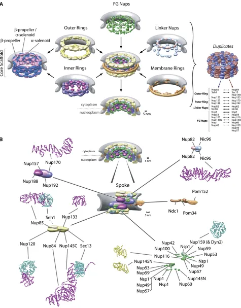
Tomographic electron cryomicroscopy and high-resolution scanning electron microscopy on rotary-shadowed speci-mens have shown that the yeast NPC shares its overall architectural features with those studied in other eukar-yotes, although it is somewhat smaller, being 100 nm in diameter and40 nm in height as compared with130 nm ·80 nm for its vertebrate counterpart (Yanget al.1998) (Figure 1). The core of the NPC consists of an octagonally symmetrical cylinder, the axis of which lies perpendicular to the plane of the NE. This core is made of coaxial inner, outer, and membrane rings surrounding a central channel (or“ cen-tral transporter”) of40-nm diameter through which virtu-ally all nucleocytoplasmic trafficking occurs (membrane proteins excepted). The circular membrane of the nuclear pores actually passes between the membrane ring and the outer/inner rings, thus anchoring the NPC firmly into the NE. Eight short filaments can be seen to project from the core into the cytoplasm, and, similarly, eight filaments extend 50 nm into the nucleoplasm, where they conjoin distally to form a structure said to resemble a “basket” on the nuclear face of the NPC (Fahrenkrog et al.1998; Yang et al. 1998; Kiseleva et al. 2004) (Figure 1). Starting in 1990, researchers took advantage of yeast genetic screens as well as cross-reacting monoclonal antibodies made against vertebrate NPCs to identify the first NPC compo-nents (termed nucleoporins or “Nups”) in yeast. Genetic and biochemical methods then steadily and rapidly filled in the list of yeast Nups (reviewed in Wente and Rout 2010), ultimately culminating in the yeast NPC being the
times, and some NPC-associated proteins also extend their functions and localizations beyond the NPC (Arib and Akhtar 2011).
The sheer size andflexibility of the NPC make it difficult to fully solve its molecular architecture by conventional techni-ques. Therefore, an orthogonal approach has been taken; large and diverse sets of proteomic data were amassed and a computational method for using these data was developed to define the relative positions and proximities of the yeast NPC’s constituent proteins. A corresponding average protein density map represents the position of every Nup with a precision of 5 nm, sufficient to resolve the molecular organization of the entire NPC (Alberet al.2007a,b) (Figure 2A). The resulting map agrees with complementary data in both yeast and verte-brates (reviewed in Strambio-de-Castilliaet al.2010).
Overall Composition
Computational fold predictions and biochemical domain mapping have analyzed the fold composition of every Nup (Devoset al.2004, 2006; Dokudovskayaet al.2006), show-ing that the NPC is surprisshow-ingly simple in terms of fold com-position, consisting of a few, highly repetitive fold types (Figure 3). Thisfinding, supported by recently solved crystal structures for several Nups (Figure 2B) (see below), indi-cates that the bulk of its structure has evolved through ex-tensive gene duplication from a simple precursor set of only a few proteins. Indeed, each spoke can be divided into two parallel columns, in which every Nup in the first column carries a similarly positioned homolog in the adjacent col-umn (Figure 2A). This pattern has been interpreted as resulting from at least one (and likely more) ancient ge-nome duplication events, which gave rise to the two col-umns composing each spoke. S. cerevisiaeis one of several related yeast that have undergone a whole-genome duplica-tion with subsequent gene loss (Wolfe and Shields 1997; Kelliset al.2004). Yeast-specific homologous Nup pairs such
as Nup116p/Nup100p, Nup157p/Nup170p, and Nup53p/
Nup59p, which exist only as single proteins in vertebrate
NPCs (Cronshawet al.2002), are segregated pairwise into the two columns in each spoke and so enhance the partition-ing of Nups to these columns, although the functional rea-son for maintaining these proteins as homologous pairs in yeast is still not clear (Figure 2). Most of the motifs, domains and even domain arrangements, and Nup types found in yeast NPCs are also found in vertebrate NPCs, and even in the NPC from the most divergent eukaryote studied so far (Trypanosoma; member of the kinetoplastida), albeit with some variations (DeGrasseet al.2009). This implies strongly that the basic structural elements of the NPC are conserved across all eukaryotes and that the NPCs of the last common ancestor of all eukaryotes had most of the major attributes of modern NPCs. It also means that studies of the yeast NPC are reasonably representative of the Eukaryota as a whole and are relevant for medically significant organisms, includ-ing ourselves.
Membrane Ring
The first class of NPC components is the set of three membrane proteins, called“poms,”which compose the mem-brane ring forming a distinct subcomplex in the NPC (Alber et al.2007b; Onischenkoet al.2009) (Figure 2). All are pre-dicted to carry transmembrane a-helices, which likely help to anchor the NPC in the NE, interact with core components and one another, and have functional redundancy perhaps as
membrane domain anchors and stabilizers (Chialet al.1998; Miaoet al.2006). This is consistent with a network of inter-actions formed between these three proteins (Onischenko et al. 2009). The first to be discovered and largest pom is
Pom152p, a type II transmembrane protein (Wozniak et al.
predicted to form homophilic binding interfaces (Bryant and Stow 2004) and likely explains the oligomeric lumenal ring. Much less is known aboutPom34. It is a small protein con-taining two transmembrane helices and two small domains both facing into the core scaffold. As yet, no function has been assigned to Pom34—even cells lacking both Pom34 and
Pom152are viable—and no nonfungal homolog has yet been
found. However, like Pom152,Pom34 genetically interacts with several core scaffold proteins in ways that suggest these poms’functions partially overlap (see below) (Madridet al. 2006; Miaoet al.2006).Ndc1p(nucleardivisioncycle 1) is also a pom but was named differently because it was actually
first characterized due to the effect of one of its mutants on the assembly of the spindle. Indeed, it turns out that, like several other Nups,Ndc1pplays at least two roles in the cell: in the case ofNdc1p, one in the NPC and one in the spindle pole body (SPB)—the mitotic spindle organizer. In yeast, both macromolecular assemblies are embedded in the NE, and it seems thatNdc1phelps to insert and attach both into their respective nuclear pores.Ndc1phas (at least) six transmem-brane helices and a carboxy-terminal domain that interacts with the core scaffold and other poms (Alber et al.2007b; Onischenkoet al.2009) and has confirmed homologs in both Schizosaccharomyces pombe[i.e., cut11, also with known roles in spindle assembly and an NPC component (West et al. 1998)] and metazoa [i.e., NDC1 (Lauet al.2004; Mansfeld et al.2006; Stavruet al.2006)]. Dissecting the NPC function
ofNdc1pfrom its SPB function has been difficult, but several
lines of genetic (Lau et al. 2004) and molecular biological (Onischenkoet al.2009) evidence suggest thatNdc1pplays an important role, with the help of the other poms, in NPC assembly (see also below).
Core Scaffold: Inner and Outer Rings
The second class of NPC components comprises the core scaffold proteins. This scaffold is composed of an inter-locking lattice of roughly a dozen evolutionarily conserved structural proteins that link together to form a core layer giving the NPC shape and strength (Figure 2). Fold compo-sition analyses revealed that the core scaffold consists of Nups composed of only two fold types in three arrange-ments: consisting almost entirely of a b-propeller fold, or almost entirely of a-solenoid-like/helix-turn-helix repeat folds, or ab-propeller followed by ana-solenoid-like domain (Figure 3). These fold types together are characteristic of components of the clathrin, COPI and COPII membrane ves-icle-coating complexes (reviewed in Field and Dacks 2009), and related complexes such as the intraflagellar transport complex (Jekely and Arendt 2006) and the HOPS/CORVET complexes (Nickerson et al. 2009). The latter b-propeller/ a-solenoid combination is particularly characteristic of this family, as these protein types interlock in a variety of related ways into a lattice forming their vesicle coats. These simi-larities were recently further underscored by atomic struc-tures solved for several core scaffold Nups (Figure 2B)
(Boehmer et al. 2003; Berkeet al. 2004; Hsia et al.2007; Jeudy and Schwartz 2007; Brohawnet al.2008; Debleret al. 2008; Schrader et al. 2008; Brohawn and Schwartz 2009; Leksaet al.2009; Nagyet al.2009; Seoet al.2009; Whittle and Schwartz 2009) as well as clathrin (reviewed in Owen et al.2004) and COPII and COPI components (Stagget al. 2006; Fathet al.2007; Lee and Goldberg 2010; reviewed in Stagg et al.2007, 2008). This fold similarity, analyzed ini-tially in yeast, led to the“protocoatomer hypothesis,”which proposes that a simple membrane-curving module, made primarily fromb-propeller anda-solenoid folds, was a com-mon ancestor for NPCs and coated vesicles that originated in the precursors to the ancient last ancestor common to all eukaryotes (Devoset al. 2004, 2006; Alberet al.2007a,b). This protcoatomer gave these ancestors the ability to gener-ate internal membrane systems by invagination of the plasma membrane and then to manipulate and elaborate these systems, eventually leading to the evolution of the ER, Golgi, and nucleus that characterize modern eukaryotes (Field and Dacks 2009).
The core scaffold somewhat resembles a vesicle coat, as it forms a discrete layer completely following the curve of the pore membrane, effectively coating it (Figure 2). Thus, it defines the size of the central tube/transporter of the NPC and the height of the NPC core, and all other Nups and poms are attached to either the inner or the outer face of this coat (Figure 1). Biochemical studies have shown that the core scaffold Nups compose several subcomplexes that appear to function as “building blocks”during NPC assembly and can even exchange with a soluble pool in mature NPCs (Lutzmann et al. 2002; D’Angelo et al. 2006; Makio et al. 2009). Morphologically, the scaffold is made of two inner rings, sandwiched between and interconnected with two outer rings, such that the nuclear and cytoplasmic halves of the NPC have one inner and one outer ring each (Figure 2). Although there is still some debate on the matter, a con-sensus remains that in both yeast and vertebrates these in-ner rings are compositionally, as well as morphologically, distinct (Tran and Wente 2006; Alber et al.2007b).
Four large Nups, each just under 200 kDa in size, compose the inner rings:Nup188pandNup192p(primarily composed ofa-solenoid-like folds) and the homologousNup170pand
Nup157p(Figure 2) (made from the clathrin-like pattern of
Miaoet al.2006). Moreover, on both the nuclear and cyto-plasmic sides of each spoke, one copy of the NupNic96p is anchored through Nup192p and a second copy through
Nup188p, linking the inner ring to the other internal
struc-tures of the NPC (Figure 2) (see below).
Structurally, the most extensively studied set of core scaffold proteins are the seven Nups that compose the yeast outer rings, first identified and characterized by the Hurt laboratory; Nup133p, Nup120p,Nup145Cp,Nup85p,
Nup84p, Seh1p, and Sec13p form a discrete complex that
can be biochemically isolated and is termed theNup84 com-plex (Figure 2B) (Siniossoglouet al.1996, 2000; Lutzmann et al.2002; Flemming et al.2009). Importantly, the evolu-tionary link between NPCs and vesicle-coating complexes is supported by the fact thatSec13pis shared with theSec13/ 31 COPII vesicle-coating complex (Siniossoglouet al.1996; Salama et al. 1997; Devos et al. 2004). Moreover, just like the inner-ring proteins, all Nup84 complex proteins are formed almost entirely by a b-propeller fold (Seh1p,
Sec13p), an a-solenoid-like fold (Nup85p, Nup84p,
Nup145Cp), or an N-terminal b-propeller and a
carboxy-terminal a-solenoid-like fold (Nup133p, Nup120p), again common to vesicle-coating complexes (Devos et al. 2004, 2006) (Figure 2B and Figure 3). Excitingly, crystal structures primarily from the Blobel and Schwartz laboratories have begun to piece this complex together at the atomic level (Figure 2B) (Hsiaet al. 2007; Brohawnet al. 2008; Debler et al.2008; Brohawn and Schwartz 2009; Leksaet al.2009; Nagy et al. 2009; Seo et al. 2009; Whittle and Schwartz 2009). Electron microscopy studies have shown that the
Nup84complex formed an extended Y structure (Siniossoglou
et al. 2000; Lutzmann et al. 2002; Kampmann and Blobel 2009), and pioneering work from the Hurt laboratory re-constituted this complex from bacterially expressed pro-teins and showed that the two short arms of this Y are composed, respectively, of Nup120p and Nup85p+Seh1p, whileNup133p,Nup84p, and Nup145Cp/Sec13p form the main stalk (Siniossoglouet al.2000; Lutzmannet al.2002). There is evidence that this complex isflexible (Kampmann and Blobel 2009), perhaps reflecting the known flexibility of the NPC in response to changes in NE shape and during its assembly (see below).
Few interactions have been found between the compo-nents of the Nup84 complex and the rest of the NPC, al-though it connects with the inner rings through, for example, aNup157p–Nup120pconnection (Lutzmannet al. 2005; Alberet al.2007a,b). Mutations in any of these seven Nups are often characterized by temperature sensitivity, messenger RNA (mRNA) and pre-ribosomal export prob-lems, and aberrant NPC assembly. In particular, a so-called
“clustering” phenotype was first described in mutants of
theNup84complex components in which the NPCs can be
seen to cluster into one or a few patches in the NE (Doye et al. 1994; Aitchison et al. 1995a; Heath et al. 1995; Li et al. 1995; Pemberton et al. 1995; Goldstein et al. 1996; Siniossoglou et al. 1996). That outer-ring Nup mutants
cause mislocalization of otherwise reasonably functional NPCs in the plane of the NE may point to a role for this structure in keeping the NPC stably located in the pore membrane. The outer rings are strategically placed at the point where the pore membrane joins the coplanar outer and inner NE membranes, and it seems reasonable that a major role for them is to ensure the smooth transition of the pore membrane into the inner and outer NE membranes (Figure 2) (Alberet al.2007b).
Phenylalanine-Glycine Nups
It was thefield’s catalog of the composition of the yeast NPC that led to a surprise. No homologs of mechanochemical proteins or NTPases of any kind that could physically drive a gating process were found as components of the NPC. Instead, strikingly, the cataloging revealed that over one-third of Nups in the NPC shared a highly characteristic repetitive motif, consisting of multiple repeated phenylala-nine-glycine (FG) pairs spaced by 20 mainly polar amino acids (Figure 3). Although these proteins are found in all eukaryotes studied, once again they were first sequenced from yeast (Hurt 1988; Davis and Fink 1990; Nehrbass et al.1990), and the yeast“FG Nups”remain the best stud-ied. On the basis initially of work in yeast, twoflavors of FG Nups were described: FxFG Nups typified by Nsp1p and GLFG Nups typified byNup100p(on the basis of the typical sequence of their FG repeat), the former having some charged amino acids in their spacers and the latter having relatively uncharged spacers (reviewed in Rout and Wente 1994). These two flavors of FG repeat also appear to be conserved (Figure 3), although by examination of orthologs in syntenic yeasts it was shown that the spacer sequences between each of the FG repeats evolved more rapidly than did other Nups (Denning and Rexach 2007), a situation common to the FG-repeat regions of all eukaryotes (DeGrasse et al. 2009). On the basis of various physical measurements of purified and bacterially expressed pro-teins, a consensus has emerged that the FG-repeat regions of FG Nups take on a natively unfolded structure bothin vitro and in vivo, such that they form long, disordered flexible
filaments (Denning et al. 2003; Denning and Rexach 2007; Lim et al. 2006a,b, 2007a, 2008; Patel et al.2007; Yamadaet al.2010); the lack of structural constraints there-fore likely explains the lower evolutionary constraints on sequence conservation of these repeat regions (Denning and Rexach 2007).
of the scaffold is lined with FG Nups whosefilamentous FG-repeat regions fill the central channel and extend into the nucleoplasm and cytoplasm (Figure 2). On both the cyto-plasmic and nucleocyto-plasmic sides of each spoke two copies of
Nic96pcarry the FG NupsNsp1p,Nup57p, andNup49pand
another two copies form interactions to additional copies of
Nsp1p, such that these FG Nups face both the nucleus and
the cytoplasm. At the cytoplasmic side, Nup82p associates
with Nsp1p as well as with the cytoplasmically facing FG
Nups Nup159p, Nup116p,Nup100p, andNup42p (Grandi
et al. 1995; Belgarehet al.1998; Bailer et al.2000, 2001; A. K. Hoet al.2000; Routet al.2000; Alberet al.2007a,b). There are also the FG NupsNup145Np,Nup1p, andNup60p found on the nucleoplasmic side, connecting mainly to the inner-ring Nups. In addition, Nup53p and Nup59p are at-tached to Nup170p and Nic96p, and both face the pore membrane (Figure 2). The latter two Nups may also belong to the class of FG Nups, as they can bind transport factors and carry degenerate FG-repeat regions that are predicted to be natively unfolded (Marelliet al.1998; Fahrenkroget al. 2000b; Lusket al.2002; Makhnevychet al.2003; Alberet al. 2007b). One FG Nup, Nup145p, uniquely cleaves itself in half at its Phe605-Ser606 peptide bond to produce two sep-arate Nups,Nup145Np(carrying the FG-repeat region and the autoproteolytic b-sandwich domain at its new carboxy-terminus) andNup145Cp(a mainlya-solenoid-like protein that forms a major component of theNup84complex) (Fig-ure 3) (Wente and Blobel 1994; Teixeiraet al.1997, 1999; Rosenblum and Blobel 1999). Although not essential in yeast (Emtageet al.1997), this cleavage event appears con-served in vertebrates (Fontoura et al. 1999; Hodel et al. 2002; Sun and Guo 2008). Nup145Cp and Nup145Np re-main linked as a dynamic complex such thatNup145Npcan shuttle between the NPC and the nuclear interior, as does its vertebrate counterpart (Griffis et al. 2002; Ratner et al. 2007). The entirety ofNup145Npis highly conserved with the homologous yeast nucleoporinsNup100pandNup116p, neither of which undergoes autoproteolysis as they lack a ho-mologous counterpart forNup145Cp. It seems that lineage-specific gene duplications of an ancestral Nup145N-like gene gave rise to the truncated versions Nup100p and
Nup116p, and likely other FG Nups also originated from
such, sometimes more ancient, duplication events (Mans et al.2004; Devoset al.2006).
Collectively, the anchored FG Nups form the business end of the NPC, as the FG-repeat domains form low-affinity, high-specificity interactions with transport factors involved in active transport through the NPC and so actually form the selective barrier in and around the central tube by providing the binding sites for transport factors that facilitate their exchange across the NE while excluding the passage of macromolecules not destined for nucleocytoplasmic transport. In one sense, the NPC can be considered a framework that provides the correct positioning of the FG repeats,flanking andfilling the central tube while defining the upper diameter of the central tube and the cargoes that transit through it (Figures 1 and 2) (Rout and
Aitchison 2001; Rout et al.2003; Hetzer and Wente 2009; Walde and Kehlenbach 2010).
Cytoplasmic Filaments and Nuclear Basket
While not as morphologically prominent as their vertebrate counterparts, both cytoplasmic filaments and nuclear bas-kets have been seen to project from the yeast NPC (Figure 1) (Kiselevaet al.2004).Nup159p,Nup82p, andNup42pseem to contribute to the cytoplasmic filaments (Kraemer et al. 1995; Hurwitz et al.1998; Strahm et al.1999; Rout et al. 2000; Alber et al.2007b) and function in the last stages of export from the NPC (below). Curiously, the proteinDyn2p, a light chain component of the dynein microtubule motor, binds toNup159pand helps form a rigidfilamentous struc-ture that may stiffen the cytoplasmic filament projecting it out from the core scaffold (Stelteret al.2007). Yeast lack an obvious homolog of the vertebrate Nup358, which is be-lieved to produce the more prominent cytoplasmicfilaments in the latter (Wuet al.1995; Matuniset al.1998). In verte-brates, the bulk of the nuclear basket seems to be made of Tpr (Cordeset al.1997; Haseet al.2001; Frosstet al.2002; Krullet al.2004; Qiet al.2004). Tpr is a conserved 200-kDa protein made mainly of extensive coiled-coil domains that dimerize into long rods forming the basket struts. Two Tpr homologs,Mlp1pandMlp2p, exist in yeast and localize to the region of the nuclear basket (Strambio-de-Castillia et al. 1999). Unlike metazoa, no lamina lies interwoven between NPCs beneath the NE, but bothMlp1pandMlp2p spread out along the inner face of the NE to form a delicate network interconnecting yeast NPCs, although they are ex-cluded where the dense crescent of the nucleolus presses against the NE (Strambio-de-Castillia et al. 1999). Mlp2p is additionally associated with the SPB (Niepel et al. 2005). In yeast,Mlp2p is the result of the specific genome duplication; however, a spindle organizer-specific copy of Tpr homologs has been independently reinvented several times in evolution for reasons that are still unclear (Jimenez et al. 2000; DeGrasse et al. 2009). Overall, a bewildering array of functionalities have been ascribed to the Mlp net-work, including roles in recruitment of transport factors, late processing of transcripts, and epigenetic regulation of gene expression, as will be discussed below.
Shuttling Nucleoporins
The definition of nucleoporins becomes more difficult when one considers the dynamics of some of the classically defined nucleoporins.Nup2p, for example, was defined as a nucleo-porin as early as 1993 on the basis of its localization to the NPC; however, fluorescence microscopy, subcellular frac-tionation, and experiments monitoring its dynamicsin vivo (Dilworthet al.2001) revealed thatNup2pactually cycles on and off the nuclear basket and in this sense behaves more like a soluble transport factor. Similarly,Yrb2p(yeastranbinding
FG repeats, yet it only transiently associates with the NPC (Floer and Blobel 1996). So far, all such rapidly “shuttling” nucleoporins belong to the FG Nup family. As well as an FG-repeat region, bothNup2pandYrb2pcarry a consensus Ran-binding motif and may have a role in promoting the disassembly of transport cargos; in this way, shuttling nucle-oporins may act as mediators between the stationary and soluble phases of transport (see below) (Dilworth et al. 2001, 2005; Gilchristet al.2002). In vertebrates, the dynam-ics of Nups have been comprehensively examined, revealing varying half-lives of each Nup on the NPC, and it seems likely that this will be borne out in yeast (Tran and Wente 2006). For example,Nup145Nphas a localization that is biased to, but not exclusively on, the nuclear face, while Nup116p
and Nup100pare similarly biased to the cytoplasmic face
(Suntharalingam and Wente 2003). This variation compli-cates efforts to define a “stoichiometry”for components of the NPC, as any number for these more dynamic Nups will be an average of what may be a stochastic variation in Nup number and location. We expect that, the closer we look, the more difficult it will be to consider the NPC an autono-mous structure; rather, perhaps it should be considered a dy-namic assembly of proteins which to varying degrees, cycle between the stationary and soluble phases during transport and assembly, and functionally link the NPC to numerous other dynamic cellular activities (see below).
NPC Assembly
NPCs are not static structures. They are assembled, and their components appear to be capable of turning over during the NPC’s lifetime. In many organisms, NPCs disassemble upon NE breakdown at the beginning of mitosis or meiosis and reassemble coordinately with the NE around the newly seg-regated chromosomes at its end. However, yeast has a
“closed”mitosis in which the NE remains intact, such that the NPCs remain assembled throughout the life cycle of the cell and negate the need for NE and NPC disassembly—in sharp contrast to the elaborate mitotic nuclear disassembly and reassembly processes seen in metazoans (Suntharalingam and Wente 2003). Careful analyses of serially sectioned yeast confirmed that NPC assembly occurs continuously throughout the entire cell cycle with a typical haploid NE containing between 70 NPCs just after mitosis to 140 NPCs in late anaphase (Wineyet al.1997). How this assem-bly occurs is still unclear, despite much work in both yeast and vertebrate model systems, with most of that work in metazoan cells (because researchers generally studied the synchronized assembly of NPCs in mitosis), and some of the details are only just beginning to emerge (as reviewed in Fernandez-Martinez and Rout 2009; Hetzer and Wente 2009). Nevertheless, the processes of NPC and NE assem-bly—and the reasons why some species opt for a closed mitosis while other related species opt for variants of an open mitosis [compare the ascomycetes Saccharomycesand Aspergillus (De Souza et al.2004; Osmani et al. 2006; Liu
et al. 2009)], remain somewhat mysterious. Work in verte-brate cell-free systems has established, finally, that new NPCs are indeed insertedde novointo the NE (rather than, e.g., “budding off” from existing NPCs) (D’Angelo et al. 2006). In yeast, it is primarily genetic approaches that have given some of these insights. As a yeast cell grows, the nu-cleus also grows in volume and the NE enlarges its surface area, during which time new NPCs are inserted into the NE (Winey et al. 1997). Although not proven, it seems likely that this process in yeast is similar to interphase NPC assem-bly in vertebrates, which has been shown to occur through de novoassembly of precursor building blocks recruited from both the nucleoplasm and cytoplasm into the regions of the NE between pre-existing NPCs (D’Angeloet al.2006). The continued assembly of the NPC and NE throughout the yeast cell cycle has been used as a basis for genetic screens, select-ing for mutants that caused mislocalization of tagged Nups. Initially, mutants in various Nups produced phenotypes that (if not lethal) gave a puzzling collection of different pheno-types that were difficult to interpret in terms of NPC assem-bly. Some made the NPCs cluster (above), whereas others led to herniations of the NE extending over the cytoplasmic face of NPCs to seal them (Wente and Blobel 1993, 1994). However, more recent approaches have given more inter-pretable phenotypes. By using a photoconvertable Dendra tag in cells blocked and then released in NPC assembly, it was shown that some pre-assembly Nup complexes congre-gate on both the inner and the outer membranes of the NE, including cytoplasmic-facing Nups on the cytoplasmic face of the NE and nucleoplasmic/basket Nups on the nuclear face, whereas symmetrically disposed Nups were found to accumulate on both NE faces (Makio et al. 2009; Oni-schenko et al.2009). These pre-assembly complexes might correspond to the discrete complexes found to compose the NPC, such as theNup84complex (see above). Targeting of these pre-assembly Nups to the NE seems to require certain soluble transport factors normally used to chaperone and power the transport of cargoes through the NPC (see be-low), as genetic screens for conditional mutants in NPC assembly identified Ran,RanGEF, RanGAP, and Ntf2 (see Figure 4 and below) (Ryan and Wente 2002; Ryan et al. 2003, 2007). The karyopherin (Kap)Kap95pwas also iden-tified in these screens, and another karyopherin, Kap121p, seems to aid Nup53p in assembling into a complex with
Nup170p(Lusket al.2002).
Interestingly, these mutants correspond to two key com-ponents of the cargo-carrying transport factor pathways, namely the b-karyopherinsKap95p andKap121pand Ran cycle components [Ran, RanGAP, RanGEF, and Ntf2 (re-sponsible for transporting RanGDP into the nucleus)] (Lusk et al.2002; Ryan and Wente 2002; Ryanet al.2003, 2007). The reasons for the functional associations between NPC as-sembly and transport factors are still being elucidated, but similar connections have been seen in vertebrates (D’Angelo et al.2006). In yeast,Kap121phas been proposed to target
componentNup170p. Indeed, recent work has revealed the importance of the core scaffold to the early stages of NPC assembly. Thus, when the C-terminal domain of Nup170p is overexpressed, what appear to be intermediates of NPC assembly accumulate both in the cytoplasm and at the NE (Flemming et al. 2009). Similarly, in strains lacking both
Nup53p and its paralogNup59p, depletion of Nup170por
either of two transmembrane nucleoporins that connect
with Nup170p—Pom152porPom34p—also caused the
ac-cumulation of such intermediates in yeast cells (Onischenko et al.2009).
For an NPC to be inserted into the intact NE, both the inner and the outer NE membranes must approach at a site and fuse to give rise to the pore membrane, upon which the core scaffold and the rest of the NPC can then assemble. It is curious, therefore, that two of the three poms (Pom152p
and Pom34p) are not essential and so are dispensable for
NPC assembly and that all three poms (includingNdc1p) are not required for NPC assembly in the closely related fungi, Aspergillus (Liu et al. 2009). Taken together, this suggests that there must be other transiently or dynamically associ-ating membrane proteins that play key roles in initiassoci-ating the NPC assembly process and fusion of the inner nuclear mem-brane (INM) and outer nuclear memmem-brane (ONM) to form the pore membrane.
Indeed, there has been a growing cadre of proteins that, while not strictly Nups, play a key role in yeast NPC assembly. As well as Ran, Ran cofactors, and the Kaps (above; Lusk et al.2002; Ryanet al.2003, 2007), the two yeast reticulons
Rtn1p andRtn2pand their interacting partnerYop1phave
been implicated in NPC assembly (Dawsonet al.2009). Rtns and Yop1/DP1 proteins can deform and mold membranes, having been shown to have roles in both dynamically restruc-turing and maintaining tubular ER (De Craene et al.2006; Voeltzet al.2006; Huet al.2008) and, in metazoans, even in postmitotic NE shaping (Anderson and Hetzer, 2008b). Retic-ulons have a segment that can insert into one leaflet of a membrane, which may promote or induce membrane curva-ture (Oertleet al.2003; De Craeneet al.2006; Voeltzet al. 2006; Shibata et al. 2008); indeed, they are depleted in regions of flat membrane, such as the NE between NPCs, and are found to concentrate in curved membrane regions such as tubular ER (De Craeneet al.2006; Voeltzet al.2006; Anderson and Hetzer, 2008a,b). The apparent absence of these proteins in the mature NPC suggests that they play only a transient role at the beginning of the assembly process, perhaps helping thefirst NE membrane curving and fusion step to make the pore membrane. Similarly, the NE/ER pro-teinsApq12pandBrr6pare genetically linked to each other and are necessary for normal NPC assembly and distribution. This work indicates that both proteins are involved in main-taining lipid homoeostasis in the ER, which is necessary for proper NPC insertion and distribution in the NE (Scarcelli et al.2007; Hodgeet al.2010).
Another candidate NPC assembly factor is Pom33p, iso-lated in a genetic screen for genes that are essential in cells
lackingNup133p(Chadrinet al.2010). The transmembrane protein Pom33pand its paralog Per33p are found in both the ER and the NE, although Pom33pshows a preferential dynamic localization at NPCs.Pom33, but notPer33, genet-ically interacts with Nup84 complex components and the interacting proteins Nup170p and Ndc1p and physically forms a direct complex with Rtn1p (Chadrin et al. 2010). These data, plus the fact that depletion of bothNup170and
Pom33 significantly impaired assembly of NPCs, point to
a role forPom33pin NPC assembly or maintenance of the NE (Chadrinet al.2010).Pom33pthus potentially links the reticulon membrane bending and manipulation machinery with the assembling NPC, which together possibly either help the transmembrane nucleoporins during the initial membrane fusion event required for the start of NPC assem-bly or facilitate the stabilization of the nascent nuclear pore. Following this initial pore formation, assembly to form the mature NPC must proceed extremely rapidly, as no naturally occurring intermediates have been found.
Of course, the NPC core scaffold is composed almost entirely of homologs of vesicle-coating proteins, whose function is to mold and fuse membranes into curved vesicles. On the basis of this similarity it has been suggested that theNup84complex and theNup170inner-ring complex (which interacts directly with poms) could be directly in-volved, after recruitment to the NE, in forming a coat some-what like those in coated vesicles that produces the nascent pore membrane and pinches the inner and outer NE mem-branes together in a manner analogous to pinching off a curved vesicle (reviewed in Fernandez-Martinez and Rout 2009; Hetzer and Wente 2009).
recruitment of the inner-ring components would recruit the outer-ring components, permitting the assembly of the en-tire membrane-coating core scaffold in the pore (Alberet al. 2007b). Rapid association of the remaining FG Nups, other NPC components, and the nuclear basket would then com-plete the process. However, this sequence of events remains strictly speculative, and much remains to be understood about the mechanism of NPC assembly in yeast or in any other eukaryote.
It seems possible that other NE-associated structures share at least some aspects of the NPC’s assembly process. Curi-ously,Nup60pandPom152pare also required for the assem-bly and repair of the SPB (Greenlandet al.2010). Recall that the pore membrane component,Ndc1p, has been shown to be a shared component of both the NPC and the SPB and is required for the assembly of both (Chialet al.1998; Lauet al. 2004). The functional connection between the SPB and NPC is underscored by the putative nuclear basket component
Mlp2p, which may associate withNup60(Zhaoet al.2008)
and connects to both NPCs and SPBs (Niepelet al.2005). It seems that several proteins are found at both locales, raising the possibility that, as both NPCs and SPBs are inserted into a membranous grommet formed from the fusion of the inner and outer nuclear membranes, there are some commonalities in their assembly mechanisms.
Turnover of NPCs
No repair mechanism, as such, has been found for the NPC. Rather, it seems that a combination of some pre-emptive replacement of components by constant turnover and di-lution of “old”NPCs by new ones through cell growth and division are the tactics taken to rejuvenate the NPC popula-tion in a growing yeast populapopula-tion. The turnover rates of yeast Nups are not yet precisely known, although certainly some FG Nups exchange very quickly (Dilworthet al.2001; Tran and Wente 2006). Moreover, there is some uncertainty about how “old”and “new”NPCs are partitioned between the mother and the daughter cells upon budding. While evi-dence was originally presented that the old NPCs are re-tained preferentially in mother cells, potentially ensuring that the daughters receive a fresh supply of new NPCs (Shcheprovaet al.2008), more recent work indicates instead that new and old NPCs partition equally between mother and daughter at mitosis (Khmelinskiiet al.2010, 2011). As NPC segregation and turnover have direct implications for aging studies (Kaeberlein 2010), this and related topics will doubt-less be areas of intensive future investigation.
Soluble Phase of Transport: Transport Signals and Carriers
While NPC-mediated gating does not require an energy input, nucleocytoplasmic transport and the accumulation of cargoes in the nucleus and cytoplasm are driven by the formation and maintenance of concentration gradients
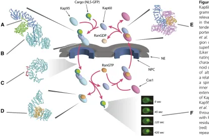
across the NE by GTPases and ATPases in the nucleoplasm and cytoplasm (Figures 4 and 5) (reviewed in Rout et al. 2003; Strambio-de-Castilliaet al.2010). Moreover, as is typ-ical for protein sorting throughout eukaryotic cells, proteins synthesized in the cytoplasm that are destined for the nu-cleus carry targeting signals [generally termed nuclear lo-calization signals (NLSs)] that are recognized by soluble receptors, which mediate their transport. Thefirst transport factors to be identified and purified to homogeneity were karyopherina, karyopherinb, and a small Ras-like GTPase called Ran. Through classic biochemical fractionation com-bined with in vitroimport assays, these proteins were
puri-fied to homogeneity from mammalian systems and shown to mediate transport of reporter proteins carrying an NLS from the SV40 large T antigen (reviewed in Pemberton and Paschal 2005; Wente and Rout 2010). Because the yeast genome had recently been completed, it was then a straight-forward matter to identify orthologs in yeast (reviewed in Wozniaket al.1998), and soon work on the mechanisms of nucleocytoplasmic transport was progressing in both yeast and mammalian systems. These studies established that kar-yopherin a (Kap60p in yeast) binds to the NLS and that karyopherin b (Kap95p in yeast) enhances (or stabilizes) the interaction and in turn binds to FG-repeat-containing nucleoporins. Ran-GTP provides an important source of en-ergy to the reaction by binding to karyopherinbas it enters the nucleus with karyopherinaand cargo in tow, releasing the cargo to the nucleoplasm (Figures 4 and 5). Importantly, the versatility of yeast as a model system rapidly led to complementary approaches and insights beyond those im-mediately possible in mammalian systems.
It was clear that not all proteins destined for the nucleus contain an NLS typified by SV40 large T antigen. The diversity of cargoes and complexes that traverse the NPC is huge, ranging from proteins to RNAs and ribonucleoproteins (RNPs), including mRNPs and ribosomes, to viruses. Analysis of the yeast genome revealed family transport factors structurally related to karyopherinb(and more distantly to karyopherin a). Other eukaryotes studied, even the most evolutionarily divergent, seem to retain this same family of Kaps (DeGrasse et al. 2009; Masonet al. 2009). Members of the b-Kap family are generally large (molecular weight of 100–125 kDa) proteins that share20% sequence identity with each other. Each is typified by the presence of up to20 HEAT repeats (amphipathic helix-loop-helix motifs) that form a large helical solenoid (with a single extended hydro-phobic core) (Figure 5) (Cansizogluet al.2007). There are apparently 14 Kaps in S. cerevisiae and at least 19 Kaps in humans (Stewart 2003), all of which differentially bind dif-ferent classes of nuclear transport signals, FG-repeat nucleo-porins, and Ran; unlike theKap60p:Kap95pdimer, all other b-Kaps bind directly to their cargos (Figure 4).
cells, Ran primarily exists in two forms: in the nucleus, Ran is maintained in its GTP-bound form by a GTP exchange factor (RanGEF; RCC1,Prp20p, orSrm1pin yeast); this protein is chromatin bound, thus signaling to the nucleocytoplasmic transport system the position of the nucleoplasm by virtue of generating a cloud of RanGTP around it. In contrast, the Ran GTPase-activating protein (RanGAP) is localized to the cytoplasm, so that Ran in the cytoplasm predominates in the GDP form. Karyopherins exploit this property during transport. As mentioned above, during an import cycle, Kaps bind to their cargoes in the cytoplasm, and when they reach the high Ran-GTP in the nucleus, are induced to release their cargoes. In contrast, exportin binding to cargoes is enhanced by the formation of a trimeric complex that includes Ran-GTP. Thus, as this complex meets the RanGAP in the cyto-plasm, the GTP is hydrolyzed and the complex falls apart. Indeed, the direction of karyopherin-mediated transport through the NPC can be reversed by inversion of the Ran gradient (Nachury and Weis 1999). Most karyopherins are thought to be recycled to their original compartments empty, but in a few instances they are believed to chaperone another cargo on their return journey (Figure 4).
Studies of prototypical interactions among constituents of these transport pathways have shed considerable light on the structural basis of transport (Figure 5). In the classical pathway, the NLS binds to a long region on the inside of
the Kap60 superhelix, made of alternating a-helical turns.
Kap95p, which is also made of alternating a-helical turns,
forms a spiral with two surfaces, and the inner surface wraps around an extended N-terminal domain ofKap60p(Figure 5) [a.k.a the importin b-binding (IBB) domain] (Cingolani et al. 1999). The interaction of Kap95p with FG Nups is mediated as the repeated Phe residues on the FG-repeat regions (see below) insert into complementary repeated pockets formed from the crevices between adjacenta-helical repeats all along the outer surface of Kap95p’s spiral. RanGTP binds toKap95p(Leeet al.2005) on the inner sur-face ofKap95’s amino-terminal solenoid spiral, which causes conformational changes that lead to release ofKap60p(and cargoes) (Figure 5).
DuringKap60pexport,Kap60pand RanGTP are bound to the inner surface of theCse1pspiral (Matsuura and Stewart 2004). In this form, the IBB domain is held tightly against the side ofKap60p, inhibiting NLS binding and leaving the outer surface free to interact with FG repeats and thereby carrying the complex through the NPC out of the nucleus (Figure 5). Once in the cytoplasm, the RanGTP hydrolyzes to RanGDP, causing the complex to dissociate. Kap60p remains bound to its IBB even when free in the cytoplasm, but binding to an NLS exposes the IBB and allowsKap95pto bind, initializing another round of import.
Karyopherins and Their Cargoes
The apparent presence of a family of karyopherins, and the knowledge that there are numerous classes of cargoes that
must be transported across the NPC, led researchers to begin to identify cargoes for each of the karyopherins. Again, yeast has been a tremendous model system for investigating this fundamental question. The mainstay approach for doing so has been to take advantage of homologous recombination techniques to genomically tag karyopherins with an epitope tag (like protein A) and to isolate the Kap and its associated cargoes (Aitchisonet al.1996). Genetic perturbations of the Kap genes have then been used to explore the consequences with respect to the potential cargo. This approach wasfirst applied to Kap104p to establish that it is responsible for importing a subclass of RNA-binding proteins (Nab2p and
Nab4p/Hrp1p) (Aitchison et al. 1996). These proteins are
major mRNA-binding proteins essential for mRNA process-ing and export (Andersonet al. 1993). They appear to ac-company the mRNA out of the nucleus, and upon reaching the cytoplasm, they are recycled for another round by
Kap104p (Lee and Aitchison 1999).Mtr10p/Kap111palso
appears dedicated to this essential function; it imports
Npl3p, another essential mRNA biogenesis factor.
Interest-ingly, while both Kaps import essential proteins, neither is essential (under the same conditions) by itself. This suggests that Kaps must have the capability to compensate for one another and bind to their cargoes with some promiscuity. This wasfirst made obvious upon examination ofKap123p. Deletion of Kap123 is virtually without phenotypic conse-quences in laboratory strains of yeast. Yet,Kap123pis per-haps the most abundant of the Kaps in yeast, conserved throughout the Eukaryota, and it binds to Lys-rich NLSs shared by a host of ribosomal proteins and ribosome assem-bly factors, which leads to their import into the nucleus prior to their assembly into ribosomes (Rout et al. 1997;Leslie et al.2002; Timneyet al.2006)—an essential process if ever there was one! Indeed, a host of genetically interacting Kaps appear to be involved in the import of proteins critical to ribosome assembly (e.g., Kap108p/Sxm1p, Kap119p/
Nmd5p, Kap121p/Pse1p) (Rosenblum et al. 1997; Rout
et al. 1997; Sydorskyyet al.2003; Caesaret al.2006), and it has been shown explicitly that, in the absence ofKap123p,
Kap121pcan bind toKap123psubstrates and import them
into the nucleus (Rout et al.1997).
Perhaps it is not surprising that structurally related Kaps can bind to structurally related NLSs, but it also appears that Kaps can recognize more than one type of NLS. For example, whileKap121pwas originally shown to bind to noncanon-ical Lys-rich NLSs (Routet al.1997; Kaffman et al.1998b; Leslie et al. 2002), it, like Kap104p, also imports proteins through rg-NLSs, which are reminiscent of structurally dis-tinct RNA-binding motifs (Dreyfuss et al. 1993; Lee and Aitchison 1999; Leslieet al.2004) characterized by repeats of Arg and Gly amino acid residues. Moreover, multiple cargo domains exist inKap114p, and it has been proposed that this Kap is capable of importing multiple cargoes simul-taneously (Hodgeset al.2005).
transit the NPC during their life cycle, and as a field, we have identified only a handful of the cargoes that they each recognize. So, while it has been proposed many times that evolution has likely exploited their overlapping specificities and potential complexity to regulate classes of cargoes by regulating the karyopherins, it remains for thefield to more comprehensively define Kap-cargo complexes to demon-strate how much this is the case and to fully appreciate how they may have done so.
Protein export from the nucleus is mediated by at least three b-karyopherins. The first (“classic”) nuclear export signal was defined in vertebrate cells in HIV-Rev protein. Rev binds specifically to unspliced and singly spliced HIV mRNA and ensures that it is exported efficiently. Studies to define this process identified a short leucine-rich region within Rev that is necessary and sufficient for nuclear ex-port. This sequence is recognized by the karyopherinXpo1/ Crm1 (Stade et al. 1997). As it turns out, there are many proteins that contain variants of the prototypical sequence and are exported by Xpo1p. These include the proteins of the 40S and 60S preribosomal subunits (J. H. Hoet al.2000; Stage-Zimmermannet al. 2000; Moy and Silver 2002); nu-merous transcriptional or signaling proteins (Ferrignoet al.
1998; Jensenet al.2001; Menezeset al.2004; Changet al. 2006; Martinet al.2006; Azevedoet al.2007; Pelaezet al. 2009); key regulators of the cell cycle [Cdc14p(Bembenek et al. 2005)], which control exit from mitosis; and certain small RNAs (Gallardo et al. 2008; Thomson and Tollervey 2010).Xpo1p/Crm1pis also, at least indirectly, required for normal mRNA production and export (Feng et al. 1999; Strasseret al.2000; Hammellet al.2002; Donget al.2007).
Msn5p has also been shown to act as a nuclear export
factor, exporting phosphorylated nuclear transcription fac-tors (Kaffman et al. 1998a; DeVit and Johnston 1999; Gorner et al. 2002; Queralt and Igual 2003; Durchschlag et al.2004; Uetaet al.2007), theHOendonuclease (Bakhrat et al.2008), andWhi5p, the yeast ortholog of Rb (Taberner et al. 2009). A consensus nuclear export signal (NES) for
Msn5p has been elusive, but its preference for
phosphory-lated proteins suggests a role for reguphosphory-lated export. Indeed, regulation of transport provides an exquisite mechanism to control gene expression. Perhaps the best-characterized ex-ample of such regulation in yeast comes from studies of
Pho4p.Pho4pis a transcription factor that induces the
However, in the presence of excess phosphate, Pho4p is phosphorylated adjacent to its NLS, inhibiting Kap121p binding and consequently its import. In addition, phosphor-ylation at two different sites promotes the factor’s nuclear export(Kaffmanet al.1998b; Komeili and O’Shea 1999). A similar shuttling activity has been described for Tor1p (a target of the immunosuppressant rapamycin), which is a pro-tein kinase that controls growth in response to nutrients through the regulation of diverse cellular processes in the cytoplasm and nucleus. Interestingly, PolIII transcription is also regulated by transport; Maf1p, a global inhibitor of PolIII, is exported byMsn5p when cells are shifted to con-ditions that favor growth and high PolIII activity (Towpik et al.2008).
Msn5p appears to be the most versatile of the Kaps. It
has also been shown to import proteins into the nucleus (Yoshida and Blobel 2001), and both Los1p and Msn5p have been shown to be capable of exporting transfer RNAs (tRNAs) from the nucleus. Both appear to bind double-stranded RNA directly (Shibata et al.2006), and (at least)
Los1pappears to play a proofreading role, ensuring that its
tRNA substrates are appropriately structured prior to their export to the cytoplasm (although splicingper seappears not to be proofread byLos1pbinding (Artset al.1998; Lipowsky et al. 1999; Cook et al. 2009; Hopper et al. 2010). The coupling of transport to function of the cargo is certainly not limited to the tRNA example; import Kaps such as
Kap114p, Kap104p, and Mtr10p release their cargoes in
the nucleus in concert with their cargoes binding to DNA and RNA. In effect, nuclear-binding sites compete with Kaps for their cargoes upon import, suggesting a mechanism for controlled release and intranuclear targeting.
Competition as a Major Factor in Nuclear Transport
Similarly, competition effects play a major role in the behavior of nuclear transport. Using anin vivoassay and by manipulating the amounts, types, and affinities of Kaps and cargos, it was shown that import ratesin vivoare governed in a straightforward manner by the concentrations of Kaps and their cargo and the affinity between them and that the main limiting factor for import (accounting for the fact that nu-clear accumulation of transported cargo was much slower than expected) was the poor ability of Kaps and cargoes to
find each other in the cytoplasm in a background of over-whelming nonspecific competition. In other words, the key rate-limiting step of the transport cycle is not transiting through the NPC itself, but is instead the formation of the Kap/cargo complex within the cell’s crowded environment (Timneyet al.2006). The importance of competition seems to extend to the mechanism of the NPC. A recent computa-tional model indicated how the selectivity of the NPC could be enhanced by the exclusion of nonspecific molecules by specific ones, due to competition for binding sites and limited space inside the channel. By using recombinant purified full-length yeast FG Nups and transport factors, it was shown that
FG Nup-functionalized nanopores behave as a nanoselective
filter, reproducing key features of trafficking through the NPC. It was also confirmed that competition between trans-port factors and nonspecific proteins is a major factor in the transport mechanism (Jovanovic-Talismanet al.2009).
Not Just Karyopherins: RNA Export
Many small RNAs, such as small nuclear RNA and tRNAs, are exported using the same RanGTP-powered karyopherin-dependent pathways used by exporting proteins, although some karyopherins seem to specialize in this function, such as the tRNA exporter Los1 (reviewed in Köhler and Hurt 2007). However, the process is considerably more compli-cated for mRNAs and ribosomal RNAs. All such large RNAs are transcribed and assembled into RNP complexes, and each RNP can be considered an intermediate along an as-sembly line as proteinsflit on and off the assembling struc-tures to trim and assemble the RNA. Consider the ribosome. Over 200 proteins are believed to be involved in the assem-bly of ribosomes as they mature during their complex bio-genesis. Similarly, each mRNA is assembled into an RNP (mRNP) particle involving a series of complex assembly intermediates, which associate with each species of RNA in a dynamic fashion to allow for precise transcript maturation (Fatica and Tollervey 2002; Hopper and Phizicky 2003; Vinciguerra and Stutz 2004).
Surprisingly, mRNA uses a Kap- and Ran-independent mechanism for export (Santos-Rosa et al. 1998; Katahira et al. 1999). Moreover, mRNP assembly and export involve strict surveillance mechanisms to ensure that only fully ma-ture and functional RNPs are transported to the cytoplasm (Palancade et al.2005; Schmid and Jensen 2008; Skruzný et al.2009). In addition, there are many different species of mRNA, each potentially with its own particular maturation pathway. This is a topic that has been both extensively researched and comprehensively reviewed, so we will only summarize these findings briefly and refer the reader to these reviews (Rondon et al. 2010; Stewart 2010; Rodriguez-Navarro and Hurt 2011).
Upon being processed and packaged into mRNP particles in the nucleus, the non-Kap transport factors Mex67pand
Mtr2passociate with and chaperone the mRNP through the
TREX-2, associates with the mRNP at the NPC, apparently assuring that it is correctly packaged for its journey across the NPC. TheMex67p-Mtr2pheterodimer, by binding both the mRNP and the FG Nups, mediates the actual mRNP transport event, which can be surprisingly rapid; recent work in vertebrates has suggested that the actual translo-cation event, even for a multi-megadalton mRNP complex, lasts only milliseconds (Grunwald and Singer 2010; Mor et al. 2010). After transiting the NPC’s central channel, the mRNP encounters Dbp5p, an ATP-driven RNA helicase tethered to the cytoplasmicfilament proteinNup159p. Reg-ulated by Nup42p-tethered Gle1p and the small molecule IP6 (inositol hexaphosphate),Dbp5p’s action on the exiting mRNP serves to release the transport factors Mex67p and
Mtr2pas well as mRNP proteins such asNab2p, both actions
preventing re-import of the mRNP (and thereby helping to confer directionality to export) and preparing the mRNA for translation (Figure 6) (reviewed in Carmody and Wente 2009; Rodriguez-Navarro and Hurt 2011).
Ribosomal subunit export is still a little less well characterized, and while clearly differing from both protein and mRNP export, oddly shares elements from both (Hage and Tollervey 2004; Zemp and Kutay 2007; Henras et al. 2008; Lo and Johnson 2009). Three transport factors have been implicated in yeast in the export of the large ribosomal subunit: Mex67p-Mtr2p (Yao et al. 2007), Crm1p (which docks to the large subunit adaptor protein Nmd3p (J. H. Ho et al.2000; Gadal et al. 2001), and the noncanonical receptor Arx1p (Bradatsch et al. 2007; Hung et al. 2008). Fusions of Mex67p, Los1p, Mtr2p, Cse1p, or Msn5p to
Nmd3p, lacking its Crm1p-dependent NES, all function in
export of 40S subunits is still poorly understood, but may be somewhat simpler than for the 60S subunit (Zemp and Kutay 2007; Maggiet al.2008; Perreaultet al.2008; Carron et al. 2011). Two different yeast nonribosomal proteins,
Dim2pandLtv1p, have been proposed to function as
adapt-ers for Crm1p-mediated 40S export in yeast (Seiser et al. 2006; Vanrobayset al.2008). Both are late-acting 40S bio-genesis factors that shuttle between the nucleus and the cytoplasm.
Mechanism of Nuclear Transport
The molecular details are beginning to emerge as to exactly how the NPC mediates the active exchange of selected macromolecules while excluding all others, although this remains a subject of vigorous debate. Nevertheless, certain basic features of the NPC as a transport machine are generally accepted.
First, the NPC defines a tube of defined width and height that connects between the nucleoplasm and cytoplasm. These dimensions delimit the upper size of the transport cargos, defined in vertebrate NPCs as35 nm, and, on the basis of morphological maps, are likely to be similar in yeast and other eukaryotes (reviewed in Strambio-de-Castillia et al.2010). Second, the tube is lined with FG-repeat regions contributed by the160 copies of the different types of FG Nups anchored in and around this tube; work in yeast in-dicated that no ATPases or GTPases are needed as compo-nents of the NPC, such that the NPC does not appear to open and shut as a physical gate, but rather behaves as a“virtual” one (Figures 1 and 2) (Routet al.2000, 2003; Peters 2009). Thus, the power for transport is generated in the nucleo-plasm and cytonucleo-plasm, and the NPC is chiefly responsible for selectivity. On the basis of mapping and deletion muta-genesis experiments, an affinity gradient of FG-binding sites between the nuclear and cytoplasmic faces of the NPC also does not seem to be essential for nuclear transport in yeast (Rout et al. 2000; Strawn et al. 2004). As the general ar-chitecture, distribution, and composition of the FG-repeat regions are similar throughout the eukaryote (see above), the mechanism of gating is likely conserved. The FG-repeat regions do not appear to fold into permanent secondary or tertiary structures, and indeed it is likely that they never form such structures. Rather, they appear highly flexible, allowing them both to assume many possible conformations and to dynamically switch between those conformations. Because they are unfolded, the FG-repeat regionsfill a vol-ume many times that of a folded protein of the same size. This means that they can extend tens of nanometers from their anchor point, such that the central tube isflanked by, and filled with,filamentous FG repeats, accounting for the
“cloud”offilaments seen to surround the yeast NPC by elec-tron microscopy (Fahrenkrog et al. 2000a; Kiseleva et al. 2004). Another advantage of disorderedfilaments as bind-ing sites is that only a little protein is needed tofill a lot of volume—a very economical way of having a small amount
of protein generate a huge binding site. As stated above, transport factors bind FG-repeat regions, and it is through this binding that they are allowed selective passage through the central channel. Regardless of their differing atomic structures, it seems that all transport factors carry numerous copies of surface-accessible hydrophobic pockets into which several of the F residues of an FG-repeat region can bind (see above). These appear to have low affinity and rapid exchange rates, although as there are several such interac-tions per transport factor (at least 14 in the case of the karyopherin transport factorCse1), the avidity of transport factors for FG Nups is expected to be high (Isgro and Schul-ten 2005, 2007). In a sense, then, the FG repeats can be thought of as antennae, reaching out in a cloud of binding sites many tens of nanometers from the nuclear and cyto-plasmic faces of NPCs to efficiently funnel transport factors and their associated cargoes into the NPC, while generating a zone of exclusion for nonspecific materials around the NPC (Figure 1) (Rout and Aitchison 2000, 2001; Routet al.2000, 2003; Macara 2001).
How does it actually work? We still do not know, but attempts have been made to describe the basic physical principles of NPC-mediated gating although this has been done without a detailed description of FG Nup behavior, considering only the consensus features of the NPC and making some basic physical assumptions, thus treating the NPC as a narrow hole lined with binding sites and allowing that molecules access and transit this hole through normal diffusion. It has been shown that a narrow channel filled with FG-repeat regions presents a significant barrier to passage across the NPC, such that the probability of a macromolecule translocating through the channel is low. However, transient trapping by a macromolecule that can bind to the FG repeats (such as a transport factor) increases the probability of that molecule remaining in the central channel and thus enhances its transport through the channel (Zilmanet al.2007, 2010). Such explanations are similar to those applied successfully to account for the transport prop-erties of other channels (e.g., Berezhkovskii and Szabo 2005; Berezhkovskii and Bezrukov 2005). More elaborate analyses consider some of the proposed biophysical proper-ties of the FG-repeat regions or invoke others (e.g., Bickel and Bruinsma 2002; Kustanovich and Rabin 2004). Molecular dynamics simulations are also beginning to shed considerable light on the likely behaviors of FG-repeat regions in the NPC (e.g., Miao and Schulten 2009, 2010), but the sheer complex-ity of computationally simulating this system remains a signif-icant challenge. However, the fact that a narrow hole filled with a selective polymer is, in principle, all that is needed at the NPC for gating has been demonstrated by chemical ana-logs (Caspi et al. 2008) and, importantly, by a nanochannel
system; thus gating in principle requires only the FG-repeat regions.
Although perhaps not needed to understand many of the basic principles of NPC-mediated gating, a full understand-ing of transport will ultimately require an understandunderstand-ing of how the FG Nups behave at the molecular level. Indeed, in the absence of any folded structure, the question of the physical form and behavior of the FG repeats in and around the NPC’s central channel comes down to what is the precise balance of the FG-repeat regions’intramolecular and inter-molecular forces. Cohesive forces have been measured be-tween at least some types of FG-repeat regions and even within individual repeat regions (Krishnanet al.2008). Con-versely, repulsive forces resulting from entropic exclusion— the tendency of the Brownian motion of a disordered polymer to sweep away other molecules from its vicinity—has also been measured for FG-repeat regions (Limet al.2006b). The distribution of FG-repeat types has recently been cataloged and characterized extensively, showing that the FxFG (and similar) repeat regions are characterized by being highly charged and in vitroadopt dynamic, extended-coil confor-mations whereas the low-charge-content GLFG regions have been reported to form more globular, collapsed coil confi g-urations in vitro (Yamada et al. 2010). Nsp1p, Nup159p,
Nup1p,Nup60p, and Nup2p carry mainly charged (FxFG)
regions while Nup100p, Nup116p, Nup145Np, Nup57p,
Nup49p, and Nup42p carry mainly uncharged regions;
however, many of these FG nucleoporins, although pre-dominantly featuring one type, actually have both types of regions next to each other, such as Nsp1p, which has an 190-amino-acid GLFG-like amino-terminus followed by an 430-amino-acid region that is canonically FxFG-like (Yamadaet al. 2010). The extended regions would tend to push away from each other and are predicted to be more mobile, while the more compact regions would tend to be cohesive and less mobile (Ader et al. 2010; Yamada et al. 2010). Thus, if intermolecular attraction forces dominate— even rigid, amyloid-like interactions as measuredin vitrofor these proteins—then the FG repeats would form a gel; such a “hydrogel” barrier has been proposed, with the central channel filled with a gel of FG Nups cross-linked by inter-molecular cohesion, excluding nonspecific molecules by sieving but through which the transport factors dissolve a tunnel by binding to the FG repeats and thereby unzipping them (Ribbeck and Gorlich 2002; Freyet al.2006; Frey and Gorlich 2007; Mohret al.2009; Aderet al.2010). If repul-sive forces such as entropic exclusion dominate, then the central channel isfilled with a polymer brush. The repulsive forces exclude nonspecific molecules but can be nullified by the binding forces of transport factors (Lim et al. 2006b, 2007a,b, 2008; Routet al. 2000, 2003). If intra- and inter-molecular cohesions are modulated by entropic exclusion effects, then various“hybrid”models are possible. One such model is the“reduction in dimensionality”(“oily spaghetti”) model that suggests that the FG-repeat regions are more compact, such that they form a layer around the inner walls
of the central channel (Macara 2001; Peters 2005). Because they bind, transport factors can enter this layer, allowing them access to the entire central channel, while nonbinding molecules can access only the narrow middle region of the channel, which is devoid of FG repeats. In addition,in vitro measurements led to the suggestion that the reversible bind-ing of transport factors causes the FG repeats to collapse and open a path (Limet al.2006b, 2007b, 2008). Similarly, it has been proposed that the more compact and more elongated FG-repeat regions are arranged in such a way as to form an organized but highly dynamic structure, forming a tubular density within the central region of the NPC (Yamadaet al. 2010), and perhaps accounting for the“central transporter” as seen in electron micrographs and tomographic recon-structions of the yeast NPC (Yang et al. 1998; Kiseleva et al. 2004). Different transport factors would have access to different portions of this transporter, allowing them to pass by each other to cross the NPC, while entropic exclu-sion effects prevent nonspecific molecules from crossing (Akey 2010; Yamadaet al.2010). Certainly, there is signif-icant evidence that the different classes of FG repeats are spatially segregated in the yeast NPC (Rout et al. 2000; Alberet al.2007a,b).