BY BJÖRN OSKARSSON, MD
T
his brief review touches on 5 relativelynew things that ought to be useful for neurologists in general practices. The chosen topics include common condi-tions, techniques that are rapidly growing in utilization, and new developments that mandate new paradigms of treatment. Furthermore, all topics have had important developments in the last 5 years. The first section discusses the very common my-algias, myopathies, and rare rhabdomyolysis occurring with statin use. Next, the recently established necrotiz-ing myopathy associated with statins is described. The third topic is muscle imaging with MRI and ultra-sound, which are techniques used in conjunction with neuromuscular examination, EMG, and muscle biop-sies, all of which remain the mainstay of diagnostic studies for muscle disease. The fourth topic reviewed is the myositis-specific autoantibodies and their relation-ship to the inflammatory myopathies. Finally, enzyme replacement therapy for Pompe disease is reviewed. This is the first limb-girdle syndrome for which we have a specific treatment and this has increased the impor-tance of firmly establishing the underlying diagnosis in patients with these phenotypes.
Before proceeding, it should be acknowledged that the review will not cover other interesting developments in the field of muscle diseases, particularly the muscular dystrophies and mitochondrial muscle diseases, where huge steps in our understanding of these conditions
have been made. Also, while the term myositis is often used interchangeably with myopathy in both clinical practice and in the literature, in this review the term myositis will be used exclusively for the inflammatory myopathies. It is important to be aware that the term often is used to refer to all myopathies, inflammatory or not, and that it can not be taken to imply an inflamma-tory etiology.
TOXIC STATIN MYOPATHIES Statin drugs are being used more and more frequently, with up to 29.7 million people using them in the United States and with $19.7 billion US dollars being spent on outpatient statin prescriptions in 2005.1
Neurolo-gists are also frequently prescribing statins because they are effective drugs for stroke prevention. Muscle complaints are common in statin users, occurring in more than 10% of this population.2There are
mil-lions of people with statin-induced muscle com-plaints and they account for a growing portion of all patients seen for muscle problems. Statins are 3-hydroxy-3-methylglutaryl-coenzyme A (HMG-CoA) reductase inhibitors that lower serum choles-terol. The best current understanding is that they exert their myotoxic effect by inhibiting protein far-nesylation and prenylation. Which affected proteins are the most important remains debatable, but ubiquinone (coenzyme Q10) is one of the prenylated proteins exhibiting reduced levels (at least in the se-rum) as a result of statin use. The net myotoxic effect of statins seems to be a dose-dependent and proapop-totic effect.3
All the statins can cause muscle problems and the risk increases along with increases in their lipophilic-ity, cholesterol-lowering potency, and dosage. Ceriv-astatin in particular has been implicated as having a higher risk and it has been withdrawn from the US market. Of the remaining statins, atorvastatin and From the Department of Neurology, University of California Davis
Medical Center, Sacramento.
Address correspondence and reprint requests to Dr. Bjo¨rn Oskarsson, UC Davis Multidisciplinary ALS Clinic, University of California Davis Medical Center, 4860 Y St., Sacramento, CA 95817; [email protected]
Author disclosures are provided at the end of the article.
Neurology®Clinical Practice2011;76 (Suppl 2):S14–S19
Myopathy
Five New Things
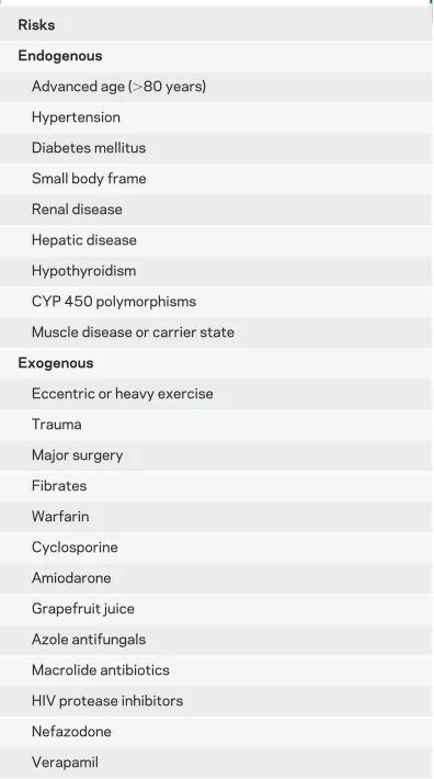
simvastatin have higher myotoxicity rates. Other nonstatin lipid-lowering agents such as niacin and fibrates also carry risks of muscle problems, particu-larly when combined with statins. While it is not possible to predict what patients will have statin-induced muscle problems, prior muscle problems may be a risk factor and should be considered when initiating statin treatment. Family history of myop-athy is relevant if a patient might be a carrier of a genetic myopathy because it could be unmasked by the added stress of statin treatment. Other risk fac-tors may include age over 80 years, low body weight, female sex, hypothyroidism, and Asian descent, as well as concomitant use of certain medications, in-cluding calcium channel blockers, macrolide antibi-otics, omeprazole, amiodarone, azole antifungals, histamine H2receptor antagonists, nefazodone, cy-closporin, HIV protease inhibitors, warfarin, and grapefruit juice. For a comprehensive list, see table 1. The most common muscle symptom caused by statins is muscle pain or myalgia and it occurs in about 7% of statin users. The myalgia can be any-where from mild to severe and is often worsened by muscle activity. If the symptom is tolerable and the indication for statin treatment strong, for ex-ample, in a patient with hypercholesterolemia and a recent myocardial infarction, continued statin treatment may be appropriate.
Baseline creatine kinase (CK) levels are not uni-formly recommended before initiation of statin treat-ment by the organizations guiding statin treattreat-ment, but CK levels can provide very useful information if muscle symptoms later develop. If a patient has mus-cle symptoms during treatment, then a CK level should be checked, and if the level is moderately ele-vated,⬎10 times the upper limit of normal, then the statin drug should be discontinued, according to the National Lipid Association Statin Safety Assessment Task Force.4 This recommendation is reasonable,
but as always the risks need to be weighted against the benefits of treatment. Many athletes and patients with muscular dystrophies live with higher, more markedly elevated CK levels without developing re-nal failure, and a high CK level is not necessarily detrimental. With escalating levels of muscle break-down products being released into the blood, how-ever, at some point renal impairment does occur.
Muscle symptoms resolve with discontinuation of the statin but symptoms can coast for months after discontinuation of the drug. A trial off drug should be 6 months long, unless symptoms improve sooner. After complete resolution of symptoms, rechalleng-ing patients with a lower dose of a statin with rela-tively low risk of muscle complications (such as fluvastatin) can be considered if a strong indication for the statin treatment exists.
Asymptomatic elevation of CK is frequently also seen and this often results in a referral to a neurolo-gist. CK values show great variability among individ-uals and normal values are dependent on sex, age, race, muscle mass, and physical activity. An under-standing of this variability is helpful for interpreting CK values. For example, muscle is a very dynamic tissue, and after muscle exercise, CK values rise to peak around 4 days after exercise and may not nor-malize until 10 –14 days from the exercise.5
Muscle weakness can also occur, and it is often fatigable in quality and combined with pain and ele-vated CK. Like most myopathies, the weakness is most pronounced proximally. Rare episodes of
rhab-“There are millions of people with
statin-induced muscle complaints and they
account for a growing portion of all
patients seen for muscle problems”
Table 1 Risk factors for statin myopathy
Risks Endogenous
Advanced age (⬎80 years)
Hypertension
Diabetes mellitus
Small body frame
Renal disease
Hepatic disease
Hypothyroidism
CYP 450 polymorphisms
Muscle disease or carrier state
Exogenous
Eccentric or heavy exercise
Trauma
Major surgery
Fibrates
Warfarin
Cyclosporine
Amiodarone
Grapefruit juice
Azole antifungals
Macrolide antibiotics
HIV protease inhibitors
Nefazodone
domyolysis have also occurred with statin therapy; these are far less frequent but can possibly be fatal.
Given the prevalence of statin use, other myopa-thies also affect statin users. If symptoms of a pre-sumably statin-induced myopathy are severe, atypical, or do not resolve quickly with discontinua-tion of statin treatment, then a more extensive workup including EMG and a muscle biopsy is indi-cated. The changes that can be seen on muscle histol-ogy that are most typical of a statin myopathy are cytochrome oxidase negative fibers, increased lipid content, and ragged red fibers.6
NECROTIZING IMMUNE-MEDIATED STATIN MYOPATHY An autoimmune necrotizing myop-athy is a rare form of statin myopmyop-athy.7In these
pa-tients, discontinuation of the statin drug does not translate into recovery even after several months off the drug. Patients have a predominantly proximal, often painless weakness. An awareness of this proba-bly infrequent condition is important because its treatment differs significantly from that of the com-mon toxic statin myopathies discussed previously.
Muscle histology reveals a bland necrotic myop-athy with limited or no inflammatory cells in the muscle (figure). A few macrophages in close relation-ship to necrotic fibers are present, but lymphocytic infiltrates are absent. The histologic features of a toxic statin myopathy are not present in the cases reported to date. The anti-200/100 autoantibody was recently described in this population8and it is
not yet commercially available; other markers of au-toimmunity are largely absent. Despite the rather
featureless appearance of the muscle histology, these patients respond to immunosuppressive agents such as prednisone and methotrexate but they may require a more aggressive combination of treatments. The other known causes of necrotizing inflammatory my-opathy include overlap syndromes, particularly in the setting of signal recognition particle (SRP) antibod-ies (discussed later in this review), and also paraneo-plastic myopathies.
IMAGINGNeither MRI nor ultrasound muscle im-aging are yet able to substitute for functional, physi-ologic, or histologic evaluations, but they can be used to provide anatomic information that sometimes is not obvious even by careful physical examination. The detailed anatomic information provided by im-aging can help in the pattern recognition of specific diseases and both modalities can distinguish active and chronic muscle changes. Imaging can also be used to target the muscle biopsy to moderately af-fected tissue, in order to maximize the yield of the biopsy; this is especially useful in diseases with patchy involvement.
MRI is the muscle imaging technique most often used in the United States. Muscle MRI is performed as a series of image sequences including T1-weighted, T2-T1-weighted, and proton density– weighted sequences. Fat-saturated and short tau inversion recovery techniques allow the distinction between fat and edema, and gadolinium enhance-ment can show increased vascularization.9 The
ab-normality most often seen in myopathies is an increase in muscle water content, referred to as mus-cle edema. Musmus-cle edema on MRI does not have a specific histologic correlate, and it is seen in most types of acute and ongoing muscle diseases including dystrophies.10The other detectable abnormalities are
fatty infiltration, hypertrophy, and atrophy, which are nonspecific chronic muscle changes.
Muscle ultrasound also is employed, but the tech-nique is still not widely used, possibly because of its being operator-dependent. However, at some centers it is used by experienced examiners with consistent results. Ultrasound is quick and painless, making it particularly popular in pediatric muscle disease cen-ters. Ultrasound examination can provide informa-tion about both muscle edema as a sign of active muscle disease and chronic changes of fatty degener-ation and replacement of muscle by connective tis-sue. Contrast-enhanced ultrasound can also give an impression of the blood flow in the imaged muscle and this is another indirect measure of an active mus-cle disease. In addition to atrophy and hypertrophy, ultrasound provides real-time information allowing visualization of fasciculations.11
Figure Necrotic muscle fibers without inflammatory cells
AUTOANTIBODIESThe field of autoantibodies re-lated to immune-mediated inflammatory myopathies has expanded in recent years and there is now a host of antibodies that have relevance to these myopa-thies. The 1975 Bohan and Peter criteria for the
clas-sification of immune-mediated inflammatory
myopathies do not reflect many newer insights, and several newer classification schemes exist, but none enjoy uniform acceptance.12 Some controversy
re-mains as to the pathophysiology behind dermatomy-ositis, but this disease is probably the most consistently defined. Conversely, polymyositis has several varied definitions, and in the Bohan and Peter criteria it was not delineated from inclusion body myopathy (IBM). The antisynthetase syndrome asso-ciated with antibodies described in this section does not cleanly sort under either the dermato- or poly-myositis labels. The inflammatory myopathies asso-ciated with SRP and 200/100 antibodies do not even necessarily have the inflammatory muscle infiltrates that we traditionally associate with inflammatory myopathies. While IBM has prominent inflamma-tory features, none of the described autoantibodies are linked to IBM, nor is immunomodulatory treat-ment of any benefit. For these and other reasons, many authorities believe IBM to be more of a myo-degenerative disease with secondary inflammation.13
Granulomatous myopathy, HIV-associated myositis,
and graft vs host disease are other immune-mediated inflammatory myopathies without associated muscle-directed antibodies.
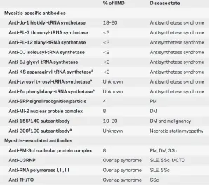
There are also the overlap syndromes in which another defined autoimmune condition exists and overlaps with a myositis. This can occur in diseases such as systemic sclerosis, rheumatoid arthritis, sys-temic lupus erythematosus, and Sjo¨gren syndrome. Distinguishing the primary inflammatory myopa-thies from the overlap syndromes is done by exclud-ing the conditions causexclud-ing overlap syndromes, but there are also autoantibodies that are almost unique to the immune-mediated inflammatory myopathies referred to as muscle-specific autoantibodies (MSA). Other antibodies are frequently seen in other con-nective tissue disorders, and these can be referred to as myositis-associated autoantibodies (MAA). All of these antibodies can help establish the diagnosis of myositis when the muscle biopsy is inconclusive, and the MSAs as well as some of the MAAs are listed in table 2.
The most prevalent MSA is the anti-Jo antibody, which is directed against histidyl-tRNA synthetase. Anti-Jo is detected in about 20% of patients with myositis in most populations. Anti-Jo can be de-tected in both dermatomyositis and polymyositis and is frequently associated with interstitial lung disease and mechanic’s hands. This clinical and laboratory constellation is referred to as the antisynthetase syn-drome. Interstitial lung disease is a potentially fatal comorbidity that often requires more aggressive im-munomodulatory treatment. Histologically, the in-flammation is often more perimysial rather than endomysial.14There are other newer antisynthetase
antibodies with similar clinical features including those that recognize threonyl-tRNA synthetase (anti-PL-7), alanyl-tRNA synthetase (anti-PL-12), glycyl-tRNA synthetase (anti-EJ), isoleucyl-glycyl-tRNA synthetase (OJ), asparaginyl-tRNA synthetase (KS), anti-tyrosyl-tRNA synthetase, and antiphenylalanyl syn-thetase (anti-Zo). These other antibodies are each present in a few percent of patients, but there is essen-tially no overlap between them and patients do not ex-press more than one antisynthetase antibody. A different type of antibody is the Anti-Mi-2 autoanti-body. This nuclear antibody is directed against a com-ponent of the nucleosome-remodeling deacetylase, is seen more often in dermatomyositis, and is infrequent in most populations.
A clinically useful antibody is the SRP antibody. This antibody can often be found when there is myonecrosis, but little or no inflammation is seen on muscle histology. Identifying the antibody can be helpful in establishing that the myopathy is in-flammatory and encourages escalating immunosup-Table 2 Myositis-specific and associated antibodies (modified from
Targoff et al.15)
% of IIMD Disease state Myositis-specific antibodies
Anti-Jo-1 histidyl-tRNA synthetase 18–20 Antisynthetase syndrome
Anti-PL-7 threonyl-tRNA synthetase ⬍3 Antisynthetase syndrome
Anti-PL-12 alanyl-tRNA synthetase ⬍3 Antisynthetase syndrome
Anti-OJ isoleucyl-tRNA synthetase ⬍2 Antisynthetase syndrome
Anti-EJ glycyl-tRNA synthetase ⬍2 Antisynthetase syndrome
Anti-KS asparaginyl-tRNA synthetasea ⬍2 Antisynthetase syndrome
Anti-tyrosyl tyrosyl-tRNA synthetasea Unknown Antisynthetase syndrome
Anti-Zo phenylalanyl-tRNA synthetasea Unknown Antisynthetase syndrome
Anti-SRP signal recognition particle 4 PM
Anti-Mi-2 nuclear protein complex 8 DM
Anti-155/140 autoantibody 10–20 DM and malignancy
Anti-200/100 autoantibodya Unknown Necrotic statin myopathy
Myositis-associated antibodies
Anti-PM-Scl nucleolar protein complex 8 PM, DM, SSc
Anti-U3RNP Overlap syndrome SLE, SSc, MCTD
Anti-RNA polymerase I, II, III Overlap syndrome SLE, SSc
Anti-TH/TO Overlap syndrome SSc
Abbreviations: DM⫽dermatomyositis; MCTD⫽mixed connective tissue disease; PM⫽
polymyositis; SLE⫽systemic lupus erythematosus; SSc⫽systemic sclerosis.
pression even if initial attempts are unsuccessful. The target of the new anti-155/140 antibody remains unknown, but this antibody is seen in dermatomyositis and is more common in paraneo-plastic dermatomyositis compared to idiopathic autoimmune dermatomyositis.15 The not yet
com-mercially available anti-200/100 autoantibody ap-pears to have specificity for the necrotizing statin myositis (discussed earlier).8In patients with a
myop-athy of unclear cause and a nondiagnostic biopsy testing, one should consider testing the Jo anti-body and a comprehensive panel of the other MSA, either sequentially or simultaneously.
POMPE DISEASE Pompe disease, also known as acid maltase deficiency, is one of many causes of adult and childhood onset limb-girdle weakness. Clinically there is often more prominent respiratory muscle involvement than in the average limb-girdle dystrophy, but the clinical phenotypes of several con-ditions overlap.
The disease is caused by a severe deficiency of the
␣-glucosidase (GAA) enzyme resulting from muta-tions of both alleles coding for the enzyme. An ab-sence of enzyme activity leads to disease onset in infancy and a severe but incomplete deficiency trans-lates into later onset. The lack of enzyme activity leads to glycogen accumulation particularly in mus-cle tissue. Now that it has been found that enzyme replacement treatment is effective for patients with later onset disease,16distinguishing this disease from
other limb-girdle syndromes is critical, as it provides guidance not only for symptomatic management and prognostic information for affected individuals, but now a disease-directed treatment. The enzyme re-placement therapy may not only halt functional de-cline but even marginally improve function. A diagnosis can be established by measuring GAA ac-tivity in blood, or in cultured fibroblasts, or by ge-netic testing, but none of these techniques are perfect. Currently several organizations are drawing up new practice parameters for the testing and treat-ment of Pompe disease.
DISCUSSION The statin myopathies may affect millions of Americans and an awareness of the details of this common statin myalgia and other muscle problems is useful, as is a basic understanding of the rare necrotic statin myopathy. Current imaging tech-niques can provide valuable anatomic information, but cannot conclusively differentiate between dis-eases. The increasing number of commercially avail-able muscle-specific antibodies provides new tools for establishing the cause of the immune-mediated myopathies. The exciting development of an enzyme replacement treatment for late-onset Pompe disease gives hope for patients with the muscular dystro-phies, mitochondrial cytopathies, and other meta-bolic myopathies, where many promising treatments currently are being explored. Hopefully more disease-directed treatments will be available in the future to supplant our current arsenal of supportive measures. An area not covered in this review includes the treatments of the inflammatory myopathies. The cornerstones remain largely unchanged, but new im-munomodulatory therapies are available and treat-ment failure or limiting side effects now often can be overcome.
DISCLOSURE
Dr. Oskarsson served on the national advisory board for Avanir Pharma-ceuticals; performs muscle biopsies and EMG and cares for patients with muscle diseases in his clinical practice (50% effort) and bills for these procedures; and receives research support from the NIH.
Received October 31, 2010. Accepted in final form December 16, 2010.
REFERENCES
1. Stagnitti MN. Trends in statins utilization and expendi-tures for the U.S. civilian noninstitutionalized popula-tion, 2000 and 2005. Rockville, MD: Quality AfHRa; 2008.
2. Bruckert E, Hayem G, Dejager S, Yau C, Begaud B. Mild to moderate muscular symptoms with high-dosage statin therapy in hyperlipidemic patients: the PRIMO study. Cardiovasc Drugs Ther 2005;19:403– 414.
3. Mammen AL, Amato AA. Statin myopathy: a review of recent progress. Curr Opin Rheumatol 2010;22:644 – 650.
4. McKenney JM, Davidson MH, Jacobson TA, Guyton JR. Final conclusions and recommendations of the National Lipid Association Statin Safety Assessment Task Force. Am J Cardiol 2006;97:89C–94C.
5. Pettersson J, Hindorf U, Persson P, et al. Muscular exercise can cause highly pathological liver function tests in healthy men. Br J Clin Pharmacol 2008;65:253–259.
6. Phillips PS, Haas RH, Bannykh S, et al. Statin-associated myopathy with normal creatine kinase levels. Ann Intern Med 2002;137:581–585.
7. Grable-Esposito P, Katzberg HD, Greenberg SA, Sriniva-san J, Katz J, Amato AA. Immune-mediated necrotizing myopathy associated with statins. Muscle Nerve 2010;41: 185–190.
Myopathy: Five New Things
• Risk of statin toxicity increases along with increases in their lipophilicity, cholesterol-lowering potency, and dosage.
• In immune-mediated statin myopathy, discontinuation does not translate into immediate recovery.
• MRI and muscle ultrasound in myopathy may provide detailed anatomic information.
• Autoantibody testing may be helpful in defining myopathies of unclear cause.
8. Christopher-Stine L, Casciola-Rosen LA, Hong G, Chung T, Corse AM, Mammen AL. A novel autoantibody recog-nizing 200-kd and 100-kd proteins is associated with an immune-mediated necrotizing myopathy. Arthritis Rheum 2010;62:2757–2766.
9. Curiel RV, Jones R, Brindle K. Magnetic resonance imaging of the idiopathic inflammatory myopathies: structural and clinical aspects. Ann NY Acad Sci 2009;1154:101–114. 10. Wattjes MP, Kley RA, Fischer D. Neuromuscular imaging in
inherited muscle diseases. Eur Radiol 2010;20:2447–2460. 11. Pillen S, Arts IM, Zwarts MJ. Muscle ultrasound in
neuro-muscular disorders. Muscle Nerve 2008;37:679 – 693. 12. Sultan SM, Isenberg DA. Re-classifying myositis.
Rheu-matology 2010;49:831– 833.
13. Askanas V, Engel WK, Nogalska A. Inclusion body my-ositis: a degenerative muscle disease associated with intra-muscle fiber multi-protein aggregates, proteasome inhibition, endoplasmic reticulum stress and decreased lysosomal degradation. Brain Pathol 2009;19:493–506. 14. Mozaffar T, Pestronk A. Myopathy with anti-Jo-1 antibodies: pathology in perimysium and neighbouring muscle fibres. J Neurol Neurosurg Psychiatry 2000;68:472– 478. 15. Targoff IN, Mamyrova G, Trieu EP, et al. A novel
autoan-tibody to a 155-kd protein is associated with dermatomyo-sitis. Arthritis Rheum 2006;54:3682–3689.
16. van der Ploeg AT, Clemens PR, Corzo D, et al. A random-ized study of alglucosidase alfa in late-onset Pompe’s dis-ease. N Engl J Med 2010;362:1396 –1406.
If you liked this article, you may be interested in ...
Neurology
Claeys et al. Phenotype of a patient with recessive centronuclear myopathy and a novel bin1 mutation.
February 9, 2010;www.neurology.org
Broccolini et al. Analysis of NCAM helps identify unusual phenotypes of hereditary inclusion-body
myopathy. July 20, 2010;www.neurology.org
Kono et al. Dominant-negative effects of a novel mutation in the filamin myopathy. August 10,
2010;www.neurology.org
Muelas et al. MYH7 gene tail mutation causing myopathic profiles beyond Laing distal myopathy.
August 24, 2010;www.neurology.org
Neurology Now
Anthony A. Amato, M.D. Ask the Experts: Polymyositis. January/February 2010;
www.neurologynow.com
Neurology Today
Kurt Samson. Small alemtuzumab trial finds molecular benefits in some myositis patients.
DOI 10.1212/WNL.0b013e31820c3648
2011;76;S14-S19
Neurology
Björn Oskarsson
Myopathy: Five New Things
This information is current as of February 14, 2011
Services
Updated Information &
http://n.neurology.org/content/76/7_Supplement_2/S14.full
including high resolution figures, can be found at:
References
http://n.neurology.org/content/76/7_Supplement_2/S14.full#ref-list-1
This article cites 15 articles, 1 of which you can access for free at:
Subspecialty Collections
http://n.neurology.org/cgi/collection/ultrasound
Ultrasound
http://n.neurology.org/cgi/collection/other_toxicology
Other toxicology
http://n.neurology.org/cgi/collection/muscle_disease
Muscle disease
http://n.neurology.org/cgi/collection/mri
MRI
http://n.neurology.org/cgi/collection/autoimmune_diseases
Autoimmune diseases
following collection(s):
This article, along with others on similar topics, appears in the
Permissions & Licensing
http://www.neurology.org/about/about_the_journal#permissions
its entirety can be found online at:
Information about reproducing this article in parts (figures,tables) or in
Reprints
http://n.neurology.org/subscribers/advertise
Information about ordering reprints can be found online:
rights reserved. Print ISSN: 0028-3878. Online ISSN: 1526-632X.
1951, it is now a weekly with 48 issues per year. Copyright Copyright © 2011 by AAN Enterprises, Inc.. All ® is the official journal of the American Academy of Neurology. Published continuously since