1
Unveiling mesenchymal stromal cells’ organizing function in regeneration
1 2
Author list:
3
Peter P. Nimiritsky1,2 #, 4
Roman Yu. Eremichev1 #, 5
Natalia A. Alexandrushkina1,2 #, MD 6
Anastasia Yu. Efimenko1,2, MD, PhD 7
Vsevolod A. Tkachuk1-3, DSc 8
Pavel I. Makarevich* 1,2, MD, PhD 9
10
Author affiliations
11
1 – Institute for Regenerative Medicine, Medical research and education center, 12
Lomonosov Moscow State University, Moscow, Russia 13
2 – Faculty of Medicine, Lomonosov Moscow State University, Moscow, Russia 14
3 – Laboratory of Molecular Endocrinology, National medical research center of 15
cardiology, Moscow, Russia 16
* corresponding author: [email protected] 17
# - equal contribution 18
Abstract
21
Regeneration is a fundamental process much attributed to functions of adult 22
stem cells. In last decades delivery of suspended adult stem cells is widely adopted 23
in regenerative medicine as a leading mean of cell therapy. However, adult stem 24
cells can not complete the task of human body regeneration effectively by 25
themselves as far as they need a receptive microenvironment (the niche) to engraft 26
and perform properly. Understanding of mechanisms underlying mammalian 27
regeneration lead us to an assumption that improved outcomes of cell therapy 28
requires a specific microenvironment generated in damaged area prior to stem cell 29
delivery. To certain extent it may be achieved by delivery of mesenchymal stromal 30
cells (MSC), not in dispersed form, but rather self-organized in cell sheets (CS) – 31
tissue-like structures comprising of viable cells and microenvironment components: 32
extracellular matrix and soluble factors deposited in the matrix. 33
In this communication we highlight a potential role of mesenchymal stromal 34
cells (MSC) as regeneration organizers and speculate that this function emerges in 35
CS. This concept shifts our understanding of therapeutic mechanism underlying a 36
widely known CS-based delivery method for regenerative medicine. 37
Keywords: stem cell, stromal cell, mesenchymal stromal cell, regeneration, 38
cell sheet, cell delivery 39
1. The challenge for stem cell therapy: importance of microenvironment
41
In last decades stem cell therapy has moved from use of fetal and embryonic 42
material to adult stem cell application pursuing the goal of damaged tissue structural 43
and functional restoration [1]. Adult stem cells localized in majority of organs 44
driving their physiological renewal and recovery after damage or disease [2]. This 45
natural mechanism of human body regeneration provided a promising tool for 46
regenerative medicine and resolved ethical problems related to fetal material use as 47
well as major safety concerns connected with use of undifferentiated and/or 48
potentially teratogenic cells [3, 4]. 49
Despite initial promise of stem cell therapy it showed limited efficacy in 50
numerous clinical trials [5] and, thus, put to doubt initial concept portraying adult 51
stem cells as regenerative “commando engineers” that can engraft, differentiate and 52
rebuild fully functional tissue once delivered viable and in sufficient amounts [6]. 53
Eventually, accumulated data led to a conclusion that most of delivered stem cells 54
fail to integrate and give rise to new tissue elements and work as “bystanders” 55
releasing paracrine factors and disappearing within several weeks [7-9]. 56
Discussing reason of disappointing results of stem cell therapy we may 57
question what can be improved to overcome these hurdles in the way of the 58
promising approaches in biomedicine. To answer that crucial role of 59
microenvironment should be considered – from location of cell to set of signals 60
required to maintain finely tuned physiological functions [10]. This issue is known 61
since Raymond Schofield’s seminal work suggesting that stem cells are “non-62
autonomous” and depend on microenvironment known as niche [11]. The latter 63
controls, directs and supports balanced stages of stem cell life cycle and includes 64
other cells (stromal, parenchymal, vascular etc.), extracellular matrix (ECM), 65
soluble proteins and peptides, extracellular vesicles, small molecules and chemical 66
factors (pH, oxygen pressure etc.) [12]. 67
Importance of microenvironment for success of stem cell-driven body repair 68
has been supported by investigation of regeneration in animals with ability to rebuild 69
these creatures “separated” adult stem cells (neoblasts) themselves lack ability to 71
drive full-scale regeneration and require certain amount of tissues to preform 72
properly [13]. 73
Human adult stem cells administered to damage site in suspension are 74
deprived of crucial stimuli required for their regenerative potential to unfold. They 75
lack appropriate intercellular contacts, nutrition and regulatory signals. Under such 76
unfavorable conditions formation of mature tissue elements from transplanted stem 77
cells is an extremely rare event hardly providing significant therapeutic impact [14, 78
15]. 79
In this paper we summarize our vision on problems that stand in the way of 80
successful application of stem cell therapies. We focus on a cornerstone role of 81
microenvironment formed during regeneration and contribution of mesenchymal 82
stromal cells (MSC) to this process. We suggest that tissue engineered constructs 83
known as cell sheets (CS) present a feasible tool to unfold potential of MSC as 84
organizers of regeneration. 85
86
2. Shifting the focus to microenvironment: feeder needed!
87
To illustrate the possible reason of stem cell therapies failure and support our 88
accent on importance of receptive microenvironment for success of therapeutic 89
approach we shall start with a metaphor to compare mammalian tissue regeneration 90
with repopulation of an ecosystem after a natural catastrophe. 91
It is well-known that structure of ecological systems has hierarchy based on 92
food chains. It is typically portrayed as a pyramid reflecting dependency of high-93
order consumers on lower-order and down to the ground level of energy-absorbing 94
producers – plants and microorganisms (left part of Figure 1). 95
After disaster hierarchy of ecosystem and its structure recovers stage by stage 96
from the foundation starting with the most adaptive and viable species - producers. 97
When they generate a necessary trophic substrate then more demanding species can 98
enter this system and populate it. Gradually the species diversity of entire ecosystem 99
ecosystem is restored and recovery occurs [16]. However, the cornerstone of this 101
process is – and it should be emphasized – formation of a “ground level” of 102
producers required for other species to survive [17]. 103
This metaphor was used to show why therapies using stem cells may fail. In 104
acute phase of response to injury we deliver them to a microenvironment lacking 105
necessary elements and regulatory framework that existed before damage. We 106
speculate that stages of ecosystem recovery might portray regeneration in humans 107
and illustrate that stem cells are capable to unveil their potential only when adequate 108
microenvironment is generated prior to that. Indeed, after damaged area is cleansed 109
by inflammatory cells, they leave “ground zero” with disrupted structure of the 110
tissues down to molecular level. Under such conditions stem cell fails to rebuild the 111
tissue as it depends on other elements – ECM, soluble factors, endothelium, stromal 112
cells and neural terminals disrupted by damage [10-12]. 113
In ecological terms this may be described as a high-order consumer entering 114
the vast field of ashes after a forest fire before the lower levels are re-populated. 115
Failing to find food and mate eventually he will be leaving the unsuitable habitat. 116
Following this idea basal layer of right side of the pyramid - the feeder becomes of 117
paramount importance for microenvironment rebuilding to involve specific 118
progenitors (right part of Figure 1) similarly to the ecological dogma that producers 119
presence is obligatory prior to consumers’ population appears. 120
Figure 1. Critical role of feeder in sequential regeneration of human
122
body.
123
Both in ecosystem and human body “ground” and “feeder” levels respectively 124
are generated by the most adaptive and universal inhabitants. They have critical 125
importance as they provide foundation for subsequent interactions between elements 126
and the system structure recovery (see text for detail). 127
128
We define the feeder as a transient heterogenous structure formed in vivo at 129
early stage of regeneration to re-create disrupted components of microenvironment 130
which allows other cell types to perform properly in recovery of structure and 131
function. In our concept this putative feeder may contain stromal, immune and 132
probably vascular cells and intercellular components of microenvironment: ECM 133
and soluble factors predominantly anchored in the matrix to render paracrine effects 134
and create gradients of stimuli mapping further stages of regeneration. 135
Similar requirement for feeder has been described in zoology prior to 136
formation of blastema after amputation of a limb in salamander. Immediately after 137
injury a layer of epidermal cells covers the wound and becomes an apical epithelial 138
cap providing ground for attraction of fibroblasts and other cells that dedifferentiate 139
to form the blastema and eventually regrow the limb [18]. 140
An encouraging example of whole organ recapitulation from delivered stem 141
cells in humans is bone marrow transplantation [19]. However, in this procedure 142
after myeloablative stage the stromal component of bone marrow remains relatively 143
intact providing a functional niche for transplanted hematopoietic cells supporting 144
the idea that receptive microenvironment is critical for regeneration allowing stem 145
cells to rebuild tissue but not work as secretome-producing “bystanders” [14, 15]. 146
Typical example of a non-receptive microenvironment that can be formed 147
after damage is scar – avascular bulk of ECM packed to hold the tissues together 148
and barely undergoing resolution over time. Stem cell therapies have shown little to 149
no effect in stimulation of fibrosis resolution yet in some disorders like cystic 150
progress [20]. Nevertheless, these effects were once again generally attributed to 152
cells’ paracrine function rather than engraftment and tissue restoration [8]. 153
154
3. Re-building the feeder: focus on mesenchymal stromal cells (MSC)
155
Thus, we come to a conclusion that stem cell therapies do not achieve their 156
goal until a receptive microenvironment is being formed. Due to a defined order of 157
events during regeneration after injury we suggest to focus not only on tissue-158
specific stem cells but on the basal layer for which we provide the term feeder (in 159
particular, similarly to cell culture technique used to support stem cells and other 160
cell types). Below we speculate on participation of MSC in its formation in vivo and 161
address their biological and physiological functions that support this point. 162
Among numerous cell types of human body MSC remain a prominent 163
candidate for therapeutic use and in our concept they comprise an important (yet not 164
the only) component of putative feeder. In human body MSC are involved in 165
maintaining of structure and homeostasis [21] comprising an ubiquitous stromal 166
component in majority of organs. In quiescent state they are localized in perivascular 167
compartments supporting vascular permeability, secrete growth factors, support 168
ECM production and turnover. These functions are important for interaction with 169
local tissue progenitors and stem cells to retain organs architecture by maintaining 170
gradients of stimuli. In intestinal crypt niche MSC create a gradient of bone 171
morphogenic proteins (BMP) that controls resident stem cell fate and differentiation 172
[22]. However, this tissue function of MSC is not limited to stem cell niches and in 173
intact organs they participate in formation of microenvironment for all cells located 174
nearby [23]. 175
After damage MSC migrate to the lesion site where they actively proliferate 176
[24]. Their ability to survive under stress resulted in excellent cultural properties of 177
isolated MSC. Upon cease of acute phase of inflammation MSC attenuate intensity 178
of residual inflammation. They secrete a wide range of anti-inflammatory factors 179
(IL-10, TSG-6, PGE2) and, thus, switch the microenvironment to a pro-regenerative 180
Further, MSC produce ECM proteins, and factors of matrix turnover (matrix 182
metalloproteases etc.), as well as cytokines and growth factors to attract and support 183
other cell types (endothelial, tissue-specific progenitors etc.) [25-27]. 184
This guided to investigate the composition of MSC secretome and our group 185
as well as others found that physiological potency of secretome is sufficient to 186
trigger events crucial for regeneration [28-30]. It activates proliferation and survival 187
of different cells under stress, protects tissue from excessive fibrosis, induces 188
angiogenesis and nerve growth, regulates immune response etc. leading to idea of 189
secretome therapeutic application known as “cell-free cell therapy” [31-33]. Our 190
further efforts were focused on functionalization of MSC secretome by viral delivery 191
of growth factors genes or pre-treatment by recombinant proteins. And we have 192
shown that it results in significant shift of MSC functional activity and improvement 193
of therapeutic effect [34-36]. However, we did not find increase of engraftment after 194
functionalization leading us to a generally supported conclusion that MSC therapies 195
succeed to deliver secretome produced by stem cells over time rather than replenish 196
native tissue elements by their differentiation. 197
198
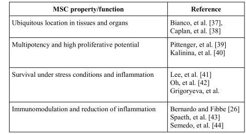
Table 1. Biological properties of MSC that contribute to their potential
199
as participants of feeder formation
200
MSC property/function Reference
Ubiquitous location in tissues and organs Bianco, et al. [37], Caplan, et al. [38]
Multipotency and high proliferative potential Pittenger, et al. [39] Kalinina, et al. [40]
Survival under stress conditions and inflammation Lee, et al. [41] Oh, et al. [42] Grigoryeva, et al.
Immunomodulation and reduction of inflammation Bernardo and Fibbe [26] Spaeth, et al. [43]
Active pleiotropic secretome comprising of ECM components, growth factors, cytokines and
extracellular vesicles
Kalinina, et al. [28] Konala, et al. [29] Vizoso, et al. [32]
Ability to support and regulate other cell types in vivo Rubina, et al. [27] Battiwalla, et al. [45]
201
Thus, after injury damage-activated MSC mitigate inflammatory response and 202
produce basic elements of the putative feeder: ECM and soluble trophic factors to 203
attract other cells, to interact with them, to form a basis for regeneration. 204
Nevertheless, one may wonder why MSC delivered by injection fail to form 205
interactions between each other and drive regeneration. Available answer is that 206
negative influence of ECM and contacts disruption due to harvesting from 207
monolayer followed by injection makes MSC unable to perform their functions [46]. 208
It is predominantly related to stress due to loss of microenvironment that existed in 209
vitro, namely – adhesive surface, ECM and soluble factors in culture medium.
210
Certain reports claim that after injection up to 70% of MSC undergo cell death 211
within the next 48-72 hours after delivery [40]. 212
Thus, MSC therapy is limited to bystander effects similar to other stem cell 213
therapies. However, MSC are able to produce basic components of their own 214
microenvironment: ECM and pro-survival soluble factors. Moreover, they are able 215
to organize this microenvironment and generate a tissue-like structure in vitro – 216
namely, cell sheet (CS). The latter is generally used to facilitate cell delivery and 217
transplant MSC in an organized manner resolving the problem of dispersed cells’ 218
inability to survive without appropriate microenvironment. Below we shall provide 219
our point of view and data that support CS-based MSC delivery as a more 220
physiological approach potentially providing ground for effective human 221
regeneration. 222
223
4. Cell sheet technology: basics and application
224
Cell sheets are scaffold-free tissue-like constructs consisting of living cells, 225
CS varies in time (3-14 days), depends on ECM and soluble factors production, cells 227
proliferation and their ability to integrate to a multilayered construct that detaches 228
from thermo-responsive coating [47] or after treatment by chelating agents and/or 229
mechanical peeling [48]. 230
This technology quickly found application in many areas of regenerative 231
medicine, primarily due to advantages over direct injection. Delivery by CS is 232
positioned as a method to deliver cells along with microenvironment formed in vitro. 233
Many preclinical and clinical studies report CS efficacy to treat diseases of the 234
bones, cartilage [49, 50], skin, urinary bladder, heart, blood vessels, esophagus, 235
cornea etc. [51]. To better understand details and therapeutic promise of CS 236
technology in regenerative medicine we may address the Reader to a number of 237
recent seminal reviews [52-56]. 238
Experimental studies to treat wounds, limb ischemia [57], nerve damage [58] 239
and myocardial infarction [59] have been published by our group during last decade. 240
Besides delivery of primary MSC sheets we have studied endothelial cells, cardiac 241
stem cells [48] and viral modification of CS to enhance therapeutic outcomes [57, 242
58, 60] and our data along with published works of other researchers allows two 243
general conclusions regarding CS as a delivery platform for MSC therapy. 244
1. Delivery of MSC assembled as CS results in strong and reproducible 245
increase of efficacy in models of different tissue lesions.
246
Comparison of efficacy after delivery of equivalent amount of MSC by 247
injection or by CS transplantation has shown that sheets are superior to injected cells. 248
In our studies CS have performed better than suspended MSC for treatment of 249
ischemia, nerve repair [57, 58, 61], deep wound healing [62] and pressure ulcer (data 250
unpublished). Recent papers describing use of CS for liver regeneration [63] or bone 251
healing [64] often omit direct “head to head” comparison of outcomes after CS or 252
suspended delivery yet most of them report long-term survival of transplanted cells 253
suggesting that it mediates therapeutic outcome. 254
2. MSC delivered as CS show signs of engraftment including long-term 255
This point was illustrated in one of our works using subcutaneous 257
implantation of labeled MSC sheets in mice indicating CD31-positive capillaries and 258
larger caliber blood vessels in CS mass as well as Ki-67-positive MSC detected 2 259
weeks after transplantation [57]. Our results were supported by data from other 260
groups using MSC sheets [65-67] and by own data using other cell types – namely 261
cardiac stem cell sheets transplanted to treat myocardial infarction in a rat model 262
[48, 59]. 263
Points mentioned above support our suggestion that assembly of CS from 264
MSC facilitates engraftment and, thus, delivery of a stromal part of suggested feeder 265
that may become a putative basis for further regeneration. However, to do this MSC 266
must interact with other cell types and integrate in an organotypic structure. 267
Potential mechanism for that is generation of stimuli gradients which will be 268
explained further. 269
270
5. Role of MSC in structure organization in development and
271
regeneration
272
Early stages of human development are driven by embryonic stem cells’ 273
(ESCs) ability to interconnect and integrate in community [68]. Crosstalk between 274
ESCs dynamically changes form and structure resulting in embryogenesis. 275
Mesenchyme appears during gastrulation phase and gets involved in subsequent 276
organogenesis. Interactions of mesenchymal cells with epithelial cells are crucial in 277
development of teeth [69], lungs [70], pancreas [71], heart [72], kidney [73, 74] and 278
liver [75]. 279
Critical aspect of mentioned processes in development is formation of stimuli 280
gradients including those produced by mesenchymal cells [76]. Soluble factors, 281
ECM and cell-to-cell contacts have been postulated to participate in accurate 282
organogenesis of many organs and role of mesenchymal cells acting as main 283
structure organizers is well-established as well [77, 78]. 284
In postnatal period MSC (despite losing many traits they featured in 285
basis of this ability is generation of stimuli gradients once regenerative program is 287
activated by injury. This supports our concept of MSC role in generation of putative 288
feeder and explain how they can facilitate and “map” next stages of regeneration. 289
290
6. MSC as organizers of other cell types: ex vivo evidence
291
MSC’s ability to support and organize other cells in vitro has been reported 292
previously for epithelial cells from embryonic salivary gland. In this study MSC but 293
not conventionally used feeder (NIH/3T3 fibroblasts) supported self-assembly of 294
primary embryonic epithelial cells and subsequent branching with buds formation 295
in 3D culture resembling mesenchyme function in development [79]. 296
One of the strongest evidences of MSC ability to facilitate organogenesis in 297
vitro is a work by Takebe et al. [80] describing MSC-driven condensation of
iPS-298
derived progenitors to form bud-shaped organoids. Spectrum of organoids that can 299
be successfully assembled under influence of MSC in vitro included liver, intestine, 300
lung, kidney, heart and brain (!). Finally, endothelium-containing pancreatic and 301
renal buds derived under influence of MSC were transplanted to animals where they 302
rapidly underwent vascularization and self-organized into functional, tissue-specific 303
structures. 304
Organization of such complex organ(oid)s as pancreas or kidney should 305
employs the same principles as in organogenesis relying on focal stimuli provided 306
by MSC to organize other cell types in a proper manner. At this point our recent data 307
describing heterogeneity in human MSC-derived CS provides a new outlook on this 308
delivery mode and its potential impact on regeneration [81, 82]. 309
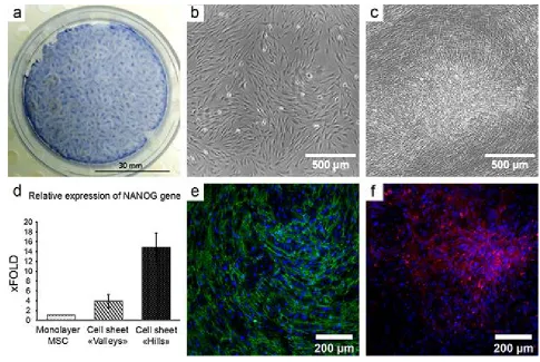
In our study a CS from human adipose-derived MSC showed significant 310
morphological heterogeneity with 2 distinct compartments of high and low density 311
of cells (“hills” and “valleys respectively – see Figure 2, a-c). High-density regions 312
were formed as a result of MSC proliferation cycles followed by coordinated 313
contraction and migration of cells (see Supplementary video 1) which resembles a 314
previously described contraction phenomenon that accompanies MSC-driven self-315
317 318
Figure 2. Self-organization of MSC in cell sheets results in formation of
319
heterogeneous microenvironment.
320
a. Heterogeneous distribution of MSC in CS at day 12 of cultures; hematoxylin 321
staining; 322
b-c. Microphotographs taken in the same field of view of MSC culture at day 1 (b) 323
and day 12 (c). Formation of a high-density “hill” is observed as a result of MSC 324
self-organization in CS; time-lapse microscopy, phase-contrast. 325
d. MSC subpopulations were picked by laser microdissection. MSC in the “hills” 326
show increased expression of stemness-associated factor (NANOG); real-time 327
qPCR (n=3); data expressed as mean ± standard deviation are shown. 328
e-f. MSC in CS organise EDA-fibronectin in regular network of fibres (green, 329
panel e) while laminin (red, panel f) is deposited primarily in “hills”, but not in 330
“valleys” creating foci of ECM deposition in CS (f); immunofluorescent 331
microscopy, nuclei are stained by DAPI (blue); from Nimiritsky et al. [81]. 332
For details of experimental procedures please address supplementary Materials and 333
Mesenchyme-mediated guidance in development relies on ECM differential 335
distribution that has crucial importance as it facilitates attraction and retain of 336
different cell types [77]. We found that in MSC sheets two ECM proteins – EDA-337
fibronectin and laminin are distributed uneven with higher density in “hills” and 338
lower – in “valleys” (Fig. 2, e and f). Condensed laminin deposition was detectable 339
only in “hills” of CS and in monolayer of MSC no acquirable signal was detected 340
(data not shown). Laminin is known to mediate interaction of mesenchymal and 341
epithelial cells leading to assumption that heterogeneity of its in CS reflects 342
formation of guiding ECM stimuli that can orchestrate organization of endothelial, 343
epithelial and mature stromal cells on this in vitro formed feeder. 344
Differential morphology and ECM deposition in CS suggest that different 345
MSC subtypes main retained in them so we used laser microdissection to separate 346
“hills” and “valleys” at day 12 to evaluate relative expression of stemness-related 347
gene NANOG in MSC from different microenvironments. 348
We compared laser-dissected “hills” vs. remaining “valley” portion of CS and 349
found significantly (up to 15-fold) increase of NANOG expression compared to 350
“valleys” of same CS (Figure 2, d). It suggested that heterogeneity of CS results in 351
either sorting of different cells to different compartments of the construct or local 352
shift of stemness in situ resembling “mapping” function of mesenchyme known in 353
development and suggesting that MSC sheets actually can be considered as a guiding 354
construct rather than just delivery tool. 355
Previously MSC have been successfully used to generate pre-vascularized 356
structures and endothelial cells spontaneously organized in tubes once were co-357
seeded with MSC for sheet formation [79, 83]. This provided a method for 358
fabrication of tissue-like 3D structures by stacking pre-vascularized CS together 359
without loss of nutrition inside the construct. Nevertheless, our data suggests more 360
attention to potential use of MSC-based CS as an effective driver of organoid or 361
tissue self-assembly as it possesses an important “mapping” trait required for 362
Previous data obtained by our group and other authors using single cell 364
analysis found numerous subpopulations in primary MSC with varying sensitivity 365
to hormones, differentiation ability and signaling [84-86]. However, origin of 366
stimuli gradients and CS heterogeneity is subject for elucidation taking into account 367
data on mesenchymal cells’ role in differentiation control during development and 368
organogenesis ex vivo. 369
370
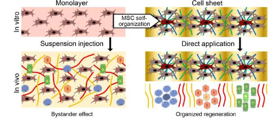
Figure 3. Self-organization of MSC as a basis of its organizing function
371
in regeneration.
372
Conventionally used injection of MSC cultured in monolayer (left part) is 373
limited to paracrine (bystander) effects of delivered cells. Self-organization during 374
CS formation results in retain of microenvironment with gradient of stimuli that 375
organize regeneration after delivery (right part). 376
377
7. Concluding remarks
378
Present paper reflects our vision on how stem cell delivery might be improved 379
basing on knowledge of human development, system biology and – in particular – 380
limitations of existing cell therapy approaches. 381
To our knowledge regeneration after injury has not been previously compared 382
to the recovery of ecosystem structure. Terminology of ecological and evolutionary 383
we consider our approach reasonable to explain importance of putative feeder 385
formed in early stages of regeneration for its outcome. 386
In our view participation and recruitment of MSC during formation of feeder 387
has physiological rationale. Our point is supported by “mapping” role of 388
mesenchymal cells in development and by prominent examples suggesting that adult 389
MSC retain this ability even ex vivo driving functional organoid formations and 390
vascularization in a dish. The concept of feeder as a base layer for successful 391
regeneration might be a subject for discussion yet we suggest it as a summary of our 392
knowledge about regeneration and data from other groups. 393
This concept overall shifts our understanding of MSC-based CS as a delivery 394
platform for regenerative medicine. We believe that CS is not only a mean of “cells 395
+ ECM” deployment, but may present a unique object for study of mammalian 396
regeneration, organoid assembly or tissue modelling. MSC within CS may retain or 397
reproduce certain traits of mesenchymal cells in development to orchestrate 398
regeneration. Origins and consequences of heterogeneity we observed in CS as well 399
as its influence on other cells (including tissue-specific progenitors and terminally 400
differentiated cells) is a subject for further investigation. 401
402
Funding statement
403
The study is supported by RFBR grants (№17-04-01452 and №18-315-404
20053) as a part of state assignment of Lomonosov Moscow State University. 405
Experimental procedure described were carried out with use of equipment purchased 406
under Moscow University Development program. 407
408
References
409
1. Rao, M.; Mason, C.; Solomon, S., Cell therapy worldwide: an incipient 410
revolution. Regen Med 2015, 10, (2), 181-91. 411
2. Macrin, D.; Joseph, J. P.; Pillai, A. A.; Devi, A., Eminent Sources of Adult 412
Mesenchymal Stem Cells and Their Therapeutic Imminence. Stem Cell Rev 413
2017, 13, (6), 741-756. 414
3. King, N. M.; Perrin, J., Ethical issues in stem cell research and therapy. Stem 415
Cell Res Ther 2014, 5, (4), 85.
4. Volarevic, V.; Markovic, B. S.; Gazdic, M.; Volarevic, A.; Jovicic, N.; 417
Arsenijevic, N.; Armstrong, L.; Djonov, V.; Lako, M.; Stojkovic, M., 418
Ethical and Safety Issues of Stem Cell-Based Therapy. Int J Med Sci 2018, 419
15, (1), 36-45. 420
5. Schroeder, I. S., Stem cells: are we ready for therapy? Methods Mol Biol 421
2014, 1213, 3-21. 422
6. Baksh, D.; Song, L.; Tuan, R. S., Adult mesenchymal stem cells: 423
characterization, differentiation, and application in cell and gene therapy. J 424
Cell Mol Med 2004, 8, (3), 301-16.
425
7. Gharaibeh, B.; Lavasani, M.; Cummins, J. H.; Huard, J., Terminal 426
differentiation is not a major determinant for the success of stem cell therapy 427
- cross-talk between muscle-derived stem cells and host cells. Stem Cell Res 428
Ther 2011, 2, (4), 31.
429
8. Savukinas, U. B.; Enes, S. R.; Sjoland, A. A.; Westergren-Thorsson, G., 430
Concise Review: The Bystander Effect: Mesenchymal Stem Cell-Mediated 431
Lung Repair. Stem Cells 2016, 34, (6), 1437-44. 432
9. Breunig, J. J.; Arellano, J. I.; Macklis, J. D.; Rakic, P., Everything that 433
glitters isn't gold: a critical review of postnatal neural precursor analyses. 434
Cell Stem Cell 2007, 1, (6), 612-27.
435
10. Morrison, S. J.; Spradling, A. C., Stem cells and niches: mechanisms that 436
promote stem cell maintenance throughout life. Cell 2008, 132, (4), 598-437
611. 438
11. Schofield, R., The relationship between the spleen colony-forming cell and 439
the haemopoietic stem cell. Blood Cells 1978, 4, (1-2), 7-25. 440
12. Scadden, D. T., The stem-cell niche as an entity of action. Nature 2006, 441, 441
(7097), 1075-9. 442
13. Tanaka, E. M.; Reddien, P. W., The cellular basis for animal regeneration. 443
Dev Cell 2011, 21, (1), 172-85.
444
14. Kurtz, A., Mesenchymal stem cell delivery routes and fate. Int J Stem Cells 445
2008, 1, (1), 1-7. 446
15. Eggenhofer, E.; Luk, F.; Dahlke, M. H.; Hoogduijn, M. J., The life and fate 447
of mesenchymal stem cells. Front Immunol 2014, 5, 148. 448
16. Young, J. A.; Evans, R. A., Population Dynamics after Wildfires in 449
Sagebrush Grasslands. Journal of Range Management 1978, 31, (4), 283-450
289. 451
17. Sutherland, E. F.; Dickman, C. R., Mechanisms of recovery after fire by 452
rodents in the Australian environment: a review. Wildlife Research 1999, 26, 453
(4), 405-419. 454
18. McCusker, C. D.; Gardiner, D. M., Positional Information Is Reprogrammed 455
in Blastema Cells of the Regenerating Limb of the Axolotl (Ambystoma 456
mexicanum). PLoS ONE 2013, 8, (9). 457
19. Thomas, E. D.; Lochte, H. L., Jr.; Lu, W. C.; Ferrebee, J. W., Intravenous 458
infusion of bone marrow in patients receiving radiation and chemotherapy. 459
N Engl J Med 1957, 257, (11), 491-6.
20. Loi, R.; Beckett, T.; Goncz, K. K.; Suratt, B. T.; Weiss, D. J., Limited 461
restoration of cystic fibrosis lung epithelium in vivo with adult bone 462
marrow-derived cells. Am J Respir Crit Care Med 2006, 173, (2), 171-9. 463
21. Klimczak, A.; Kozlowska, U., Mesenchymal stromal cells and tissue-464
specific progenitor cells: their role in tissue homeostasis. Stem cells 465
international 2016, 2016.
466
22. Kosinski, C.; Li, V. S.; Chan, A. S.; Zhang, J.; Ho, C.; Tsui, W. Y.; Chan, T. 467
L.; Mifflin, R. C.; Powell, D. W.; Yuen, S. T., Gene expression patterns of 468
human colon tops and basal crypts and BMP antagonists as intestinal stem 469
cell niche factors. Proceedings of the National Academy of Sciences 2007, 470
104, (39), 15418-15423. 471
23. Kfoury, Y.; Scadden, D. T., Mesenchymal cell contributions to the stem cell 472
niche. Cell stem cell 2015, 16, (3), 239-253. 473
24. Caplan, A. I., Mesenchymal Stem Cells: Time to Change the Name! Stem 474
Cells Transl Med 2017, 6, (6), 1445-1451.
475
25. Dimarino, A. M.; Caplan, A. I.; Bonfield, T. L., Mesenchymal stem cells in 476
tissue repair. Front Immunol 2013, 4, 201. 477
26. Bernardo, M. E.; Fibbe, W. E., Mesenchymal stromal cells: sensors and 478
switchers of inflammation. Cell stem cell 2013, 13, (4), 392-402. 479
27. Rubina, K.; Kalinina, N.; Efimenko, A.; Lopatina, T.; Melikhova, V.; 480
Tsokolaeva, Z.; Sysoeva, V.; Tkachuk, V.; Parfyonova, Y., Adipose stromal 481
cells stimulate angiogenesis via promoting progenitor cell differentiation, 482
secretion of angiogenic factors, and enhancing vessel maturation. Tissue 483
Engineering Part A 2009, 15, (8), 2039-2050.
484
28. Kalinina, N.; Kharlampieva, D.; Loguinova, M.; Butenko, I.; Pobeguts, O.; 485
Efimenko, A.; Ageeva, L.; Sharonov, G.; Ischenko, D.; Alekseev, D.; 486
Grigorieva, O.; Sysoeva, V.; Rubina, K.; Lazarev, V.; Govorun, V., 487
Characterization of secretomes provides evidence for adipose-derived 488
mesenchymal stromal cells subtypes. Stem Cell Res Ther 2015, 6, 221. 489
29. Konala, V. B.; Mamidi, M. K.; Bhonde, R.; Das, A. K.; Pochampally, R.; 490
Pal, R., The current landscape of the mesenchymal stromal cell secretome: 491
A new paradigm for cell-free regeneration. Cytotherapy 2016, 18, (1), 13-492
24. 493
30. Lopatina, T.; Bruno, S.; Tetta, C.; Kalinina, N.; Porta, M.; Camussi, G., 494
Platelet-derived growth factor regulates the secretion of extracellular 495
vesicles by adipose mesenchymal stem cells and enhances their angiogenic 496
potential. Cell Commun Signal 2014, 12, 26. 497
31. Sagaradze, G. D.; Basalova, N. A.; Kirpatovsky, V. I.; Ohobotov, D. A.; 498
Grigorieva, O. A.; Balabanyan, V. Y.; Kamalov, A. A.; Efimenko, A. Y., 499
Application of rat cryptorchidism model for the evaluation of mesenchymal 500
stromal cell secretome regenerative potential. Biomed Pharmacother 2019, 501
109, 1428-1436. 502
32. Vizoso, F. J.; Eiro, N.; Cid, S.; Schneider, J.; Perez-Fernandez, R., 503
Mesenchymal Stem Cell Secretome: Toward Cell-Free Therapeutic 504
33. Sagaradze, G. D.; Nimiritsky, P. P.; Akopyan, Z. A.; Makarevich, P. I.; 506
Efimenko, A. Y., “Cell-Free Therapeutics” from Components Secreted by 507
Mesenchymal Stromal Cells as a Novel Class of Biopharmaceuticals. In 508
Biopharmaceuticals, Chen, M.-K. Y. a. Y.-C., Ed. InTechOpen: 2018.
509
34. Zubkova, E. S.; Beloglazova, I. B.; Makarevich, P. I.; Boldyreva, M. A.; 510
Sukhareva, O. Y.; Shestakova, M. V.; Dergilev, K. V.; Parfyonova, Y. V.; 511
Menshikov, M. Y., Regulation of Adipose Tissue Stem Cells Angiogenic 512
Potential by Tumor Necrosis Factor-Alpha. J Cell Biochem 2016, 117, (1), 513
180-96. 514
35. Shevchenko, E. K.; Makarevich, P. I.; Tsokolaeva, Z. I.; Boldyreva, M. A.; 515
Sysoeva, V. Y.; Tkachuk, V. A.; Parfyonova, Y. V., Transplantation of 516
modified human adipose derived stromal cells expressing VEGF165 results 517
in more efficient angiogenic response in ischemic skeletal muscle. J Transl 518
Med 2013, 11, 138.
519
36. Zubkova, E. S.; Semenkova, L. N.; Dudich, I. V.; Dudich, E. I.; Khromykh, 520
L. M.; Makarevich, P. I.; Parfenova, E. V.; Men'shikov, M., [Recombinant 521
human alpha-fetoprotein as a regulator of adipose tissue stromal cell 522
activity]. Bioorg Khim 2012, 38, (5), 524-34. 523
37. Bianco, P.; Cao, X.; Frenette, P. S.; Mao, J. J.; Robey, P. G.; Simmons, P. J.; 524
Wang, C.-Y., The meaning, the sense and the significance: translating the 525
science of mesenchymal stem cells into medicine. Nature medicine 2013, 526
19, (1), 35. 527
38. Caplan, A. I., Mesenchymal Stem Cells: The Past, the Present, the Future. 528
Cartilage 2010, 1, (1), 6-9. 529
39. Pittenger, M. F.; Mackay, A. M.; Beck, S. C.; Jaiswal, R. K.; Douglas, R.; 530
Mosca, J. D.; Moorman, M. A.; Simonetti, D. W.; Craig, S.; Marshak, D. R., 531
Multilineage potential of adult human mesenchymal stem cells. science 532
1999, 284, (5411), 143-147. 533
40. Kalinina, N. I.; Sysoeva, V. Y.; Rubina, K. A.; Parfenova, Y. V.; Tkachuk, 534
V. A., Mesenchymal stem cells in tissue growth and repair. Acta Naturae 535
2011, 3, (4), 30-7. 536
41. Lee, S.; Choi, E.; Cha, M.-J.; Hwang, K.-C., Cell adhesion and long-term 537
survival of transplanted mesenchymal stem cells: a prerequisite for cell 538
therapy. Oxidative medicine and cellular longevity 2015, 2015. 539
42. Oh, E. J.; Lee, H. W.; Kalimuthu, S.; Kim, T. J.; Kim, H. M.; Baek, S. H.; 540
Zhu, L.; Oh, J. M.; Son, S. H.; Chung, H. Y., In vivo migration of 541
mesenchymal stem cells to burn injury sites and their therapeutic effects in a 542
living mouse model. Journal of Controlled Release 2018, 279, 79-88. 543
43. Spaeth, E.; Klopp, A.; Dembinski, J.; Andreeff, M.; Marini, F., 544
Inflammation and tumor microenvironments: defining the migratory 545
itinerary of mesenchymal stem cells. Gene therapy 2008, 15, (10), 730. 546
44. Semedo, P.; Palasio, C. G.; Oliveira, C. D.; Feitoza, C. Q.; Gonçalves, G. 547
M.; Cenedeze, M. A.; Wang, P. M.; Teixeira, V. P.; Reis, M. A.; Pacheco-548
acute kidney injury. International immunopharmacology 2009, 9, (6), 677-550
682. 551
45. Battiwalla, M.; Hematti, P., Mesenchymal stem cells in hematopoietic stem 552
cell transplantation. Cytotherapy 2009, 11, (5), 503-15. 553
46. Kushida, A.; Yamato, M.; Isoi, Y.; Kikuchi, A.; Okano, T., A noninvasive 554
transfer system for polarized renal tubule epithelial cell sheets using 555
temperature-responsive culture dishes. Eur Cell Mater 2005, 10, 23-30; 556
discussion 23-30. 557
47. Okano, T.; Yamada, N.; Sakai, H.; Sakurai, Y., A novel recovery system for 558
cultured cells using plasma-treated polystyrene dishes grafted with poly(N-559
isopropylacrylamide). J Biomed Mater Res 1993, 27, (10), 1243-51. 560
48. Dergilev, K. V.; Makarevich, P. I.; Tsokolaeva, Z. I.; Boldyreva, M. A.; 561
Beloglazova, I. B.; Zubkova, E. S.; Menshikov, M. Y.; Parfyonova, Y. V., 562
Comparison of cardiac stem cell sheets detached by Versene solution and 563
from thermoresponsive dishes reveals similar properties of constructs. 564
Tissue Cell 2017, 49, (1), 64-71.
565
49. Chen, L.; Xing, Q.; Zhai, Q.; Tahtinen, M.; Zhou, F.; Chen, L.; Xu, Y.; Qi, 566
S.; Zhao, F., Pre-vascularization Enhances Therapeutic Effects of Human 567
Mesenchymal Stem Cell Sheets in Full Thickness Skin Wound Repair. 568
Theranostics 2017, 7, (1), 117-131.
569
50. Roh, J. L.; Lee, J.; Kim, E. H.; Shin, D., Plasticity of oral mucosal cell 570
sheets for accelerated and scarless skin wound healing. Oral Oncol 2017, 571
75, 81-88. 572
51. Kirby, G. T. S.; Michelmore, A.; Smith, L. E.; Whittle, J. D.; Short, R. D., 573
Cell sheets in cell therapies. Cytotherapy 2018, 20, (2), 169-180. 574
52. Li, M.; Ma, J.; Gao, Y.; Yang, L., Cell sheet technology: a promising 575
strategy in regenerative medicine. Cytotherapy 2018. 576
53. Masuda, S.; Shimizu, T., Three-dimensional cardiac tissue fabrication based 577
on cell sheet technology. Adv Drug Deliv Rev 2016, 96, 103-9. 578
54. Hannachi, I. E.; Yamato, M.; Okano, T., Cell sheet technology and cell 579
patterning for biofabrication. Biofabrication 2009, 1, (2), 022002. 580
55. Matsuura, K.; Utoh, R.; Nagase, K.; Okano, T., Cell sheet approach for 581
tissue engineering and regenerative medicine. J Control Release 2014, 190, 582
228-39. 583
56. Dergilev, K. V.; Makarevich, P. I.; Menshikov, M. Y.; Parfyonova, E. V., 584
Application of tissue engineered constructs on the basis of cell sheets for 585
restoration of tissues and organs. Genes and Cells 2016, 11, (3), 23-32. 586
57. Makarevich, P. I.; Boldyreva, M. A.; Gluhanyuk, E. V.; Efimenko, A. Y.; 587
Dergilev, K. V.; Shevchenko, E. K.; Sharonov, G. V.; Gallinger, J. O.; 588
Rodina, P. A.; Sarkisyan, S. S., Enhanced angiogenesis in ischemic skeletal 589
muscle after transplantation of cell sheets from baculovirus-transduced 590
adipose-derived stromal cells expressing VEGF165. Stem cell research & 591
therapy 2015, 6, (1), 204.
592
58. Hsu, M. N.; Liao, H. T.; Li, K. C.; Chen, H. H.; Yen, T. C.; Makarevich, P.; 593
by hybrid baculovirus for prolonged GDNF expression and improved nerve 595
regeneration. Biomaterials 2017, 140, 189-200. 596
59. Dergilev, K.; Tsokolaeva, Z.; Makarevich, P.; Beloglazova, I.; Zubkova, E.; 597
Boldyreva, M.; Ratner, E.; Dyikanov, D.; Menshikov, M.; Ovchinnikov, A.; 598
Ageev, F.; Parfyonova, Y., C-Kit Cardiac Progenitor Cell Based Cell Sheet 599
Improves Vascularization and Attenuates Cardiac Remodeling following 600
Myocardial Infarction in Rats. Biomed Res Int 2018, 2018, 3536854. 601
60. Yeh, T. S.; Fang, Y. H.; Lu, C. H.; Chiu, S. C.; Yeh, C. L.; Yen, T. C.; 602
Parfyonova, Y.; Hu, Y. C., Baculovirus-transduced, VEGF-expressing 603
adipose-derived stem cell sheet for the treatment of myocardium infarction. 604
Biomaterials 2014, 35, (1), 174-84.
605
61. Makarevich, P. I.; Boldyreva, M. A.; Dergilev, K. V.; Gluhanyuk, E. V.; 606
Gallinger, J. O.; Efimenko, A. Y.; Tkachuk, V. A.; Parfyonova, Y. V., 607
Transplantation of cell sheets from adipose-derived mesenchymal stromal 608
cells effectively induces angiogenesis in ischemic skeletal muscle. Genes 609
and Cells 2015, 10, (3), 68-77.
610
62. Alexandrushkina, N. A.; Makarevich, O. A.; Nimiritskiy, P. P.; Efimenko, 611
A. Y.; Danilova, N. V.; Parfyonova, Y. V.; Makarevich, P. I.; Tkachuk, V. 612
A., Adipose mesenchymal stromal cell sheets accelerate healing in rat model 613
of deep wound. Hum Gene Ther 2017, 28, (12), A86-A86. 614
63. Tatsumi, K.; Okano, T., Hepatocyte Transplantation: Cell Sheet Technology 615
for Liver Cell Transplantation. Curr Transplant Rep 2017, 4, (3), 184-192. 616
64. Zhou, Y.; Chen, F.; Ho, S. T.; Woodruff, M. A.; Lim, T. M.; Hutmacher, D. 617
W., Combined marrow stromal cell-sheet techniques and high-strength 618
biodegradable composite scaffolds for engineered functional bone grafts. 619
Biomaterials 2007, 28, (5), 814-24.
620
65. Chen, G.; Qi, Y.; Niu, L.; Di, T.; Zhong, J.; Fang, T.; Yan, W., Application 621
of the cell sheet technique in tissue engineering. Biomed Rep 2015, 3, (6), 622
749-757. 623
66. Li, L.; Chen, X.; Wang, W. E.; Zeng, C., How to Improve the Survival of 624
Transplanted Mesenchymal Stem Cell in Ischemic Heart? Stem Cells Int 625
2016, 2016, 9682757. 626
67. Tano, N.; Narita, T.; Kaneko, M.; Ikebe, C.; Coppen, S. R.; Campbell, N. 627
G.; Shiraishi, M.; Shintani, Y.; Suzuki, K., Epicardial placement of 628
mesenchymal stromal cell-sheets for the treatment of ischemic 629
cardiomyopathy; in vivo proof-of-concept study. Mol Ther 2014, 22, (10), 630
1864-71. 631
68. Li, N.; Xie, T.; Sun, Y., Towards organogenesis and morphogenesis in vitro: 632
harnessing engineered microenvironment and autonomous behaviors of 633
pluripotent stem cells. Integrative Biology 2018, 10, (10), 574-586. 634
69. Grobstein, C., Epithelio‐mesenchymal specificity in the morphogenesis of 635
mouse sub‐mandibular rudiments in vitro. Journal of Experimental Zoology 636
70. Herriges, M.; Morrisey, E. E., Lung development: orchestrating the 638
generation and regeneration of a complex organ. Development 2014, 141, 639
(3), 502-13. 640
71. Golosow, N.; Grobstein, C., Epitheliomesenchymal interaction in pancreatic 641
morphogenesis. Developmental biology 1962, 4, (2), 242-255. 642
72. Wessels, A.; Anderson, R. H.; Markwald, R. R.; Webb, S.; Brown, N. A.; 643
Viragh, S.; Moorman, A. F.; Lamers, W. H., Atrial development in the 644
human heart: an immunohistochemical study with emphasis on the role of 645
mesenchymal tissues. Anat Rec 2000, 259, (3), 288-300. 646
73. Hatini, V.; Huh, S. O.; Herzlinger, D.; Soares, V. C.; Lai, E., Essential role 647
of stromal mesenchyme in kidney morphogenesis revealed by targeted 648
disruption of Winged Helix transcription factor BF-2. Genes & development 649
1996, 10, (12), 1467-1478. 650
74. Hum, S.; Rymer, C.; Schaefer, C.; Bushnell, D.; Sims-Lucas, S., Ablation of 651
the renal stroma defines its critical role in nephron progenitor and 652
vasculature patterning. PloS one 2014, 9, (2), e88400. 653
75. Yin, C.; Evason, K. J.; Asahina, K.; Stainier, D. Y., Hepatic stellate cells in 654
liver development, regeneration, and cancer. J Clin Invest 2013, 123, (5), 655
1902-10. 656
76. Gilbert, S. F., Developmental Biology. Sinauer: 2016. 657
77. C Birchmeier, a.; Birchmeier, W., Molecular Aspects of Mesenchymal-658
Epithelial Interactions. Annual Review of Cell Biology 1993, 9, (1), 511-540. 659
78. Green, J. B.; Smith, J. C., Growth factors as morphogens: do gradients and 660
thresholds establish body plan? Trends in genetics : TIG 1991, 7, (8), 245-661
50. 662
79. Ren, L.; Ma, D.; Liu, B.; Li, J.; Chen, J.; Yang, D.; Gao, P., Preparation of 663
three-dimensional vascularized MSC cell sheet constructs for tissue 664
regeneration. Biomed Res Int 2014, 2014, 301279. 665
80. Takebe, T.; Enomura, M.; Yoshizawa, E.; Kimura, M.; Koike, H.; Ueno, Y.; 666
Matsuzaki, T.; Yamazaki, T.; Toyohara, T.; Osafune, K.; Nakauchi, H.; 667
Yoshikawa, H. Y.; Taniguchi, H., Vascularized and Complex Organ Buds 668
from Diverse Tissues via Mesenchymal Cell-Driven Condensation. Cell 669
Stem Cell 2015, 16, (5), 556-65.
670
81. Nimiritsky, P. P.; Alexandrushkina, N. A.; Sagaradze, G. D.; Tyurin-671
Kuzmin, P. A.; Efimenko, A. Y.; Makarevich, P. I., MSC self-organization 672
in vitro is concordant with elevation of regenerative potential and 673
characteristics related to stem cell niche function. Hum Gene Ther 2018, 29, 674
(12), A62-A62. 675
82. Nimiritsky, P. P.; Makarevich, O. A.; Sagaradze, G. D.; Efimenko, A. Y.; 676
Eremichev, R. Y.; Makarevich, P. I.; Tkachuk, V. A., Evaluation of 677
mechanisms underlying regenerative potential of cell sheets from human 678
mesenchymal stromal cells. Hum Gene Ther 2017, 28, (12), A100-A100. 679
83. Zhou, Y.; Dong, L. Q.; Liu, C.; Lin, Y. H.; Yu, M. F.; Ma, L.; Zhang, B.; 680
composite cell sheet by light-induced cell sheet technology. Rsc Adv 2017, 682
7, (52), 32468-32477. 683
84. Bianco, P., “Mesenchymal” Stem Cells. Annual Review of Cell and 684
Developmental Biology 2014, 30, (1), 677-704.
685
85. Kotova, P. D.; Sysoeva, V. Y.; Rogachevskaja, O. A.; Bystrova, M. F.; 686
Kolesnikova, A. S.; Tyurin-Kuzmin, P. A.; Fadeeva, J. I.; Tkachuk, V. A.; 687
Kolesnikov, S. S., Functional expression of adrenoreceptors in mesenchymal 688
stromal cells derived from the human adipose tissue. Biochim Biophys Acta 689
2014, 1843, (9), 1899-908. 690
86. Tyurin-Kuzmin, P. A.; Fadeeva, J. I.; Kanareikina, M. A.; Kalinina, N. I.; 691
Sysoeva, V. Y.; Dyikanov, D. T.; Stambolsky, D. V.; Tkachuk, V. A., 692
Activation of beta-adrenergic receptors is required for elevated alpha1A-693
adrenoreceptors expression and signaling in mesenchymal stromal cells. Sci 694
Rep 2016, 6, 32835.
695
87. Gatenby, R. A., Population ecology issues in tumor growth. Cancer Res 696
1991, 51, (10), 2542-7. 697
88. Nagy, J. D., The ecology and evolutionary biology of cancer: a review of 698
mathematical models of necrosis and tumor cell diversity. Math Biosci Eng 699
2005, 2, (2), 381-418. 700
89. Gatenby, R. A.; Grove, O.; Gillies, R. J., Quantitative imaging in cancer 701
evolution and ecology. Radiology 2013, 269, (1), 8-15. 702
90. Papayannopoulou, T.; Scadden, D. T., Stem-cell ecology and stem cells in 703
motion. Blood 2008, 111, (8), 3923-30. 704