Dissertation zur Erlangung des Doktorgrades
der Fakultät für Chemie und Pharmazie
der Ludwig-Maximilians-Universität München
Identifizierung und funktionelle
Charakterisierung des neuen
MAR-bindenden Proteins, SATB2
Gergana Dobreva
aus
Sofia, Bulgarien
Erklärung
Diese Dissertation wurde im Sinne von §13 Abs. 3 bzw. 4 der Promotionsordnung vom 29. Januar 1998 von Prof. Dr. Rudolf Grosschedl betreut.
Ehrenwörtliche Versicherung
Diese Dissertation wurde selbstständig, ohne unerlaubte Hilfe erarbeitet.
München, 4.12.2003
Gergana Dobreva
Table of Contents
List of Figures 1
List of Abbreviations 3
Summary 5
Acknowledgements 6
I. Introduction 7
1. Nuclear matrix 7
2. Matrix attachment (association) regions (MARs) 7 2.1. MARs of the lymphocyte receptor genes 9
2.1.1. The MAR of the Igκgene 9 2.1.2. The IgH gene locus MARs 10 2.1.3. The TCRβ gene locus MAR 11 2.1.4. The TCR α/δ gene locus MARs 12
3. MAR-binding proteins 12
3.1. Cux/CDP (CCAAT displacement protein) 13 3.2. Bright (B cell regulator of IgH transcription) 14 3.3. SATB1 (special AT-rich sequence binding protein 1) 15 4. Post-translational protein modifications. SUMO modification 19
4.1. SUMO (small ubiquitin-related modifier) 19
4.2. The SUMO conjugation pathway 21
4.3. SUMO protein ligases 23
4.4. SUMO-specific proteases (SUSPs) 24 4.5. Biological functions of SUMO modification 26 4.5.1. Nuclear pore complex shuttling 26 4.5.2. Changes in subnuclear localization and
targeting to PML bodies 27 4.5.3. Modulation of protein-protein interactions 28 4.5.4. Regulation of DNA binding 28 4.5.5. Modulation of the activity of transcription
4.5.6. Antagonism of ubiquitination 29
II. Results 31
1. Synthetic multimerized MAR-binding sites mimic the natural
MARs in augmenting the transcription of the IgH gene 31 2. A homologue of the MAR-binding protein SATB1 is expressed
in pre-B cells 33
2.1. SATB2 has a high homology and a similar domain
organization to SATB1 33
2.2. Expression pattern of SATB2 35
2.3. SATB2 is a component of the nuclear matrix 36 2.4. SATB2 binds to the core-unwinding element of
the IgH enhancer MARs in vitro 37 2.5. SATB2 binds to the MARs of the endogenous µ locus 40 2.6. SATB2 is a B cell specific transcriptional activator 40
2.6.1. SATB2 mediates transcriptional activation
under multimerized MAR consensus sites 40 2.7.2. SATB2 mediates transcriptional activation
under the natural MARs of the immunoglobulin
enhancer 43
2.7.3. SATB2 augments immunoglobulin
transcription 43
3. SATB2 is posttranslationally modified 46
3.1. SATB2 is SUMO-modified 46
3.1.1. SATB2 is SUMO-conjugated at two
SUMO-consensus sites 49 3.1.2. SATB1 is not SUMO-modified 49 3.1.3. SATB2 is not proteolytically cleaved by
caspase 6 51
3.2. PIAS1 is the E3 ligase for SATB2 53 3.2.1. SATB2 interacts specifically with PIAS1 53 3.2.2. PIAS1 stimulates SUMO conjugation to SATB2
3.2.3. PIAS1 is an E3 ligase for SATB2 55 3.3. SUMO conjugation antagonizes SATB2 mediated
gene activation 56
3.3.1. Mutations of the sumoylation sites of SATB2 augment its activation potential 56 3.3.2. Covalent attachment of SUMO1 and SUMO-3 antagonizes SATB2-dependent transcription 58 3.3.3. Wild-type and SATB2-dlmut, but not the
SUMO-SATB2 fusions, augment
immunoglobulin Cα gene transcription 58 3.4. Mutations of the sumoylation sites of SATB2 augment the association with MAR sequences of the
endogenous immunoglobulin heavy chain locus. 60 3.5. Covalent fusion of SUMO1 and SUMO3 to
SATB2 does not affect its DNA binding in vitro 62 3.6. SUMO modification does not inhibit the
dimerization of SATB2 63
3.7. SUMO modification alters the subnuclear
localization of SATB2 64
3.8. SUMO conjugation is stimulated by stress conditions 66
III. Discussion 68
1.Synthetic multimerized MAR-binding sites mimic the natural
MARs in augmenting the transcription of the IgH gene 68 2. SATB2 is a novel MAR-binding protein 69 3. SATB2 augments immunoglobulin gene expression 69
3.1. Possible mechanisms of SATB2-mediated
transcriptional activation – comparison with other
MAR-binding proteins 70
4. Regulation of SATB2 function 72
4.1. SATB2 is SUMO-modified 72
transcriptional activation 75 4.4. SUMO modification of SATB2 reduces its chromatin
association 76
4.5. Sumoylation alters the subnuclear localization of
SATB2 77
5. Perspectives 79
IV. Materials and Methods 80
1. Reagents 80
1.1. Chemicals 80
1.2. Enzymes 80
2. General buffers 80
3. Cloning and related techniques 82
3.1. Cloning of SATB2 constructs 82
3.2. Cloning of the luciferase reporter constructs 83 3.3. Preparation of competent E. coli 83 3.4. Transformation and growth of transformed bacteria 83 3.5. “Mini” and “maxi” plasmid preparation 84
3.6. Restriction digests 84
3.7. Ligation of DNA 84
3.8. Agarose gel electrophoresis 84
3.9. Point mutant generation 85
4. Tissues culture and related techniques 85
4.1. Cell lines 85
4.2. Culture conditions 86
4.3. Calcium phosphate transfection 87 4.4. Transfection by electroporation 88 4.5. Stable cell line establishment 88 4.6. Nuclear matrix preparation 88
4.7. Immunofluorescensce 89
4.8. Microscopy 90
5.1. Isolation of total and poly- A+ RNA 91
5.1.1. Preparation of total RNA 91 5.1.2. Preparation of poly-A+RNA 92
5.2. Northern blot hybridization 92 5.2.1. Agarose gel electrophoresis 92 5.2.2. Transfer of RNA to nylon membrane 93
5.2.3. Preparation of probes 93
5.2.4. Hybridization 94
5.3. RT-PCR 95
6. DNA isolation, purification and analysis 96
6.1. Isolation of genomic DNA 96
6.2. Southern blot hybridization 96 6.2.1. Restriction digest and agarose gel
electrophoresis 96
6.2.2. Transfer of the digested DNA to nylon
membrane 97
6.2.3. Preparation of probes 97
6.2.4. Hybridization 97
6.3. PCR (Polymerase chain reaction) 98
7. Protein purification and analysis 98
7.1. Expression and purification of recombinant proteins 98
7.2. Preparation of total protein extracts 100
7.2.1. Cell extracts 100
7.2.2. Organ extracts 101
7.3. Measurement of protein concentration 101 7.4. Sodium dodecylsulphate polyacrylamide gel electro-
phoresis (SDS-PAGE) 101
7.5. Coomassie staining of polyacrylamide gels 102 7.6. Silver staining of polyacrylamide gels 102 7.7. Western blotting (immunoblotting) 102 8. Analysis of protein-protein and protein-DNA interactions 103
8.1. Protein-protein interactions 103 8.1.1. Co-immunoprecipitation 103
8.2. Protein-DNA interactions 105 8.2.1. Electromobility shift assay (EMSA) 105 8.2.2. Chromatin Immunoprecipitation 105
9. Sequence analysis 106
V. Publications 108
VI. References 109
List of Figures
Figure 1. High-resolution structure of the nuclear matrix. 8 Figure 2. Physical location of MARs in lymphoid receptor genes. 9
Figure 3. Domain organization of SATB1. 15
Figure 4. SATB1 forms a three-dimensional cage-like network,
surrounding dense regions of chromatin. 16 Figure 5. Sequence alignment of SUMO family members and ubiquitin. 21
Figure 6. The SUMO conjugation pathway. 22
Figure 7. Functional significance of SUMO-modification. 26 Figure 8.Synthetic multimerized MAR-binding sites mimic the natural
MARs in augmenting transcription of the IgH gene. 32
Figure 9. Alignment of SATB2 and SATB1. 34
Figure 10. Domain organization of SATB2. 35
Figure 11. SATB2 has a cell type and tissue specific distribution. 36 Figure 12. SATB2 is a component of the nuclear matrix. 38 Figure 13. SATB2 binds to nuclear matrix attachment region (MAR) DNA. 39 Figure 14. SATB2 binds to the MAR of the endogenous µ locus. 41 Figure 15. SATB2 mediates transcriptional activation under the
multimerized MAR consensus sites. 42
Figure 16. SATB2 mediates transcriptional activation under the natural
MARs of the immunoglobulin enhancer. 44
Figure 17. SATB2 augments the transcription of the immunoglobulin
gene. 45
Figure 26. PIAS1 stimulates SUMO conjugation to SATB2 in vitro. 56 Figure 27. Sumoylation antagonizes SATB2-mediated transcriptional
activation. 57
Figure 28. Covalent attachment of SUMO-1 and SUMO3 antagonizes
SATB2-dependent transcription. 59
Figure 29. Wild-type and SATB2-dlmut augment immunoglobulin
Cα gene transcription. 60
Figure 30. The SATB2 double mutant has a stronger association with the
immunoglobulin intronic enhancer MARs. 61
Figure 31. Covalent fusion of SUMO to SATB2 does not affect
its DNA binding in vitro. 62
Figure 32. Covalent fusion of SUMO1 and SUMO3 to SATB2 do not
affect the dimerization of the protein. 64 Figure 33. SUMO-modification alters the subnuclear localization
of SATB2. 65
Figure 34. SATB2 SUMO modification is augmented by stress-inducing
List of Abbreviations
A - ampere
A260, A280, A595 - absorbance at 260, 280 and 595 nm, respectively
APS - ammonium persulphate ARID - AT-rich interaction domain ATP - adenosine 5'-triphosphate
BES - N,N-Bis(2-hydroxyethyl)-2-aminoethanesulfonic acid b / bp - base(s) / base pair(s)
BSA - bovine serum albumin BUR(s) - base unpairing region(s)
Cα - immunoglobulin alpha constant heavy chain
cDNA - complementary deoxyribonucleic acid CDP - CCAAT displacement protein
CoIP - co-immunoprecipitation CUE - core unwinding element dH2O - distilled water
D - dalton
dlmut - double sumoylation mutant (K233R/K350R) of SATB2 dNTP(s) - deoxynucleoside triphosphate(s)
DEPC - diethyl pyrocarbonate DMSO - dimethyl sulfoxide DNA - deoxyribonucleic acid DTT - dithiothreitol
E. coli - Escherichia coli
EDTA - ethylenediamine tetraacetic acid
(E)GFP - (enhanced) green fluorescent protein
EGTA - ethylene glycol-bis(β-aminoethyl ether)-N,N,N',N'-tetraacetic acid EMSA - electrophoretic mobility shift assay
EST - expressed sequence tag
FBS = FCS - foetal bovine serum = foetal calf serum Fig. - figure
g - gram
GTP - guanosine 5'-triphosphate
HEPES - N-(2-hydroxyethyl)piperazine-N'-(2-ethanesulfonic acid) His - hexahistidine tag
h. - hour(s) l - liter
M - molar (mol/l)
MAR(s) - matrix-attachment region(s) min. - minute(s)
Mw - molecular weight NM - nuclear matrix OD - optical density
PBS - phosphate-buffered saline PCR - polymerase chain reaction PFA - paraformaldehyde
PIAS - protein inhibitor of activated STAT
PIPES - Piperazine-N,N′-bis(2-ethanesulfonic acid) PML - promyelocytic leukemia (protein)
RanGAP - Ran GTPase-activating protein RNA - ribonucleic acid
RT - room temperature s. - second(s)
SATB1/2 - special AT-rich sequence binding protein 1/2 SDS - sodium dodecylsulphate
SDS PAGE - sodium dodecylsulphate polyacrylamide gel electrophoresis SUMO - small ubiquitin-related modifier
SUSP(s) - SUMO-specific protease(s) TBE - Tris/borate/EDTA (buffer)
TEMED - N,N,N’,N’-Tetramethylethylenediamine Tris - Tris(hydroxymethyl)aminomethane
UE(s) - unwinding element(s) V - volt(s)
Summary
The regulation of gene expression is governed in large part by transcription factors that bind to enhancers and promoters. The functions of transcription factors involve both the modulation of chromatin accessibility via the recruitment of histone-modifying enzymes or nucleosome-remodeling complexes, and the stimulation of RNA polymerase via an interaction with the mediator complex. In addition to enhancers and promoters, nuclear matrix attachment regions (MARs) have been implicated in the regulation of gene expression by altering the organization of eukaryotic chromosomes and augmenting the potential of enhancers to act over large distances.Although a lot is known about the function of MARs, the precise mechanism of their action is still obscure and probably diverse. One proposed model stipulates that their function is accomplished through the action of transcription factors, which are components of the nuclear matrix.
Here, we identify and characterize a novel cell-type specific MAR-binding protein, SATB2, which binds to the MARs of the endogenous immunoglobulin
Acknowledgements
First, I would like to thank Prof. Rudolf Grosschedl, who welcomed me at the Gene Center and provided expert guidance and extensive supervision during my PhD work. I would also like to thank many people in his lab, which created a great social and scientific atmosphere.
I am specially grateful to Matthias and Quinn from whom I learned a lot during our scientific and non-scientific discussions and also for providing a great spirit and working atmosphere.
I also want to say a special “thank you” to my “children”. In particular, Julia and Harry, who together with me created the rocking “MAR team” and transformed the lab in a never ending place for work and entertainment; they were also always ready to dip in the night life of Munich and rock around the clock. Of course it would never have been the same without the new additions - Tatjana, Tamara, Magda, Denis and Bjorn, which helped to maintain the great atmosphere in and outside the lab. I will never forget our long nights spent in “Soul City” (only on Thursdays!!), and barbeques on the banks of he Isar. In addition, I would like to thank Bjorn for the DVD sessions that helped us to be in line with the time.
I express my special gratitude to Heidi, who is one of the nicest persons I have ever known. She was always solving the problems with a smile and great tolerance.
I would also like to thank Wera, Claudio, Steffi and Henrik for the help and many interesting discussions on all possible topics.
Of course I would like to thank all my friends outside the lab and especially Andi, Karim, Penelope, Edward, who have always been a lovely company and have helped me to feel not so lonely living in a foreign country.
I. Introduction
1. Nuclear matrix
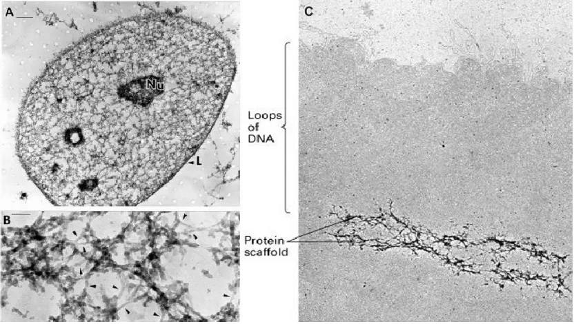
DNA within the eukaryotic nucleus is organized into higher-order looped domains ranging from 5 to 200 kb in length (Zlatanova and van Holde, 1992). The domain organization of the nucleus is thought to be mediated by a proteinacous intranuclear framework called "nuclear matrix" (Berezney and Coffey, 1974) or "nuclear scaffold" (Mirkoritch et al., 1984; Ivanchenko et al., 1992). The nuclear matrix is composed of two parts: a peripheral nuclear lamina and an internal filamentous network that transects the nuclear remnant (Luderus et al., 1992). This network is built up of branched core filaments, which provide a supporting structure for the formation of DNA loops and participate in diverse matrix-associated processes such as DNA replication, transcription, recombination, RNA processing and transport, as well as signal transduction and apoptotic events. The association of DNA with the nuclear matrix (skeleton) on the one hand serves to structurally define the borders of chromatin domains at the sites of attachment, and on the other hand participates in the regulation of transcription (Razin, 1987, 2001; Bode et al., 2000). The development of a method based on LIS (2,5-Lithium diiodosalicilate) detergent extraction of the nuclear matix allowed the isolation of nuclear matrix-associated DNA sequences (Mirkovitch et al., 1984). The scaffold-bound DNA fragments obtained by this extraction procedure were termed MARs (matrix attachment regions) or SARs (scaffold attached regions).
2. Matrix attachment (association) regions (MARs)
et al., 1992; 1996). Although prokaryotes do not contain MAR sequences (as evidenced by the fact that prokaryotic DNA, even in large excess, does not interfere with MAR/nuclear matrix reassociation in vitro), some plasmids possess two intensively studied narrow unwinding elements (UEs) that flank the ampicillin resistance gene (Benham, 1997). These sites are insufficient to mediate matrix attachment, but they serve as convenient internal standards for measuring the properties of the eukaryotic MARs. Eukaryotic MARs form extended base unpairing regions (BURs), which usually consist of multiple UEs, that compete strongly with the above-mentioned standards. The UEs typically include a major core unwinding element (CUE), which is the nucleation center for base unpairing (Bode et al., 1996).
MARs are evolutionarily conserved and are often found at the borders of chromatin domains and in close association with certain enhancers or in introns (Gasser and Laemmli, 1986; Cockerill and Garrard, 1986a,b). MARs are located either in non-transcribed regions or within transcription units, but rarely if ever in coding regions (Bode et al., 2000).
C C
Figure 1. High-resolution structure of the nuclear matrix. The nuclear matrix was visualized by resinless section electron microscopy, following high salt extraction and DNase I digestion. The nuclear matrix of an interphase nucleus consists of two parts – the nuclear lamina (L) and a network of internal nuclear fibres (Nu - nucleoli, L - lamina). (A) Overview.
The best studied matrix attachment regions are the ones found in the genes for lymphocyte receptors. Our main interest at the onset of this work was the functional characterization of the immunoglobulin µ gene MARs. Therefore, the following section will focus on the current knowledge about the properties and functions of the matrix attachment regions from the immunoglobulin and other lymphocyte receptor genes.
2.1. MARs of the lymphocyte receptor genes
2.1.1. The MAR of the Igκ gene
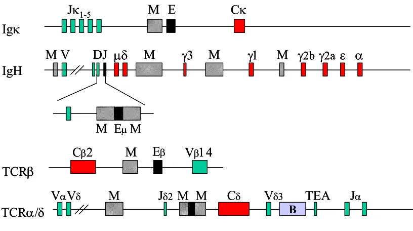
The first identified MAR was in the Igκ receptor gene locus (Fig. 2) (Cockerill and Garrad, 1986b).
Igκ
IgH
Jκ1-5
TCRβ
TCRα/δ
M E Cκ
M M M M M M M
M V DJ µδ γ3 γ1 γ2b γ2a ε α
Cβ2 Eβ Vβ14
VαVδ
Eµ
Vδ3 Jα
Jδ2
B
Cδ TEA
M M
Igκ
IgH
Jκ1-5
TCRβ
TCRα/δ
M E Cκ
M M M M M M M
M V DJ µδ γ3 γ1 γ2b γ2a ε α
Cβ2 Eβ Vβ14
VαVδ
Eµ
Vδ3 Jα
Jδ2
B
Cδ TEA
M M
Figure 2. Physical location of MARs in lymphoid receptor genes. MARs are shown as cross-hatched boxes, exons in green; B stands for boundary element, TEA for T-early α. (Based on Scheuermann and Garrad, 1999.)
Xu et al., 1989). It has also been demonstrated that the Igκ MAR is required for the demethylation of in vitro methylated Igκ constructs, following their stable integration into plasmocytoma cell lines (Kirillov et al., 1996; Lichtenstein et al., 1994). In addition, the Igκ gene MAR is involved in regulating somatic hypermutation of Igκ transgenes, since transgenes lacking the MAR showed an impaired ability to serve as a substrate for hypermutation (Goyenechea et al., 1997). However, in mice with a targeted MAR deletion no significant defects in the levels of κ gene rearrangement, B cell populations or antibody production were detected (Yi et al., 1999). More detailed studies later demonstrated that in a fraction of B cells, Vκ-Jκ joining occurred earlier during development at MAR deleted alleles. Thus, it is possible that this MAR can act to insulate the enhancer activity in pro-B but not in pre-B cells. Supporting the transgenic mice studies, in ∆MAR mice the extent of somatic hypermutation in germinal centers is significantly reduced (Yi et al., 1999).
2.1.2. The IgH gene locus MARs
The immunoglobulin heavy chain (IgH) gene locus has multiple MARs. The most extensively studied ones are the 5' and 3' MARs flanking the Igµ gene intronic enhancer (see Fig. 2). Initial experiments involving transient transfection or stable integration of reporter constructs suggested that these MARs might play a role in repressing the Igµ gene intronic enhancer in non-B cells (Scheuermann and Chen, 1989). The IgH MARs are found to activate or repress transcription of reporter genes when the appropriate MAR-binding proteins are co-expressed in transient transfection experiments (Herrscher et al., 1995; Wang et al., 1999).
expression of the same constructs in stably transfected tissue culture cells (Forrester et al., 1994). In addition, the transgene with deleted MARs is susceptible to chromosomal position effects and is expressed at low and variable levels, ranging between 0.1 and 3% relative to the level of the wild-type enhancer transgene (Forrester et al., 1994). The requirement for MAR function in transgenic animals, but not in cell lines or animals created from blastocyst fusions, suggests a function of MAR in chromatin remodeling during early development or passage through the germline (Sakai et al., 1999). In fact, methylation of the µ gene constructs in vitro, prior to stable transfection into murine B cells, abrogates the ability of the core enhancer alone to activate the Vh promoter over a distance of 1.2 kb (Forrester et al., 1999). In most of the clones the pre-methylated DNA templates were demethylated, which is in line with the findings of other groups that the presence of MARs in the immunoglobulin µ enhancer region correlates with the demethylation of the adjacent DNA (Lichtenstein et al., 1994; Kirillov et al., 1996). Furthermore, it was shown that MARs facilitate long-range chromatin accessibility (Jenuwein et al., 1997) and generate an extended domain of histone acetylation (Forrester et al., 1999). The effect of the MARs on the extended acetylation of histone H4 is independent of transcription and together with the finding that the H4 C-terminal tail interacts with the neighboring nucleosome (Luger and Richmod, 1998) implies that acetylation could result in disorganization of the higher order chromatin fibers and may account for the active transcription in wild-type µ transgenes.
2.1.3. The TCRβ gene locus MAR
The TCRβ MAR is required for reporter gene transactivation in transiently transfected T-cell lines (Chattopadhyay et al., 1998a). Deletion of the TCRβ
2.1.4. The TCR α/δ gene locus MARs
Recent studies led to the identification of three MARs in the TCR α/δ gene locus. Two of these flank the TCRδ transcriptional enhancer in a similar manner to the MARs in the IgH intronic enhancer (see Fig. 2). It was demonstrated that they are required for efficient VD to J recombination (Zhong et al., 1999).
Overall, the presence of MARs in close proximity to enhancers is a conserved feature of lymphoid receptor genes, arguing for the functional importance of these elements. MAR sequences are also found within the immunoglobulin VH
and Vκ loci at a much higher frequency than throughout the rest of the
genome (Goebel et al., 2002). It has been proposed that variations in nuclear matrix factors binding to these MARs could potentially influence the extent of localized accessibility to V(D)J recombination and thus could play a role in the unequal rearrangement of individual V genes. The MAR sites could also contribute to the effective transcription of immunoglobulin genes in mature and activated B cells, bringing both the promoter and the enhancer regions into close proximity at the nuclear matrix (Goebel et al., 2002).
3. MAR-binding proteins
Following the initial description of the nuclear matrix as a general structure a number of specific proteins were identified as matrix components. They are referred to as MAR-binding proteins, due to their ability to associate with matrix attachment regions.
Factor Expression
pattern Target MAR Putative biochemical functions Expression function
Cux/CDP Ubiquitous IgHE, Vh, TCRβ,CD8a, TCRα
Displaces MAR-BP1, Bright, binds SATB1, stimulates histone deacetylation
Transcriptional
repression, antagonizes Bright transactivation
SATB1 Thymocytes IgHE, Igκ, TCRβ, CD8a, TCRδ
Influences base
unpairing Transcriptional repression
Bright Mature B and plasma cells
IgHE, Vh Chromatin remodeling Transcriptional activation
SMAR1 Ubiquitous, abundant in thymus
TCRβ (MARβ) Unknown Transcriptional modulation
HMGI(Y) Ubiquitous IgHE Displaces histone H1 Derepression
MeCP2 Ubiquitous IgHE, chicken
lysozyme MAR Recruits histone deacetylases Transcriptional repression
CTCF Ubiquitous Unknown Forms topologically independent chromatin loops that may support gene silencing.
Transcriptional repression
MAR-BP1 Ubiquitous IgHE, Igκ Stimulates nuclear
matrix attachment Unknown
hnRNP-A1/B1 Ubiquitous IgHE DNA splicing helicase/RNA Antagonizes Cux/CDP repression AP1
(Fos/Jun)
Ubiquitous Igκ Trans-activation Transcriptional activation
Nucleolin Ubiquitous IgHE Unknown rRNA transcription and ribosome assembly
Table 1. Characteristics of selected MAR-binding proteins. (Based on Scheuermann and Garrad, 1999.)
3.1. Cux/CDP (CCAAT displacement protein)
located near the carboxy terminus. Separately, each of the CUT and the HOX domains are able to bind DNA with high AT-content (Harada et al., 1994; 1995). However, full-length Cux/CDP appears to recognize DNA in a unique way that depends more on tertiary DNA structure rather than the linear nucleotide sequence, because a consensus motif is not found.
Further experiments have shown that Cux/CDP binds in a cooperative manner to multiple sites in the two MARs flanking the IgH intronic enhancer, suggesting that multiple DNA-binding subunits are involved in the formation of a higher-order complex (Scheuermann and Chen, 1989). Transactivation studies have revealed that it is involved in the negative regulation of IgH enhancer-mediated transcriptional activation (Zong et al., 1995; Wang et al., 1999).
3.2. Bright (B cell regulator of IgH transcription)
Bright has a restricted cell-type expression, being present predominantly in mature B cells, splenic B cells and a population of small pre-B cells in bone marrow. Its expression in splenic cells can be augmented following LPS stimulation (Webb et al., 1999). Bright is retained in the nucleus following nuclear matrix preparation and co-localize with PML bodies, which also associate with the nuclear matrix (Webb et al., 1999).
The DNA binding region of the protein is represented by an ARID domain (AT-rich interaction domain) (Kortschak et al., 2000), that has a homology to the
3.3. SATB1 (special AT-rich sequence binding protein 1)
SATB1 PDZ-like CUT CUT HOX 764
Caspase 6 cleavage site
MAR binding domain
Figure 3. Domain organization of SATB1. (See the text for details.)
SATB1 is highly expressed in thymus and binds selectively to matrix-associated regions (MARs) of DNA. Specific mutations that diminish the unwinding potential of MAR sequences greatly reduce their binding affinity for SATB1 (Dickinson et al., 1992). The DNA-binding domain consists of two CUT repeats, also referred to as the MAR-binding domain (Dickinson et al., 1997), a homeodomain (HOX) and a dimerization (PDZ-like) domain (Galande et al., 2001; see Fig. 3). The isolated MAR-binding domain recognizes a certain DNA sequence context within MARs that is highly potentiated for base unpairing. Unlike the MAR-binding domain, the isolated homeodomain binds poorly and with low specificity to DNA. However, the combined action of the MAR-binding domain and the homeodomain allows SATB1 to specifically recognize the core-unwinding element within the base-unpairing region of MARs (Dickinson et al., 1997). The core-unwinding element is critical for MAR structure, since point mutations within it abolish the unwinding propensity of the MAR (Dickinson et al., 1997).
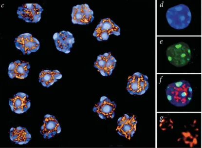
Figure 4. SATB1 forms a three-dimentional cage-like network, surrounding dense regions of chromatin. The image shows thymocyte nuclei, which were salt-extracted, digested with DNase I, and stained with DAPI (blue, d), with an antibody against the heterochromatin-associated protein M31 (green, e), and an antibody against SATB1 (red, g). f shows the merged image of d and e with the SATB1 staining (in red). c displays a 3-D reconstruction of the SATB1 network in several thymocyte nuclei. (From Cai et al, 2003.)
Cux/CDP and SATB1. The binding site was further shown to be a nuclear matrix attachment region, referred to as MARβ (See Fig. 2) (Chattopadhyay et al., 1998a). Binding sites for SATB1 are also found in the CD8B gene locus and are thought to be important for the epigenetic regulation of CD8 expression (Kieffer et al., 2002).
Transient transfection reporter assays revealed a role for SATB1 in modulating the expression of multiple genes. For example, SATB1 is a component of a large complex called γ−PE (for β-globin promoter and enhancer binding factor) that binds to five sites located 5' and 3' of the human
the NURD chromatin remodelling complex to a SATB1-binding site in the IL-2Rα locus, and mediates the specific deacetylation of histones in a large domain within the locus (Yasui et al., 2002). However, Cai et al., (2003) showed by histone-modification analyses across a gene-enriched genomic region of 70 kb that acetylation of histone 3 at Lys9 and Lys14 peaks at a SATB1-binding site and extends over a region of roughly 10 kb covering genes regulated by SATB1, and that this closely correlates with SATB1-mediated transcriptional augmentation. These controversial results could be explained by different associations of SATB1 with the histone modifying and chromatin remodeling machinery at different binding sites and will correlate with the observed opposing effects on the transcription of different genes. Thus, it was proposed that SATB1 is a new type of gene regulator with a novel nuclear architecture, providing sites for tissue-specific organization of DNA sequences and regulating region-specific histone modification (Yasui et al., 2002).
In conclusion, it should be pointed out that although a lot has been learned about the function of MARs, the precise mechanism through which they influence gene expression is still obscure and probably diverse. One possible model involves the MAR-binding protein HMGI(Y) that could depelete histone H1 and cause chromatin opening. MARs also mediate the targeting of DNA to the nuclear matrix, which is enriched in RNA polymerases and splicing complexes (Berezney et al., 1995). Another mechanism could rely on the MAR-induced changes in DNA topology, since matrix attachment regions have been shown to interact with topoisomerase II and contain sequences, which unwind easily under superhelical tension (Bode et al., 1992; Benham et al., 1997). Yet another pathway may involve MeCP2 - a ubiquitously expressed protein that interacts with both methylated CpG dinucleotides and MAR sequences. MeCP2 associates with histone deacetylases and might therefore confer repression upon MAR-containing regions of chromatin. Finally, certain MAR sequences are also recognized by tissue-specific transcriptional regulators, such as Bight and SATB1, which can modulate transcription at the sites of binding.
4. Post-translational protein modifications. SUMO modification
4.1. SUMO (small ubiquitin-related modifier)
mono-ubiquitination, unlike poly-ubiquitination, is not involved in the protein degradation pathway, but plays a role in at least three distinct cellular processes, such as histone regulation, endocytosis, and budding of retroviruses from the plasma membrane (Terrell et al., 1998; Hofmann and Pickart, 1999; Deng et al., 2000; Hicke, 2001).
During the past few years, several proteins have been discovered that have sequence similarity to ubiquitin. These ubiquitin-like proteins form two separate classes: ‘ubiquitin-like modifiers’ (UBLs) and ‘ubiquitin-domain proteins’ (UDPs). UBLs function as modifiers in a manner analogous to that of ubiquitin. This class is comprised of SUMO (small ubiquitin-related modifier), Rub1 (also called Nedd8), Apg8 and Apg12. UDPs harbour domains that are homologous in sequence to ubiquitin but are otherwise unrelated to each other. In contrast to UBLs, these proteins are not conjugated to other proteins (Jentsch et al., 2000; Hochstrasser et al., 2000). This class includes parkin, RAD23 and DSK2.
sequence is not known. SUMO2 and SUMO3, analogously to SUMO1, can also be conjugated to target proteins (Kamitani et al., 1998). Further experiments demonstrated that the target proteins for SUMO1 and SUMO2/3 are different and the conjugation of SUMO-2 and -3 can be promoted by stress-inducing stimuli, such as acute temperature shift and high osmolarity (Saitoh and Hinchey, 2000). Moreover, SUMO2/3 can form poly-SUMO chains (Tatham et al., 2001). It was shown that both 2 and -3, but not SUMO-1, contain a consensus motif, ψKXE (ψ standsfor a hydrophobic amino acid and X for any amino acid), in their N-terminal regions that is used for sequential SUMO conjugation. Point mutation of the lysine residue in the above sequence abolishes the poly-SUMO chain formation. The yeast SUMO homologue, Smt3p, also contains a similar sequence motif, and can form poly-SUMO chains (Johnson and Gupta, 2001) but until now the functional significance of the poly-SUMO chain formation is unknown.
SUMO-1 1 MSD----QEAKPSTEDLGDKKEG-EYIKLKVIGQDSS-EIHFKVKMTTHLKKLKESYCQRQGV 57 SUMO-2 1 MAD----E--KP---KEGVKTENNDHINLKVAGQDGS-VVQFKIKRHTPLSKLMKAYCERQGL 53 SUMO-3 1 MSE----E--KP---KEGVKTEN-DHINLKVAGQDGS-VVQFKIKRHTPLSKLMKAYCERQGL 52 SMT3 1 MSDSEVNQEAKPEV-KPEVKPET--HINLKVS--DGSSEIFFKIKKTTPLRRLMEAFAKRQGK 58 Ubiquitin 1 MQIFVKTL--TGK-TITLEVEPSDTIENVKAKIQDKEGI 36
SUMO-1 58 PMNSLRFLFEGQRIADNHTPKELGMEEEDVIEVYQEQTGGHSTV 101 SUMO-2 54 SMRQIRFRFDGQPINETDTPAQLEMEDEDTIDVFQQQTGGVY 95 SUMO-3 53 SMRQIRFRFDGQPINETDTPAQLEMEDEDTIDVFQQQTGGVPESSLAGHSFL 104 SMT3 59 EMDSLRFLYDGIRIQADQTPEDLDMEDNDIIEAHREQIGGATY 101 Ubiquitin 37 PPDQQRLIFAGKQLEDGRTLSDYNIQKESTLHLVLRLRGG 76
Figure 5. Sequence alignment of SUMO family members and ubiquitin. Sequence comparison of the human SUMO family members (SUMO-1, -2, and -3) to the yeast SUMO homologue SMT3 and to human ubiquitin. Identities are shown in blue, similarities in yellow. The scissors symbol indicates the sites of precursor processing, which occurs in the carboxy-terminus after the double-glycine motif. (Based on Müller et al., 2001.)
4.2. The SUMO conjugation pathway
Figure 6. The SUMO conjugation pathway. The SUMO precursor is subjected to proteolysis to expose the C-terminal Gly residue to the E1 activating enzyme (a dimer of SAE1/SAE2 (Aos1/Uba2 in yeast)), which forms a highly reactive thioester bond with SUMO. SUMO is subsequently transferred to the E2 conjugating enzyme, Ubc9, and with the help of the substrate specific E3 ligase is conjugated to target proteins at ψKXE consensus motifs. The resulting isopeptide bond is stable and SUMO-deconjugating enzymes are required for hydrolysis. (From Verger et al., 2002.)
of a highly reactive thioester bond between the E1 enzyme and SUMO. In contrast to the ubiquitin-activating enzyme, the SUMO E1 is a heterodimer consisting of two subunits: Aos1 and Uba2. Interestingly, Aos1 shows sequence and structural similarity to the amino-terminal part of Uba1 - the E1 enzyme for ubiquitin, whereas Uba2 is related to the carboxy-terminal region of Uba1. The Uba2 subunit harbours the ‘active site’ cysteine residue required for the formation of SUMO–E1 enzyme thioesters; nevertheless both subunits are required for SUMO activation in vitro and in vivo.
In vitro experiments demonstrated that the E1 activating and E2 conjugating enzymes (together with ATP and SUMO) are sufficient for a relatively robust modification of proteins at precisely the lysines that are preferred in vivo. In contrast, in the ubiquitin pathway an additional factor, called E3 ligase or ubiquitin-protein ligase, is almost always necessary for efficient substrate ubiquitination, both in vitro and in vivo (Hershko and Ciechanover, 1998).
4.3. SUMO protein ligases
The SUMO E3 ligases can be separated in two classes. The first class comprises Siz1, Siz2 and the PIAS family. One common feature in this class is the presence of a RING-like domain that is known to mediate interaction of E3 with E2 in the ubiquitin system (Freemont, 2000). Mutations in the RING-like domain of Siz1 and PIAS1 result in the loss of binding of the E3-RING-like enzyme to Ubc9 (Takahashi et al., 2001a,b; Kahyo et al., 2001). Thus, the RING-like domain of the E3-like enzymes might function in a similar manner to the RING domain of E3 ubiquitin ligases. The second class consists of RanBP2 and Pc2, which have no significant homology to the RING-like ligases. RanBP2 (Ran binding protein 2) contains a RanGAP-binding domain, and repeats responsible for the nuclear transport receptor binding together with a cyclophilin homology region, but has no similarity to the other E3 SUMO ligases (Pichler et al., 2002). Pc2 is a member of the Polycomb group (PcG) proteins, which were first described in Drosophila as factors responsible for maintaining the transcriptionally repressed state of Hox/homeotic genes in a stable and heritable manner throughout development (Otte et al., 2003). The PcG complex forms unique nuclear structures, termed PcG bodies (Saurin et al., 1998). These domains vary in number and structure and it is currently unclear whether they are storage compartments or are directly involved in silencing (Spector, 2001).
4.4.SUMO-specific proteases (SUSPs)
SUMO-protein conjugates are highly susceptible to deconjugation in cell extracts and different subcellular fractions, indicating the presence of several isopeptidases.
In yeast, two SUMO-specific proteases - Ulp1 and Ulp2 were identified (Li and Hochstrasser, 1999, 2000; Schwienhorst et al., 2000). In vitro, both Ulp1 and Ulp2 can catalyze the C-terminal processing of the SUMO precursor and both enzymes can remove SUMO from isopeptide-linked conjugates. Genetic studies have shown that like SUMO conjugation, SUMO de-conjugation is needed for viability in the budding yeast (Li and Hochstrasser, 1999, 2000; Schwienhorst et al., 2000). It was further demonstrated that Ulp1, but not Ulp2, is essential for viability. Analysis of an Ulp1 temperature sensitive mutant showed an arrest of the cells at the G2/M boundary when shifted to the restrictive temperature, suggesting that a SUMO-conjugated protein needs to be deconjugated in order that the cell could progress into mitosis. In addition, it is suggested that Ulp1 is involved in the processing of the SUMO precursor molecule, because the lethality of Ulp1 deletion can be partially overcome by expressing mature SUMO. Disruption of Ulp2, although not absolutely lethal, leads to several abnormalities including slow and thermosensitive growth, defects in cell morphology, chromosome instability and sporulation defects. Both Ulp1 and Ulp2 mutant strains revealed an increased accumulation of SUMO conjugates. Interestingly, the overall pattern of the SUMO conjugated proteins differs in each of the individual mutants, suggesting that the two enzymes act on distinct substrates. Surprisingly, however, inactivation of Ulp2 partially rescues the defects caused by Ulp1 deficiency, and the double mutant accumulates fewer SUMO conjugates than either of the single mutants. One suggested explanation is that Ulp1 and Ulp2 control the modification levels of proteins in opposing pathways (Li and Hochstrasser, 2000).
The sequence similarity between Ulp1, Ulp2, SUSP1 and SENP1 is restricted to a 200 amino acid sequence, termed ULP domain, which harbours the catalytically active center. Remarkably, no sequence or structural similarity exists between the SUSPs and the de-ubiquitinating enzymes, although both belong to the cysteine protease superfamily. Moreover, these proteins show similarity to the adenovirus L3 protease, suggesting that SUSPs may use a catalytic mechanism similar to that of viral proteases (Li and Hochstrasser, 1999; Andres et al., 2001).
4.5. Biological functions of SUMO modification
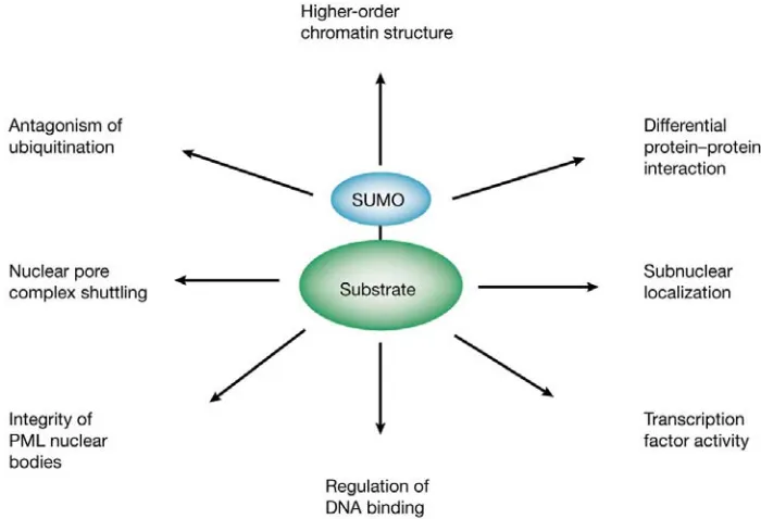
Figure 7. Functional significance of SUMO modification. (From Verger et al., 2002.)
4.5.1. Nuclear pore complex shuttling
Blobel, 1993). Sumoylation, almost exclusively with SUMO1, is required for the stable association of RanGAP1 and RanBP2. Since SUMO-1 itself cannot bind to RanBP2 (Mahajan et al., 1997), sumoylation most likely induces a conformational change to create a binding surface for RanBP2 in the C-terminal domain of RanGAP1. It was further demonstrated that RanBP2 is the E3 ligase for RanGAP1 (Pichler et al., 2002).
Indirect evidence for the role of SUMO in nuclear import processes comes from studies of Drosophila melanogaster harbouring a loss-of-function mutation in the UBC9 gene (semushi) (Epps et al., 1998). In these mutants, nuclear import of the transcription factor BICOID is prevented, leading to defects in embryogenesis. However, it remains unclear whether sumoylation is indeed involved in the control of the nuclear protein import pathway.
4.5.2. Changes in subnuclear localization and targeting to PML bodies
suggested (Lallemand-Breitenbach et al., 2001). It was shown that sumoylation at a specific residue triggers the proteasome-dependent degradation of PML and that the mature nuclear bodies harbor 11S proteasome components, suggesting that these structures might be the site of intranuclear proteolysis.
4.5.3. Modulation of protein-protein interactions
Sp100 is an interferon-inducible protein, which was initially characterized as an antigen reactive with antibodies from patients with autoimmune disorders (Szostecki et al., 1990). SUMO-conjugation of Sp100 promotes its interaction with members of the HP1 (heterochromatin protein 1) family of non-histone chromosomal proteins, suggesting its regulatory role in chromatin organization and in the functional interplay between the nuclear bodies and chromatin (Lehming et al., 1998; Sternsdorf et al., 1999; Seeler et al., 1998, 2001).
4.5.4. Regulation of DNA binding
HSF1 and HSF2 (heat shock factors 1 and 2) are transcription factors that mediate the induction of heat shock protein gene expression under environmental stress conditions (Cotto and Morimoto, 1999). Electromobility shift assays demonstrated that SUMO modification enhances the DNA-binding ability of both proteins. Furthermore, mutation of the SUMO target lysine on HSF1 results in a significant decrease in stress-induced transcriptional activity of the protein in vivo (Goodson et al., 2001; Hong et al., 2001).
4.5.5. Modulation of the activity of transcription factors
TEL to nuclear speckles in a cell cycle-specific manner (Chakrabarti et al., 2000). The transcriptional repressor Daxx can be recruited to nuclear bodies upon sumoylation of PML, thereby relieving the Daxx-mediated transcriptional repression of its target genes (Ishov et al., 1999; Lehembre et al., 2001). Sumoylation of PML is also responsible for the recruitment of p53 to the nuclear bodies and promotes the transcriptional and pro-apoptotic activities of p53 (Fogal et al., 2000). In addition, recruitment of p53 to the nuclear bodies has been suggested to trigger post-translational modifications (e.g. acetylation), which also stimulate the transcriptional activity of p53 (Giaccia and Kastan, 1998; Pearson et al., 2000). Later experiments revealed that p53 is also a target for SUMO-1 modification and that the SUMO conjugation is induced by UV irradiation (Gostissa et al., 1999; Rodriguez et al., 1999; Müller et al., 2000). The target lysine residue for sumoylation, however, is different from the one conjugated to ubiquitin. Therefore SUMO conjugation at Lys-386 does not affect the stability of p53. Furthermore, the apoptotic potential of a mutant form of p53, in which Lys-386 is replaced by arginine (K386R), is moderately impaired, implying that sumoylation of p53 is necessary for exerting its full apoptotic activity (Müller et al., 2000). In addition, SUMO modification is a common mechanism for regulating the transcriptional activity of the steroid receptor superfamily. Point mutations of lysine residues that are targets for SUMO conjugation in the androgen receptor enhance its transcriptional activity, suggesting that sumoylation negatively regulates the activity (Tian et al., 2002). SUMO modification was shown to be a common mechanism for repressing a variety of transcription activators (e.g. LEF1, Sp3 and CtBP) by altering their subnuclear localization and recruiting them to nuclear matrix-associated PML bodies (Sachdev et al., 2001; Ross et al., 2002; Kagey et al., 2003).
4.5.6. Antagonism of ubiquitination
and Henkel, 1994; Siebenlist et al., 1994; Beg and Baldwin, 1993; Verma et al., 1995). Upon stimulation with proinflammatory cytokines, phorbol esters, oxidants, or viral infection, IκBα is rapidly phosphorylated by a signal-inducible IκB kinase (IKK) complex (Mercurio et al., 1997; Woronicz et al., 1997; Zandi et al., 1997). Once IκBα is phosphorylated, it is recognized by the ubiquitin-conjugating machinery, which leads to the formation of poly-ubiquitin chains that target the protein for rapid degradation by the 26S proteasome (Alkalay et al., 1995; DiDonato et al., 1996). This results in the liberation of NFκB, which then translocates from the cytosol to the nucleus to activate the transcription of target genes. Interestingly, SUMO can be conjugated to the same lysine residue as ubiquitin, thus leading to stabilization of IκBα against degradation by the 26S proteasome (Desterro et al., 1998). Sumoylated forms of IκBα have been detected in a number of cell types, and are resistant to TNFα-induced degradation. Another example of the antagonistic role of sumoylation against ubiquitination is Mdm2 (Buschmann et al., 2001). Mdm2 is an E3 ligase catalyzing the ubiquitination of p53, as well as of itself (Honda et al., 1997). In normal cells, most of the Mdm2 proteins are sumoylated at Lys-446, which is the same site as the one for self-ubiquitination (Buschmann et al., 2001). SUMO-conjugation protects Mdm2 from destabilization, but increases p53 ubiquitination and degradation. It is suggested that upon DNA damage Mdm2 is desumoylated and subsequently conjugated to ubiquitin, which targets the protein for degradation. This results in a dramatic increase of the activity of p53, leading to cell cycle arrest or apoptosis (Oren, 1999).
II. Results
1. Synthetic multimerized MAR-binding sites mimic the natural MARs in augmenting the transcription of the IgH gene
Previous experiments indicated that the nuclear matrix-binding regions (MARs) that flank the intragenic µ enhancer augment markedly the expression of a rearranged µ gene in pre-B cells derived from transgenic mice (Forrester et al., 1994). Furthermore, stable cell lines established with µ gene constructs, premethylated at all CpG dinucleotides, revealed that the MARs are responsible for the generation of an extended domain of histone acetylation, which could account for the long-range function of the µ enhancer in combination with MARs. Since the 5' and 3' MAR regions are fairly long (approximately 500 bp for the 5' MAR and 300 bp for the 3' MAR regions) and contain consensus sites for binding of general transcription factors, we decided to examine whether shorter, synthetic multimerized MAR consensus sites could mimic the natural MARs in augmenting IgH transcription. To answer that question, a construct - pµ∆MAR-SBS7, in which the core
immunoglobulin enhancer is surrounded by 7 copies of the MAR consensus sequences (Dickinson et al., 1992) was generated (Fig. 8A). pµ, pµ∆MAR or pµ∆MAR-SBS7 plasmid DNAs were subjected to in vitro CpG methylation with SssI DNA methylase (Fig. 8B) and were transfected in J558L cells. Twelve stable cell lines for each of the transfections were established and DNA and RNA were isolated in parallel (four representative clones are shown). The stable integration of the constructs was analyzed by Southern blotting with a probe specific for the rearranged VDJ region of the µ gene construct (Grosschedl et al., 1984; Fig. 8D). The transcriptional activity was monitored by Northern blot analysis. As shown previously, the transcriptional activity of the pre-methylated pµ∆MAR construct was dramatically decreased (Fig. 8C,
pµ
H H S
VDJ Cµ
E
Vh probe
H H S
VDJ Cµ
E N N
Enhancer probe pµ∆MAR
pµ∆MAR-SBS7
H H S
VDJ Cµ
E
Vh probe
A
pµ X XH H S
VDJ Cµ
E
Vh probe
H H S
VDJ Cµ
E N N
Enhancer probe pµ∆MAR
pµ∆MAR-SBS7
H H S
VDJ Cµ
E
Vh probe
A
X XC
D
Cell lines:
RNA blot DNA blot
pµ
1 2 3 4
pµ
1 2 3 4 pµ∆MAR
1 2 3 4
pµ∆MAR 1 2 3 4 pµ∆MAR-SBS7
pµ∆MAR-SBS7
1 2 3 4 1 2 3 4
B
HpaII Msp I HpaI I Msp I HpaI I Msp I pµ∆MAR pµ
pµ∆ MAR-SBS7
C
D
Cell lines:
RNA blot DNA blot
pµ
1 2 3 4
pµ
1 2 3 4 pµ∆MAR
1 2 3 4
pµ∆MAR 1 2 3 4 pµ∆MAR-SBS7
pµ∆MAR-SBS7
1 2 3 4 1 2 3 4 pµ∆MAR-SBS7
pµ∆MAR-SBS7
1 2 3 4 1 2 3 4
B
HpaI I Msp I HpaI I Msp I HpaI I Msp I pµ∆MAR pµpµ∆ MAR-SBS7
Figure 8.Synthetic multimerized MAR sites mimic the natural MARs in augmenting the transcription of the IgH gene. (A) Schematic diagram of the rearranged pµ wild-type, pµ∆MAR and pµ∆MAR-SBS7 constructs. The intragenic locus control region (LCR), contains the µ enhancer (black bar), flanked by matrix attachment regions (MARs - hatched bars). The exons are shown as gray boxes, and the transcription start site of the Vh promoter is indicated by an arrow. Relevant restriction sites: (S) Sac I; (E) EcoRV; (H) HpaII/Msp; (N) NotI sites, used to introduce the artificial enhancer in the vector pµ∆2, lacking the 1 kb XbaI fragment of the intragenic LCR; (X) XbaI sites, flanking the intragenic LCR. (B) Analysis of the methylation pattern of the SssI pre-methylated µ genes. The pre-methylated DNA was digested to completion with either Msp (M) or HpaII (H) and run on a 1% agarose gel. (C)
binding protein might be involved in the regulation of IgH transcription in B cells.
2.A homologue of the MAR-binding protein SATB1 is expressed in pre-B cells
Our data revealed that multimerized MAR consensus sites mimic the function of the natural MARs and suggested that MAR-binding proteins might be involved in the regulation of immunoglobulin transcription. Although two previously cloned MAR-binding proteins, SATB1 and Bright, were shown to interact with the same set of five binding sites in the µ MARs in vitro
(Dickinson et al., 1992; Herrscher et al., 1995), neither of these proteins are expressed in early B cells, in which the enhancer and MARs collaborate to augment Vh promoter activity. In particular, SATB1 is predominantly expressed in T lymphocytes, whereas Bright is expressed in activated and terminally differentiated B cells. Therefore, we began to search for additional MAR-binding proteins that are expressed in pre-B cells.
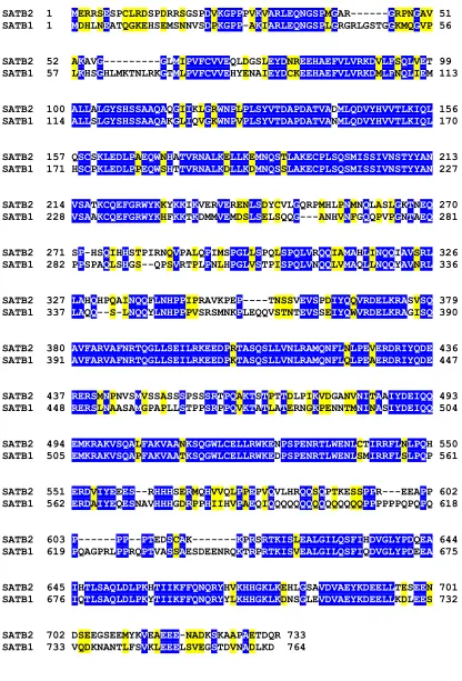
To identify MAR-binding proteins that are expressed predominantly in early stages of the B cell lineage, we searched the murine and human databases for sequences that show homologies to known MAR-binding proteins and examined their expression in pre-B cells. We identified two clones that displayed 71% and 70% overall sequence homology with SATB1 and Bright, respectively (Fig. 9 and data not shown). Analysis of the expression patterns of these clones revealed a ubiquitous expression for the Bright homologue and a predominantly pre-B cell specific pattern for the SATB1 homologue, which we termed SATB2 (Fig. 11 and data not shown).
2.1. SATB2 has a high homology and a similar domain organization to SATB1
SATB2 1 MERRSESPCLRDSPDRRSGSPDVKGPPPVKVARLEQNGSPMGAR---GRPNGAV 51 SATB1 1 MDHLNEATQGKEHSEMSNNVSDPKGPP-AKIARLEQNGSPLGRGRLGSTGGKMQGVP 56
SATB2 52 AKAVG---GLMIPVFCVVEQLDGSLEYDNREEHAEFVLVRKDVLFSQLVET 99 SATB1 57 LKHSGHLMKTNLRKGTMLPVFCVVEHYENAIEYDCKEEHAEFVLVRKDMLFNQLIEM 113
SATB2 100 ALLALGYSHSSAAQAQGIIKLGRWNPLPLSYVTDAPDATVADMLQDVYHVVTLKIQL 156 SATB1 114 ALLSLGYSHSSAAQAKGLIQVGKWNPVPLSYVTDAPDATVANMLQDVYHVVTLKIQL 170
SATB2 157 QSCSKLEDLPAEQWNHATVRNALKELLKEMNQSTLAKECPLSQSMISSIVNSTYYAN 213 SATB1 171 HSCPKLEDLPPEQWSHTTVRNALKDLLKDMNQSSLAKECPLSQSMISSIVNSTYYAN 227
SATB2 214 VSATKCQEFGRWYKKYKKIKVERVERENLSDYCVLGQRPMHLPNMNQLASLGKTNEQ 270 SATB1 228 VSAAKCQEFGRWYKHFKKTKDMMVEMDSLSELSQQG---ANHVNFGQQPVPGNTAEQ 281
SATB2 271 SP-HSQIHHSTPIRNQVPALQPIMSPGLLSPQLSPQLVRQQIAMAHLINQQIAVSRL 326 SATB1 282 PPSPAQLSHGS--QPSVRTPLPNLHPGLVSTPISPQLVNQQLVMAQLLNQQYAVNRL 336
SATB2 327 LAHQHPQAINQQFLNHPPIPRAVKPEP----TNSSVEVSPDIYQQVRDELKRASVSQ 379 SATB1 337 LAQQ--S-LNQQYLNHPPPVSRSMNKPLEQQVSTNTEVSSEIYQWVRDELKRAGISQ 390
SATB2 380 AVFARVAFNRTQGLLSEILRKEEDPRTASQSLLVNLRAMQNFLNLPEVERDRIYQDE 436 SATB1 391 AVFARVAFNRTQGLLSEILRKEEDPKTASQSLLVNLRAMQNFLQLPEAERDRIYQDE 447
SATB2 437 RERSMNPNVSMVSSASSSPSSSRTPQAKTSTPTTDLPIKVDGANVNITAAIYDEIQQ 493 SATB1 448 RERSLNAASAMGPAPLLSTPPSRPPQVKTATLATERNGKPENNTMNINASIYDEIQQ 504
SATB2 494 EMKRAKVSQALFAKVAANKSQGWLCELLRWKENPSPENRTLWENLCTIRRFLNLPQH 550 SATB1 505 EMKRAKVSQAPFAKVAATKSQGWLCELLRWKEDPSPENRTLWENLSMIRRFLSLPQP 561
SATB2 551 ERDVIYEEES--RHHHSERMQHVVQLPPEPVQVLHRQQSQPTKESSPPR---EEAPP 602 SATB1 562 ERDAIYEQESNAVHHHGDRPPHIIHVPAEQIQQQQQQQQQQQQQQQPPPPPPQPQPQ 618
SATB2 603 P---PP--PTEDSCAK---KPRSRTKISLEALGILQSFIHDVGLYPDQEA 644 SATB1 619 PQAGPRLPPRQPTVASSAESDEENRQKTRPRTKISVEALGILQSFIQDVGLYPDEEA 675
SATB2 645 IHTLSAQLDLPKHTIIKFFQNQRYHVKHHGKLKEHLGSAVDVAEYKDEELLTESEEN 701 SATB1 676 IQTLSAQLDLPKYTIIKFFQNQRYYLKHHGKLKDNSGLEVDVAEYKDEELLKDLEES 732
SATB2 702 DSEEGSEEMYKVEAEEE-NADKSKAAPAETDQR 733 SATB1 733 VQDKNANTLFSVKLEEELSVEGSTDVNADLKD 764
The alignment of murine SATB1 and SATB2 shows that the two proteins are 59% identical and 71% similar (Fig. 9). Sequence analysis of SATB2 revealed the presence of two CUT domains and a Hox domain, which are highly conserved with the corresponding DNA-binding domains of SATB1 (Fig. 10; Nakagomi et al.,1994; Dickinson et al., 1997). A unique feature of the protein is a 23 amino acid stretch in the N-terminus of the protein, which is not homologous to any sequence in SATB1, suggesting that this portion may confer some specific properties on the molecule.
SATB2 CUT CUT HOX
86% 82% 85%
733
SATB1 PDZ-like CUT CUT HOX 764
PDZ-like
Figure 10. Domain organization of SATB2. Schematic linear representation of the domain organization of SATB1 and SATB2 (drawn to scale). The two CUT repeats (black boxes) represent the MAR-binding domain; HOX (yellow boxes) - homeodomain; PDZ-like dimerization domain (red boxes). The numbers indicate the percentage of identity between the respective domains.
2.2. Expression pattern of SATB2
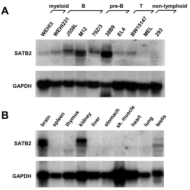
To examine the tissue and cellular distribution of SATB2, RNA from different mouse organs and cell lines was isolated and a Northern blot was performed, using the full-length cDNA as a probe. The membrane was subsequently re-hybridized with a probe recognizing the transcript of the house-keeping gene glyceraldehyde-3-phosphate dehydrogenase (GAPDH), as a control for loading and transfer.
although differently sized transcripts were detected at a lower abundance in thymus and testis (Fig. 11B).
A
sple en thym us brai n liver kidn ey hear t sk. muscl e
stom ach
lung test is SATB2 GAPDH WEH I231 BW 1514 7 EL4 38B 9 J558 L
M12 70Z/ 3
ME L
B pre-B T
WEH I3 293
B
SATB2 GAPDH non-lymphoid myeloidFigure 11. SATB2 has a cell type and tissue-specific distribution. Full-length SATB2 coding sequence was used in Northern blot to probe RNA isolated from various cell lines (A)
and mouse organs (B). Abundant expression of ~6.3 and ~5.4 kb transcripts that hybridize with a 32P-labeled SATB2 DNA probe can be detected in B cells, brain and kidney (A, B; top panels). The quality and quantity of RNA was confirmed by hybridization with a GAPDH probe (A, B; bottom panels).
2.3. SATB2 is a component of the nuclear matrix
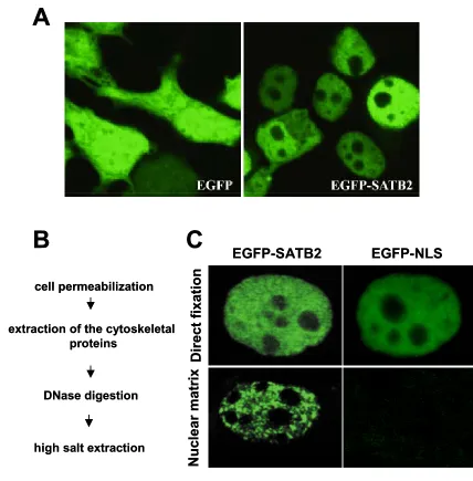
transfection, EGFP-SATB2 and all other SATB2 constructs (FLAG-SATB2 and SATB2-myc, tested by indirect immunofluorescence) localized to the nucleus (Fig. 12A).
Previous studies have shown that SATB1 is a component of the nuclear matrix (de Belle et al., 1998; Cai et al., 1999). To examine whether SATB2 can also associate with the nuclear matrix, we transfected 293T cells with plasmids encoding either an EGFP-SATB2 fusion protein or an EGFP-NLS control protein. 36 h. post transfection the cells were directly fixed or a nuclear matrix preparation was performed. In brief, nuclear matrices were prepared by removing the membrane and soluble proteins with detergent treatment, followed by DNaseI digestion and high salt extraction of the chromatin fraction (Fig. 12B). As shown in Fig. 12C, when cells were directly fixed, both EGFP-SATB2 and EGFP-NLS were localized in the nucleus; however, following high salt extraction SATB2 remained associated with the nuclear matrix, in contrast to EGFP-NLS, which was no longer found in the nucleus.
2.4. SATB2 binds to the core-unwinding element of the IgH enhancer MARs in vitro
A
B
EGFP-SATB2 EGFP-NLSDir
ect fixation
Nuclear matrix
cell permeabilization
extraction of the cytoskeletal proteins
DNase digestion
high salt extraction
C
A
B
EGFP-SATB2 EGFP-NLSDir
ect fixation
Nuclear matrix
cell permeabilization
extraction of the cytoskeletal proteins
DNase digestion
high salt extraction
C
To this end, we examined the DNA binding ability of SATB2 by electrophoretic mobility shift assays with purified recombinant SATB2 protein. Efficient binding was detected with the wild-type MAR consensus sequence (Dickinson et al., 1992), but not with a mutant oligonucleotide (Fig. 13; lane 2 and 12). We confirmed the specificity of DNA binding by the addition of excess unlabelled competitor DNA, which impaired DNA binding in a dose-dependent manner (Fig.13; lane 3-6). In contrast, the binding could not be competed with increasing amounts of nonspecific competitor (lane 7-10). Thus, SATB2 resembles SATB1 in its ability to bind the wt MAR consensus sequence, derived from the core unwinding element of the IgH enhancer MARs.
mut wt SATB2
Competitor
MAR-wt MAR-mut
Probe
SATB2-DNA
Free probe
1 2 3 4 5 6 7 8 9 10 11 12
2.5. SATB2 binds to the MARs of the endogenous µ locus
Our in vitro data demonstrated that SATB2 binds to the core-unwinding element of the IgH enhancer MARs. This raised the question whether the same is also true in vivo. To address this issue, we performed chromatin immunoprecipitations (ChIP). Due to the lack of antibodies that immunoprecipitate SATB2, we generated a pre-B cell line that had been stably transfected with a SATB2-TAPtag gene construct. After a two-step affinity purification (Rigaut et al., 1999) of SATB2-TAPtag protein that had been cross-linked to DNA in vivo, the immunoprecipitated DNA was amplified in serial dilutions by polymerase chain reactions with primers specific for the 5' MAR region of the intronic µ enhancer (Fig. 14, lanes 1-6), or with primers specific for β-globin gene sequences as a control (Fig. 14, lanes 7-12). Significant enrichment (approximately 100-fold) of µ 5' MAR sequences was detected with immunoprecipitated DNA from the SATB2-TAPtag-expressing cell line (lanes 1-6, top panels), but not with the immunoprecipitated DNA from the parental pre-B cell line (lanes 1-6, bottom panels). No enrichment of β -globin sequences was detected (lanes 7-12, top panels), indicating that SATB2 is specifically bound to the MAR flanking the intragenic enhancer of the endogenous µ gene.
2.6. SATB2 is a B cell-specific transcriptional activator
2.6.1. SATB2 mediates transcriptional activation under multimerized MAR consensus sites
38B9:SATB2-TAPtag
38B9
input DNA
ChIP input DNA
ChIP
µ5´MAR β-globin
µ 5´MAR
1 2 3 4 5 6
β-globin
7 8 9 10 11 12 38B9:SATB2-TAPtag
38B9
input DNA
ChIP input DNA
ChIP
µ5´MAR β-globin
µ 5´MAR
1 2 3 4 5 6
β-globin
7 8 9 10 11 12
Figure 14. SATB2 binds to the MAR of the endogenous µ locus. Chromatin immunoprecipitations (ChIP) of extracts from stably transfected 38B9 cells, carrying a SATB2-TAPtag expression plasmid, and from control 38B9 cells were performed by affinity-purifying chromatin fragments that have been cross-linked to SATB2-TAPtag. The immunoprecipitated DNA was subsequently analyzed by semi-quantitative PCR amplification with primers located in the 5' MAR region of the immunoglobulin µ enhancer (lanes 1-6) or in the β-globin locus (lanes 7-12). The levels of enrichment in the immunoprecipitations were estimated by comparison with the amplification products of DNA isolated from the bulk chromatin extracts (input DNA). Template DNA was used in a linear dilution (3, 1, 0.3, 0.1, 0.03, 0.01 ng) to allow for a semi-quantitative determination in the PCR assays.
To examine the functional activity of SATB2, we transfected a fos-luciferase reporter construct containing multimerized wild-type or mutated SATB2-binding sites, together with a β-galactosidase control plasmid and increasing amounts of a SATB2 expression plasmid into J558L plasmacytoma cells (Fig. 15A). 48 h. post transfection the cells were collected and luciferase and β -galactosidase assay were performed. The results were presented as a relative luciferase number, normalized to the β-galactosidase values.
A
µg transfected SATB2
pfosluc-wtSBS7 pfosluc-mutSBS8 luciferase fos wtSBS7 mutSBS8
µg transfected SATB2 0
2 4 6 8
0 3 10 30
293T fold ac tiv ation 0 20 40 60 80 100 120 140
0 3 10 30
J588L fold ac tiv ation
B
A
µg transfected SATB2
pfosluc-wtSBS7 pfosluc-mutSBS8 luciferase fos wtSBS7 mutSBS8
µg transfected SATB2 0
2 4 6 8
0 3 10 30
293T fold ac tiv ation 0 20 40 60 80 100 120 140
0 3 10 30
J588L
fold ac
tiv
ation
B
Figure 15. SATB2 mediates transcriptional activation under the multimerized MAR consensus sites.(A) Schematic linear representation of the reporter constructs used for the experiments in (B). Seven copies of the wt MAR consensus sequence were multimerized and cloned in front of a minimal fos promoter, driving the expression of a luciferase gene. (B)
mutated MAR sequences, which correlates with the results from EMSA experiments indicating that SATB2 could also bind, albeit with significantly lower efficiency, to the mutant MAR sequences, when they are multimerized (data not shown).
2.7.2. SATB2 mediates transcriptional activation under the natural MARs of the immunoglobulin enhancer
Next we wanted to examine whether SATB2 could also mediate transcriptional activation via the natural MARs of the immunoglobulin enhancer. Co-transfection experiments using luciferase reporter constructs, carrying either the µ enhancer or the core enhancer, lacking the MAR regions (pfosluc-µ∆MAR) in front of the fos promoter (Fig. 16A) and increasing amounts of SATB2 in Jurkat and J558L cells were performed. Transfection of increasing amounts of SATB2 in Jurkat cells revealed a slight activation of the transcription of the luciferase reporter construct (Fig. 16B). In contrast, when the experiment was performed in J558L plasma cells a huge transcriptional activation of around 450 fold was observed (Fig. 16C), implying that a B cell-specific co-activator may be involved in SATB2-mediated gene activation.
2.7.3. SATB2 augments immunoglobulin transcription
Following the demonstration that SATB2 can activate transcription we tested in co-transfection experiments whether SATB2 could augment the transcription of a rearranged µ gene, where the enhancer is more than 1 kb away from the Vh promoter. J558L cells were transfected with the rearranged