Scholarship@Western
Scholarship@Western
Electronic Thesis and Dissertation Repository
12-18-2014 12:00 AM
Measures of Acoustic Reflexes in Typically Developing Children
Measures of Acoustic Reflexes in Typically Developing Children
and Children with Suspected Auditory Processing Disorder
and Children with Suspected Auditory Processing Disorder
Udit Saxena
The University of Western Ontario Supervisor
Dr Prudence Allen
The University of Western Ontario
Graduate Program in Health and Rehabilitation Sciences
A thesis submitted in partial fulfillment of the requirements for the degree in Doctor of Philosophy
© Udit Saxena 2014
Follow this and additional works at: https://ir.lib.uwo.ca/etd
Part of the Communication Sciences and Disorders Commons
Recommended Citation Recommended Citation
Saxena, Udit, "Measures of Acoustic Reflexes in Typically Developing Children and Children with Suspected Auditory Processing Disorder" (2014). Electronic Thesis and Dissertation Repository. 2609.
https://ir.lib.uwo.ca/etd/2609
This Dissertation/Thesis is brought to you for free and open access by Scholarship@Western. It has been accepted for inclusion in Electronic Thesis and Dissertation Repository by an authorized administrator of
Disorder
(Thesis format: Integrated Article)
by
Udit Saxena
Graduate Program in Health & Rehabilitation Sciences
A thesis submitted in partial fulfillment
of the requirements for the degree of
Doctor of Philosophy
The School of Graduate and Postdoctoral Studies
The University of Western Ontario
London, Ontario, Canada
ii
Abstract
A series of studies were conducted to examine the acoustic reflex in normal hearing
adults, typically developing children and children with suspected auditory processing
disorder (APD). Elevated acoustic reflex thresholds (ART) and shallower acoustic
reflex growth functions (ARGF) were found in children with suspected APD in
comparison to typically developing children and normal hearing adults. These effects
were strongest in the crossed condition. There were no group differences for
acoustic reflex latency (ARL) or acoustic reflex decay (ARD).
In all studies the children with suspected APD were divided into two groups
based on the diagnosis made on the basis of a behavioral APD battery; (1) APD
which included children who received APD diagnosis and (2) Clinical non-APD who
did not receive APD diagnosis. Children in the clinical non-APD and APD groups
had similar ART and ARGF abnormalities highlighting a potential weakness in
relying strictly on behavioral tests in the assessment of children suspected of APD.
The effect of acoustic reflex activation on middle ear absorbance (MEA) and
middle ear resonant frequency (MERF) was also investigated. It was found that the
activation of the acoustic reflex resulted in a decrease of MEA between 226 and
1000 Hz, an increase MEA between 1000 and 2000 Hz and shift of MERF to a
higher frequency. These changes in middle ear function may be critical to speech in
noise perception. The effect of reflex activation was diminished in children with
iii
Across studies, acoustic reflex measures including ART, ARGF and the effect
of the reflex on MEA and MERF showed a trend suggesting age-related changes but
the trends did not reach statistical significance. However, a significant
developmental trend in ARTs was found when corrected for ear canal volume
differences. These results suggest that acoustic reflex measures in clinical children
should be compared with those of typically developing children rather adults.
Keywords
Acoustic reflex, acoustic reflex threshold, acoustic reflex growth function, acoustic
reflex latency, acoustic reflex decay, auditory processing disorder, middle ear
iv
Lists of Abbreviations
A
AAA American Academy of Audiology
ANSI American National Standards Institute
APD Auditory Processing Disorder
ARD Acoustic Reflex Decay
ARGF Acoustic Reflex Growth Function
ARL Acoustic Reflex Latency
ART Acoustic Reflex Threshold
ASHA American Speech-Language and Hearing Association
C
cc Cubic centimetre
CN Cochlear Nucleus
D
dB Decibel
D-ART Difference between crossed and uncrossed ART
E
ECV Ear Canal Volume
H
HL Hearing Level
M
MEA Middle Ear Absorbance
MERF Middle Ear Resonant Frequency
ml Millilitre
msec Millisecond
MSO Medial Superior Olive
P
PVCN Posterior Ventral Cochlear Nucleus
peSPL Peak Equivalent Sound Pressure Level
R
RET SPL Reference Equivalent Threshold Sound Pressure Level
RECD Real Ear to Coupler Difference
v S
SD Standard Deviation
SE Standard Error
SL Sensation Level
SMN Stapedius motor neuron
vi
Co-Authorship Statement
This thesis is comprised of six chapters. I am the main author for all the chapters as
I was responsible for designing the methods of the experiments, collecting the data,
statistical analysis of the results and writing the manuscripts. Drs Chris Allan and
Prudence Allen are co-authors in chapters 2-5 as they participated in study design
vii
Dedication
I dedicate this work to the two most wonderful ladies
Drs Prudence Allen and Chris Allan
For all the support, motivation, guidance and the motherly warmth
AND
My Angel “Suhani”
&
viii
Acknowledgments
I would to acknowledge my advisors Drs David Purcell and Hanif Ladak for their
valuable advices during the course of this research.
Drs Ewan Macpherson, Susan Scollie and Vijay Parsa for clearing my scientific
doubts I had in their areas of expertise.
David Grainger, David Lee, Lucy Keiffer and Steve Beaulac for all the technical and
administrative support.
Sangam for critical discussions during coffee breaks.
The University of Western Ontario.
ix
Table of Contents
Abstract ... ii
Dedication ... vii
Acknowledgments... viii
Table of Contents ... ix
List of Tables ... xiii
List of Figures ... xiv
Chapter 1 ... 1
1 Introduction: Auditory Processing Disorder and Acoustic Reflex ... 1
1.1 Auditory processing disorder ... 1
1.1.1 Children with suspected APD: Symptoms and characteristics ... 1
1.1.2 APD: Prevalence in school age children and etiologies ... 2
1.1.3 APD: Diagnosis ... 2
1.1.4 APD: Neural basis... 3
1.2 The acoustic reflex ... 5
1.2.1 Anatomy of the acoustic reflex pathway ... 6
1.2.2 Functions of the acoustic reflex ... 8
1.2.3 Measures of acoustic reflex ... 10
1.3 Thesis purpose and chapter outline ... 13
1.4 References ... 15
Chapter 2 ... 22
2 Acoustic Reflexes in Normal Hearing Adults, Typically Developing Children and Children with Suspected Auditory Processing Disorder: Thresholds, Real Ear Corrections and the role of Static Compliance on Estimates ... 22
2.1 Introduction ... 22
x
2.2 Study aims ... 26
2.3 Study 1: Reflex Threshold Estimates in Crossed and Uncrossed Pathways for Normal Hearing Adults, Typically Developing Children and Children with Suspected Auditory Processing Disorder... 27
2.3.1 Methods... 27
2.3.2 Results ... 30
2.3.3 Discussion ... 33
2.4 Study 2: Effect of Real Ear Correction and Static Compliance on Uncrossed Acoustic Reflex Thresholds in Normal Hearing Children and Adults ... 36
2.4.1 Methods... 36
2.4.2 Results ... 40
2.4.3 Discussion ... 45
2.5 General conclusions of this chapter ... 48
2.6 Reference ... 49
Chapter 3 ... 54
3 Crossed and Uncrossed Acoustic Reflex Growth Functions in Normal Hearing Adults, Typically Developing Children and Children with suspected Auditory Processing Disorder ... 54
3.1 Introduction ... 54
3.2 Method ... 57
3.2.1 Participants ... 57
3.2.2 Procedure ... 58
3.3 Results ... 60
3.4 Discussion ... 65
3.5 Conclusion ... 70
3.6 References ... 71
xi
Children and Children with Suspected Auditory Processing Disorders: Latency and
Decay... 75
4.1 Introduction ... 75
4.1.1 Acoustic reflex latency ... 75
4.1.2 Acoustic reflex decay ... 78
4.1.3 Children with suspected APD ... 79
4.2 Methods... 81
4.2.1 Participants ... 81
4.2.2 Signals & measurements ... 82
4.3 Results ... 85
4.3.1 Acoustic reflex latencies ... 85
4.3.2 Acoustic reflex decay ... 90
4.4 Discussion ... 92
4.4.1 Acoustic reflex latencies ... 92
4.4.2 Acoustic reflex decay ... 93
4.5 General conclusion... 94
4.6 References ... 95
Chapter 5 ... 99
5 Effect of the Activation of Acoustic Reflex on Middle Ear Functioning in Normal hearing adults, Typically Developing Children and Children with Suspected Auditory Processing Disorder ... 99
5.1 Introduction ... 99
5.2 Method ... 103
5.2.1 Participants ... 103
5.2.2 Procedure ... 103
5.2.3 Data analysis for absorbance ... 104
xii
5.3.2 Effect of reflex activation on resonant frequency ... 113
5.4 Discussion ... 114
5.4.1 Effect of reflex activation on absorbance ... 114
5.4.2 Effect of reflex activation on resonant frequency ... 119
5.5 Conclusion ... 120
5.6 Reference ... 121
Chapter 6 ... 125
6 Summary, Implications, Strengths, Limitations and Future Directions ... 125
6.1 Summary ... 125
6.2 Implications... 128
6.3 Strengths ... 130
6.4 Limitations and future directions ... 130
6.5 References ... 131
Appendices ... 134
xiii
List of Tables
Table 2.1: Pearson Correlation Coefficient for static compliance and reflex
thresholds (Uncorrected and corrected for ear canal volume differences). ... 44
Table 4.1: F and p values for ear, group, condition and frequency effect on 10% On
Latency, 90% On Latency, 90% Off Latency, 10% Off Latency, rise time and fall time
xiv
List of Figures
Figure 1.1: The acoustic reflex pathway ... 7
Figure 2.1: Mean of crossed and uncrossed reflex thresholds at 500, 1000 and 2000
Hz in normal hearing adults, typically developing children, APD and clinical non-APD
averaged for right and left ears. Uncrossed and crossed reflex thresholds are
represented with filled and open diamonds respectively. Error bars show +1
standard error. ... 31
Figure 2.2: Mean of differences between crossed and uncrossed ART (D-ART) at
500, 1000 Hz and 2000 Hz (averaged for right and left ears). Squares, circles,
diamonds and triangles represent normal hearing adults, typically developing
children, APD and clinical non-APD, respectively. Error bars show +1 standard error.
... 32
Figure 2.3: Example of a probe check measurement in a child’s ear canal with an
ear canal volume of 0.87 cc and in a 2 cc coupler. The continuous and broken lines
represent the probe measurements in the ear canal and 2 cc coupler, respectively.38
Figure 2.4: Example of measurements used to estimate RECD at 500, 1000 and
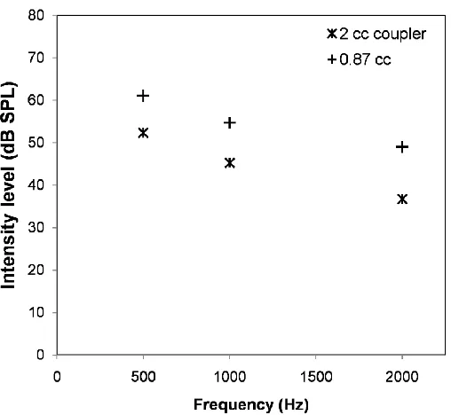
2000 Hz in a participant with an ear canal volume of 0.087 cc. Stars and plus signs
represent the sound intensity levels measured in the 2 cc coupler and in the
participant’s ear canal, respectively. ... 40
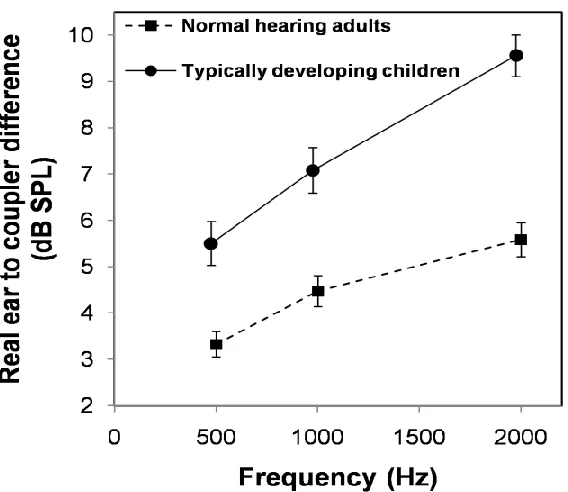
Figure 2.5: Mean of RECD values at 500, 1000 and 2000 Hz. Circles and squares
represent typically developing children and normal hearing adults, respectively. Error
bars show +1 standard error. ... 41
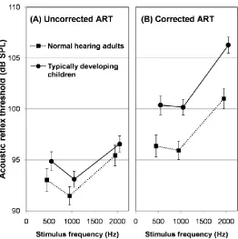
Figure 2.6: Mean of uncorrected (A) and corrected (B) acoustic reflex thresholds at
500, 1000 and 2000 Hz for the typically developing children and normal hearing
adults. Typically developing children and normal hearing adults are shown by the
xv
Figure 2.7: Corrected acoustic reflex thresholds plotted against static compliance.
Adults and children are shown in top and bottom panel, respectively... 45
Figure 3.1: Mean slopes of the reflex growth functions at 500, 1000 and 2000 Hz.
Separate panels show data from the adults, typically developing children, children
with an APD diagnosis and clinically referred children who did not receive an APD
diagnosis. Slopes in the uncrossed and crossed conditions are shown by the filled
and open symbols, respectively. Right and left ears are shown by the circles and
diamonds, respectively. Error bars show +1 standard error. ... 63
Figure 3.2: Ratio of crossed to uncrossed reflex growth function slopes plotted
against the slope of the uncrossed reflex growth functions. Data from the normal
hearing adults, typically developing children, APD and clinical non-APD are shown in
separate panels. Each data point shows an individual ratio. Data measured at 500,
1000 or 2000 Hz are shown by the diamonds, squares and circles, respectively.
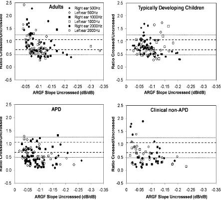
Filled and open symbols show data in right and left ears, respectively. ... 64
Figure 3.3: Average ratio of crossed to uncrossed reflex growth function slopes for
each participant plotted against the average slope of the uncrossed growth function.
Ratios were averaged for each individual across ears and frequencies. Data from
the normal hearing adults and typically developing children are shown by the filled
squares and circles, respectively. Data from children in the clinical groups, APD and
non-APD are shown by open diamonds and triangles, respectively. ... 70
Figure 4.1: Parameters of reflex latencies measured in this study ... 84
Figure 4.2: Mean of 10% On Latency, 90% On Latency, 90% Off Latency and 10%
Off Latency averaged across right and left ear. Normal hearing adults, typically
developing children, APD and clinical non-APD are shown in squares, circles,
diamonds and triangles respectively. Acoustic reflex latencies for crossed and
uncrossed reflexes are represented by open and filled symbols respectively. Error
bars show +1 standard error. ... 87
Figure 4.3: Mean and standard error of rise time and fall time for acoustic reflexes
xvi
children, APD and clinical non-APD are shown in squares, circles, diamonds and
triangles respectively. Acoustic reflex latencies for crossed and uncrossed reflexes
are represented by open and filled symbols respectively. Error bars show +1
standard error. ... 89
Figure 4.4: Mean of crossed and uncrossed acoustic reflex decay for right ear in
normal hearing adults, typically developing children, APD and clinical non-APD.
Error bars show +1 standard error. ... 91
Figure 4.5: Mean and standard error of crossed and uncrossed acoustic reflex decay
for left ear in normal hearing adults, typically developing children, APD and clinical
non-APD Error bars show +1 standard error. ... 92
Figure 5.1: Example of baseline absorbance and absorbance measured with
activation of the acoustic reflex at threshold (A). Also shown in the figure is the
difference between the same absorbance measured with and without activating
reflex (B) ... 105
Figure 5.2: Mean difference in absorbance measured with and without the activation
of crossed reflex between 226 and 4000 Hz. Results from normal hearing adults,
typically developing children and children with suspected APD are shown in the first,
second and third columns respectively. Absorbance change at different activator
intensity levels i.e. ART, ART + 5dB and ART +10 dB are shown in first, second, and
third rows respectively. Mean change in absorbance is represented by the solid
black line and individual data with grey lines. ... 108
Figure 5.3: Maximum decrease between 226 and 1000 Hz (top panel) and increase
between 1000 and 2000 Hz (bottom panel) in absorbance for different activator
intensity levels i.e. ART, ART + 5dB and ART +10 dB. Children with suspected APD,
typically developing children and normal hearing adults are shown by unfilled black
diamonds, filled black circles and filled black squares respectively. Means of
maximum decrease or increase in absorbance in children with suspected APD,
typically developing children and normal hearing adults are shown by red diamonds,
xvii
Figure 5.4: Mean and standard error of resonant frequency measured without
activating the reflex and under the influence of reflex activation at two reflex activator
Chapter 1
1
Introduction: Auditory Processing Disorder and Acoustic
Reflex
1.1 Auditory processing disorder
The term “Auditory Processing Disorder” (APD) suggests difficulties in the
processing of auditory information by the central auditory nervous system
(American Speech-Language-Hearing Association [ASHA], 2005). Individuals
with APD form a highly complex group with large individual differences. APD can
affect children, adults, or elderly persons and its etiology and symptoms may
vary across individuals (American Academy of Audiology [AAA], 2010; ASHA,
2005).
1.1.1
Children with suspected APD: Symptoms and characteristics
Children with suspected APD are often described as having difficulty hearing
even in the presence of normal hearing sensitivity. Difficulty understanding
speech in the presence of background noise, being easily distracted in complex
acoustic environments, problems following multiple commands and slow
comprehension of simple auditory information are frequently reported symptoms
(Benson, Seaton & Johnson, 1997; Keith, 1999; Jerger & Musiek, 2000). APD
may lead to, or may be associated with, attention, language, reading, learning
and cognitive disorders, however the nature of the relationships are not well
understood (AAA, 2010; ASHA, 1996, 2005). The combination of APD and its
possible comorbid conditions have the potential to negatively impact a child’s
1.1.2
APD: Prevalence in school age children and etiologies
APD has an estimated prevalence of 2% to 7% in school aged children (Bamiou,
Musiek & Luxon, 2001; Chermak & Musiek, 1997). Chermak (2002) in
summarizing possible etiologies of APD in children based on previous reports
(e.g. Chermak & Musiek, 1997, Musiek, Baran & Pinheiro, 1992; Musiek,
Gollegly & Ross, 1985; Musiek, Kibbe & Baran, 1984) suggested that
neuromorphologic disorders are the likely cause behind 65% to 70% of the
problem of children who are diagnosed with APD. Neuromaturational delay may
account for 25% to 30%, and neurologic disorders, disease or damage for 5%.
1.1.3
APD: Diagnosis
Diagnosing APD in a child can be a challenging task for the audiologist. ASHA
(2005) recommended a test battery approach that includes tasks to assess
sound localization and lateralization, auditory discrimination, auditory pattern
recognition, temporal processing and performance in presence of competing
acoustic signals. A positive diagnosis of APD is to be made if a child performs
poorly (> 2 standard deviations below age expected values) on at least 2 auditory
tests. Since there is no gold standard on the selection of tests, audiologists can
choose from a wide range of tests. This could lead to a high variability in the
criterion for APD diagnosis across clinics and in research (Allen & Allan, 2014).
Also, the most commonly used tests are behavioral (Emanuel, Ficca & Korczak,
2011) and may be strongly linked to underlying language and cognitive abilities
(Allen & Allan, 2014). Other possible limitations of behavioral tests include the
response required by the test may not be appropriate, and a lack of attention and
motivation in young children may limit performance. Many of the behavioral tests
are unavailable in languages other than English and many do not have normative
data for very young children (AAA, 2010; ASHA, 2005; Jerger & Musiek, 2000).
Objective tests have been recommended by AAA (2010), ASHA (2005)
and Jerger and Musiek (2000) but these tests have not been the preferred choice
among audiologists for APD assessment (Emanuel et al., 2011). However the
ability of the objective tests to estimate a specific site of dysfunction and the fact
that objective tests are not influenced by factors such as language or procedure
can make them highly effective in the assessment of APD, especially in children.
1.1.4
APD: Neural basis
The central auditory pathway stretches from the neural fibers originating in the
cochlea to the auditory cortex. Each anatomical nucleus along this pathway
serves one or more central auditory processes and auditory processing disorders
can result from deficit in one or more of these neural structures (Bamiou, Musiek
& Luxon, 2001; Moore, 2006). The auditory brainstem is the locus of the earliest
processing of auditory information as it ascends the auditory tract. Trouble with
the processing of sound at the brainstem level may lead to poor decoding at
higher neural centres and thus result in perceptual difficulty.
Objective measurements of acoustic reflexes, contralateral suppression of
otoaoustic emissions and auditory brainstem responses have indicated auditory
suppression of transient evoked otoacoustic emissions is reported in children
with suspected APD when compared to normal hearing children (Muchnik et al.,
2004; Sanches & Carvallo, 2006). However, Butler, Purcell and Allen (2011)
contradict these findings as they found similar contralateral suppression of
distortion product otoacoustic emission in children with APD and normal hearing
children.
Abnormalities in auditory brainstem responses in children with suspected
APD are demonstrated in several studies. Significantly reduced amplitude of the
binaural interaction component of the auditory brainstem responses (Gopal &
Pieral, 1999) shallower slopes of waves I through V (Gopal & Kowalski, 1999)
and delayed wave V (Jisra, 2001) have been found in children with suspected
APD in comparison to normal hearing children. Kraus and colleagues (Banai,
Nicol, Zecker & Kraus, 2005; Cunningham, Nicol, Zecker, Bardlow & Kraus,
2001; King, Warrier, Hayes & Kraus, 2002; Wible, Nicol & Kraus, 2004) have also
suggested atypical speech evoked auditory brainstem responses in one third of
the children with language learning disorders who also have symptoms of APD.
Allen and Allan (2007, 2014) investigated acoustic reflex and auditory
brainstem responses in children with suspected APD. They reported 65% of the
children tested showed clinically significant abnormalities in either acoustic reflex
or auditory brainstem responses. High percentages of reflex abnormalities in
children with suspected APD are also reported by Meneguello et al. (2001) and
Based on the studies described it is apparent that many children with
suspected APD may have abnormalities in brainstem function. The acoustic
reflex is a sensitive measure of auditory brainstem dysfunction (Gelfand, 2005;
Jerger & Jerger, 1977;Silman, 1984) but only limited literature on acoustic
reflexes, specific to the reflex thresholds, is available in children with suspected
APD. Detailed studies of the acoustic reflex measures in children with suspected
APD can provide important information about the auditory brainstem in this
clinical population. These studies will also provide insight into the relationship
between the suggested functional role of acoustic reflexes in perceiving speech
in the presence of noise and children with suspected APD.
1.2 The acoustic reflex
The middle ear muscle reflex is one of the primary feedback mechanisms of the
auditory system (Liberman & Guinan, 1998). The reflex results largely from the
contraction of the stapedius and tensor tympani muscles following acoustic
stimulation of the ears. In most animals both the stapedius and tensor tympani
muscles contribute to the reflex in response to auditory stimuli (Moller 1984;
Mukerji, Windsor & Lee, 2010). In humans, it is predominantly the stapedius
muscle while the contraction of the tensor tympani muscle occurs primarily during
the startle response to intense sounds or to non-auditory stimuli (Borg, 1968;
1.2.1
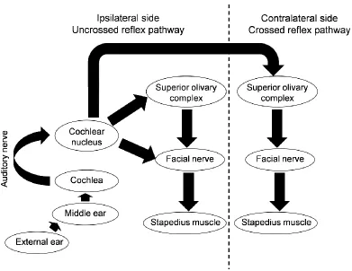
Anatomy of the acoustic reflex pathway
The acoustic reflex occurs from stimulation to both crossed and uncrossed reflex
pathways. The anatomy of the reflex pathway is well described in the literature
(Moller, 1984; Mukerji et al., 2010). Anatomical structures include the peripheral
auditory system (external ear, middle ear and the cochlea), the auditory nerve,
two nuclei of the auditory brainstem (the cochlear nucleus [CN] and the superior
olivary complex [SOC]), the facial motor nucleus and nerve and the stapedius
muscle (Figure 1.1). The central segment of the acoustic reflex arc initiates with
the projection of type I spiral ganglion neurons (afferents from inner hair cells) to
the cochlear nucleus. Interneurons responsible for the acoustic reflex lie in the
posterior ventral cochlear nucleus (PVCN). Interneurons from the PVCN
innervate the stapedius motor neuron (SMN) of the facial nerve through direct
and indirect projections. Direct projection involves the innervation of SMN directly
by PVCN interneurons. Indirect projection includes projection of PVCN
interneurons to the SMN through the superior olivary complex. It is the medial
superior olive (MSO) that is primarily involved in the acoustic reflex. The PVCN
supplies second order neurons to the ipsilateral MSO and contralateral MSO.
The MSO finally sends third order neurons to the SMN of the ipsilateral facial
nerve (for uncrossed reflex) and SMN of the contralateral facial nerve (for
Figure 1.1: The acoustic reflex pathway
The main function of the auditory brainstem is to preserve and extract
spectral and temporal information for processing in the higher auditory system
(Irvine, 1992). The superior olivary complex is also the first nucleus of the
auditory system where binaural inputs interact (Brugge, 1992). In the presence of
a normal peripheral auditory system any abnormality in the acoustic reflex may
indicate a deficit in the functioning of the auditory nerve, the cochlear nucleus or
the superior olivary complex. Therefore the information provided by the acoustic
reflex testing in individuals with auditory processing deficits can be useful in
1.2.2
Functions of the acoustic reflex
The functional importance of the acoustic reflex has been discussed for many
years. Several hypotheses are offered regarding its role including primarily its
roles in protecting against inner ear damage from loud sounds and its facilitation
in speech perception in the presence of noise (Borg et al., 1984). It has also
been suggested to aid in the perception of faint sounds, it improves temporal
resolution and it enhances auditory attention.
Support for the protective function of acoustic reflex comes from studies
that investigated the relationship between elicitation of the acoustic reflex and
temporary threshold shift following noise exposure (Cohen & Bauman, 1964;
Mills & Lilly, 1971; Ward, 1962; Zakrisson, Borg, Liden & Nilsson, 1980). Studies
have shown that the presence of a normal acoustic reflex is associated with
reduced temporary threshold shifts. But the protective role of the acoustic reflex
is not universally supported (Fletcher, 1962; Henderson, Subramaniam,
Papazian & Spongr, 1994; Ryan, Bennett, Woolf & Axelsson, 1994). It is
suggested that the acoustic reflex may provide only limited protection for loud
sounds because its onset is most often over 100 msec (Borg , 1982; Gorga &
Stelmachowicz, 1983; Hung & Dallos, 1972; Qiu & Stucker, 1998).This delay
makes the acoustic reflex relatively meaningless in preventing damage from
impulse noise or stimulus onsets. Also the acoustic reflex undergoes adaptation
if the sound is present for very long durations (Gelfand, 2005) and therefore the
The role of the reflex in enhancing speech perception in noise is more
likely. Simmons (1964) explained that the acoustic reflex helps in improving
speech intelligibility especially in the presence of noise by attenuating low
frequency information. The reflex modulates the amplitude and frequency of
sounds which therefore may increase alertness in listeners, allow better
separation of background noise and signal and enhance attention to the signal.
Aiken, Andrus, Bance and Phillips (2013) suggested that the acoustic reflex may
help in speech perception in noise by preventing upward spread of masking at
moderate noise levels. De Andrade et al. (2011) and Colletti, Fiorino, Verlato
and Carner (1992) found that the acoustic reflex is important for better
performance in speech discrimination and frequency selectivity tasks,
respectively. Dorman, Cedar, Hannley and Leek (1986) reported that the
activation of the acoustic reflex in normal hearing listeners improves their vowel
recognition. On the contrary, Phillips, Stuart and Carpenter (2002) found no role
of the reflex in word recognition in quiet but suggested that role of reflex in
speech perception could be restricted to the adverse listening conditions
including listening in noise environment. Borg and Zakrisson (1974) found
greater masking in the ears with acute stapedius muscle paralysis in comparison
to the ear with normal acoustic reflexes when the stimulus was presented above
reflex thresholds. Similar masking was reported in both the ears for the stimulus
1.2.3
Measures of acoustic reflex
There are several measurable characteristics of the acoustic reflex, each of
which provides important details about the reflex activity. The reflex threshold is
the minimum intensity level of the reflex activator stimulus at which the acoustic
reflex activates. At threshold, the magnitude of the reflex is observable as a small
change in the acoustic compliance of the middle ear. Presentation of stimuli
above the threshold results in a greater magnitude. The magnitude of the reflex
increases with increase in stimulus level until an asymptote, or maximum
compliance change is reached. This generally occurs within 30 dB of reflex
threshold. The relationship between reflex magnitude and activator stimulus level
can be described by an acoustic reflex growth function. Measures can also be
made of the time course of the reflex activation. Acoustic reflex latency refers to
the time taken by the stapedius muscle to contract after the onset of the stimulus.
The amplitude of the reflex reaches its maximum magnitude after the activator is
presented for around 250 msec. The reflex then undergoes adaptation and its
amplitude decreases if the stimulation continues for a longer duration. This
characteristic of acoustic reflex is known as acoustic reflex decay.
Individuals with the disorders of auditory nerve and auditory brainstem
lesions have shown absent or elevated reflex thresholds (Anderson, Barr &
Wedenberg, 1970; Johnson, 1977; Mangham, Lindeman & Dawson, 1980), low
reflex amplitudes (Mangham et al., 1980), shallower growth functions (Harrison,
Silman & Silverman, 1989; Mangham et al., 1980; Silman, Popelka & Gelfand,
1983; Mangham et al., 1980) and greater or earlier reflex decay (Anderson et al.,
1970; Mangham et al., 1980; Olsen, Noffsinger & Kurdziel, 1975)
Absent or elevated reflexes would indicate no reflex activity or that reflex
activity is only initiated at higher stimulus levels. Low reflex amplitude and
shallower growth of the reflex magnitude may suggest that the acoustic reflex is
weak. Longer reflex latencies would mean a delay in the activation of acoustic
reflex and greater or earlier decay may suggest that the reflex is only providing
limited benefit. Abnormalities in one or more of these characteristics of the reflex,
if present, may also therefore suggest limited benefit in speech in noise
perception.
Despite the importance of the acoustic reflex in assessing auditory nerve
and brainstem disorders and its potential importance for speech perception in the
presence of noise, investigations of reflex in children with suspected APD are
limited. Published studies report only reflex thresholds (Allen & Allan, 2007,
2014; Meneguello et al., 2001; Thomas et al., 1985). Further Investigations of
other characteristics of the reflexes may provide greater information about the
potential role in children with suspected APD.
Adult and child differences in acoustic reflexes have been investigated for
reflex threshold, amplitude and decay. Habener and Snyder (1974) found lower
reflex amplitude and elevated reflex thresholds in normal hearing children (aged
3 to 19 years) when compared to the young adults (aged 19 to 29 years) but no
(1972), Jerger, Hayes, Anthony and Mauldin (1978) and Osterhammel and
Osterhammel (1979) have suggested higher thresholds in children (aged 7 to 15
years) when compared to adults. The reason for adult-children differences in
reflex amplitudes and threshold are not well understood. A possible explanation
could be the differences in the characteristics of the ear canal and middle ear
static compliance that develop until puberty (Abdala & Keefe, 2012; Obake,
Tanaka, Hamada, Miura & Funai, 1988). However the relationship between these
factors and the acoustic reflex has not been investigated.
The acoustic reflex is bilateral with stimulation to either ear its effect can
be measured in a crossed and uncrossed configuration referencing stimulus or
measurement ear. Differences in crossed and uncrossed measures are reported
in some studies and the suggestion is generally that the crossed pathways are
weaker showing higher reflex thresholds (Fria, LeBlanc, Kristensen & Alberti,
1975; Gelfand, 2005; Jerger et al., 1978; Moller, 1961, 1962). The growth of the
reflex with changes in stimulus magnitude is reported to be shallower for crossed
stimulation in comparison to those with uncrossed responses (Jerger et al., 1978;
Moller, 1961). Decay also differs in crossed and uncrossed condition. Lilly,
Mekaru and Chudnow (1983, cited in: Wilson, Shanks & Lilly, 1984) reported that
uncrossed reflexes had an earlier onset of reflex decay than reflexes in the
crossed condition. Oviatt and Kileny (1979) suggested greater reflex decay for
uncrossed stimulation in comparison to crossed stimulation but a significant
difference was not found. Allen and Allan (2014) highlighted that acoustic reflex
crossed pathways in children with this clinical disorder. This is similar to reports
of reflexes in brainstem disorders which also shown abnormalities specific to the
crossed pathways (Griesen & Rasmussen, 1970; Jerger & Jerger, 1977). These
findings reflect the importance of the estimation of acoustic reflex measures in
both crossed and uncrossed condition while using acoustic reflex in the auditory
assessment.
1.3 Thesis purpose and chapter outline
Previous reports have suggested auditory brainstem abnormalities may be seen
in some children with suspected APD. Acoustic reflexes have proven to be an
important measure to assess auditory brainstem function. But investigations into
acoustic reflexes in children with suspected APD are few and largely limited to
the measure of acoustic reflex thresholds. The primary aim of this thesis is to
better understand the relationship between acoustic reflex measures and
children with suspected APD. In the first study (chapter 2), acoustic reflex
thresholds were investigated in children with suspected APD to confirm previous
findings of abnormal thresholds in children with suspected APD. This study also
examined real ear corrections on threshold estimates and the role of static
compliance. The second study was aimed at understanding the acoustic reflex
growth function (chapter 3) which may be more sensitive to auditory pathology
than a single threshold estimate. In the third study acoustic reflex latencies and
decay were investigated to determine if there were pathology or age-related
examined the impact of the acoustic reflex on the absorbance and resonant
frequency of the middle ear (chapter 5).
The diagnosis of APD can be a difficult task because there is no gold
standard for diagnosis. Although professional associations suggest a test battery
approach, individual clinicians are free to select tests from a large number that
are available and often clinicians limit their test selection to behavioral measures,
often examining some aspects of degraded speech perception or temporal
pattern recognition (Emanuel et al. 2011). Yet Allen and Allan (2014), found that
using a battery of such tests often failed to diagnose children as APD when
referred for listening difficulties yet these children were found to show clinically
significant abnormalities in auditory brainstem responses or reflex data
suggesting some level of neural dysfunction that was missed with a test battery
restricted to behavioral speech and pattern recognition tests. Therefore, in the
studies included in this thesis, children with suspected APD were divided into two
groups: (1) the APD group included children who were diagnosed as having APD
based on a behavioral test battery of tests including Staggered Spondaic Word
test (Katz, 1998), the Pitch Pattern Sequence Test (Pinheiro, 1977), the Words in
Ipsilateral Competition test (Ivey, 1969, 1987) and two custom tests of frequency
discrimination and gap detection; and (2) the clinical non-APD group included
children who were referred for APD assessment but who were not diagnosed as
APD based on this typical clinical battery. This provided the opportunity to
investigate auditory brainstem functioning using acoustic reflexes in both the
Because there is generally a lack of published data on acoustic reflexes in
children, each study also included a group of typically developing children and
normal hearing adults. Most published studies and clinical normative have
compared acoustic reflexes in clinical populations to those of normal hearing
adults. The inclusion of typically developing children as well as adults allowed for
the evaluation of potential age related effects.
1.4 References
Abdala, C., & Keefe, D. H. (2012). Morphological and functional ear
development. In L. A. Werner, R. R Fay & A. N. Popper (Eds.), Human Auditory Development (pp. 19-59). New York: Springer.
Aiken, S. J., Andrus, J. N., Bance, M., & Phillips, D. P. (2013). Acoustic Stapedius Reflex Function in Man Revisited. Ear and hearing, 34(4), e38-e51.
Allen, P. & Allan, C. (2007). Auditory processing disorders: Putting the “neural” back into sensorineural hearing loss. In R.C. Seewald & J.M. Bamford (Eds.),A Sound Foundation Through Early Amplification 2007:
Proceedings of the Fourth International Conference (pp. 221-233). Stäfa, Switzerland: Phonak AG.
Allen, P. & Allan, C. (2014). Auditory Processing Disorders: relationship to cognitive processes and underlying auditory neural integrity.
International Journal of Paediatric Otorhinolaryngology, 78, 198-208.
American Academy of Audiology [AAA]. (2010). American Academy of Audiology Clinical Practice Guidelines:Diagnosis, Treatment and Management of Children and Adults with Central auditory Processing Disorder. Retrieved from http:// www.audiology.org/resources/
American Speech-Language-Hearing Association. (1996). Central auditory processing: Current status of research and implications for clinical practice. American Journal of Audiology, 5, 41-54.
Anderson, H., Barr, B. &Wedenberg, E. (1970). Early diagnosis of VIIIth-nerve tumours by acoustic reflex tests. ActaOto-Laryngologica Supplement, 263, 232-237.
Banai, K., & Kraus, N. (2007). Neurobiology of (central) auditory processing disorder and language-based learning disability. In G. D. Chermak & F. E. Musiek (Eds.), Handbook of (central) auditory processing disorders (pp. 89-116). San Diego: Plural Publishing.
Banai, K., Nicol, T., Zecker, S. G., & Kraus, N. (2005). Brainstem timing: implications for cortical processing and literacy. The Journal of Neuroscience,25(43), 9850-9857.
Bamiou, D.E., Musiek, F.E. &Luxon, L.M. (2001). Aetiology and clinical
presentations of auditory processing disorders – a review. Archives of Disease in Childhood, 85, 361-365.
Benson, P. V., Seaton, J. B., & Johnson, C. D. (1997). Educational audiology handbook. San Diego, CA: Singular.
Borg, E. (1968). A quantitative study of the effect of the acoustic stapedius reflex on sound transmission through the middle ear of man. ActaOto-laryngologica, 66, 461-472.
Borg, E. (1982). Time Course of the Human Acoustic Stapedius Reflex: A Comparison of Eight Different Measures in Normal-hearing Subjects.
Scandinavian audiology, 11(4), 237-242.
Borg, E. Counter, S. A. &Rosler, G. (1984). Theories of middle ear muscle function. In: S. Silman (Ed.), The acoustic reflex: Basic principle and clinical applications. Orlando: Academic press.
Borg, E., & Zakrisson, J. E. (1974). Stapedius reflex and monaural masking.
Actaoto-laryngologica, 78(1-6), 155-161.
Brugge, J. F. (1992). An overview of central auditory processing. In: A. N. Popper & R.R. Fay (Eds.), The mammalian auditory pathway: Neurophysiology (pp. 153-231). New York: Springer
Butler, B. E., Purcell, D. W., & Allen, P. (2011). Contralateral inhibition of distortion product otoacoustic emissions in children with auditory
processing disorders. International journal of audiology, 50(8), 530-539.
Chermak, G. (2002). Deciphering auditory processing disorders in children.
Otolaryngologic Clinics of North America, 35, 733-749.
Clemis, J.D. &Sarno, C.N. (1980). The acoustic reflex latency test: Clinical application. Laryngoscope, 90, 601-611.
Cohen, A., & Baumann, K. C. (1964). Temporary Hearing Losses following Exposure to Pronounced Single‐Frequency Components in Broad‐Band Noise.The Journal of the Acoustical Society of America, 36(6), 1167-1175.
Colletti, V., Fiorino, F.G., Verlato, G. &Carner, M. (1992). Acoustic reflex in frequency selectivity: Brain stem auditory evoked response and speech discrimination. In J. Katz, N.A. Stecker& D. Henderson (Eds.). Central Auditory Processing: A Transdisciplinary View(pp. 39-46). St. Louis, MO: Mosby - Year Book, Incorporated.
Cunningham, J., Nicol, T., Zecker, S. G., Bradlow, A., & Kraus, N. (2001). Neurobiologic responses to speech in noise in children with learning problems: deficits and strategies for improvement. Clinical
Neurophysiology, 112(5), 758-767.
De Andrade, K.C.L., Camboim, E.D., Soares, I.D.A., Peixoto, M.V.D.S., Neto, S.C., Menezes, P.D.L. (2011). The importance of acoustic reflex for communication. American Journal of Otolaryngology – Head and Neck
Medicine and Surgery, 32, 221-227.
Dorman, F., Lindholm, J. M., Hannley, M. T., & Leek, M. R. (1987). Vowel
intelligibility in the absence of the acoustic reflex : Performance-intensity characteristics. The Journal of the Acoustical Society of America81(2),
562–564.
Emanuel, D.C., Ficca, K.N., &Korczak, P. (2011). Survey of the diagnosis and management of auditory processing disorder. American Journal of Audiology, 20, 48-60.
Fletcher, J. L. (1962). Reflex response of middle-ear muscles : Protection of the ear from noise. Sound, 1 (2), 17–23.
Fria, T., LeBlanc, J., Kristensen, R., &Alberti, P. W. (1974). Ipsilateral acoustic reflex stimulation in normal and sensorineural impaired ears: a
preliminary report. Canadian journal of otolaryngology, 4(4), 695-703.
Gelfand, S.A. (2005). The acoustic reflex. In J. Katz (Ed.), Handbook of clinical audiology: Fifth Edition (pp: 205-232). Baltimore, MD: Lippincott Williams & Wilkins.
Gopal, K.V. & Kowalski, J. (1999). Slope analysis of auditory brainstem responses in children at risk of central auditory processing disorders.
Gopal, K.V. &Pierel, K. (1999). Binaural interaction component in children at risk for central auditory processing disorders. Scandinavian Audiology, 28, 77-84.
Greisen, O., & Rasmussen, P. E. (1970). Stapedius muscle reflexes and oto-neurological examinations in brain-stem tumors.Acta
oto-laryngologica,70(5-6), 366-370.
Habener, S. A., & Snyder, J. M. (1974). Stapedius reflex amplitude and decay in normal hearing ears. Archives of Otolaryngology, 100(4), 294-297.
Harrison, T., Silman, S. & Silverman, C.A. (1989). Contralateral acoustic reflex growth function in a patient with a cerebellar tumor: A case study.
Journal of Speech and Hearing Disorders, 54, 505-509.
Henderson, D., Subramaniam, M., Papazian, M., &Spongr, V. P. (1994). The role of middle ear muscles in the development of resistance to noise induced hearing loss. Hearing Research,74, 22–28.
Hung, I. J. &Dallos, P. (1972). Study of the acoustic reflex in human beings. I. Dynamic characteristics. The Journal of the Acoustical Society of America, 52 (4), 1972-80.
Irvine, D. R. (1992). Physiology of the auditory brainstem. In: A. N. Popper & R.R. Fay (Eds.), The mammalian auditory pathway:
Neurophysiology (pp. 153-231). New York: Springer
Ivey, R. (1969). Words in Ipsilateral Competition (WIC), (Unpublished Masters Thesis), Colorado State University, Fort Collins, CO.
Ivey, R.G. (1987). [Words in ipsilateral competition (WIIC) – Version 2]. Unpublished Normative Study.
Jerger, J., Jerger, S., & Mauldin, L. (1972). Studies in impedance audiometry: I. Normal and sensorineural ears. Archives of Otolaryngology, 96, 513-523.
Jerger, J., Hayes, D., Anthony, L. & Mauldin, L. (1978). Factors influencing prediction of hearing level from the acoustic reflex. Monographs in Contemporary Audiology, 1, 1-20.
Jirsa, R.E. (2001). Maximum length sequences – auditory brainstem responses from children with auditory processing disorders. Journal of the
American Academy of Audiology, 12, 155-164.
Jerger, S., & Jerger, J. (1977). Diagnostic value of crossed vs uncrossed acoustic reflexes: Eighth nerve and brain stem disorders. Archives of Otolaryngology, 103(8), 445-453.
Jerger, J., & Musiek, F. (2000). Report of the consensus conference on the diagnosis of auditory processing disorders in schoolaged children.
Journal of American Academy Audiology, 11, 467–74.
Johnson, E.W. (1977). Auditory test results in 500 cases of acoustic neuroma.
Archives of Otolaryngology, 103, 152-158.
Katz, J. (1998). The Staggered Spondaic Word Test (SSW) Fifth Edition, Vancouver, WA: Precision Acoustics.
Keith, R.W. (1999). Clinical issues in central auditory processing disorders.
Language, Speech, and Hearing Services in Schools, 30, 339-344.
King, C., Warrier, C. M., Hayes, E., & Kraus, N. (2002). Deficits in auditory brainstem pathway encoding of speech sounds in children with learning problems. Neuroscience letters, 319(2), 111-115.
Liberman, M. C., &Guinan, J. J. (1998). Feedback control of the auditory periphery: anti-masking effects of middle ear muscles vs.
olivocochlearefferents. Journal of Communication Disorders, 31(6), 471–82. Linares, A. E. &Carvallo, R. M. M. Acoustic reflex latency in children with auditory prossessing disorder. (2004). International Archives of Otorhinolaryngology. 8(1).
Mangham, C.A. & Lindeman, R.C. (1980). The negative acoustic reflex in retrocochlear disorders. Laryngoscope, 90, 1753-1761.
Meneguello, J., Domenico, M. L. D., Costa, M. C. M., Leonhardt, F. D.,
Barbosa, L. H. F., & Pereira, L. D. (2001). Occurrence of acoustic reflex changes in auditory processing disorders. Brazilian Journal of
Otorhinolaryngology. 67 (7), 2001.
Mills, J. H., & Lilly, D. J. (1971). Temporary threshold shifts produced by pure tones and by noise in the absence of an acoustic reflex. The Journal of the Acoustical Society of America, 50(6B), 1556-1558.
Moller, A. R. (1961). Bilateral contraction of the tympanic muscles in man examined by measuring acoustic impedance-change. The Annals of otology, rhinology, and laryngology, 70, 735.
Moller, A. R. (1984). Neurophysiological basis of the acoustic middle ear reflex. In: S. Silman (Ed.), The acoustic reflex: Basic principle and clinical applications. Orlando: Academic press.
Moore, D. R. (2006). Auditory processing disorder (APD): Definition, diagnosis, neural basis, and intervention. Audiological Medicine, 4(1), 4-11.
Muchnik, C., Roth, D.A., Othman-Jebara, R., Putter-Katz, H., Shabtai, E.L. &Hildesheimer, M. (2004). Reduced medial olivocochlear bundle system function in children with auditory processing disorders.
Audiology & Neuro-Otology, 9, 107-114. doi: 10.1159/000076001
Mukerji, S., Windsor, A. M., & Lee, D. J. (2010). Auditory brainstem circuits that mediate the middle ear muscle reflex. Trends in Amplification, 14(3), 170–91.
Musiek, F. E. & Lee, W. W. (1997). Conventional and maximum length sequences: Middle latency response in patients with central nervous system lesions. Journal of American Academy of Audiology, I8:173-180.
Okabe, K., Tanaka, S., Hamada, H., Miura, T., & Funai, H. (1988). Acoustic impedance measurement on normal ears of children. Journal of the Acoustical Society of Japan (E), 9(6), 287-294.
Olsen, W.O., Noffinger, D. &Kurdziel, S. (1975). Acoustic reflex and reflex decay. Occurrence in patients with cochlear and eighth nerve lesions.
Archives of Otolaryngology, 101, 622-625.
Osterhammel, D., &Osterhammel, P. (1979). Age and sex variationsfor the normal stapedial reflex thresholds and tympanometric compliance values. Scandinavian Audiology, 8, 153-158.
Oviatt, D. L., &Kileny, P. (1984). Normative characteristics of ipsilateral acoustic reflex adaptation. Ear and hearing, 5(3), 145-152.
Pinheiro, M. (1977). Pitch Pattern Sequence Test. Auditec of St. Louis.
Ryan, A. F., Bennett, T. M., Woolf, N. K., &Axelsson, A. (1994). Protection from noise-induced hearing loss by prior exposure to a nontraumatic
stimulus : Role of the middle ear muscles. Hearing Research, 72, 23–
28.
Silman, S. (1984).The acoustic reflex: Basic principle and clinical applications.
Orlando: Academic press.
Silman, S., Popelka, G.R. &Gelfand, S.A. (1978). Effect of sensorineural
hearing loss on acoustic stapedius reflex growth functions. The Journal of the Acoustical Society of America, 64, 1406-1411.
Simmons, F. B. (1964). Perceptual theories of middle ear muscle function. The Annals of otology, rhinology, and laryngology, 73, 724-739.
Thomas, W.G., McMurry, G. & Pillsbury, H.C. (1985). Acoustic reflex
abnormalities in behaviorally disturbed and language delayed children.
Laryngoscope, 95, 811-817.
Ward, W. D. (1962). Damage‐Risk Criteria for Line Spectra. The Journal of the Acoustical Society of America, 34(10), 1610-1619.
Wible, B., Nicol, T., & Kraus, N. (2004). Atypical brainstem representation of onset and formant structure of speech sounds in children with
language-based learning problems. Biological psychology, 67(3), 299-317.
Chapter 2
2
Acoustic Reflexes in Normal Hearing Adults, Typically
Developing Children and Children with Suspected
Auditory Processing Disorder: Thresholds, Real Ear
Corrections and the role of Static Compliance on
Estimates
2.1 Introduction
The Acoustic Reflex Threshold is defined as the minimum stimulus intensity at
which the stapedius muscle contracts. Reflex thresholds are used diagnostically
in clinical audiology, often to determine if a hearing loss is of cochlear or
retrocochlear origin, but lesions anywhere in the auditory system can cause
reflex abnormalities (Gelfand, 1984, 2005). Abnormal reflexes are usually
interpreted along with the results of other auditory tests in order to determine the
site of dysfunction. Abnormalities in reflexes thresholds due to middle ear or
cochlear dysfunction are generally interpreted based on the results of pure-tone
audiometry and tympanometry. An air-bone gap of as little as 30 dB in the
stimulus ear may make it impossible to elicit a reflex simply because a sufficient
excitation level cannot be reached within the limits of most equipment. In the
probe ear even a mild middle ear pathology may be sufficient to make it
impossible to measure change in impedance associated with reflex activation
even if it occurs. With cochlear pathologies, reflexes are generally within the
expected range unless a severe loss of hearing is present in which case
Reflex abnormalities associated with disorders of the auditory nerve or
brainstem, i.e. retrocochlear pathology, may be more complex and often requires
comparison of crossed and uncrossed responses (Jerger & Jerger, 1977). When
the auditory nerve is affected, reflexes are often elevated or absent with
stimulation to the affected ear regardless of the degree of hearing loss (e.g.
Anderson, Barr & Wedenberg, 1970; Ferguson et al., 1996; Jerger, Harford,
Clemis & Alford, 1974; Mangham, Lindeman & Dawson, 1980; Prasher & Cohen,
1993; Thomsen & Terkildsen, 1975). When there is an elevation or absence in
thresholds in the presence of significant sensorineural hearing loss the diagnosis
of cochlear versus retrocochlear pathology may be more difficult. Generally reflex
thresholds of 95 dB HL or higher are taken as an indication of retrocohlear
pathology (Anderson, Barr and Wedenberg (1969) cited in: Silman & Gelfand,
1981). Gelfand and colleagues (Silman & Gelfand,1981; Gelfand, Scehwander &
Silman, 1990) estimated the 90th percentile cut-off for reflex thresholds at 500,
1000 and 2000 Hz in normal hearing adults and adults with cochlear impairment
to fall at 95, 100 and 100 dB HL, respectively if the hearing thresholds are within
normal limits (< 15 dB HL). Presently, reflex thresholds beyond these cutoff
values are used in clinics for determination of reflex abnormalities of
retrocochlear origin (Gelfand, 2005).
Recently, Allen and Allan (2007, 2014) reported abnormal reflexes in
children with suspected auditory processing disorders (APD), often absent or
elevated, particularly in the crossed configuration. Meneguello et al. (2001) found
(1985) suggested abnormal reflex thresholds in 32% of children with language
delay, learning disability and who were suspected of APD but did not find any
correlation between acoustic reflex abnormalities and children of suspected APD.
Despite of the high incidences of reflex threshold abnormalities in children with
suspected APD, measurements of reflex threshold are not typically included as
diagnostic indicators in the assessment of auditory processing. In order to
improve the accuracy of reflex threshold testing in the assessment of children
with auditory processing disorders, normative data from children is preferable to
that from adults as age-effects have sometimes been reported. As well, different
norms for crossed and uncrossed reflexes should be used as thresholds for
uncrossed reflexes are most often lower than for crossed reflexes in adult
listeners (Fria, LeBlanc, Kristensen & Alberti, 1975; Gelfand, 2005; Jerger,
Hayes, Anthony & Mauldin, 1978; Moller, 1961, 1962). However, developmental
differences in crossed and uncrossed thresholds are unknown.
The morphology and functional characteristics of the conductive
mechanism mature from birth to puberty (Abdala & Keefe, 2012; Obake, Tanaka,
Hamada, Miura & Funai, 1988). Both ear canal volume and static compliance are
smaller in children than in adults (Abdala & Keefe, 2012; Barlow et al., 1988;
Jerger et al., 1978; Obake et al., 1988) and could influence the measurement
and interpretation of reflex thresholds when comparing results between children
2.1.1
Real ear correction and reflex thresholds
Real ear corrections for differences in ear canal volume are common in many
measures of hearing in children. When evaluating behavioral threshold or
measuring hearing aid gain these corrections are nearly universally recommend
(American Academy of Audiology [AAA], 2013). However, similar corrections
have not been applied to reflex threshold measurements. Calibration of the
stimulus used to elicit reflex thresholds is typically completed using a 2 cc
coupler (Grason-Stadler, 2005; Interacoustics, 2011). It is known that sounds
with similar intensities can result in different sound pressure levels (SPL) in ear
canals with different volumes (Martin, 2003). It is likely that the reflex activator
presented at a fixed intensity level result in a higher SPL in the ear canal with a
smaller volume than in an ear with a larger volume. This could result in
erroneous measurements of reflex thresholds in individuals with ear canal
volume smaller than 2 cc such as children. While it is well accepted that real ear
to coupler differences (RECD) in individuals with a small ear canal volume show
larger RECD values and vice versa (Barlow et al., 1988; Feigin, Kopun,
Stelmachowicz & Gorga, 1989; Martin, Westwood & Bamford, 1996), there has
been no systematic investigation into the influence of real ear correction on reflex
threshold measurements.
2.1.2
Static compliance and reflex thresholds
Age-related differences in static compliance could also impact estimates of
children’s acoustic reflex thresholds. Static compliance represents an estimate
compliance values are often lower in children than in adults (Jerger et al., 1972;
Obake, et al., 1979). Near threshold, the reflex causes only a small change in the
compliance of the middle ear. Smaller static compliance values could possibly
make it difficult to measure this very small change. At higher stimulus levels the
contraction of the stapedius muscle is stronger resulting in a larger change in
compliance that may be easier to measure. This measurement parameter, which
may vary developmentally, could lead to a higher estimate of reflex thresholds in
children.
Wilson (1979) examined the impact of static compliance on acoustic reflex
thresholds in normal hearing adults. He reported a low correlation between
crossed thresholds and static compliance. But the participants in his study had
histories of negative middle ear pressure which could have influenced
measurements of reflex thresholds and static compliance and only crossed
thresholds were measured. Correlating crossed reflex thresholds with static
compliance of the measurement ear rather than the stimulus ear may have
contributed to the lower correlation.
2.2 Study aims
The aim of this study was to replicate previous studies showing elevated reflex
thresholds in children with suspected APD and to compare their crossed and
uncrossed threshold estimates to those from typically developing children and
normal hearing adults. Because ear canal volume and static compliance have
shown developmental effects and because these factors could affect the
the effect of real ear corrections for stimulus levels on thresholds and the
relationship between static compliance and threshold estimates in both adults
and children.
2.3 Study 1: Reflex Threshold Estimates in Crossed and
Uncrossed Pathways for Normal Hearing Adults,
Typically Developing Children and Children with
Suspected Auditory Processing Disorder.
2.3.1
Methods
2.3.1.1
Participants
Participants in this study included 20 normal hearing adults (18-30 years of age),
28 typically developing children (7 to 15 years of age) and 66 children (aged 7 to
15 years) suspected of having an auditory processing disorder. The children
suspected of having an auditory processing disorder were referred to the Child
Hearing Research Laboratory by caregivers, teachers, parents, or physicians for
an auditory processing assessment. All participants had normal otoscopic
examination, normal hearing thresholds (American Speech-Language-Hearing
Association [ASHA], 2005a), normal middle function (ASHA, 1988) and no history
of neurologic disorder.
Children referred for the evaluation of suspected APD underwent a behavioral
assessment that included the Staggered Spondaic Word test (Katz, 1998), the
Pitch Pattern Sequence test (Pinheiro, 1977), the Words in Ipsilateral
Competition test (Ivey, 1969, 1987) and two custom tests of signal feature
adaptive 3-alternative forced-choice procedure designed to track the 70.7%
correct threshold level. As suggested by ASHA (2005b), children who performed
at least 2 standard deviations below age expectations on at least 2 of these tests
were classified as APD. Those that did not meet the criterion but who reported
listening difficulties were classified as clinical non-APD (Allen & Allan, 2014).
Forty two of the children were therefore classified as APD and 24 as clinical
non-APD.
2.3.1.2
Instrumentation
Otoscopic examination was conducted using a hand-held Welch Allyn otoscope.
Pure tone audiometry and the auditory processing evaluation were administered
using a Grason Stadler 61 (GSI 61) diagnostic audiometer and a JVC XL Z32 CD
player. A GSI Tympstar Middle Ear Analyzer version 2 was used to evaluate
middle ear function and obtain reflex thresholds. It was professionally calibrated
for probe tone frequency, probe tone level, compliance, stimulus intensity level,
volume and pressure according to American National Standard Institute [ANSI] S
3.39 (1987) standard.
2.3.1.3
Procedure
All impedance and reflex measurements were obtained with a 226 Hz probe
tone. A proper hermetic seal was sustained during the testing. Crossed and
uncrossed reflex thresholds were obtained using 500, 1000 and 2000 Hz
pure-tone activator stimuli. For both conditions, reflex threshold measurements were
made in 5 dB steps. A reflex amplitude of 0.02 ml or more was considered as
same stimulus level in order to validate the threshold estimates. For the statistical
analyses reported in this study, the Greenhouse-Geisser corrected values are
2.3.2
Results
Reflexes were absent in 3 typically developing, 6 APD and 4 clinical non-APD
children in one or more measurement conditions. Therefore they were not
included in the statistical analysis. Repeated measures analysis of variance
(RM-ANOVA) showed no effect of stimulus ear on reflex thresholds [F (1, 100) =
1.575, p = 0.212], therefore data from right and left ear were averaged for each
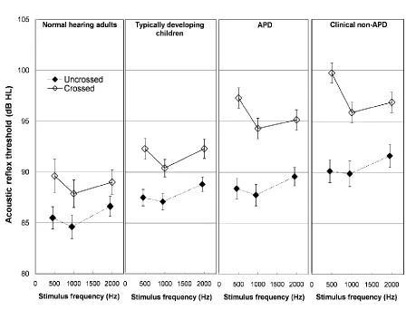
individual at each frequency and condition combination. Figure 2.1 shows the
mean and standard error of reflex thresholds measured at 500, 1000 and 2000
Hz for the uncrossed and crossed conditions in all groups averaged across ears.
Error bars show +1 standard error. Thresholds in crossed and uncrossed
Figure 2.1: Mean of crossed and uncrossed reflex thresholds at 500, 1000 and 2000 Hz in normal hearing adults, typically developing children, APD and clinical non-APD averaged for right and left ears. Uncrossed and crossed reflex thresholds are represented with filled and open
diamonds respectively. Error bars show +1 standard error.
Overall, reflex thresholds were higher in crossed than in uncrossed
conditions [F (1, 97) = 204.945, p < 0.001]. Consistent with previous reports
(Allen & Allan, 2014), thresholds also varied across groups [F (3, 97) = 9.470, p <
0.001] and there was a significant group by condition interaction [F (3, 97) =
thresholds and a larger crossed-uncrossed difference in the two groups of clinical
children when compared to the adults and typically developing children. To better
visualize the group-condition interaction differences between the crossed and
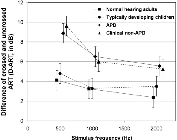
uncrossed reflex thresholds (D-ART) were calculated. The mean and standard
error of these differences (D-ART) at 500, 1000 and 2000 Hz are shown in Figure
2.2 for each group.
Figure 2.2: Mean of differences between crossed and uncrossed ART (D-ART) at 500, 1000 Hz and 2000 Hz (averaged for right and left ears). Squares, circles, diamonds and triangles represent normal hearing adults, typically developing children, APD and clinical non-APD, respectively. Error bars show +1 standard error.
A Bonferroni corrected post hoc t-test confirmed that typically developing
children and normal hearing adults had similar D-ARTs (p = 1.000). Normal
hearing adults had smaller D-ARTs in comparison to both clinical groups of
children, [APD (p = 0.002) and clinical non-ADP (p = 0.009)]. Typically
developing children also showed significantly different D-ARTs in comparison to
the APD (p = 0.007) and clinical non-APD (p = 0.030) groups. There were no
significant differences between the 2 clinical groups of children (p = 1.000).
These results indicate that, in comparison to the uncrossed reflex thresholds, the
crossed reflex thresholds were elevated to the greatest degree in the clinical
groups of children.
There was a significant effect of stimulus frequency on the reflex
thresholds [F (1.725, 167.282) = 18.452, p < 0.001] and a significant interaction
between stimulus frequency and condition [F (1.837, 178.224) = 25.339, p <
0.001]. In the crossed condition, thresholds at 1000 Hz were significantly lower
than those at 500 Hz (p < 0.001) or 2000 Hz (p = 0.001) and thresholds at 500
Hz were higher than 2000 Hz (p = 0.009). In the uncrossed condition, 500 and
1000 Hz had similar thresholds (p = 0.131) but significantly higher reflex
thresholds were recorded at 2000 Hz when compared to 500 (p = 0.001) and
2000 Hz (p < 0.001).
2.3.3
Discussion
Crossed and uncrossed reflex thresholds were measured in normal hearing
adults, typically developing children and children with suspected APD. The latter
group of children included those who received a diagnosis of APD based upon a
battery of clinically accepted behavioral tests (APD) and those who did not
(clinical non-APD). For the participants in this study, there was no right-left ear
difference on reflex thresholds which is consistent with previous reports