Developing peptide based capture agents
for diagnostics and therapeutics
Thesis by Arundhati Nag
In Partial Fulfillment of the Requirements for the degree of
Doctor of Philosophy
CALIFORNIA INSTITUTE OF TECHNOLOGY Pasadena, California
2013
DEDICATIONS
I have been lucky to work with several exceptional undergraduate students, Abdul Ahad Tariq, Mary Beth Yu and Belen Alvarez during my years at Caltech. Their work has been instrumental in finishing my dissertation.
During my research I have used resources from other research laboratories as well as Caltech central facilities. I thank Dr. Jost Vielmetter of the Protein Expression Center and Professors David A. Tirrell for the use of the SPR. I thank Professor David A. Baltimore for ready access to the ELISA plate reader. I thank Dr. Mona Shahgholi for assistance with MALDI-MS measurements. I thank Professor Carl S. Parker for the unconditional lab access he has provided and thank the Caltech Protein/Peptide Micro Analytical Laboratory for acquiring ESI-TOF spectra.
I would like to thank the Institute for Collaborative Biotechnologies, the Army Research Office, the National Cancer Institute and the Bill and Melinda Gates Foundation for funding my research.
5.2
Materials and Methods ... 130
5.2.1
Materials ... 130
5.2.2
Methods ... 130
5.2.2.1
Synthesis of randomized peptide library and on bead cyclization ... 130
5.2.2.2
Synthesis of scrambled phospho-epitope and target phospho-epitope ... 133
5.2.2.3
IR spectra ... 133
5.2.2.4
Screening cyclic OBOC library with peptide epitope ... 133
5.2.2.5
ELISA for cyclic peptide Cy(yytytTz4) against full length Akt2 protein .... 134
5.2.2.6
Non-radioactive kinase assay to evaluate effect of ligands on Akt2 kinase activity… ... 135
5.3
Results and discussion ... 136
5.3.1
Verification of on bead cyclization by Edman Peptide Sequencing ... 136
5.3.2
IR verification of on bead cyclized peptide formation by CuAAC reaction ... 137
5.3.3
Using on bead cyclic peptide library for ligand discovery ... 138
5.3.4
Determination of best cyclic binder to the target Akt2 C terminal fragment ... 139
5.3.5
Effect of cyclization on binding specificity ... 139
5.3.6
Effect of ring size on binding ... 140
5.3.7
Characterization of the cyclic ligand Cy(YYTYT-Tz4) ... 142
5.3.8
Effect of cyclic monoligand on Akt2 kinase activity ... 143
5.3.9
Optimization of monoligand anchor ... 144
Figure 5.2: Cyclic peptide ligands isolated by screening against target peptide epitope. ... 136
Figure 5.3: IR spectra of linear peptide L-Pra-VFAKV-L-Az4 and the cyclic peptide Cy(VFAKVTz4). ... 138
Figure 5.4: Selectivity of the cyclic peptide Cy(YYTYT-Tz4) for the target peptide. ... 139
Figure 5.5: The cyclized ligand Cy(YYTYT-Tz4) shows significant binding to the epitope compared to its linear version L-Pra-YYTYT-L-Az4. ... 140
Figure 5.6: Ring size affects cyclization efficiency. ... 141
Figure 5.7: Effect of ring size on epitope selectivity. ... 141
Figure 5.8: Determination of binding affinity of cyclic ligand Cy(YYTYT-Tz4) for Akt2 protein ... 142
Figure 5.9: Cy(YYTYT-Tz4) is selective for the Akt2 protein over the Akt1 and Akt3 proteins. ... 143
Figure 5.10: Cyclic ligand Cy(YYTYT-Tz4) inhibits Akt kinase activity. ... 143
Figure 5.11: Monoligand anchor candidate peptides developed for biligand screen. ... 145
(28) Vivanco, I.; Sawyers, C. L. Nat Rev Cancer 2002, 2, 489. (29) Manning, B. D.; Cantley, L. C. Cell 2007, 129, 1261.
(30) Yang, J.; Cron, P.; Good, V. M.; Thompson, V.; Hemmings, B. A.; Barford, D. Nat Struct Biol 2002, 9, 940.
Chapter 2:
Development of branched capture agents against bovine
dissolved in 40% aqueous THF. NaHCO3 (10 eq.) and Fmoc-OSu (1.1 eq.) were added 127.1, 127.7, 127.8, 130.4, 141.3, 143.6, 143.7, 155.8, 157.9.
31: 1H-NMR (400MHz, CDCl3): δ 1.40-1.52 (m, 4H), 1.60-1.68 (m, 2H), 1.76-1.82 (m, 2H), 120.0, 125.1, 127.1, 127.8, 130.4, 141.3, 143.7, 158.3.
13C-NMR (100MHz, CDCl3): δ 36.9, 47.1, 50.7, 54.7, 67.1, 67.3, 69.8, 70.2, 114.8, 120.0, 125.1, 127.1, 127.8, 130.4, 141.3, 143.7, 155.8, 157.9, 175.8
33: 1H-NMR (400MHz, CDCl 120.0, 125.1, 127.1, 127.8, 130.4, 141.3, 143.8, 155.8, 157.9, 175.4, 176.8.
Table 2.3: Hit sequences from the first biligand screen BB1.
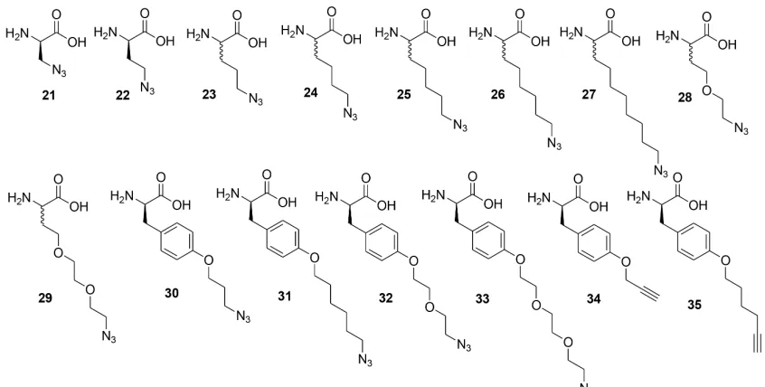
The library A of form X1X2X3X4X5X6-TG (where Xi= D-Trp, D-Lys, D-Ile, Gly, D-Leu, D-Val, D/L- azidolysine TG = tentagel S NH2 resin, diversity 117649) is screened with anchor peptide
37 with the Alexa-647 labeled bCAII protein.
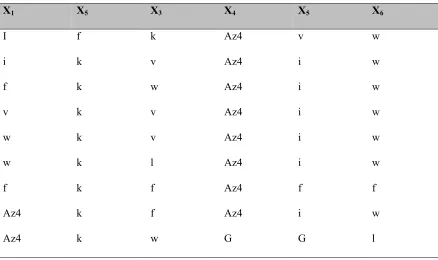
X1 X5 X3 X4 X5 X6
I f k Az4 v w
i k v Az4 i w
f k w Az4 i w
v k v Az4 i w
w k v Az4 i w
w k l Az4 i w
f k f Az4 f f
Az4 k f Az4 i w
Table 2.4: Hit sequences from the second generation biligand screen BB2.
The library B of form X1X2X3-D/L-Az4-X5X6-D-Met-TG (where Xi = Trp, Lys, Leu, D-Val, D-Ile, Gly, Met=L-Methionine, TG = tentagel S NH2 resin, 15552unique sequences) is screened with anchor peptide 37 with the Alexa-647 labeled bCAII protein in the biligand screen.
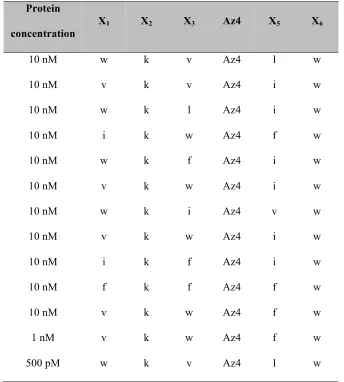
Protein
concentration
X1 X2 X3 Az4 X5 X6
10 nM w k v Az4 l w
10 nM v k v Az4 i w
10 nM w k l Az4 i w
10 nM i k w Az4 f w
10 nM w k f Az4 i w
10 nM v k w Az4 i w
10 nM w k i Az4 v w
10 nM v k w Az4 i w
10 nM i k f Az4 i w
10 nM f k f Az4 f w
10 nM v k w Az4 f w
1 nM v k w Az4 f w
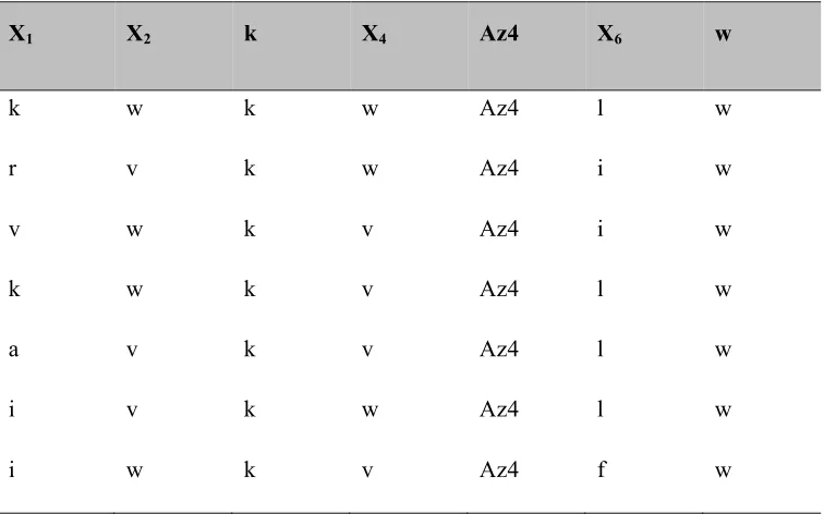
Table 2.5: Hit sequences from the third generation biligand screen BB3.
The library C of form X1X2kX4-Az4X6w-D-Met-TG (where X1= all 18 D amino acids except D Met and D-Cys; X2= D-Lys(Alloc)-OH, D-Trp, D-Val; X4= Arg, Asn, Gln, Asp, D-Lys, D-Ser, D-Thr, D-His, D-Ala, Gly, D-Val, D-Trp; X6 = D-Arg, D-Asn, D-Gln, D-Asp, D-D-Lys, D-Ser, D-Thr, D-His, D-Ala, Gly, D-Leu, D-Phe) is screened with anchor peptide 37 and Alexa-647 labeled bCAII protein .
X1 X2 k X4 Az4 X6 w
k w k w Az4 l w
r v k w Az4 i w
v w k v Az4 i w
k w k v Az4 l w
a v k v Az4 l w
i v k w Az4 l w
19 yifflr 58 kfhrrk 97 ftvllr
20 lraylr 59 yafflr 98 riyvrv
21 wrrfrr 60 kfyyrv 99 lrkwlw
22 ytGlfk 61 kiryfr 100 rfvkvf
23 pypyl 62 nwkwrk 101 wpherd
24 nrGnhr 63 khwrrr 102 pfdlw
25 Iiiyrs 64 rkawlr 103 fyyrk
26 yylvkr 65 fitrkf 104 pwfwG
27 llhltk 66 kwvver 105 rfvkvf
28 wpvpvf 67 ywlvkr 106 fkrkir
29 frvysf 68 lffrwv 107 wriyir
30 nfyyri 69 fafyvr 108 lfirly
31 rwklrr 70 wirirk 109 vfvkkl
32 kwtrei 71 hifirk 110 llrlay
33 wirGfy 72 rifvfr 111 rlrfhk
34 vyrkyk 73 llfyrk 112 prfyky
35 iyifrk 74 hyrkkw 113 rvkwkk
36 yrwrkf 75 ywflkk
Table 2.8: Hit sequences from the first generation screen TRI3 using the product screening method.
The library F of form D/L-Az4- X1X2X3X4X5-TG (Xi = all 18 D amino acids except D-Met and D-Cys) is screened with biligand anchor peptide 40 with the Alexa-647 labeled bCAII protein in the triligand screen.
Az4 X1 X2 X3 X4 X5
Az4 h d t f y
Az4 h d t G f
Az4 h d e G G
Az4 y s q w a
2.2.2.6 Dot blots
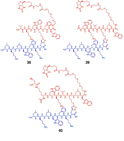
anchor to the middle of the second peptide arm. X2 and X6 positions of the hit sequences were also found to be highly conserved. A second focused library was synthesized, where the position of the D/L-azidolysine was fixed at X4. The library was screened against three different concentrations (10 nM, 1 nM and 500 pM) of Alexa 647-labeled bCAII in presence of peptide 37. The beads with highest fluorescence intensity were picked. This yielded eleven beads from the 10 nM screen, and one bead each from the 1 nM and 500 pM screens (Table 2.4). The trends noticed in the first screen were even more noticeable in this screen, yielding two motifs, wkX-Az4-Xw and vkX-Az4-Xw. Biligands 38 and 39 were synthesized in which vkw-Az4-lw and wkv-Az4-lw were clicked to the peptide 37.
Figure 2.3: Molecular structures of the reported monoligand 36 and the monoligand anchor 37 used in the branched biligand screen.
Figure 2.5: Motifs obtained by screening against bovine carbonic anhydrase II in presence of monoligand anchor peptide 37.
Figure 2.6: Molecular structures of branched biligands 38 and 39 and biligand anchor 40.
In the branched biligands 38 and 39, the 2° arm isolated through screens (in red) is linked by CuAAC chemistry to the monoligand anchor 37 (in blue). An alkyne is appended to the biligand
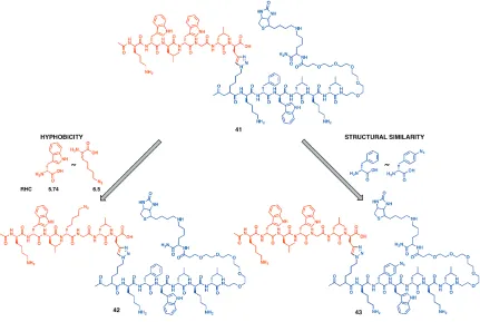
The other strategy of amino acid replacement commonly followed in literature is to replace an amino acid with an artificial derivative having similar molecular structure. Following this strategy, D-phenylalanine of the biligand 41 was replaced with 4-azido-D-phenylalanine to synthesize biligand 43. The binding affinities of biligands 42 and 43 for bCAII as determined by SPR were similar to the binding affinity of linear biligand 41 for bCAII. So both replacement strategies do not compromise the binding affinity of the modified biligand in SPR measurements (Figure 2.10). However, the yield of 43 was not high due to instability of phenyl azide under the harsh deprotection conditions during peptide synthesis.
Figure 2.9: Amino acid substitution in the linear biligand to develop branched biligands.
Figure 2.10: SPR response sensograms of the branched biligands developed by amino acid substitution of the linear biligand.
2.3.3
Branched triligand development with substituted biligand
The biligand 42 obtained by modifying the linear biligand 41 using the hydrophobicity approach was utilized as a new anchor peptide for a branched triligand peptide development. In the in situ click target screen, the Alexa Fluor 647 labeled bCAII pretreated with biligand 42 was screened against library D. The beads with high fluorescence intensity are automatically sorted using COPAS plus and sequenced by Maldi TOF/TOF24 (Table 2.6). The 8 most frequently occurring amino acids were used to generate the focused library. Screening this focused library gave highly homologous peptide sequences (Table 2.7). The hit sequences are found to contain into three motifs (denoted by red, green and blue colors in the sequences). One peptide was chosen from each of the three motifs as the triligand arm. Triligand 45 was constructed with the sequence Ac-(D-Pra)fkifvr as the third peptide arm. Other two triligands 46 and 47 were synthesized with Ac-(D-Pra)irlflk and Ac-(D-Pra)kyffrf as the third peptide arm respectively.
Figure 2.11: Schematic representation of the in situ click target screens TRI1 and TRI2 for developing branched triligand.
reaction of the biligand anchor with triligand arm hdtgf, hdtfy, hdegg and Az4-ysqwa, respectively.
Figure 2.14: Scheme for in situ click product screen TRI3 for development of branched triligand.
Figure 2.15: Molecular structures of branched triligands developed through product screen against the bovine carbonic anhydrase II.
Figure 2.17: ELISA demonstrating relative binding affinities of biligands and triligands.
Binding of linear biligand 41 (purple), branched biligand 42 (black), linear triligand 44 (red), and branched triligands 46 (blue) and 47 (green) for the CAII protein. The biotinylated ligands are immobilized on a SA plate and treated with different concentrations of the protein. The individual absorbance values are denoted as points and the fits are denoted as solid lines.
2.3.6
Substitution of triazole linkage by triazole mimic
challenging to synthesize the 1,5 disubstituted triazole linked peptide biligand in bulk for different assays. To overcome this difficulty, a mimic of the 1,5 disubstituted triazole linker was synthesized. As proline has been successfully replaced by the 1,5 disubstituted triazole molecule in proteins25, we reversed the concept to replace 1,5 disubstituted triazole with proline. To recreate the length of the 1,5-triazole link that is formed between L-azidolysine and D-propargyl glycine, as in the in situ protein click reaction, D-Pro and D-Asp was coupled to the acid side chain of L-diaminobutyric acid.
Figure 2.18: Molecular structure comparison of 1,5 disubstituted triazole linked biligand and its mimic.
Chapter 3:
Epitope targeting strategy: developing capture agents
a cocktail of TFA, TES and double distilled water (95:2.5:2.5), precipitated in ice cold ether and lyophilized. The crude solid was used in further synthesis. 2 equivalents of zinc acetate was dissolved in methanol and added to 1 equivalent of compound 2 and stirred overnight at room temperature. The solvent was removed under reduced pressure and the solid was purified using a gradient of water and acetonitrile and 0.1% TFA on the RP-HPLC (Beckman Coulter System Gold 126 Solvent Module and 168 Detector) using a C18 reversed phase semi-preparative column (Phenomenex Luna 10 µm, 250 × 10 mm). Calculated mass: [M].2H2O 1369.45, Observed mass: [M].2H2O 1369.
3.2.2.2 Verification of binding of Zn2L-Az4-PEG2-Biotin to phospho-amino acids and a
phosphate containing peptide
Figure 3.1: Synthesis of dinuclear Zn chelator Zn2L-Az4-PEG2-Biotin.
Figure 3.2: Evidence of binding of dinuclear zinc chelator to phospho amino acids and phospho peptide.
Table 3.3: Hit sequences from the biligand (bi-L) screen with 25 nM target peptide.
X1 X2 X3 X4 X5 D-Pra
h n G i i D-Pra
h n G r e D-Pra
h r y y G D-Pra
v n r r f D-Pra
h n G G d D-Pra
a y p h f D-Pra
G f r r f D-Pra
r G f f l D-Pra
h n G y G D-Pra
Table 3.4 Hit sequences for biligand screen with 10 nM target peptide.
X1 X2 X3 X4 X5 D-Pra
v y y r h D-Pra
h n G a I D-Pra
f h y y y D-Pra
f y h k h D-Pra
p f q h f D-Pra
s h f y t D-Pra
Peptides generally were synthesized on the Titan 357 Automatic Peptide Synthesizer (AAPPTec, Louisville, KY). Overnight coupling steps were performed manually in 8 ml fritted polysterene tubes. The protocols for the peptide general synthesis, cleavage, Cu catalyzed on bead click reaction, and HPLC protocols followed are described in chapter 2. The synthesis and characterization of the individual peptides follow:
Target phospho-peptide:
The 32mer target peptide sequence, amino acids 450-481 of Akt2, ITPPDRYDSLGLLELDQRTH-FPQF(pS)YSASIRE was synthesized on Rink Amide MBHA resin, using the Titan 357 peptide synthesizer. Fmoc-Ser(OPO3Bzl)-OH (AaPPTec) was used for the incorporation of phosphoserine in the peptide. Calculated mass: [M+H]+ 3832. Observed mass: [M+H]+ 3831.97
Figure 3.3: Sequence of the phospho peptide used as target epitope.
The target phospho-peptide, amino acids 450-481 of Akt2, is phosphorylated at Ser474. The hydrophobic motif is highlighted in red.
Synthesis of mono-L:
(Anaspec) following standard Fmoc SPPS synthesis protocol. 1.5 equivalents of S1 were then coupled to the peptide. After TFA cleavage the mono-L was purified using a gradient of water and acetonitrile and 0.1% TFA on the RP-HPLC. Calculated mass: 1494.8. Observed mass: 1494.6
Figure 3.4: Synthesis of the intermediate S1 for the bulk synthesis of the monoligand peptide mono-L.
Figure 3.5: Structure of the monoligand peptide mono-L developed against the target peptide.
Synthesis of compound 4:
Fmoc-NH-PEG2-OH was coupled using standard Fmoc protocol on Biotin Novatag resin. 1.5 equivalent of D,L-Fmoc-azidolysine was coupled on the resin followed by coupling of 1.5 equivalent of compound 2. The resin was then subjected to on bead Cu catalyzed click reaction with Fmoc - D-Pra-OtBu. The excess copper was removed by washing with the copper chelating solution. 5-Azido-pentanoic acid was then coupled. The resulting peptide S2 was TFA cleaved and lyophilized. The crude was used in further synthesis.
Figure 3.6: Synthesis of the zinc chelator - monoligand complex compound 4 used in biligand screen of Akt2.
Synthesis of bi-L:
standard Fmoc SPPS synthesis. After TFA cleavage the biligand was purified using a gradient of water and acetonitrile and 0.1% TFA on the RP-HPLC. Mass calculated: 2309.7 Mass observed: 2309.4
Figure 3.7: Synthesis of intermediate S3 for bulk synthesis of the biligand bi-L, biligand anchor anchor-3Cand the triligand C– term–tri-L.
S3 is used in the synthesis of the bi-L, Anchor-3C and C– term–tri-L.
Figure 3.10: Structure of biligand anchor peptides Anchor-3N and Anchor-3C.
A. Anchor-3N is used to the Akt2 protein before screening against a alkyne containing OBOC library A. Anchor-3C is used to the Akt2 protein before screening against a alkyne containing OBOC library.
Synthesis of N-term-tri-L:
Figure 3.11: Molecular structure of the triligand N-term-tri-L.
Synthesis of N-term-tri-dimer:
Figure 3.13: Molecular structure of the triligand N-term-tri-dimer.
Synthesis of D-Lys (pentyne) amide:
Boc-D-Lys(Fmoc)-OH was coupled with rink amide resin. Then 4-pentynoic acid was coupled with it, after standard piperidine deprotection. The dried resin was cleaved with TFA cocktail and purified using a gradient of water and acetonitrile and 0.1% TFA on the prep-HPLC. Mass calculated: (M+H) 225 Mass observed: 226
Synthesis of C-term-tri-L:
was carried out with Fmoc-D-Pra -OtBu. After washes with copper chelating solution, the peptide was further extended to hngyf on the N terminal using standard Fmoc SPPS synthesis. The dried resin was cleaved with TFA cleavage solution and purified using a gradient of water and acetonitrile and 0.1% TFA on the RP-HPLC. Mass calculated: 3199.6 Mass observed: 3198.4.
Figure 3.14: Molecular structure of the triligand C-term-tri-L.
3.3
Results and discussion
3.3.1
Synthesis of the dinuclear zinc chelator Zn
2L-Az4-PEG
2-Biotin and
verification of its binding to phospho amino acids and phosphopeptide
aromatic Mannich reaction of 2.5 equivalents of N,N –di(2-picolyl)amine and 3.75 equivalents of formaldehyde with ethyl 4-hydroxobenzoate under reflux conditions. Alkaline hydrolysis of the ester yielded 3,5-bis((bis(pyridin-2-ylmethyl)amino)methyl)-4-hydroxybenzoic acid (compound
3, Figure 3.1), a heptadentate ligand. The carboxylic acid group on 2 is compatible with solid phase Fmoc based peptide synthesis. To add an azido tag and a biotin tag to 2, Fmoc -azidolysine was coupled using standard SPPS protocols to Biotin PEG Novatag resin, followed by the coupling of 3 to the resin. After cleaving the compound off the resin with trifluoroacetic acid and subsequent HPLC purification, a treatment in solution with Zinc acetate yielded the desired ligand Zn2L-Az4-PEG2-Biotin(Figure 3.1). The property of Zn2L-Az4-PEG2-Biotinto selectively bind to phospho-serine, phospho-tyrosine and a phospho-peptide (pSrc substrate) was demonstrated by incubating Zn2L-Az4-PEG2-Biotin with the respective amino acids or peptide in borate buffer, followed by Maldi TOF in a positive mode, that yielded the peaks for Zn2L-Az4-PEG2-Biotin -pSer, Zn2L-Az4-PEG2-Biotin -pTyr and Zn2L-Az4-PEG2-Biotin -pSrc complexes23 (Figure 3.2).
3.3.2
Modification of peptide fragment of Akt2
3.3.3
Identification of a 1
oligand targeted against Akt2 C terminal fragment
Figure 3.16: Screening strategy for developing a capture agent targeting the C terminal hydrophobic motif of the Akt2 protein.
Zn2L-Az4-PEG2-Biotin is coupled with the Akt2 32-mer C-terminal fragment containing p-S474 through the interaction to create Complex-1. Complex-1is screened against Library A to yield 1o ligand candidates, from which a consensus 1o ligand (mono-L) is identified. In the next round of ligand development, compound 4 is coupled to the C-terminal fragment through the Zn chelator to create Complex-2. Complex-2 is screened against Library B to identify candidate 2o ligands, from which a consensus biligand (bi-L) is prepared.
3.3.4
Identification of a biligand targeted against Akt2 C terminal fragment
We used a similar strategy to identify candidate 2o ligands. The Zn chelator Zn2L-Az4-PEG2-Biotin was modified with peptide wkvkl and an azide on the N terminal to form compound
Figure 3.17: Scheme describing triligand screens using anchor-3N and anchor-3C.
Anchor-3N and Anchor-3C are synthesized by modifying bi-L with an azide group at the N- terminal and an alkyne group at the C-terminal of the peptide, respectively. Anchor-3N and Anchor-3Care separately screened against Library A and Library C in the presence of full length Akt2 to identify candidate 3o ligands, from which the consensus triligands are identified. The screens involve various other steps (not shown) that were designed to remove false positive hits. All hit beads are identified using alkaline-phosphatase (AP)-labeled anti-biotin antibody as a means of detecting the on-bead triazole linked product.
3.4
Conclusion
insert a click handle. In fact, our preliminary data with that method yielded sequences similar to the followed method. The epitope targeting strategy is a particularly valuable strategy when the target protein has posttranslational modifications like glycosylation or phosphorylation, which are difficult to express or purify.
Chapter 4:
Characterization of protein capture agents developed
polypeptide Akt2 fragment (corresponding to residues 346 through 378) that is similarly surface exposed and unstructured in the full length protein. All the three capture agents exhibited a high selectivity for the C-terminal 32-mer relative to the other polypeptide fragment (Figure 4.3)
The selectivity of the N-term-tri-L and C-term-tri-L for Akt2 over its isoform Akt1 and Akt3 was determined at the epitope level and against the full-length protein (Figure 4.4). One of the triligands, C-term-tri-L is highly selective for the peptide epitope of Akt2, and bind significantly less to the highly homologous C terminal epitopes of isomeric proteins Akt1 and Akt3. The same trend is observed in the binding of the ligands to the full-length Akt isoform proteins.
Figure 4.1: Immunoprecipitation and Western Blot assays showing the performance of the various PCC Agents.
Akt2 is pulled down from the lysate of an ovarian cancer cell line Acetyl-glycine-biotin is used as the control.
Figure 4.2: ELISA assays demonstrating use of PCC Agents.
Figure 4.3: Epitope specificity assay.
The various triligand PCC agents are used as capture agents for the peptide epitope. The biotinylated capture agents are immobilized on a Streptavidin plate, and then probed with the His6 tagged target or control peptides. The target peptide (Akt2 450-481) with pS474 shows a distinct binding affinity for the capture agents compared to the control peptide (Akt2 346-378). The C-term-tri-L also significant selectivity for the Akt2 epitope compared to the corresponding regions in Akt1 (449-480) and Akt3 (448-479).
Figure 4.4: Selectivity of triligands for the full length Akt isoform proteins.
Akt1 fragmentAkt2 fragmentAkt3 fragmentAkt2 off-target 0.0
0.5 1.0
Fr
a
c
tion bound
Figure 4.5: SPR measurements of triligands.
A. Sensograms from Surface Plasmon Resonance (SPR) experiment, immobilizing N–term-tri-L on SA chip and using Akt2 protein as the analyte. B. Steady state dissociation constant is measured by taking an average of the RUs in steady state over 3 time points, 150s, 200s and 250s, for each concentration of the protein. C. Sensograms from Surface Plasmon Resonance (SPR) experiment, immobilizing N –term-tri-L-dimer on SA chip and using Akt2 protein as the analyte. Because of the dimeric nature of the ligand, the sensograms fit best with the heterogenous ligand parallel model.
A.
C.
4.3.2
Inhibitory Characteristics of the Akt2 Capture Agents
to overlap at least partially with a commercial mAb that was directed at the C-terminus of the protein. As the phosphorylated hydrophobic motif around S474 (near the C-terminus) of Akt2 has been identified to act as an allosteric activator, we hypothesized that a ligand that was targeted near S474 would influence the enzymatic activity of Akt2. In this section, we explore the inhibitory characteristics of the various ligands developed here.
Figure 4.6: Influence of the PCC Agents on the enzymatic activity of the Akt2 kinase.
Chapter 5:
Development of CuAAC cyclized one bead one compound
library, all the beads in the linear library were subjected to an on bead CuAAC reaction, for 6 hours at room temperature with 1.5 equivalents of CuI, 2.5 equivalents of ascorbic acid in 20% piperidine in DMF (Figure 5.1). After washes to remove the adsorbed Cu, the library was washed with DMF, methanol and DCM and dried. Random beads were picked from the library to be sequenced. The rest of the library was stored in NMP. The structure and details of the three libraries are shown in Table 5.1.
wells were washed with binding buffer five times. Serial dilutions, from 1.15 nM to 450 nM, of active His6 tagged Akt2 (Abcam) were made in the binding buffer. 100 µl of each solution was added per well the plate shaken overnight at 4°C. The wells were washed three times with the binding buffer, then treated for an hour with a 1:1000 dilution of anti His6 mouse monoclonal antibody. A 1:10,000 dilution of goat anti mouse antibody-Horse Radish Peroxide conjugate (Abcam) in binding buffer was added to the wells for an hour. The plate was washed, treated with TMB and quenched with H2SO4, and the absorbance at 450 nm wavelength measured. From the A450 value of each protein concentration, the corresponding blank A450 (with no ligand, same amount of protein) was subtracted. All the A450 values were normalized w.r.t the maximum A450 value. They were then fitted by non-linear regression in Graphpad Prism 6.
5.2.2.6 Non-radioactive kinase assay to evaluate effect of ligands on Akt2 kinase activity
Figure 5.2: Cyclic peptide ligands isolated by screening against target peptide epitope.
A) Cy(YVYKS-Tz4), B) Cy(VFAKV-Tz4), C) Cy (IRYYS-Tz4), D) Cy(YYTYT-Tz4)
5.3
Results and discussion
5.3.1
Verification of on bead cyclization by Edman Peptide Sequencing
Figure 5.3: IR spectra of linear peptide L-Pra-VFAKV-L-Az4 and the cyclic peptide Cy(VFAKVTz4).
5.3.3
Using on bead cyclic peptide library for ligand discovery
are washed with protein denaturants and then screened with the target phospho-peptide epitope. Libraries A and B on screening in this protocol did not yield any hit beads against the target phospho-epitope. Only library C yielded four hits. The hits are listed in Table 5.2.
5.3.4
Determination of best cyclic binder to the target Akt2 C terminal
fragment
The four peptide candidates were tested for their capacity to capture the target peptide in a sandwich ELISA platform. Biotinylated capture agents were immobilized on Streptavidin plates and titrated with 1 µM of either the His6 tagged target peptide or the scrambled peptide. Binding was detected by mouse anti His6 tag antibody. Only peptide Cy(YYTYT-Tz4) showed binding to the target peptide and no binding to the scrambled peptide.
Figure 5.4: Selectivity of the cyclic peptide Cy(YYTYT-Tz4) for the target peptide.
5.3.5
Effect of cyclization on binding specificity
Figure 5.6: Ring size affects cyclization efficiency.
L-Pra – YYTYT– L-Az2 cyclizes on bead as the major product while L-Pra – YYTYT– L-Az1 does not cyclize efficiently on bead
Figure 5.7: Effect of ring size on epitope selectivity.
5.3.7
Characterization of the cyclic ligand Cy(YYTYT-Tz4)
To determine the affinity of the cyclic monoligand for full length Akt2 protein, the ligand was titrated against immobilized protein. The ligand acts as a detection agent and has a low nanomolar (124 nM) binding affinity compared to the low micromolar affinity (3.6 µM) of the linear monoligand previously developed against Akt. Thus there is a ~ 30 fold improvement in the binding affinity using a cyclic instead of linear peptide monoligand for Akt2. It will be interesting to see if the same improvement can be attained in the biligand stage using a cyclic peptide as the 2 °arm.
We demonstrate the selectivity of the cyclic monoligand for Akt2 protein by comparing the binding of the ligand to the proteins Akt1, Akt2 and Akt3. As the proteins are highly homologous, even in the targeted C terminal region, it is not surprising that binding to all the three proteins is observed to some extent. However there is a distinct preference for Akt2.
Figure 5.9: Cy(YYTYT-Tz4) is selective for the Akt2 protein over the Akt1 and Akt3 proteins.
5.3.8
Effect of cyclic monoligand on Akt2 kinase activity
To evaluate if the cyclic ligand has any effect on the kinase activity of the Akt2 protein, we performed the non-radioactive kinase assay described in Chapter 4. Briefly, the protein is treated with the ligand, ATP and an enzyme substrate, in this case, GST-GSK-3α/β fusion protein. The reaction mixture is analyzed using western blot to detect the amount of substrate phosphorylated. Under normal conditions (as in the DMSO control) the GST-GSK-3α/β fusion protein will be phosphorylated by Akt2. If, however, the ligand acts as an inhibitor, it will decrease the formation of phospho-Ser21/9- GST-GSK-3α/β fusion protein. The cyclic monoligand, Cy (YYTYT-Tz4) inhibits the phosphorylation of the substrate. As this was a preliminary experiment, we used a high concentration (2mM) of the cyclic ligand. Further verification with lower concentrations of the cyclic peptide is required.
5.3.9
Optimization of monoligand anchor
Figure 5.11: Monoligand anchor candidate peptides developed for biligand screen.
Figure 5.12: Comparison of HPLC traces to determine best monoligand anchor candidate.
5.4