Immunosorbent Assay and Virus Neutralization Assay To Detect
Antibodies to Viral Hemorrhagic Septicemia Virus
Anna Wilson,aTony Goldberg,aSusan Marcquenski,bWendy Olson,cFrederick Goetz,dPaul Hershberger,eLucas Hart,e Kathy Toohey-Kurtha,f
Department of Pathobiological Sciences, University of Wisconsin, Madison, Wisconsin, USAa; Wisconsin Department of Natural Resources Bureau of Fisheries Management, Madison, Wisconsin, USAb; Department of Pathology, Medical College of Wisconsin, Milwaukee, Wisconsin, USAc; National Oceanic and Atmospheric Administration (NOAA), Manchester Research Station, Port Orchard, Washington, USAd; U.S. Geological Survey (USGS), Western Fisheries Research Center, Marrowstone Marine Field Station, Nordland, Washington, USAe; Wisconsin Veterinary Diagnostic Laboratory, University of Wisconsin-Madison, Madison, Wisconsin, USAf
Viral hemorrhagic septicemia virus (VHSV) is a target of surveillance by many state and federal agencies in the United States.
Currently, the detection of VHSV relies on virus isolation, which is lethal to fish and indicates only the current infection status.
A serological method is required to ascertain prior exposure. Here, we report two serologic tests for VHSV that are nonlethal,
rapid, and species independent, a virus neutralization (VN) assay and a blocking enzyme-linked immunosorbent assay (ELISA).
The results show that the VN assay had a specificity of 100% and sensitivity of 42.9%; the anti-nucleocapsid-blocking ELISA
de-tected nonneutralizing VHSV antibodies at a specificity of 88.2% and a sensitivity of 96.4%. The VN assay and ELISA are
valu-able tools for assessing exposure to VHSV.
V
iral hemorrhagic septicemia (VHS) is one of the most
patho-genic viral diseases of fish worldwide and affects a wide range
of host species (
1–7
). Of the four genotypes, the North American
strains of VHS virus are designated types IVa and IVb. Type IVa
was originally isolated from asymptomatic marine salmonids in
the Pacific Northwest in 1988 (
8
); it is now known to be endemic
throughout the northeast Pacific, where it is highly virulent to
populations of Pacific herring (
Clupea pallasii
) and other marine
fishes (
9
). A new freshwater strain, type IVb, was isolated from a
muskellunge (
Esox masquinongy
) collected from Lake St. Clair,
MI, in 2003 (
10
). This distinctive sublineage has been isolated
from 31 species of fish in the Great Lakes (
11
) and has been
asso-ciated with significant die-off events of freshwater drum (
Aplodi-notus grunniens
), muskellunge (
Esox masquinongy
), gizzard shad
(
Dorosoma cepedianum
), round gobies (
Apollonia melanostomus
),
and yellow perch (
Perca flavescens
) in the Great Lakes between
2005 and 2008 (
2
,
10
,
12–16
). By 2009, the virus had spread to all
of the Great Lakes and several inland lakes. The introduction and
spread of this pathogen and the threat it poses to a broad range of
hosts resulted in increased surveillance of the virus in Wisconsin
and other states within the Great Lakes Basin.
Currently, the surveillance methods for VHS virus (VHSV)
detection include virus isolation in cell culture, followed most
commonly by confirmation by reverse transcription-PCR
(RT-PCR). Fish are tested for VHSV according to the guidelines
out-lined in the American Fisheries Society Fish Health Section Blue
Book (
17
) and the World Organisation for Animal Health (OIE)
Manual of Diagnostic Tests for Aquatic Animals (
18
). Both
ap-proved methods detect the virus but do not detect antibodies
in-dicative of previous virus exposure.
Clinical signs of disease are not consistent among
VHSV-sus-ceptible species, and VHSV IVb is not always isolated from
clini-cally affected fish, especially salmonids (
19
,
20
). Differences in
susceptibility and mortality rates among different populations of
yellow perch have been reported recently (
21
). The clinical signs
and severity of infection also depend on water temperature at the
time of infection, stress level, host age, and other environmental
factors (
15
,
22
). These variables can affect the narrow window of
opportunity to detect VHSV by virus isolation; therefore, diseased
or recovered individuals may easily be missed during surveillance
efforts.
Methods to detect neutralizing antibodies to VHSV have been
developed for surveillance using a complement-dependent
neu-tralization test (50% plaque neuneu-tralization test [PNT]) and have
been highly sensitive and specific for trout (
23–25
). However,
PNT requires overlay and plaque enumeration steps; further, this
method is best suited for small sample sizes. A
microneutraliza-tion format without the use of overlay might lead to a 50%
reduc-tion in the resources and labor required to perform the assay.
Another advantage of a virus neutralization assay is that the
indi-cator system is a susceptible cell line for the target virus, which
makes the assay inherently species independent.
Competitive and blocking enzyme-linked immunosorbent
as-says (ELISAs) for the detection of antibodies against mammalian
viruses have been in use for decades. Indirect ELISAs have been
available for VHSV since 1988 (
26
). A highly sensitive (92%)
in-direct ELISA for detecting nonneutralizing antibodies for the
sur-veillance of VHS in farmed rainbow trout (
Oncorhynchus mykiss
)
has also been described (
25
), but it requires a species-specific
sec-ondary fish antibody. Thus, these tests are not practical for
mul-tispecies VHSV surveillance in the wild, because there are at least
31 species known to be susceptible to VHSV IVb.
Received22 October 2013Returned for modification20 November 2013
Accepted10 January 2014
Published ahead of print15 January 2014
Editor:W. R. Waters
Address correspondence to Kathy Toohey-Kurth, [email protected]. Copyright © 2014, American Society for Microbiology. All Rights Reserved.
doi:10.1128/CVI.00675-13
on August 17, 2020 by guest
http://cvi.asm.org/
Here, we describe the development and evaluation of a
modi-fied virus neutralization (VN) assay and a blocking enzyme-linked
immunosorbent assay (ELISA). Both tests were adapted from
pre-viously described methods in the World Organisation for Animal
Health (OIE) manual (
27
) and use blood serum samples from
uninfected fish and VHS survivors. Although the development of
the anti-VHSV nucleocapsid monoclonal antibody used in our
ELISA was published in 1988 (
26
), no competitive ELISA methods
have been reported until now. These serological methods broaden
the window of detection by demonstrating whether previous
ex-posure to the virus had occurred, which might alleviate the time
constraints of surveillance efforts using virus isolation.
MATERIALS AND METHODS
Cell lines.The epithelioma papulosum cyprini (EPC) cell line (American Type Culture Collection, Manassas, VA), originating from fathead min-now (Pimephales promelas) epithelial cells (28), was cultured at 25°C ac-cording to detailed protocols (18,29), and the medium was supplemented with tryptose phosphate broth (Teknova; Hollister, CA, USA), 5% fetal bovine serum (FBS) (PAA Laboratories, Inc., Etobicoke, Ontario, Can-ada), 200 mML-glutamine (Life Technologies), and buffered with 7.5% sodium bicarbonate solution (Life Technologies). The Chinook salmon embryo (CHSE-214) cell line (American Type Culture Collection, Manas-sas, VA) was cultured at 20°C, and the medium was supplemented with 10% FBS (PAA Laboratories, Inc.).
Virus isolate.The Great Lakes strain of VHSV (type IVb) was isolated on the EPC cell line. The isolate was obtained from pooled kidney and spleen tissue samples from a freshwater drum in Lake Winnebago during a VHS outbreak in 2007 and confirmed by real-time RT-PCR (30,31).
Virus propagation and purification.Virus was adsorbed to the EPC cells at a multiplicity of infection (MOI) of 0.01 for 30 min at room temperature and then supplemented with cell culture medium. To prop-agate virus, flasks were incubated at 15°C for 5 to 7 days or until the first signs of cytopathic effect (CPE) were observed. A plaque-purified stock of VHSV-infected EPC cell supernatant was clarified by the removal of EPC cells, aliquoted for one-time use, and stored at⫺80°C. Postfreezing, an aliquot was thawed, and the titer of the batch was determined.
CHSE cells were used to propagate virus for ELISA antigen coating. The flasks for propagation were inoculated using the same methods as for the EPC cells but virus was adsorbed for 1 h at room temperature. After two freeze-thaw cycles, cell debris was removed by centrifugation at 4,000⫻gfor 15 min at 4°C in a Sorvall ST40R centrifuge (Thermo) and clarified. The supernatant was purified and concentrated according to the manufacturer’s protocols using a Fast-Trap virus purification and con-centration kit (Millipore, Billerica, MA). Eluted virus was aliquoted and stored at⫺80°C. A mock infection was performed in a similar manner to provide cell lysates for determining an optical density baseline in unin-fected CHSE cells. Antigen was treated with 10% MEGA-10 detergent (Sigma-Aldrich) for an hour at room temperature prior to diluting in coating buffer for use in the ELISA.
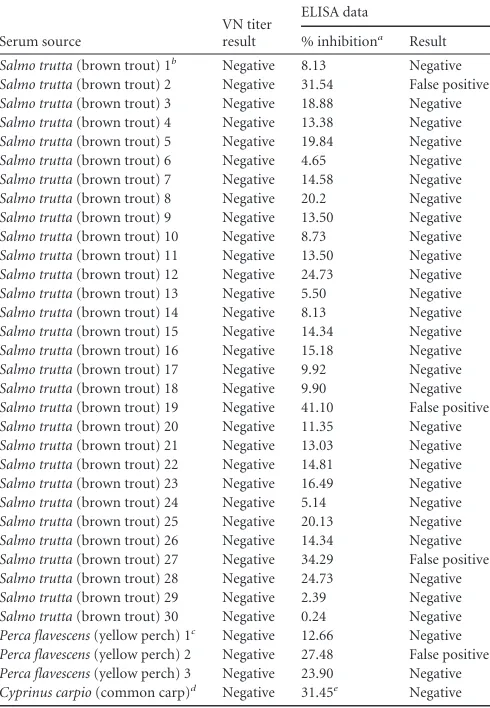
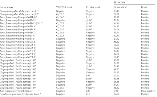
Sera from fish of known infection status.Blood serum samples were obtained from 33 uninfected fish (Table 1), including brown trout (Salmo trutta) and yellow perch. A blood serum sample with antibodies to spring viremia of carp virus (SVCV) was obtained from a common carp ( Cypri-nus carpio) (Table 1). The serum samples were collected 4 to 5 months after an SVCV epizootic occurred in May 2002 in Cedar Lake, WI, and tested positive for neutralizing antibodies to SVCV at the Center for En-vironment, Fisheries, and Aquaculture Science (CEFAS) in Weymouth, United Kingdom, using a competitive ELISA (32). Serum samples were obtained from 28 experimentally infected or wild-caught fish that had survived exposure to VHSV (Table 2), including grass carp ( Ctenopharyn-godon idella), yellow perch, Pacific herring (C. pallasii), muskellunge, and freshwater drum. All serum samples were stored frozen at⫺80°C and
then heated to 45°C for 30 min to inactivate complement before use in assays.
All yellow perch used in the study were hatched and reared at the University of Wisconsin, Milwaukee (UWM) School of Freshwater Sci-ences (SFS) Aquaculture Research Facility, according to previously de-scribed methods (33). They were exposed to VHS virus strain IVb (MI03) by intraperitoneal (i.p.) injection at a titer of 1⫻104PFU/fish (J-Z fish) or 1⫻102PFU/fish (H-Y fish). At 28 days post-VHSV injection, all yellow perch exhibited mild clinical signs of the disease, such as exophthalmia and hemorrhaging. The fish euthanized on day 64 appeared healthy, ex-hibiting no clinical signs of VHS, and all plaque assay results were nega-tive.
Hyperimmunized Pacific herring (C. pallasii) were produced from laboratory-reared specific-pathogen-free (SPF) colonies (34). Briefly, SPF herringⱖ5 years of age were immersed in waterborne VHSV (1.5⫻103 TABLE 1Virus neutralization and blocking ELISA results for VHS-negative group
Serum source
VN titer result
ELISA data
% inhibitiona Result Salmo trutta(brown trout) 1b
Negative 8.13 Negative
Salmo trutta(brown trout) 2 Negative 31.54 False positive
Salmo trutta(brown trout) 3 Negative 18.88 Negative
Salmo trutta(brown trout) 4 Negative 13.38 Negative
Salmo trutta(brown trout) 5 Negative 19.84 Negative
Salmo trutta(brown trout) 6 Negative 4.65 Negative
Salmo trutta(brown trout) 7 Negative 14.58 Negative
Salmo trutta(brown trout) 8 Negative 20.2 Negative
Salmo trutta(brown trout) 9 Negative 13.50 Negative
Salmo trutta(brown trout) 10 Negative 8.73 Negative
Salmo trutta(brown trout) 11 Negative 13.50 Negative
Salmo trutta(brown trout) 12 Negative 24.73 Negative
Salmo trutta(brown trout) 13 Negative 5.50 Negative
Salmo trutta(brown trout) 14 Negative 8.13 Negative
Salmo trutta(brown trout) 15 Negative 14.34 Negative
Salmo trutta(brown trout) 16 Negative 15.18 Negative
Salmo trutta(brown trout) 17 Negative 9.92 Negative
Salmo trutta(brown trout) 18 Negative 9.90 Negative
Salmo trutta(brown trout) 19 Negative 41.10 False positive
Salmo trutta(brown trout) 20 Negative 11.35 Negative
Salmo trutta(brown trout) 21 Negative 13.03 Negative
Salmo trutta(brown trout) 22 Negative 14.81 Negative
Salmo trutta(brown trout) 23 Negative 16.49 Negative
Salmo trutta(brown trout) 24 Negative 5.14 Negative
Salmo trutta(brown trout) 25 Negative 20.13 Negative
Salmo trutta(brown trout) 26 Negative 14.34 Negative
Salmo trutta(brown trout) 27 Negative 34.29 False positive
Salmo trutta(brown trout) 28 Negative 24.73 Negative
Salmo trutta(brown trout) 29 Negative 2.39 Negative
Salmo trutta(brown trout) 30 Negative 0.24 Negative
Perca flavescens(yellow perch) 1c Negative 12.66 Negative
Perca flavescens(yellow perch) 2 Negative 27.48 False positive Perca flavescens(yellow perch) 3 Negative 23.90 Negative Cyprinus carpio(common carp)d
Negative 31.45e
Negative a
Results determined positive atⱖ25% inhibition for test sera diluted 1:2 andⱖ35% inhibition for undiluted test sera.
b
Brown trout 1 to 30 were captive broodstock from Westfield, Wisconsin State Fish Hatchery that were never exposed to VHSV.
c
Yellow perch 1 to 3 were lab-reared at the Great Lakes Water Institute in Milwaukee, WI, and never exposed to VHSV.
d
Wild-caught from Cedar Lake, WI, following spring viremia of carp virus (SVCV) epizootic in May 2002. The serum sample was positive for neutralizing antibodies to SVCV at the Weymouth Laboratory, Weymouth, United Kingdom, using standard methods (40).
e
Result from ELISA with undiluted serum, in which the positive threshold isⱖ35% inhibition.
on August 17, 2020 by guest
http://cvi.asm.org/
PFU/ml) for 1 h. The survivors were reexposed to VHSV by i.p. injection after 49 days (2.9⫻102PFU/fish) and 77 days (2.8⫻101PFU/fish). Serum samples were collected from the hyperimmunized survivors 112 days after the initial waterborne exposure.
Virus neutralization.The VN assay to detect VHSV-neutralizing an-tibodies was modified from the mammalian VN assay protocol based on previously described methods (27). The VHSV VN assay was performed as follows: first, epithelioma papulosum cyprini (EPC) cells were pre-seeded onto sterile microtiter plates, typically 2 days prior to inoculation to achieve 100% confluence. Next, 50l of 100⫻the 50% tissue culture infective dose (TCID50) of virus (35,36) was mixed with 2-fold serial dilutions of serum starting at 1:16 in 96-well cell culture microtiter plates (BD Biosciences, San Jose, CA) and incubated at 15°C for 24 h. A back titration plate with 10-fold dilutions of the working dilution of virus (100⫻TCID50) was included to confirm the correct virus concentrations. Serum controls (serum without virus) were performed for each sample as well as an antibody positive and negative control on each plate. The cells were treated with 7% polyethylene glycol (PEG) for 10 min (37). Lastly, serum-virus mixtures were inoculated onto the PEG-treated cells,
cov-ered, and incubated at 15°C for 5 days. The virus neutralization titers were read as the last serum dilution showing protection of the cell monolayer. Cross-reactivity of anti-VHSV nucleocapsid monoclonal antibody with SVCV.Mouse anti-VHSV nucleocapsid monoclonal IgG antibody (Aquatic Diagnostics, Stirling, Scotland) (26) was purchased for use in the blocking ELISA. The specificity of the anti-VHSV nucleocapsid monoclo-nal antibody to VHSV nucleocapsid was assessed by performing a West-ern blot, as previously described (38,39). Spring viremia of carp virus (SVCV) is a rhabdovirus that is closely related to VHSV and that also causes disease during the spring season. The lysates were obtained from an isolate circulating during an SVCV epizootic in wild common carp (C. carpio) in northwestern Wisconsin (40). VHSV and SVCV lysates were separated on 4 to 20% gradient gels by SDS-PAGE and transferred onto nitrocellulose membranes (Bio-Rad) using a wet transfer Mini Trans-Blot cassette according to the manufacturer’s protocols in the Mini-PROTEAN Precast Gels Instruction Manual and Application Guide (Bio-Rad), with the following modifications. The membranes were soaked in Tris-buffered saline containing 0.05% (vol/vol) Tween 20 (TBS-T) with 5% StartingBlock (PBS) blocking buffer (Thermo) overnight at 4°C. The TABLE 2Real-time RT-PCR, virus neutralization, and ELISA results for VHS-positive group
Serum source VHS PCR result VN titer result
ELISA data
% inhibitiona Result
Ctenopharyngodon idella(grass carp) 1b Negative Negative 75.12 Positive
Ctenopharyngodon idella(grass carp) 2b C
T, 39.5 Negative 58.38 Positive
Perca flavescens(yellow perch) HI-14c C
T, 36.5 1:16 72.97 Positive
Perca flavescens(yellow perch) J2-13d Negative p1:16j 78.26 Positive
Perca flavescens(yellow perch) J1-13/J3-11d C
T, 37.8 p1:16
j 46.96 Positive
Perca flavescens(yellow perch) H4 Ac C
T, 35.7 p1:16
j 36.92 Positive
Perca flavescens(yellow perch) H4 Bc C
T, 35.7 p1:16
j 54.04 Positive
Perca flavescens(yellow perch) H4 Cc C
T, 38.6 Negative 55.93 Positive
Perca flavescens(yellow perch) J4 Ad C
T, 32.6 Negative 42.30 Positive
Perca flavescens(yellow perch) J4 Bd Negative Negative 27.62 Positive
Perca flavescens(yellow perch) Z1-2e Negative Negative 81.42 Positive
Perca flavescens(yellow perch) Z2-1e Negative Negative 95.99 Positive
Perca flavescens(yellow perch) Z2-2e Negative Negative 57.22 Positive
Perca flavescens(yellow perch) Y1-2f Negative Negative 49.47 Positive
Perca flavescens(yellow perch) Y3-1f Negative Negative 47.60 Positive
Perca flavescens(yellow perch) Y3-3f Negative Negative 56.15 Positive
Clupea pallasii(Pacific herring) 140g Negative p1:16j 42.22 Positive
Clupea pallasii(Pacific herring) 141g Negative Negative 56.22 Positive
Clupea pallasii(Pacific herring) 142g Negative p1:16j 56.91 Positive
Clupea pallasii(Pacific herring) 143g Negative Negative 41.23 Positive
Clupea pallasii(Pacific herring) 144g Negative Negative 30.95 Positive
Clupea pallasii(Pacific herring) 145g Negative 1:32 27.23 Positive
Clupea pallasii(Pacific herring) 146g Negative p1:16j 43.88 Positive
Clupea pallasii(Pacific herring) 147g Negative Negative 26.15 Positive
Clupea pallasii(Pacific herring) 148g Negative p1:16j 34.67 Positive
Clupea pallasii(Pacific herring) 149g C
T, 38.9 Negative 41.82 Positive
Esox masquinongy(muskellunge)h Negative 1:80 7.94 False negative
Aplodinotus grunniens(freshwater drum)i Negative 1:16 32.32 Positive aResults determined positive atⱖ25% inhibition for test serum diluted 1:2.
b
Injected intraperitoneally (i.p.) with 200l of 106
PFU/fish VHSV IVb, serum collected 21 days post-i.p. injection. cInjected i.p. with 1⫻102PFU/ml VHSV IVb, held at 12°C, serum collected 28 days post-i.p. injection. d
Injected i.p. with 1⫻104
PFU/ml VHSV IVb, held at 12°C, serum collected 28 days post-i.p. injection. eInjected i.p. with 1⫻104PFU/ml VHSV IVb, held at 12°C, serum collected 64 days post-i.p. injection. f
Injected i.p. with 1⫻102
PFU/ml VHSV IVb, held at 12°C, serum collected 28 days post-i.p. injection.
gHyperimmunized Pacific herring were exposed to 1.5⫻103PFU/ml VHSV IVa by waterborne immersion for 1 h (day 0). The survivors were reexposed by i.p. injection after 49
days (2.9⫻102
PFU/fish) and 77 days (2.8⫻101
PFU/fish). Serum samples were collected from the hyperimmunized survivors after 112 days.
hSurvived infection with VHSV IVb. The reference serum was received already diluted at 1:20 and used as the starting dilution for the VN assay. A new aliquot was obtained and used at 1:2 in the ELISA.
iWild-caught on Lake Winnebago in Wisconsin on 9 May 2012. Kidney and spleen tissues tested positive for VHSV by real-time PCR according to previously described methods (31).
jp1:16, partial neutralization at this dilution.
on August 17, 2020 by guest
http://cvi.asm.org/
membranes were then incubated with anti-VHSV nucleocapsid monoclo-nal antibody diluted 1:100 in StartingBlock (PBS) blocking buffer at room temperature for 1 h with constant agitation. Three 5-min wash steps with TBS-T were performed after each antibody incubation step. The mem-branes were then incubated with peroxidase-rabbit anti-mouse IgG (H⫹L) (Invitrogen) at 1:1,000 in StartingBlock (PBS) blocking buffer for 1 h. A CN/DAB substrate kit (Thermo) was used for chromogenic detec-tion of horseradish peroxidase-bound antibodies and stopped with deion-ized water.
Blocking enzyme-linked immunosorbent assay.A blocking ELISA was developed using modifications to a previous ELISA method (41). The anti-VHSV nucleocapsid monoclonal antibody utilized in our assay was previously shown to lack neutralizing activity (26). The antibody was purified and conjugated to horseradish peroxidase (HRP) using a com-mercial laboratory (American Qualex, San Clemente, CA). Alternating rows of purified MEGA-10 detergent-treated VHSV antigen and mock-infected MEGA-10 detergent-treated antigen diluted 1:100 in carbonate-bicarbonate buffer (pH 9.6) (Sigma) were adsorbed to 96-well Immulon 2 HB microtiter plates (Thermo) for 24 h at 21°C in an EchoTherm IN20 incubator (Torrey Pines Scientific) and then blocked with 200l Start-ingBlock (PBS) blocking buffer for 2 h at 20°C. Antigen and blocking buffer were aspirated from the wells using an ELx405 microplate washer (BioTek). Fifty microliters of fish test serum (either straight or diluted 1:2) was added to the wells containing VHSV antigen and mock-infected an-tigen and incubated for 30 min at 37°C. Directly after incubation (without washing or removal of test sera), 50l of the HRP-conjugated monoclo-nal antibody, diluted 1:5,000 in StartingBlock (PBS) blocking buffer, was added to the wells and incubated with the test sera for an additional 90 min at 37°C. The plates were then washed 3 times with phosphate-buff-ered saline (PBS) (pH 7.2) containing 0.05% Tween 20 (Sigma) to remove unbound antibodies. PBS was made by diluting 18.46 g of FTA hemag-glutination buffer (BD, Chicago, IL) in 2 liters of deionized water. Sure-Blue 3,3=,5,5=-tetramethylbenzidine (TMB) 1-component microwell per-oxidase substrate (KPL, Gaithersburg, MD) was used as an enzyme substrate and chromogen for development of the assay. One hundred microliters of enzyme substrate was added to each well, and the assay was developed for 15 min at 37°C. The reaction was terminated by adding 100 l of 1% HCl TMB Stop Solution (KPL, Gaithersburg, MD) per well and the optical density (OD) at 450 nm was measured in an ELx808 absor-bance microplate reader (BioTek). Multiple modified checkerboard ex-periments were performed to determine the optimal working dilution for the HRP-conjugated monoclonal antibody and the antigen concentra-tions for the coating plates.
The serum samples were tested both undiluted and at a 1:2 dilution in PBS wash solution. All OD readings for the samples and controls were adjusted by subtracting the background OD levels in the mock-infected wells. The percent inhibition (%I) was calculated using the formula %I⫽ 100⫺(100⫻sampleOD/negative controlOD).
The presence of blue color after incubation with enzyme substrate indicated an absence of anti-nucleocapsid antibodies in a well. A higher concentration of anti-VHSV nucleocapsid serum antibodies in a well re-sulted in the absence of blue color and therefore higher percent inhibition of the mouse anti-VHSV nucleocapsid monoclonal binding to the VHSV antigen.
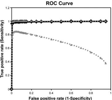
ROC analysis.A receiver operating characteristic (ROC) analysis was performed over a range of possible percent inhibition cutoff points for the ELISA (42). The thresholds were based on the percent inhibition values for the infected and uninfected fish.
Viral RNA analysis of serum by real-time RT-PCR.Two published real-time RT-PCR assays were used to detect viral RNA. At the Great Lakes Water Institute, University of Wisconsin (UW)-Milwaukee, RNA was extracted, and real-time PCR (21,43) was performed on all yellow perch (Table 1). At UW-Madison, RNA was extracted, and real-time RT-PCR was performed (31) on the remaining fish. Any samples crossing the cycle threshold before cycle 40 were considered positive.
RESULTS
Prevalence of neutralizing antibodies in control serum by VN
assay.
The VN assay was modified from a previously described
protocol (
27
). The results from the pilot studies (not shown)
in-dicated that 24-h incubation of the virus and serum prior to
inoc-ulation onto the cells produced more significant neutralization of
the virus versus a 30-min or 1-h incubation described in
previ-ously developed plaque neutralization test protocols for VHSV I
and IVb (
24
,
44
). The pilot studies also showed considerable
tox-icity at dilutions of 1:2 to 1:8. Thus, an initial dilution of 1:16 was
used. Serum samples showing partial or complete protection were
considered positive and were designated p1:16 (partial) or 1:16
(complete). If no protection was observed at 1:16, a serum sample
was considered to be negative.
Neutralizing antibody titers were not detected in any of the
serum samples from fish in the VHS-negative group (
n
⫽
34). Low
VHSV-neutralizing titers were detected in 43% (12/28) of the fish
from the VHS-positive group (
Table 2
), with titers ranging from
p1:16 to 1:80. Thus, the VN assay had a specificity of 100% (95%
confidence interval, 89.6% to 100%) and a sensitivity of 42.9%
(95% confidence interval, 24.5% to 62.8%).
The addition of naive brown trout serum as complement was
evaluated in our VN assay and was found to have no effect on
neutralization (data not shown). A methylcellulose overlay was
also evaluated for the isolation of plaques but was not necessary in
reading the last serum dilution showing protection of the
mono-layer for determining the neutralizing antibody titer in the VN
assay (data not shown). Three antibody-positive controls and one
antibody-negative control were used to compare results with and
without the addition of overlay, and no difference in antibody titer
was observed.
Cross-reactivity of anti-VHSV nucleocapsid monoclonal
an-tibody with SVCV.
A Western blot under reduced conditions
showed staining only with the nucleocapsid protein of VHSV
us-ing the anti-VHSV nucleocapsid monoclonal antibody, showus-ing
the specificity of the antibody to this protein (results not shown).
No staining occurred with the SVCV lysate in a Western blot using
the anti-VHSV nucleocapsid monoclonal antibody, indicating no
cross-reactivity between our monoclonal detection antibody and
SVCV. Specifically, these results show that there is no
cross-rec-ognition between the linear epitopes of the N proteins of VHSV
and SVCV.
Analysis of anti-VHSV nucleocapsid monoclonal antibody
in ELISA.
A blocking ELISA is well suited for testing diagnostic
samples from wildlife species because a secondary antibody is not
required. At the time of assay development, no effective
monoclo-nal antibody against the VHSV glycoprotein was available
com-mercially. The anti-VHSV nucleocapsid monoclonal antibody
used in this study was commercially available and effective. The
anti-VHSV nucleocapsid monoclonal antibody has an advantage
in that it detects persistent antibodies directed against the
nucleo-capsid. ELISA plates coated with intact viral particles revealed
in-complete blocking. Treating the virus with MEGA-10 detergent
prior to coating the plates was a critical step to allow for accurate
identification of infected and noninfected fish. Presumably, this
treatment reveals the target epitope of the nucleocapsid protein
and allows the binding of the anti-nucleocapsid monoclonal
an-tibody (
45–47
).
The efficacy of the VHSV nucleocapsid monoclonal
on August 17, 2020 by guest
http://cvi.asm.org/
body was evaluated by testing serum samples from the 34
unin-fected and 28 previously inunin-fected fish. The serum samples were
tested both undiluted and at a 1:2 dilution. The serum samples
were tested at a 1:2 dilution to eliminate high background issues
with hemolyzed serum.
Thirty of 34 serum samples (88.2%) from the VHS-negative
group fish diluted 1:2 were negative by ELISA (
Table 1
).
Twenty-seven of the 28 serum samples (96.4%) from the VHS-positive
group fish diluted 1:2 were positive by ELISA (
Table 2
).
ROC analysis for ELISA.
A receiver operating characteristic
(ROC) curve was performed to derive the optimal percent
inhibi-tion threshold for detecting VHSV antibodies in fish serum (
Fig.
1
). Undiluted samples were considered positive at 35 to 100%
inhibition and negative at
⬍35% inhibition. The samples tested at
a 1:2 dilution were considered positive at 25 to 100% inhibition
and negative
⬍25%, based on the ROC analysis. The area under
the ROC curve was 0.994, confirming that the ELISA diagnostic
performance characteristics under these thresholds were well
cor-related with the true status of each serum sample. These cutoff
values demonstrated 88.2% specificity (95% confidence interval,
72.5% to 96.6%) and 96.4% sensitivity (95% confidence interval,
81.6% to 99.4%) with serum diluted 1:2. The percent inhibition
values from uninfected fish ranged from 0.24% to 41.1% (average,
15.66%) and 7.94% to 95.99% (average, 49.21%) for previously
infected fish. The positive predictive value of the ELISA for
exper-imentally infected fish is 87.1% (95% confidence interval, 70.2%
to 96.3%) and the negative predictive value is 96.8% (95%
confi-dence interval, 83.2% to 99.5%) (
Table 3
and
4
).
Viral RNA detection by real-time RT-PCR.
Serum samples
were tested for VHSV by RT-PCR to determine if there was viral
infection at the time blood was collected for fish exposed to VHSV
and to determine if the inhibition of antibody binding was
occur-ring in our tests due to antibodies being complexed with virus in
the serum.
VHSV RNA was not detected by real-time RT-PCR in sera
from uninfected fish. Sera from yellow perch H1-14, J1-13/J3-11,
H4 A, H4 B, H4 C, and J4 A were positive, with threshold cycle
(
C
T) values ranging from 32.6 to 39.5 (
Table 2
). Serum from grass
carp 2 was positive, with a
C
Tof 39.5, and that from Pacific herring
149 was positive, with a
C
Tof 38.9. All other sera from fish in the
VHS-positive group tested negative for viral RNA.
DISCUSSION
We successfully developed a virus neutralization assay and a
blocking ELISA to detect neutralizing and nonneutralizing
anti-bodies against VHSV, respectively. The VN assay has the
advan-tage of recognizing antibodies that likely confer protective
immu-nity to VHSV and can indicate recent exposure to the virus (
25
).
The blocking ELISA is valuable for identifying nonneutralizing
anti-nucleocapsid antibodies, which may persist longer and
there-fore extend the opportunity to detect VHSV antibodies after
ini-tial virus exposure (
25
,
48
). These assays complement viral
detec-tion methods by providing a means for determining the exposure
histories of wild populations.
Previous studies have described the use of
complement-depen-dent 50% plaque neutralization tests and indirect ELISAs to detect
VHSV antibodies in trout (
24
,
25
). These methods are reliable but
not practical for screening large populations of fish from multiple
species. There are currently no commercially available diagnostic
tests in the United States for detecting antibodies to VHSV.
Sur-veillance efforts by virus isolation are labor-intensive, must occur
within narrow water temperature windows, and are costly.
Al-though real-time PCR assays are available, these methods
gener-ally need to be performed in high-throughput laboratories for
large sample sizes. Serological assays, such as our ELISA and VN
assay, provide efficient and less costly methods for evaluating the
VHSV exposure histories of samples from large wild fish
popula-tions or waterbodies.
Our VN assay is different from the traditional 50% plaque
neutralization test (
24
,
44
,
49
) in that it is performed in a
micro-neutralization format and the antibody titers are read as the last
serum dilution showing complete protection of the cell
mono-layer from VHSV. Additionally, we determined that
methylcellu-lose overlay is not necessary in our VN assay because plaques are
not counted to determine the titers. Complement has been shown
to enhance neutralization in 50% PNTs when applied to trout
serum (
23
,
48
,
50
). However, neutralization was not enhanced by
the addition of complement in our assay, which may indicate the
presence of a different immune mechanism specific to trout. It
should be noted that the reduced sensitivity observed in our VN
FIG 1Receiver operating characteristic (ROC) curve of blocking ELISA using 1:2 diluted serum from VHSV-infected fish (n⫽28) and uninfected fish (n⫽
34). The dashed lines indicate upper and lower 95% confidence intervals for the positive fraction, and the solid line indicates the curve for the true-positive rate.
TABLE 3Results of blocking ELISAa
VHS infection status
No. of known positives (n⫽28)
No. of known negatives (n⫽34)
Positive 27 4
Negative 1 30
a
n⫽62. The sensitivity is 96.4% and the specificity is 88.2%, both calculated from fish in the VHS-negative and VHS-positive groups.
TABLE 4Results of VN assaya
VHS infection status
No. of known positives (n⫽28)
No. of known negatives (n⫽34)
Positive 12 0
Negative 16 34
an⫽62. The sensitivity is 42.9% and the specificity is 100%, both calculated from fish in the VHS-negative and VHS-positive groups.
on August 17, 2020 by guest
http://cvi.asm.org/
assay may be due to VHSV forming a complex with neutralizing
antibodies in the serum, which reduces the availability of
antibod-ies for binding to virus neutralization epitopes in the VN assay
(
45–48
). However, this concern is obviated by using the
nucleo-capsid protein as our target, because antibodies with nucleonucleo-capsid
affinity presumably do not complex with the intact viral particle,
which underscores another value of the anti-nucleocapsid ELISA.
It should be noted that four of the eight sera (50%) from our
VHS-infected group tested positive for viral RNA but negative in
the VN assay. This emphasizes the importance of utilizing parallel
assays when testing the virus exposure history of fish.
Although previous experiments have determined that
homol-ogous strains of VHSV must be used for neutralization epitopes to
be recognized (
51
,
52
), serum samples from five Pacific herring
hyperimmunized with VHSV type IVa were able to neutralize type
IVb virus in the VN assay at low titers of p1:16 to 1:32. There are 21
amino acid differences between the type IVa and IVb glycoprotein
sequences. None of these differences occur in two of the identified
glycoprotein-neutralizing epitopes. This suggests there are shared
neutralizing epitopes between types IVa and IVb (
53
,
54
). Type
IVa glycoprotein epitopes may be similar enough to those of type
IVb to react in our VN assay. Indeed, a similar phenomenon was
noted when Pacific herring vaccinated with the glycoprotein gene
isolated from VHSV type Ia were protected from VHSV type IVb
(
55
). Further investigation is needed to determine whether our
VN assay may detect different antibody titers in herring exposed
to virus that is homologous to that used in the VN assay.
Addi-tionally, although we demonstrate the ability to detect antibodies
in hyperimmunized Pacific herring that likely experienced
artifi-cially high antibody titers, further investigations are needed to
determine the sensitivities of these assays in wild Pacific herring or
in those surviving more realistic VHSV exposure histories.
Our new blocking ELISA is a suitable nonlethal method for
detecting exposure to VHSV. Considering the broad host range of
VHSV type IVb (
11
), the advantage of a species-independent
ELISA is significant for the surveillance of VHSV. The assay can
measure the concentrations of antibodies directed against the
nu-cleocapsid in any freshwater species since it does not require a
secondary antibody. Furthermore, the ability of the monoclonal
antibody to bind to a single viral epitope results in high specificity.
We demonstrated by Western blotting that the nucleocapsid
monoclonal antibody binding was specific to VHSV versus SVCV,
another rhabdovirus that is present in Wisconsin. Previous studies
showed a lack of cross-reaction between the antibody used herein
and spring viremia of carp virus, infectious hematopoietic
necro-sis virus, pike fry rhabdovirus, or rhabdovirus anguilla (
26
,
56
).
According to the manufacturer of the antibody (Aquatic
Diagnos-tics, Stirling, Scotland), no cross-reaction of the antibody occurs
with nodavirus, infectious salmon anemia virus, koi herpesvirus,
salmon alphavirus (1, 2, and 3), or
Piscirickettsia salmonis
infected
cells. We were not able to test positive sera from transboundary
VHSV strains (type I, II, and III); however, previous efforts
indi-cate the anti-nucleocapsid monoclonal antibody detects
anti-nu-cleocapsid antibodies against all strains of VHSV (
26
).
A feature crucial to the function of our ELISA is the treatment
of the viral antigen with MEGA-10 detergent prior to coating the
plates (
47
). Repeated trials showed that treatment of the virus with
detergent allowed for better attachment of the
nucleocapsid-spe-cific antibody to the virus. This result is probably due to the ability
of the detergent to expose the nucleocapsid epitope and make it
available for antibody binding.
The large difference in sensitivities between our blocking
ELISA (96.4%) and VN assay (42.9%) may be attributed to the
immune response kinetics at the time of serum collection. Studies
have shown that neutralizing antibodies do not persist as long as
nonneutralizing antibodies in trout (
25
,
48
), and neutralizing
an-tibody titers peak at 6 weeks postinfection in rainbow trout
in-fected with VHSV I (
24
) and at 11 to 16 weeks in muskellunge
infected with VHSV IVb (
44
). It was observed that the majority of
VHSV-exposed fish with serum samples collected prior to 6 weeks
postinfection had no or low neutralizing antibody titers. Those
with low titers may have still been clearing virus while producing
protective antibodies, indicated by the presence of viral RNA in
the serum sample of a portion of our VHSV-exposed fish.
Inves-tigation into the kinetics of viral replication and the related
im-mune response in multiple species are therefore important for
further study.
A limitation of this study is the number of serum samples
avail-able from VHSV-uninfected and -infected fish. We used serum
samples from 27 experimentally infected and one wild-caught fish
that had VHSV-positive kidney and spleen tissues as tested by
real-time RT-PCR for ELISA development. It is also important to
note that our threshold for detecting VHSV antibodies by ELISA
may require adjustment when evaluating various wild-caught
spe-cies due to differing environments and susceptibility. In this light,
we note that 4 of 34 serum samples diluted 1:2 from the
VHS-negative group were positive on the blocking ELISA. There may be
nonspecific reactions occurring that more extensive testing would
help reconcile. For the purpose of this assay as a surveillance tool,
it is more practical to keep a threshold at a level that maximizes
sensitivity.
In summary, the blocking ELISA shows high sensitivity and
acceptable specificity, whereas the VN assay has unacceptably low
sensitivity but high specificity. When used in parallel, the VN assay
and ELISAs correctly identified the VHSV exposure status of all
known uninfected and infected fish. Our results highlight that the
anti-VHSV nucleocapsid monoclonal antibody used in the
block-ing ELISA is a good indicator of past exposure to VHSV and may
be a reliable time-independent and species-independent
diagnos-tic test suitable for nonlethal surveillance of VHSV. Our nonlethal
serological assays will be valuable for assessing VHSV exposure
history and might reduce the extensive laboratory effort needed to
screen fish for VHSV using virus isolation. Use of the VN assay,
blocking ELISA, and virus isolation under actual surveillance
con-ditions is needed to fully demonstrate the interplay between the
assays. The collection of additional reference samples is required
for continued assay validation to further assess the sensitivity and
specificity and determine repeatability, robustness, and
rugged-ness. Our serological assays might supplement existing VHS
sur-veillance protocols, which might have regulatory implications for
fish movement between VHSV-positive and -negative locations in
certain jurisdictions or geographic regions.
ACKNOWLEDGMENTS
This work was supported by the University of Wisconsin Sea Grant Insti-tute under a grant from the National Oceanic and Atmospheric Admin-istration, U.S. Department of Commerce, CFDA no. 11.417. Herring blood plasma was made available through the support of the Exxon Val-dez Oil Spill Trustee Council (project no. 10100123-I).
on August 17, 2020 by guest
http://cvi.asm.org/
The views contained in this document are those of the authors and should not be interpreted as necessarily representing the official policies of the U.S. government.
We thank Ronald Bruch, Kendall Kamke, Ryan Koenigs, Bob Hoodie, Robert Olynyk, Eric Eikenberry, Rachel Koehler, Will Mustas, Jeremy Bartz, Andrew Braasch, Tom Van Effen, Todd Rice, and Mike Staggs of the Wisconsin Department of Natural Resources for their technical assis-tance in collecting fish and tissues for this study. We thank Sara Crawford, Suzanne Burgener, Roberta Riedy, and Mark Matenaer for assisting in sample collection. We also thank Ann Hennings, Hui-Min Hsu, Audrey Dikkeboom, Todd McCoy, Jennifer Cooper, Cristina Vaughan, Georgia Wolfe, Meaghan Broman, Melissa Behr, Doug Lyman, and Peter Vander-loo of the Wisconsin Veterinary Diagnostic Laboratory for assisting with sample collection. We greatly appreciate the generous donation of serum from Mohamed Faisal and Elena Millard of Michigan State University, East Lansing, MI, and Andrew Goodwin of the U.S. Fish and Wildlife Service and Emily Marecaux of the University of Arkansas at Pine Bluff. Lastly, we greatly appreciate the invaluable review of the manuscript pro-vided by Peter McIntyre of the University of Wisconsin, Madison, WI.
REFERENCES
1.Wolf K.1988. Fish viruses and fish viral diseases. Cornell University Press, Ithaca, NY.
2.Kim R, Faisal M.2011. Emergence and resurgence of the viral hemor-rhagic septicemia virus (Novirhabdovirus,Rhabdoviridae, Mononegavi-rales). J. Adv. Res.2:9 –23.http://dx.doi.org/10.1016/j.jare.2010.05.007. 3.Hedrick RP, Batts WN, Yun S, Traxler GS, Kaufman J, Winton JR.
2003. Host and geographic range extensions of the North American strain of viral hemorrhagic septicemia virus. Dis. Aquat. Organ.55:211–220.
http://dx.doi.org/10.3354/dao055211.
4.Ross K, McCarthy U, Huntly PJ, Wood BP, Stuart D, Rough EI, Smail DA, Bruno DW. 1994. A outbreak of viral hemorrhagic septicaemia (VHS) in turbot (Scophthalmus maximus) in Scotland. Bull. Eur. Assoc. Fish Pathol.14:213–214.
5.Dixon P, Feist S, Kehoe E, Parry L, Stone D, Way K.1997. Isolation of viral haemorrhagic septicaemia virus from Atlantic herringClupea haren-gusfrom the English Channel. Dis. Aquat. Organ.30:81– 89.http://dx.doi .org/10.3354/dao030081.
6.Mortensen HF, Heuer OE, Lorenzen N, Otte L, Olesen NJ. 1999. Isolation of viral haemorrhagic septicaemia virus (VHSV) from wild ma-rine fish species in the Baltic Sea, Kattegat, Skagerrak and the North Sea. Virus Res.63:95–106.http://dx.doi.org/10.1016/S0168-1702(99)00062-3. 7.Takano R, Nishizawa T, Arimoto M, Muroga K.2000. Isolation of viral haemorrhagic septicaemia virus (VHSV) from wild Japanese flounder,
Paralichthys olivaceus. Bull. Eur. Ass. Fish Pathol.20:186 –192.
8.Hopper K.1989. The isolation of VHSV from chinook salmon at Glen-wood Springs, Orcas Island, Washington. Fish Health Sect. Am. Fish. Soc. Newsl.17:1–2.
9.Garver KA, Traxler GS, Hawley LM, Richard J, Ross JP, Lovy J.2013. Molecular epidemiology of viral haemorrhagic septicaemia virus (VHSV) in British Columbia, Canada, reveals transmission from wild to farmed fish. Dis. Aquat. Organ.104:93–104.http://dx.doi.org/10.3354/dao02588. 10. Elsayed E, Faisal M, Thomas M, Whelan G, Batts W, Winton J.2006. Isolation of viral haemorrhagic septicaemia virus from muskellunge, Esox masquinongy (Mitchill), in Lake St. Clair, Michigan, USA reveals a new sublineage of the North American genotype. J. Fish Dis.29:611– 619.http: //dx.doi.org/10.1111/j.1365-2761.2006.00755.x.
11. Thompson TM, Batts WN, Faisal M, Bowser P, Casey JW, Phillips K, Garver KA, Winton J, Kurath G.2011. Emergence of viral hemorrhagic septicemia virus in the North American Great Lakes region is associated with low viral genetic diversity. Dis. Aquat. Organ.96:29 – 43.http://dx .doi.org/10.3354/dao02362.
12. Lumsden JS, Morrison B, Yason C, Russell S, Young K, Yazdanpanah A, Huber P, Al-Hussinee L, Stone D, Way K.2007. Mortality event in freshwater drumAplodinotus grunniensfrom Lake Ontario, Canada, asso-ciated with viral haemorrhagic septicemia virus, type IV. Dis. Aquat. Or-gan.76:99 –111.http://dx.doi.org/10.3354/dao076099.
13. Groocock GH, Getchell RG, Wooster GA, Britt KL, Batts WN, Winton JR, Casey RN, Casey JW, Bowser PR.2007. Detection of viral hemor-rhagic septicemia in round gobies in New York State (USA) waters of Lake
Ontario and the St. Lawrence River. Dis. Aquat. Organ.76:187–192.http: //dx.doi.org/10.3354/dao076187.
14. Winton JR, Kurath G, Batts WN.2008. Molecular epidemiology of viral hemorrhagic septicemia virus in the Great Lakes region. U.S. Geological Survey, Western Fisheries Research Center, Seattle, WA.http://www.aps. uoguelph.ca/aquacentre/files/misc-factsheets/USGS%20VHS%20Factsh eet(2008).pdf.
15. Faisal M, Shavalier M, Kim RK, Millard EV, Gunn MR, Winters AD, Schulz CA, Eissa A, Thomas MV, Wolgamood M, Whelan GE, Winton J.2012. Spread of the emerging viral hemorrhagic septicemia virus strain, genotype IVb, in Michigan, USA. Viruses4:734 –760.http://dx.doi.org/10 .3390/v4050734.
16. Kane-Sutton M, Kinter B, Dennis PM, Koonce JF.2010. Viral hemor-rhagic septicemia virus infection in yellow perch,Perca flavescens, in Lake Erie. J. Great Lakes Res.36:37– 43.http://dx.doi.org/10.1016/j.jglr.2009.11 .004.
17. American Fisheries Society-Fish Health Section (AFS-FHS).2010. FHS blue book: suggested procedures for the detection and identification of certain finfish and shellfish pathogens, 2010 edition. AFS-FHS, Be-thesda, MD.
18. World Organization of Animal for Animal Health (OIE).2009. Manual of diagnostic tests for aquatic animals. OIE, Paris, France.
19. Kim R, Faisal M.2010. Comparative susceptibility of representative Great Lakes fish species to the North American viral hemorrhagic septicemia virus sublineage IVb. Dis. Aquat. Organ.91:23–34.http://dx.doi.org/10 .3354/dao02217.
20. Kim R, Faisal M.2010. Experimental studies confirm the wide host range of the Great Lakes viral haemorrhagic septicaemia virus genotype IVb. J. Fish Dis.33:83– 88.http://dx.doi.org/10.1111/j.1365-2761.2009.01093.x. 21. Olson W, Emmenegger E, Glenn J, Winton J, Goetz F.2013.
Compar-ative susceptibility among three stocks of yellow perch,Perca flavescens
(Mitchill), to viral haemorrhagic septicaemia virus strain IVb from the Great Lakes. J. Fish Dis.http://dx.doi.org/10.1111/jfd.12068.
22. VHSV Expert Panel and Working Group. 2010. Viral hemorrhagic septicemia virus (VHSV IVb) risk factors and association measures de-rived by expert panel. Prev. Vet. Med.94:128 –139.http://dx.doi.org/10 .1016/j.prevetmed.2009.11.020.
23. Dorson M, Torchy C.1979. Complement dependent neutralization of Egtved virus by trout antibodies. J. Fish Dis.2:345–347.http://dx.doi.org /10.1111/j.1365-2761.1979.tb00176.x.
24. Olesen NJ, Jørgensen P.1986. Detection of neutralizing antibody to Egtved virus in rainbow trout (Salmo gairdneri) by plaque neutralization test with complement addition. J. Appl. Ichthyol.2:33– 41.http://dx.doi .org/10.1111/j.1439-0426.1986.tb00427.x.
25. Olesen NJ, Lorenzen N, Jørgensen P. 1991. Detection of rainbow trout antibody to Egtved virus by enzyme-linked immunosorbent as-say (ELISA), immunofluorescence (IF), and plaque neutralization tests (50% PNT). Dis. Aquat. Organ.10:31–38.http://dx.doi.org/10.3354 /dao010031.
26. Lorenzen N, Olesen NJ, Jørgensen P.1988. Production and character-ization of monoclonal antibodies to four Egtved virus structural proteins. Dis. Aquat. Organ.4:35– 42.http://dx.doi.org/10.3354/dao004035. 27. World Organization for Animal Health (OIE).2010. Manual of
stan-dards for diagnostic tests and vaccines. OIE, Paris, France.
28. Winton J, Batts W, DeKinkelin P, LeBerre M, Bremont M, Fijan N. 2010. Current lineages of theepithelioma papulosum cyprini(EPC) cell line are contaminated with fathead minnow,Pimephales promelas, cells. J. Fish Dis.33:701–704.http://dx.doi.org/10.1111/j.1365-2761.2010.01165.x. 29. Batts W, Winton J.2007. Viral hemorrhagic septicemia.InAFS-FHS
(American Fisheries Society-Fish Health Section) (ed), FHS blue book: suggested procedures for the detection and identification of certain finfish and shellfish pathogens, 2007 edition. AFS-FHS, Bethesda, MD. 30. Mitro MG, White AL.2008. Viral hemorrhagic septicemia and freshwater
fisheries: the state of the science. Technical bulletin no. 196, 2008. Depart-ment of Natural Resources, Madison, WI. http://citeseerx.ist.psu.edu /viewdoc/download?rep⫽rep1&type⫽pdf&doi⫽10.1.1.204.190. 31. Jonstrup SP, Kahns S, Skall HF, Boutrup TS, Olesen NJ.2012.
Devel-opment and validation of a novel Taqman-based real-time RT-PCR assay suitable for demonstrating freedom from viral haemorrhagic septicaemia virus. J. Fish Dis. 36:9 –23.http://dx.doi.org/10.1111/j.1365-2761.2012 .01416.x.
32. Dixon PF, Hattenberger-Baudouy AM, Way K.1994. Detection of carp
on August 17, 2020 by guest
http://cvi.asm.org/
antibodies to spring viraemia of carp virus by a competitive immunoassay. Dis. Aquat. Organ.19:181–186.http://dx.doi.org/10.3354/dao019181. 33. Rosauer DR, Biga PR, Lindell SR, Binkowski FP, Shepherd BS,
Palmquist DE, Simchick CA, Goetz FW.2011. Development of yellow perch (Perca flavescens) broodstocks: initial characterization of growth and quality traits following grow-out of different stocks. Aquaculture317: 58 – 66.http://dx.doi.org/10.1016/j.aquaculture.2011.03.037.
34. Beaulaurier J, Bickford N, Gregg JL, Grady CA, Gannam A, Winton JR, Hershberger PK.2012. Susceptibility of Pacific herringClupea pallasiito viral hemorrhagic septicemia (VHS) is influenced by diet. J. Aquat. Anim. Health 43– 48.http://dx.doi.org/10.1080/08997659.2012.668511. 35. Specter S, Hodinka RL, Young SA.2000. Clinical virology manual, 3rd
ed. American Society for Microbiology, Washington, DC.
36. Reed LJ, Muench H.1938. A simple method of estimating fifty per cent endpoints. Am. J. Epidemiol.27:493– 497.
37. Batts WN, Winton JR.1989. Enhanced detection of infectious hemato-poietic necrosis virus and other fish viruses by pretreatment of cell mono-layers with polyethylene glycol. J. Aquat. Anim. Health1:284 –290.http: //dx.doi.org/10.1577/1548-8667(1989)001⬍0284:EDOIHN⬎2.3.CO;2. 38. Laemmli UK.1970. Cleavage of structural proteins during the assembly of
the head of bacteriophage T4. Nature227:680 – 685.http://dx.doi.org/10 .1038/227680a0.
39. Towbin H, Staehelin T, Gordon J. 1979. Electrophoretic transfer of proteins from polyacrylamide gels to nitrocellulose sheets: procedure and some applications. Proc. Natl. Acad. Sci. U. S. A.76:4350 – 4354.http://dx .doi.org/10.1073/pnas.76.9.4350.
40. Dikkeboom AL, Radi C, Toohey-Kurth K, Marcquenski S, Engel M, Goodwin AE, Stone DM, Longshaw C. 2004. First report of spring viremia of carp virus (SVCV) in wild common carp in North America. J. Aquat. Anim. Health16:169 –178.http://dx.doi.org/10.1577/H03-064.1. 41. Godhardt-Cooper JA, Zoromski J, Toohey-Kurth K.2009. Evaluation of
a blocking enzyme-linked immunosorbent assay for serological diagnosis of bovine herpesvirus 1. J. Vet. Diagn. Invest.21:523–526.http://dx.doi .org/10.1177/104063870902100416.
42. Zhou X-H, Obuchowski NA, McClish DK.2002. Statistical methods in diagnostic medicine. John Wiley and Sons, Inc., New York, NY. 43. Garver KA, Hawley LM, McClure CA, Schroeder T, Aldous S, Doig F,
Snow M, Edes S, Baynes C, Richard J.2011. Development and validation of a reverse transcription quantitative PCR for universal detection of viral hemorrhagic septicemia virus. Dis. Aquat. Organ.95:97-112.http://dx.doi .org/10.3354/dao02344.
44. Millard EV, Faisal M.2012. Development of neutralizing antibody re-sponses in muskellunge,Esox masquinongy(Mitchill), experimentally ex-posed to viral haemorrhagic septicaemia virus (genotype IVb). J. Fish Dis. 35:11–18.http://dx.doi.org/10.1111/j.1365-2761.2011.01318.x. 45. Hildreth JE.1982.N-D-Gluco-N-methylalkanamide compounds, a new
class of non-ionic detergents for membrane biochemistry. Biochem. J. 207:363–366.
46. Merza MS, Linné T, Höglund S, Portetelle D, Burny A, Morein B.1989. Bovine leukaemia virus ISCOMs: biochemical characterization. Vaccine 7:22–28.http://dx.doi.org/10.1016/0264-410X(89)90006-6.
47. Klintevall I, Näslund K, Svedlundi G, Hajdu L, Linde N, Klingeborn B. 1991. Evaluation of an indirect ELISA for the detection of antibodies to bovine leukaemia virus in milk and serum. J. Virol. Methods 319 –333. 48. Encinas P, Gomez-Casado E, Fregeneda-Grandes, Olesen NJ, Lorenzen
N, Estepa A, Coll JM.2011. Rainbow trout surviving infections of viral haemorrhagic septicemia virus (VHSV) show lasting antibodies to recom-binant G protein fragments. Fish Shellfish Immunol.30:929 –935.http: //dx.doi.org/10.1016/j.fsi.2011.01.021.
49. LaPatra S.1996. The use of serological techniques for virus surveillance and certification of finfish. Annu. Rev. Fish Dis.6:15–28.http://dx.doi.org /10.1016/S0959-8030(96)90003-4.
50. Lorenzen N, Olesen NJ, Koch NC.1999. Immunity to VHS virus in rainbow trout. Aquaculture172:41– 61.http://dx.doi.org/10.1016/S0044 -8486(98)00443-8.
51. Fregeneda-Grandes JM, Olesen NJ.2007. Detection of rainbow trout antibodies against viral haemorrhagic septicaemia virus (VHSV) by neu-tralisation test is highly dependent on the virus isolate used. Dis. Aquat. Organ.74:151–158.http://dx.doi.org/10.3354/dao074151.
52. Olesen NJ, Lorenzen N, Jørgensen PEV.1993. Serological differences among isolates of viral haemorrhagic septicaemia virus detected by neu-tralizing monoclonal and polyclonal antibodies. Dis. Aquat. Organ.16: 163–170.http://dx.doi.org/10.3354/dao016163.
53. Nishizawa T, Iida H, Takano R, Isshiki T, Nakajima K, Muroga K.2002. Genetic relatedness among Japanese, American and European isolates of viral hemorrhagic septicemia virus (VHSV) based on partial G and P genes. Dis. Aquat. Organ. 48:143–148. http://dx.doi.org/10.3354 /dao048143.
54. Ito T, Kurita J, Sano M, Skall HF, Lorenzen N, Einer-Jensen K, Olesen NJ.2012. Typing of viral hemorrhagic septicemia virus (VHSV) by mono-clonal antibodies. J. Gen. Virol.93:2546 –2557.http://dx.doi.org/10.1099 /vir.0.043091-0.
55. Hart LM, Lorenzen N, LaPatra SE, Grady CA, Roon SE, O’Reilly J, Gregg JL, Hershberger PK.2012. Efficacy of a glycoprotein DNA vaccine against viral haemorrhagic septicaemia (VHS) in Pacific herring,Clupea pallasiiValenciennes. J. Fish Dis.35:775–779.http://dx.doi.org/10.1111/j .1365-2761.2012.01364.x.
56. Ristow SS, Lorenzen N, Jørgensen PEV.1991. Monoclonal-antibody-based immunodot assay distinguishes between viral hemorrhagic sep-ticemia virus (VHSV) and infectious hematopoietic necrosis virus (IHNV). J. Aquat. Anim. Health 176 –180.http://dx.doi.org/10.1577
/1548-8667(1991)003⬍0176:MABIAD⬎2.3.CO;2.