A Novel Tuberculosis DNA Vaccine in an HIV-1 p24 Protein
Backbone Confers Protection against
Mycobacterium tuberculosis
and
Simultaneously Elicits Robust Humoral and Cellular Responses to
HIV-1
Xiaoman Li,aWei Xu,band Sidong Xionga,b
Institute for Immunobiology and Department of Immunology, Shanghai Medical College, Fudan University, Shanghai, People’s Republic of China,aand Jiangsu Key Laboratory of Infection and Immunity, Institutes of Biology and Medical Sciences, Soochow University, Suzhou, People’s Republic of Chinab
Tuberculosis (TB) caused by
Mycobacterium tuberculosis
remains a major infectious disease worldwide. Moreover, latent
M
.
tuberculosis
infection is more likely to progress to active TB and eventually leads to death when HIV infection is involved. Thus,
it is urgent to develop a novel TB vaccine with immunogenicity to both
M
.
tuberculosis
and HIV. In this study, four
uncharacter-ized T cell epitopes from MPT64, Ag85A, Ag85B, and TB10.4 antigens of
M
.
tuberculosis
were predicted, and HIV-1-derived p24,
an immunodominant protein that can induce protective responses to HIV-1, was used as an immunogenic backbone.
M
.
tuber-culosis
epitopes were incorporated separately into the gene backbone of p24, forming a pP24-Mtb DNA vaccine. We
demon-strated that pP24-Mtb immunization induced a strong
M
.
tuberculosis
-specific cellular response as evidenced by T cell
prolifera-tion, cytotoxicity, and elevated frequency of gamma interferon (IFN-
␥
)-secreting T cells. Interestingly, a p24-specific cellular
response and high levels of p24-specific IgG were also induced by pP24-Mtb immunization. When the protective effect was
as-sessed after mycobacterial challenge, pP24-Mtb vaccination significantly reduced tissue bacterial loads and profoundly
attenu-ated the mycobacterial infection-relattenu-ated lung inflammation and injury. Our findings demonstrattenu-ated that the pP24-Mtb
tubercu-losis vaccine confers effective protection against mycobacterial challenge with simultaneously elicited robust immune responses
to HIV-1, which may provide clues for developing novel vaccines to prevent dual infections.
T
uberculosis (TB) is a reemerging disease that remains one of
the leading causes of mortality in humans (44). It accounts for
4 deaths every minute and 2 million deaths annually and severely
threatens the health of humankind (26). In recent years, the
prev-alence of HIV infection among tuberculosis cases has increased
dramatically (28). A latent
Mycobacterium tuberculosis
infection
(LTBI) is 20 to 30 times more likely to progress to active TB when
HIV infection is involved (14, 23). Similarly, at least one-third of
HIV-infected individuals are already infected with TB, and
one-quarter of the persons infected with HIV die from TB (16, 23).
However,
Mycobacterium bovis
bacillus Calmette-Guérin (BCG),
the only tuberculosis vaccine approved for human use, is effective
only in children, and its protective efficacy wanes significantly
over a period of 10 to 15 years (1, 37). To date, the development of
an effective HIV vaccine is still under way (4, 24). Therefore, the
serious situation of dual epidemics asks for a novel vaccine with
immunogenicity to both
Mycobacterium tuberculosis
and HIV.
It is well established that T cell responses play an important role
in the development of resistance to
M. tuberculosis
, primarily
through production of gamma interferon (IFN-
␥
) and
eradica-tion of intracellular pathogens (32, 33). Therefore, antigens to
predominantly induce T cell response are used more in the
devel-opment of TB vaccine. To avoid unfavorable components for
hu-mans and for the humoral response induced by B cell epitopes (21,
30, 36, 39), it is better to apply T cell epitopes than to apply entire
antigens in TB vaccine design. Furthermore, a strategy that
incor-porates multiple T cell epitopes into one vaccine has the potential
to induce broad T cell immunity against several
immunodomi-nant antigens (10, 31, 34).
In this study, we predicted four T cell epitopes from four
well-defined protective antigens of
M. tuberculosis
, MPT64 (
M
.
tuber-culosis
protein 64), Ag85A (
M
.
tuberculosis
antigen 85A), Ag85B,
and TB10.4 via epitope prediction software online. Epitopes are
short peptides, and their immunogenicity is low unless
intro-duced into a carrier protein (39). Here we used p24, an
immuno-dominant protein of HIV-1 widely used in the development of
HIV vaccines (15), as the protein backbone for incorporation of
M. tuberculosis
epitopes. The gene segments of these epitopes were
grafted into various regions of the p24 gene scaffold, and a
multi-epitope DNA vaccine containing immunogens from
M.
tubercu-losis
and HIV-1 was obtained. The immunogenicity of the vaccine
to both
M. tuberculosis
and HIV-1 was evaluated.
MATERIALS AND METHODS
Prediction of T cell epitopes.Potential major histocompatibility complex class I (MHC-I)- or MHC-II-binding T cell epitopes were screened from MPT64, Ag85A, Ag85B, and TB10.4 proteins using epitope prediction software online (http://www.syfpeithi.de/,http://www.ddg-pharmfac.net /mhcpred/MHCPred/, andhttp://www.imtech.res.in/raghava/propred/). Similarity was scored using position-specific scoring matrixes derived from aligned peptides. Four epitopes, including MPT6476-84 (MPT64
from amino acids 76 to 84) (KFLSAATSS), Ag85A242-250(KLIANNTRV),
Received30 December 2011Returned for modification27 February 2012 Accepted20 March 2012
Published ahead of print29 March 2012
Address correspondence to Sidong Xiong, [email protected].
Copyright © 2012, American Society for Microbiology. All Rights Reserved.
doi:10.1128/CVI.05700-11
on August 17, 2020 by guest
http://cvi.asm.org/
Ag85B184-192 (IYAGSLSAL), and TB10.474-82 (STHEANTMA), were
sorted.
Construction of pP24-Mtb DNA vaccine.Plasmids containing the gene for HIV-1 p24 were obtained from the laboratory of Robert Tycko. The four selected T cell epitopes were engineered into the p24 scaffold separately (Fig. 1D). The chimericp24-Mtbgene was constructed by gene splicing through overlap extension of several synthetic nucleotide se-quences and was then incorporated into pcDNA3.1 plasmid (Invitrogen, Carlsbad, CA) driven by a cytomegalovirus (CMV) promoter.
Preparation of antigen peptides and proteins.BCG was purchased from the Shanghai Institute of Biological Products. Polypeptides, MPT6476-84, Ag85A242-250, Ag85B184-192, and TB10.474-82were
commer-cially synthesized by GL Biochem Ltd. (Shanghai, People’s Republic of China) with a purity of⬎95%. The polypeptides were then dissolved in phosphate-buffered saline (PBS) buffer and stored at⫺80°C until use. Recombinant p24 protein and p24-Mtb protein were expressed as histi-dine-tagged proteins in the pET-32aEscherichia coliexpression system (Novagen, Madison, WI). Thep24gene andp24-Mtbgene were inserted into the pET-32a vector to construct two recombinant plasmids, pET-P24 and pET-P24-Mtb. The recombinant plasmids were then transformed into the competentEscherichia colistrain BL21(DE3). The cells were grown in a shaker at 37°C, isopropyl--D-thiogalactoside (IPTG) (Sigma, St. Louis, MO) was added to induce recombinant protein synthesis at an optical density at 600 nm (OD600) of 0.5, and incubation was continued
for another 6 h. The cells were lysed, and the proteins were purified by Ni Sepharose 6 Fast Flow (GE Healthcare, Uppsala, Sweden). The concentra-tion of the purified proteins was determined by bicinchoninic acid test using a microplate BCA (bicinchoninic acid) protein assay reagent kit (Pierce, Rockford, IL).
Western blot assay.TheE. colistrain BL21(DE3) was transformed with pET-P24 or pET-P24-Mtb and induced by IPTG. The cells were
collected and disrupted by sonication. After centrifugation, the superna-tants were collected and electrophoresed on SDS-polyacrylamide gels and transferred to polyvinylidene fluoride membranes. Each membrane was probed with anti-p24 polyclonal antibody (Abcam, Cambridge, United Kingdom), followed by horseradish peroxidase (HRP)-conjugated goat anti-mouse antibody (Southern Biotech, Birmingham, AL), and the sig-nals were developed using chemiluminescence (Fig. 1E).
Animals and vaccination.Six- to 8-week-old female BALB/c mice (H-2d) were purchased from the experimental animal center of the Chi-nese Academy of Science (Shanghai, People’s Republic of China) and remained in pathogen-free conditions. All animal experiments were per-formed according to the guidelines for the care and use of laboratory animals (28a) and the guidelines of the Laboratory Animal Ethical Com-mission of Fudan University. Endotoxin-free plasmids were prepared us-ing an EndoFree plasmid purification megaprep kit (Qiagen, Valencia, CA). Mice were immunized intramuscularly with 50g pP24-Mtb, pP24, or pcDNA3.1 (defined as vector) 4 times at biweekly intervals. Serum was collected every 2 weeks by retro-orbital bleeding and stored at⫺70°C for further analysis.
ELISA measurement of p24- or peptide-specific antibody.The wells on 96-well enzyme-linked immunosorbent assay (ELISA) plates (Corning Costar, Cambridge, MA) were coated with p24 protein or the mixedM. tuberculosispeptides at a final concentration of 10g/ml at 4°C overnight and washed with PBS containing 0.05% Tween 20 (PBST). After blocking with PBS containing 5% nonfat milk at 37°C for 2 h, serum (1:100 dilu-tion) was added in duplicate and incubated at 37°C for 1 h. After the wells were washed, HRP-conjugated goat anti-mouse IgG, IgG1, or IgG2a (Southern Biotech) was added, followed by the addition of HRP substrate. The absorbance at 450 nm was measured in a microplate reader (Bio-Rad Lab, Hercules, CA). To determine the antibody titers, 5-fold serially di-luted sera were measured.
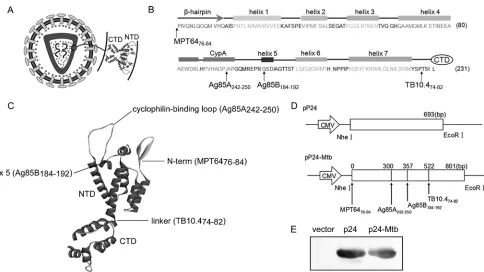
FIG 1Designation and construction of pP24-Mtb DNA vaccine. (A) Schematic model of the HIV-1 particle. p24 is composed of two domains, the N-terminal domain (NTD) and C-terminal domain (CTD). An enlargement of the two-domain structure is shown. (B) Secondary structure of the NTD. The-hairpin, CypA binding loop, and helices 1 to 7 are included in the NTD. The positions indicated by the arrows are the insertion sites of the predicted epitopes. (C) Inserted epitopes are labeled on the three-dimensional structure of p24. (D) Schematic representations of the pP24 and pP24-Mtb plasmids. (E) p24 protein and p24-Mtb protein were expressed in the pET-32aE. coliexpression system and analyzed by Western blotting with anti-p24 antibody. Individual experiments were conducted three times, with the results from one representative experiment shown for each group of mice.
on August 17, 2020 by guest
http://cvi.asm.org/
Avidity of p24-specific antibody.The avidity of the p24-specific antibody was determined by ELISA with a urea elution step (5). Briefly, sera (1:100 dilution) were tested in duplicate plates. In one of the plates, 6 M urea (Sigma) was added after incubation with samples and incubated for 10 min. Results were expressed as avidity index calcu-lated as follows: (endpoint titer in the presence of urea/endpoint titer in the absence of urea)⫻100.
IFN-␥ELISPOT assays.Enzyme-linked immunospot (ELISPOT) as-says were performed with ELISPOT assay kit (BD PharMingen, San Di-ego, CA). Briefly, wells on the plates were coated with the capture anti-IFN-␥monoclonal antibody (MAb) at 4°C overnight and then blocked with complete medium for 2 h at room temperature. Splenocytes from immunized mice were isolated, plated (1⫻106cells/well), and cultured
with the mixedM. tuberculosispeptides or p24 protein (each peptide or protein at a final concentration of 20g/ml) at 37°C for 36 h. After the plates were washed with deionized water and PBST, the biotinylated anti-IFN-␥MAb was added for 2 h at room temperature. Streptavidin-alkaline phosphatase (AP) was added to the plates and incubated for 1 h, and the color was developed by AP colorimetric substrate. An immunospot ana-lyzer (Cellular Technology, Cleveland, OH) was used to enumerate the spots.
Intracellular IFN-␥staining.Two weeks after the immunization, splenocytes were isolated and stimulatedin vitrowith the mixedM. tuber-culosispeptides or p24 protein (each peptide or protein at a final concen-tration of 20g/ml) at 37°C for 6 h. The cells were stained with anti-CD4 antibody conjugated to peridinin chlorophyll protein (anti-CD4-PerCP) and anti-CD8 antibody conjugated to fluorescein isothiocyanate (anti-CD8-FITC) (BD PharMingen). After the cells were washed, they were treated with fixation/permeabilization solution (BD PharMingen) for 30 min at 4°C and then stained with anti-IFN-␥antibody conjugated to phycoerythrin (anti-IFN-␥-PE) (BD PharMingen). The percentages of CD4⫹IFN-␥-positive (IFN-␥⫹) and CD8⫹IFN-␥⫹T cells were deter-mined by flow cytometry using a fluorescence-activated cell sorter FACSCalibur instrument (BD Biosciences, San Jose, CA).
ELISA of cytokines.Splenocytes from immunized mice were plated (5⫻106cells/well) and cultured with the mixedM. tuberculosispeptides
or p24 protein (10g/ml) for 72 h. The concentrations of IFN-␥, tumor necrosis factor alpha (TNF-␣), interleukin-4 (IL-4), and IL-10 in the cul-ture supernatants were measured by using an ELISA kit (eBioscience, San Diego, CA) following the manufacturer’s procedures.
Lymphocyte proliferation assay.Proliferation of splenocytes from immunized mice was measured 2 weeks after the last immunization. Vi-able splenocytes were adjusted to a concentration of 5⫻106cells/ml and
were added to 96-well flat-bottomed plates at 5⫻105cells/well with 10
g/ml of the mixedM. tuberculosispeptides or p24 protein. The plates were cultured at 37°C in a humidified incubator with 5% CO2for 72 h.
The reagent of the cell counting kit-8 (CCK-8; Dojindo Molecular Tech-nologies, Inc., Kumamoto, Japan) was then added to each well (10l/ well), and the plates were incubated for an additional 6 h. After incuba-tion, the absorbance of the samples at 450 nm was measured. Each sample was analyzed in triplicate. The proliferative responses of individual mice were expressed as stimulation index (SI) and calculated by the following formula: SI⫽mean OD value of the stimulated cells/mean OD value of the cells in medium alone.
Cytotoxic T lymphocyte measurement.Two weeks after the final im-munization, splenocytes were isolated and stimulatedin vitrowith the mixedM. tuberculosispeptides or p24 protein (each peptide or protein at a final concentration of 20g/ml) at 37°C in a humidified incubator with 5% CO2. The processed cells were washed and resuspended in RMPI 1640 at a concentration of 2.5⫻106/ml as effector cells. The mouse myeloma
cell line SP2/0 cells were pulsed with or without the mixedM. tuberculosis peptides or p24 protein (10g/ml) for 8 h at 37°C, washed, and resus-pended in RPMI 1640 at a concentration of 1⫻105/ml as target cells. The
cytotoxic T lymphocyte (CTL) assay was then performed according to the instructions of the manufacturer of the CCK-8 kit. Briefly, effector cells
and target cells were added in 96-well plates at an effector-to-target cell (E:T) ratio of 25:1 and incubated in an incubator for 4 h. CCK-8 reactant was added to each well and incubated for another 3 h. The absorbance of the samples at 450 nm was then measured. The cytotoxicity of CTLs was calculated by the following formula:
Cytotoxicity (%) ⫽{1 ⫺[(OD450in experiment
⫺ OD450 in effector cell control) ⁄ OD450in target cell control]} ⫻ 100
M. bovisBCG challenge and CFU determination. M. bovisBCG (Denmark strain 1331) was grown in Middlebrook 7H9 broth supple-mented with Middlebrook oleic acid-albumin-dextrose-catalase (OADC) enrichment (BD PharMingen), 0.002% glycerol, and 0.05% Tween 80 for 10 to 15 days and then stored frozen at⫺70°C. Before use, the bacteria were washed with PBS containing 0.05% Tween 80 twice and passed through a needle 10 times to disperse clumps. Immunized mice were challenged intranasally with 1⫻107CFU ofM. bovisBCG at 4 weeks
postimmunization. The number of bacteria in the spleens and lungs was determined 4 weeks postchallenge by serial dilutions of individual whole-organ homogenates in duplicate on 7H11 medium. The plates were incu-bated for 4 weeks at 37°C, and then the colonies were counted and the calculations were made. Protective efficacy is expressed as log10bacterial
count in immunized mice compared with bacterial count in the controls. Histopathology.Pulmonary tissues were harvested for histological evaluation 4 weeks afterM. bovisBCG challenge, preserved in 10% for-malin, and embedded in paraffin. Sections 4m thick were stained with hematoxylin and eosin (HE). To score lung inflammation and damage, the entire lung section was analyzed with confluent inflammatory infiltra-tion which was quantified and expressed as a percentage of the lung sur-face.
Statistical analysis.All data were given as means⫾standard devia-tions (SD). Statistical analysis of the data was performed by two-tailed independent Student’sttest using SPSS 12.0. The level of statistical sig-nificance was set atPof⬍0.05.
RESULTS
Prediction of T cell epitopes from
M. tuberculosis
antigens and
construction of pP24-Mtb DNA vaccine.
To design a novel
mul-tivalent DNA vaccine against
M. tuberculosis
and HIV-1, four
M.
tuberculosis
T cell epitopes were screened from MPT64, Ag85A,
Ag85B, and TB10.4 protein sequences and HIV-1-derived p24 was
utilized as the protein backbone for incorporation of the four
M.
tuberculosis
epitopes. As shown in Fig. 1A, p24 is composed of a
N-terminal domain (NTD) and a C-terminal domain (CTD).
Four T cell epitopes were incorporated into the NTD so as to
preserve the functional domains and three-dimensional structure
of the p24 protein. The MPT64
76-84epitope was inserted before
the NTD. Ag85A
242-250was inserted between Ile91 and Ala92.
Ag85B
184-192was inserted between Gly101 and Ser102.
TB10.4
74-82was placed between Pro147 and Thr148 (Fig. 1B and
C). The
p24
-
Mtb
chimeric gene was then placed into pcDNA3.1 to
construct a plasmid designated pP24-Mtb (Fig. 1D). The pP24
and pP24-Mtb plasmids were efficiently expressed
in vitro
as
evi-denced by the p24-specific bands (Fig. 1E).
T cell immune responses induced by pP24-Mtb
immuniza-tion to both
M. tuberculosis
and HIV-1.
T cells play a critical role
in protective immunity against mycobacterial and HIV-1
infec-tion (32, 33, 43). To measure lymphocyte proliferainfec-tion,
spleno-cytes from vaccinated mice were stimulated with the mixed
M.
tuberculosis
peptides or p24 protein
in vitro
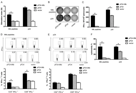
. As shown in Fig. 2A,
strong splenic T cell proliferation to
M. tuberculosis
peptides was
observed in pP24-Mtb-vaccinated mice compared to that in
pP24-treated mice (
P
⬍
0.05). In addition, splenocytes from
Protective Efficacy of pP24-Mtb Tuberculosis Vaccineon August 17, 2020 by guest
http://cvi.asm.org/
pP24-Mtb-immunized mice also showed considerable
prolifera-tion in response to p24.
IFN-
␥
ELISPOT assays and intracellular cytokine staining
as-says were performed to assess the functional T cell response. As
shown in Fig. 2B and C, pP24-Mtb vaccination significantly
in-creased the number of IFN-
␥
-secreting T cells to both
M.
tuber-culosis
peptides and p24 protein compared to pP24 vaccination
(
P
⬍
0.05), suggesting that pP24-Mtb immunization induced
po-tent
M. tuberculosis
- and p24-specific T cell responses. Similar
data were achieved when CD4
⫹IFN-
␥
⫹and CD8
⫹IFN-
␥
⫹T cells
were detected by flow cytometry (Fig. 2D and E), suggesting that
pP24-Mtb vaccination effectively produced
M. tuberculosis
-spe-cific CD4
⫹and CD8
⫹T cell responses and potent responses to
HIV-1 p24 concurrently.
Specific CTL activity was assessed by using splenocytes
stimu-lated with
M. tuberculosis
peptides or p24 protein as effector cells
and pulsed SP2/0 cells as target cells. As shown in Fig. 2F, powerful
M. tuberculosis
-specific CTL activity was observed in
pP24-Mtb-immunized mice. Moreover, splenocytes from
pP24-Mtb-immu-nized mice also showed significantly increased p24-specific CTL
activity compared with those from pP24-immunized mice (
P
⬍
0.05). These results demonstrated that pP24-Mtb vaccination
in-duced robust specific T cell responses against both
M. tuberculosis
and HIV-1.
Humoral responses specific to p24 elicited by pP24-Mtb
im-munization.
The potential of the vaccine to induce serum IgG was
also evaluated. As shown in Fig. 3B, p24-specific serum IgG was
induced, gradually increased after week 4, and peaked at week 10
in pP24-Mtb- or pP24-immunized mice with the IgG titer
reach-ing 1:4,000 and 1:2,250, respectively (Fig. 3C). In contrast, there
was no detectable IgG against
M. tuberculosis
peptides (Fig. 3A),
which was not surprising because they are T cell epitopes. In
ad-dition, an elevated IgG2a/IgG1 ratio was observed following
pP24-Mtb immunization (Fig. 3D) (
P
⬍
0.05), indicating a more
Th1-polarized response. However, the avidity indices of serum
IgG elicited by pP24-Mtb- and pP24-immunized mice were not
statistically different (Fig. 3E). These data indicated that vigorous
humoral responses to the HIV-1 protein were successfully elicited
by pP24-Mtb immunization.
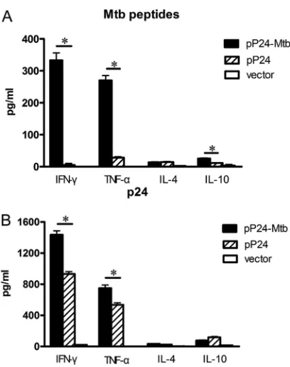
Th1 predominant response generated by pP24-Mtb
immuni-zation.
As enhanced serum IgG2a and IFN-
␥
⫹T cell response
induced by pP24-Mtb immunization indicated a Th1 immune
response, the cytokine profiles produced by
in vitro
-primed
lym-phocytes were then analyzed. After stimulation with the pooled
M.
tuberculosis
peptides, splenocytes from pP24-Mtb-vaccinated
mice produced significantly higher levels of IFN-
␥
and TNF-
␣
(
P
⬍
0.05) (Fig. 4A). Furthermore, pP24-Mtb vaccination also
showed higher levels of p24-specific IFN-
␥
and TNF-
␣
compared
FIG 2M. tuberculosis- and p24-specific cellular responses induced by pP24-Mtb immunization. Two weeks after the last immunization, splenocytes were harvested and stimulated withM. tuberculosispeptides or p24 proteinin vitro. (A) Lymphocyte proliferation was measured and expressed as stimulation index (SI). (B) IFN-␥-secreting lymphocytes were quantified by ELISPOT assay. Images represent splenic ELISPOT responses in the immunized mice. (C) Frequency of IFN-␥-secreting cells in the spleens of mice measured by ELISPOT assay. (D and E) The percentages of CD4⫹IFN-␥⫹or CD8⫹IFN-␥⫹T cells in the spleens were analyzed by flow cytometry followingM. tuberculosispeptide stimulation (D) or following p24 stimulation (E). (F)M. tuberculosis- and p24-specific CTL activity was expressed as the percent cytotoxicity. Data in the graph are from one representative experiment of three independent experiments performed. Error bars represent the means plus standard deviations (SD) (n⫽6). Values that are statistically significantly different (P⬍0.05) are indicated by a short horizontal line and an asterisk.on August 17, 2020 by guest
http://cvi.asm.org/
to pP24- or vector-treated mice (
P
⬍
0.05) (Fig. 4B). In
compar-ison, only negligible levels of IL-4 and IL-10 were generated,
indi-cating that vaccination with pP24-Mtb skewed the immune
re-sponse toward the Th1 profile.
Protective effect of the pP24-Mtb vaccine against
Mycobac-terium bovis
BCG challenge.
M. bovis
BCG, which has been
dem-onstrated to multiply well
in vivo
and induce obvious tuberculosis
pathology, has been widely used for challenge experiments (8, 25).
Here, mice were challenged intranasally with a high dose of 1
⫻
10
7CFU BCG 4 weeks after the final immunization to determine
the protective effect of the pP24-Mtb vaccination. Four weeks
after challenge, the bacterial burden in the lungs and spleens of
mice and pathological pulmonary injury were examined. As
shown in Fig. 5A and B, pP24-Mtb vaccination led to an efficient
protection both in the lung (log
10CFU
⫽
4.6) and spleen (log
10CFU
⫽
2.65) compared to pP24 or vector vaccination. These data
demonstrated that the
M. tuberculosis
-specific immune responses
elicited by pP24-Mtb led to an efficient host defense at local and
systemic tissue compartments.
When tissue pathology was observed, it was seen that lung
tissues from vector- or pP24-immunized mice showed
wide-spread and severe interstitial pneumonia, intense inflammation,
and diffuse granuloma responses after
M. bovis
BCG infection,
displaying excessive lymphocyte and macrophage infiltration
(Fig. 5C and D). However, pP24-Mtb-immunized mice exhibited
the pulmonary alveolar wall structure of healthy mice with very
mild lung inflammation, indicating dramatically attenuated
tu-berculosis pathology. The histological evidence and the bacterial
burden reduction confirmed the protective efficacy achieved by
pP24-Mtb DNA immunization.
DISCUSSION
TB remains one of the leading causes of death worldwide (44), and
HIV infection contributes to the number of deaths caused by TB
(23). On the other hand, TB is the largest single cause of death in
the AIDS setting, accounting for about 25% of AIDS-related
deaths (16). Therefore, a novel and effective tuberculosis vaccine
with immunogenicity to both
M. tuberculosis
and HIV-1 is
needed. Im et al. (19) developed a live attenuated vaccine by
con-structing a recombinant
M
.
bovis
BCG expressing an
HIV-1-de-FIG 3p24-specific serum IgG elicited by pP24-Mtb immunization. Mice were immunized 4 times with pP24-Mtb, pP24, or pcDNA3.1 (vector) at 2-week intervals. (A and B) SerumM. tuberculosis-specific IgG (A) and p24-specific IgG (B) were detected by ELISA at the indicated time points. (C to E) p24-specific antibody titers (C), serum IgG subclasses (D), and avidity (E) were determined 2 weeks after the last immunization. Each bar represents the average value plus SD (error bar) for 6 mice, measured in duplicate experiments. Values that are statistically significantly different (P⬍0.05) are indicated by a short horizontal line and an asterisk. ND, not detected.FIG 4Th1 immune response induced by pP24-Mtb immunization. The con-centrations of cytokines in the splenocyte culture supernatant were deter-mined by ELISAs. (A) Cytokine production by the splenocytes stimulated with M. tuberculosispeptides. (B) Cytokine production by the splenocytes stimu-lated with p24 protein. The data in this figure are from one representative experiment of three experiments performed and presented as the mean plus SD (n⫽6). Values that are statistically significantly different (P⬍0.05) are indicated by a short horizontal line and an asterisk.
Protective Efficacy of pP24-Mtb Tuberculosis Vaccine
on August 17, 2020 by guest
http://cvi.asm.org/
rived immunogen. It is well established that DNA vaccines are safe
and easy to prepare, as well as good at generating T cell responses,
including cytotoxic T lymphocytes and type 1 helper T cells which
are especially required for protection against
M. tuberculosis
and
HIV-1 (11, 18). In the present study, we generated a novel DNA
vaccine pP24-Mtb by incorporating four
M. tuberculosis
epitopes
into the HIV-1 p24 backbone. We demonstrated that vaccination
with pP24-Mtb inhibited the replication of
M. bovis
BCG and
mitigated the pathology of tuberculosis and simultaneously
elic-ited robust cellular and humoral responses to HIV-1. However,
though
M. tuberculosis
is a global health problem with 2 billion
people infected, most are in a latent state where mycobacteria are
thought to persist for decades (6, 7, 12). HIV infection is the
stron-gest risk factor for the progression of LTBI to active TB (14, 28).
Thus, determining whether pP24-Mtb can also protect against
LTBI or prevent LTBI from progressing to active TB would be
meaningful.
As T cell epitopes are crucial for defending against intracellular
pathogens (17, 40), such as
M. tuberculosis
, searching for epitopes
from immunodominant
M. tuberculosis
antigens is critical.
Stud-ies show that Ag85A and Ag85B stimulate strong humoral and
cell-mediated immune responses and produce significant
protec-tion against
Mycobacterium tuberculosis
H37Rv challenge (18, 20,
22, 38). In addition, MPT64, a member of the culture filtrate
pro-tein (CFP) family, can induce a strong T cell response (42).
TB10.4, a member of a subfamily of the ESAT6 family, is even
more vigorously recognized than ESAT-6 (6-kDa early secreted
antigen target of
M
.
tuberculosis
) in TB patients (35). Therefore, in
this study, we generated a vaccine harboring four uncharacterized
T cell epitopes from the four antigens. We demonstrated that the
pP24-Mtb vaccination efficiently induced
M. tuberculosis
-specific
T cell immunity and protection against mycobacterial challenge
which was evidenced by the greatly reduced bacterial burden and
lung pathology. Moreover, we also examined the immunogenicity
of individual epitopes and found that all four epitopes contributed
to
M. tuberculosis
-specific T cell responses. Of the four epitopes,
the Ag85B
184-192and Ag85A
242-250epitopes are more
immuno-genic than the TB10.4
74-82and MPT64
76-
84epitopes (data not
shown).
Many epitopes are short peptides, with poor immunogenicity
unless they are introduced into a carrier protein (39). Using a
natural protein as a scaffold is ideal for the presentation of random
sequences (13). Ofek et al. (29) grafted neutralizing antibody
epitopes into protein scaffolds and elegantly demonstrated that
epitope-specific humoral immune responses were successfully
in-duced by this strategy. In the current study, we extended previous
studies by grafting
M. tuberculosis
T cell epitopes into a p24
scaf-fold protein to form a pP24-Mtb vaccine. Strong epitope-specific
T cell proliferation, cytotoxicity, and elevated frequency of
IFN-
␥
⫹T cells clearly demonstrated that a robust cellular
re-sponse to these T cell epitopes was successfully induced by this
strategy.
As the pP24-Mtb vaccine was designed to possess
immunoge-nicity to both
M. tuberculosis
and HIV-1, it is crucial to select a
suitable immunodominant antigen to induce protective responses
to HIV-1. Studies show that HIV-1 p24 can stimulate robust
an-tigen-specific humoral and cellular responses (3, 27, 41).
There-fore, in the pP24-Mtb vaccine, the scaffold protein p24 for
incor-FIG 5Protection against mycobacterial infection initiated by pP24-Mtb immunization. Four weeks following the last immunization, mice were intranasally challenged with 1⫻107CFU ofM.bovisBCG. (A and B) Four weeks postchallenge, the bacterial loads in the lungs (A) and spleens (B) were measured. The dataare presented as the means plus SD (n⫽6) and are from one representative experiment of three separate experiments. Values that are statistically significantly different (P⬍0.05) are indicated by a short horizontal line and an asterisk. (C) Paraffin sections from lung tissues were stained with hematoxylin and eosin (HE) and evaluated for the level of lung inflammation. (D) Representative histology sections depict lung tissue of immunized mice, with a lung section of a healthy (normal) mouse as a negative control. Individual experiments were conducted three times, with the results from one representative experiment shown. Magnification,⫻200.
on August 17, 2020 by guest
http://cvi.asm.org/
poration of
M. tuberculosis
epitopes was also used as an
immunogen to induce an immune response to HIV-1. When
achieving
M. tuberculosis
-specific T immune responses, the
vac-cine also mounted strong humoral and cellular responses specific
to p24 protein as evidenced by high levels of specific antibody,
Th1-dominated immune responses, and CTL activity, indicating
the potential of this vaccine to induce protective immunity against
HIV-1. Moreover, a stronger immune response to p24 was
ob-served for pP24-Mtb vaccination than for pP24 vaccination. We
presumed that the insertion of the epitopes affected the
confor-mation of p24 or produced new epitopes in p24. These changes
may increase its recognition and presentation to better activate the
immune response, which is similar with previous work (2, 9).
However, the underlying mechanisms need further research.
In conclusion, here we assessed the feasibility and
immunolog-ical efficacy of the tuberculosis DNA vaccine in an HIV-1 p24
protein backbone. Immunization of pP24-Mtb predominantly
produced
M. tuberculosis
-specific Th1 immune responses and
CTL activity which conferred protection against mycobacterial
infection. Meanwhile, an efficient humoral and cellular immune
response to HIV-1 p24 was also elicited. This strategy may provide
a new way to design future DNA vaccines against coinfection.
ACKNOWLEDGMENTS
We thank Robert Tycko (Laboratory of Chemical Physics, National Insti-tute of Diabetes and Digestive and Kidney Diseases, National InstiInsti-tutes of Health) for donation of plasmid HIV-1 p24 and Shuo Li for technical assistance.
This work was funded by the National Science & Technology Key Projects during the Eleventh Five-Year Plan Period of China (2012ZX10003-006), China NSFC grant (81072409), Jiangsu High Level “Shuang-Chuang” Project, Program for Outstanding Medical Academic Leader (LJ06011), and grant (10JC1400900) from the Science and Tech-nology Commission of Shanghai Municipality.
REFERENCES
1.Al-Attiyah R, Mustafa AS. 2008. Characterization of human cellular immune responses to novelMycobacterium tuberculosisantigens encoded by genomic regions absent inMycobacterium bovisBCG. Infect. Immun. 76:4190 – 4198.
2.Ametani A, et al.2003. Amino acid residue substitution at T-cell deter-minant-flanking sites in beta-lactoglobulin modulates antigen presenta-tion to T cells through subtle conformapresenta-tional change. Biosci. Biotechnol. Biochem.67:1507–1514.
3.Ammaranond P, et al.2011. HIV immune escape at an immunodomi-nant epitope in HLA-B*27-positive individuals predicts viral load out-come. J. Immunol.186:479 – 488.
4.Barouch DH.2008. Challenges in the development of an HIV-1 vaccine. Nature455:613– 619.
5.Capozzo AV, et al.2006. Neonatal immunization with a Sindbis virus-DNA measles vaccine induces adult-like neutralizing antibodies and cell-mediated immunity in the presence of maternal antibodies. J. Immunol. 176:5671–5681.
6.Chan J, Flynn J.2004. The immunological aspects of latency in tubercu-losis. Clin. Immunol.110:2–12.
7.Chapman AL.2011. New tests will improve detection of latent TB. Prac-titioner255(1745):23–26.
8.Chen L, Wang J, Zganiacz A, Xing Z.2004. Single intranasal mucosal Mycobacterium bovisBCG vaccination confers improved protection com-pared to subcutaneous vaccination against pulmonary tuberculosis. In-fect. Immun.72:238 –246.
9.Culshaw A, Dong T, Rowland-Jones SL.2012. A two amino acid shift in position leads to a substantial difference in the pattern of processing of two HIV-1 epitopes. J. Acquir. Immune Defic. Syndr.59:335–339.
10. Depla E, et al.2008. Rational design of a multiepitope vaccine encoding T-lymphocyte epitopes for treatment of chronic hepatitis B virus infec-tions. J. Virol.82:435– 450.
11. D’Souza S, et al.2003. Mapping of murine Th1 helper T-cell epitopes of mycolyl transferases Ag85A, Ag85B, and Ag85C fromMycobacterium tu-berculosis. Infect. Immun.71:483– 493.
12. Fallahi-Sichani M, Flynn JL, Linderman JJ, Kirschner DE.2012. Differ-ential risk of tuberculosis reactivation among anti-TNF therapies is due to drug binding kinetics and permeability. J. Immunol.188:3169 –3178. 13.Fernandez-Carneado J, et al. 2000. Surface grafting onto
template-assembled synthetic protein scaffolds in molecular recognition. Biopoly-mers55:451– 458.
14. Gao L, Zhou F, Li X, Jin Q.2010. HIV/TB co-infection in mainland China: a meta-analysis. PLoS One5:e10736.
15. Gong X, Gai W, Xu J, Zhou W, Tien P.2009. Glycoprotein 96-mediated presentation of human immunodeficiency virus type 1 (HIV-1)-specific human leukocyte antigen class I-restricted peptide and humoral immune responses to HIV-1 p24. Clin. Vaccine Immunol.16:1595–1600. 16. Harrington M.2010. From HIV to tuberculosis and back again: a tale of
activism in 2 pandemics. Clin. Infect. Dis.50(Suppl. 3):S260 –S266. 17. Hart MK, et al.1990. Synthetic peptides containing T and B cell epitopes
from human immunodeficiency virus envelope gp120 induce anti-HIV proliferative responses and high titers of neutralizing antibodies in rhesus monkeys. J. Immunol.145:2677–2685.
18. Huygen K, et al.1996. Immunogenicity and protective efficacy of a tuberculosis DNA vaccine. Nat. Med.2:893– 898.
19. Im EJ, et al.2007. Vaccine platform for prevention of tuberculosis and mother-to-child transmission of human immunodeficiency virus type 1 through breastfeeding. J. Virol.81:9408 –9418.
20. Kamath AT, Feng CG, Macdonald M, Briscoe H, Britton WJ.1999. Differential protective efficacy of DNA vaccines expressing secreted pro-teins ofMycobacterium tuberculosis. Infect. Immun.67:1702–1707. 21. Korber B, LaBute M, Yusim K.2006. Immunoinformatics comes of age.
PLoS Comput. Biol.2:e71.
22. Launois P, et al.1994. T-cell-epitope mapping of the major secreted mycobacterial antigen Ag85A in tuberculosis and leprosy. Infect. Immun. 62:3679 –3687.
23. Lawn SD, Churchyard G.2009. Epidemiology of HIV-associated tuber-culosis. Curr. Opin. HIV AIDS4:325–333.
24. Letvin NL.2007. Correlates of immune protection and the development of a human immunodeficiency virus vaccine. Immunity27:366 –369. 25. Leversen NA, Sviland L, Wiker HG, Mustafa T. 12 January 2012.
Long-term persistence of BCG Pasteur in lungs of C57BL/6 mice following intranasal infection. Scand. J. Immunol. doi:10.1111/j.1365– 3083.2012.02683.x. [Epub ahead of print.
26. Maartens G, Wilkinson RJ.2007. Tuberculosis. Lancet370:2030 –2043. 27. Mateu MG.2009. The capsid protein of human immunodeficiency virus:
intersubunit interactions during virus assembly. FEBS J.276:6098 – 6109. 28. Meya DB, McAdam KP.2007. The TB pandemic: an old problem seeking
new solutions. J. Intern. Med.261:309 –329.
28a.Ministry of Health.1998. Care and use of laboratory animals, no. 55. Ministry of Health, Beijing, People’s Republic of China.
29. Ofek G, et al.2010. Elicitation of structure-specific antibodies by epitope scaffolds. Proc. Natl. Acad. Sci. U. S. A.107:17880 –17887.
30. Okazaki T, et al.2006. Epitope enhancement of a CD4 HIV epitope toward the development of the next generation HIV vaccine. J. Immunol. 176:3753–3759.
31. Parvanova I, Rettig L, Knuth A, Pascolo S.2011. The form of NY-ESO-1 antigen has an impact on the clinical efficacy of anti-tumor vaccination. Vaccine29:3832–3836.
32. Prabhu Anand S, Selvaraj P, Narayanan PR.2009. Effect of 1,25 dihy-droxyvitamin D3 on intracellular IFN-gamma and TNF-alpha positive T cell subsets in pulmonary tuberculosis. Cytokine45:105–110.
33. Raby E, et al.2008. The effects of HIV on the sensitivity of a whole blood IFN-gamma release assay in Zambian adults with active tuberculosis. PLoS One3:e2489.
34.Rosa DS, et al. 2011. A DNA vaccine encoding multiple HIV CD4 epitopes elicits vigorous polyfunctional, long-lived CD4⫹and CD8⫹T cell responses. PLoS One6:e16921.
35. Skjot RL, et al.2002. Epitope mapping of the immunodominant antigen TB10.4 and the two homologous proteins TB10.3 and TB12.9, which con-stitute a subfamily of the esat-6 gene family. Infect. Immun.70:5446 – 5453.
36. Song S, et al.2007. Evaluation of antitumor immunity efficacy of epitope-based vaccine with B16 cell line coexpressing HLA-A2/H-2kb and CTL multiepitope in HLA transgenic mice. Vaccine25:4853– 4860.
Protective Efficacy of pP24-Mtb Tuberculosis Vaccine
on August 17, 2020 by guest
http://cvi.asm.org/
37. Sterne JA, Rodrigues LC, Guedes IN.1998. Does the efficacy of BCG decline with time since vaccination. Int. J. Tuberc. Lung Dis.2:200 –207. 38. Tanghe A, et al.1999. Immunogenicity and protective efficacy of
tuber-culosis DNA vaccines encoding putative phosphate transport receptors. J. Immunol.162:1113–1119.
39. Tissot AC, et al. 2010. Versatile virus-like particle carrier for epitope based vaccines. PLoS One5:e9809.
40. Townsend AR, et al.1986. The epitopes of influenza nucleoprotein rec-ognized by cytotoxic T lymphocytes can be defined with short synthetic peptides. Cell44:959 –968.
41. van Bockel DJ, et al.2011. Persistent survival of prevalent clonotypes within an immunodominant HIV gag-specific CD8⫹T cell response. J. Immunol.186:359 –371.
42. Wang C, et al.2011. A DNA vaccine expressing CFP21 and MPT64 fusion protein enhances BCG-induced protective immunity againstMycobacterium tuberculosisinfection in mice. Med. Microbiol. Immunol.200:165–175. 43. Watkins DI, Burton DR, Kallas EG, Moore JP, Koff WC.2008.
Non-human primate models and the failure of the Merck HIV-1 vaccine in humans. Nat. Med.14:617– 621.
44. WHO.2010. Global tuberculosis control. WHO, Geneva, Switzerland.