ABSTRACT
RICHARDS, ANDREW LATIMER. A Dynamically Pressurized Heart Model to Facilitate the Development of Surgical Tools and Techniques for Mitral Valve Repair. (Under the direction of Dr. Gregory D. Buckner).
Background: The development of a novel surgical tool or technique used in mitral valve repair can be hampered by the cost, complexity, and time associated with performing animal trials. We sought to develop a dynamically pressurized model which detects and quantifies mitral regurgitation in intact porcine hearts in order to preliminarily evaluate the
effectiveness of mitral valve repair methods without the need for animal trials.
Methods:A computer controlled pulse duplication system was designed to accept freshly explanted porcine hearts and replicate a wide range of physiological conditions. To test the capabilities of this system in measuring mitral regurgitation, the cardiac output of four hearts was measured under two different peak left atrial pressures (120 and 150 mmHg) before and after induced mitral valve failures. Measurements were compared with clinically standard echocardiographic images.
Results:For all trials, cardiac output decreased as peak left atrial pressure was increased. After induction of mitral valve insufficiencies, cardiac output decreased, with a peak regurgitant fraction of 27%. These findings correlated well with the results from echocardiography.
Conclusions:The resulting system is able to consistently and reliably detect and quantify mitral regurgitation and serves as an effective tool for the design of mitral valve repair
A Dynamically Pressurized Heart Model to Facilitate the
Development of Surgical Tools and Techniques
for Mitral Valve Repair
by
Andrew Latimer Richards
A thesis submitted to the Graduate Faculty of North Carolina State University
in partial fulfillment of the requirements for the Degree of
Master of Science
Biomedical Engineering
Raleigh, North Carolina 2008
APPROVED BY:
__________________________ __________________________ Dr. Richard L. Goldberg Dr. Denis R. Cormier
BIOGRAPHY
Andrew Latimer Richards was born October 25, 1982 in Columbia, SC. He received his Bachelor of Science in Computer Engineering and a minor in International Engineering and Science from Clemson University (Clemson, SC) in December 2004. After a brief period of working in industry, Andy decided to further his education and began work on his Master’s Degree in the Joint Department of Biomedical Engineering at North Carolina State
ACKNOWLEDGEMENTS
I would like to thank my parents for always supporting me and giving me the encouragement to keep trying when things became tough. Thanks to all my siblings for keeping the
competition going and motivating me to try harder.
Special thanks go to Libby Long for always standing by me and keeping me sane. It certainly would not have been possible without her by my side.
I would like to thank my advisor, Dr Gregory Buckner, for his advice and guidance, Dr. Teresa DeFrancesco for her expertise in acquiring echocardiographic images, William Griffin for his assistance in the design of the system, and Nahunta Pork Center for its continued support of higher level research.
TABLE OF CONTENTS
LIST OF TABLES ... v
LIST OF FIGURES ... vi
1. Background ... 1
2. Material and methods ... 2
2.1 Heart Cart construction ... 2
2.2 Computer control ... 4
2.3 Heart preparation ... 6
2.4 Determining mitral regurgitation ... 7
2.5 Normal valve data collection ... 8
2.6 Creation of valve insufficiency ... 9
2.7 Damaged valve data collection ... 10
2.8 Echocardiographic Analysis ... 10
3. Results ... 11
3.1 Changes in cardiac output ... 12
3.2 Pressure waveforms ... 12
3.3 Echocardiographic analysis ... 13
4. Discussion ... 15
5. Limitations ... 18
LIST OF TABLES
LIST OF FIGURES
Figure 1. The Heart Cart ... 3
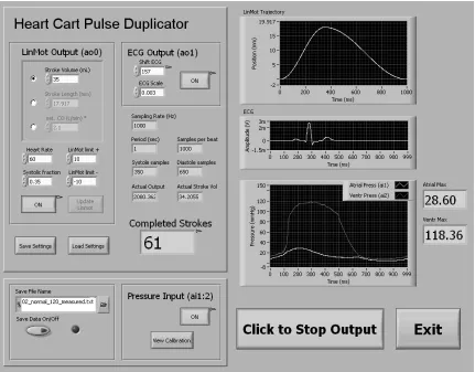
Figure 2. Heart Cart software GUI ... 5
Figure 3. Heart connected to system ... 6
Figure 4. Pressure waveform comparisons ... 13
1. Background
The last decade has witnessed marked advancements in the development of novel instruments and methods for mitral valve repair. From innovations in surgical approach, including minimally invasive [1, 2] and robotic [3] techniques, to improvements in surgical instruments and materials [4-6], options for treating mitral valve disease are expanding rapidly.
The development of new surgical tools and techniques requires extensive testing and validation before they can be introduced into the clinical setting; however, the preliminary stages of prototype development can be hampered by the cost associated with animal trials. While animal trials are the surgical standard for evaluating new technologies and approaches, and are a necessary step in product development, they are frequently inefficient choices for the initial exploration of new methods of heart valve repair. The early stages of the design process could be greatly shortened by the utilization of a cost-effective, accurate dynamic model to evaluate emerging mitral valve repair techniques, lessening the need for laboratory animal sacrifice.
and transplantation techniques. Limited time windows for consistent operation after reanimation and dependency on laboratory animals further restricts the feasibility of these systems for preliminary surgical evaluations.
In this study, we demonstrate the feasibility of a dynamically pressurized heart model to facilitate the development of surgical tools and techniques for mitral valve repair. This left heart pulse duplication system, the “Heart Cart”, dynamically pressurizes porcine hearts to replicate a wide range of physiological conditions and mitral valve deficiencies. This system uses a computer-controlled positive displacement pump and the heart’s own valves to
precisely direct the flow of saline fluid, effectively simulating the valvular dynamics of an in vivo heart. By utilizing intact hearts, the Heart Cart preserves the variability of each heart and enables surgical practice in a natural setting.
To demonstrate the effectiveness of this system in modeling and quantifying mitral regurgitation, experiments were conducted to monitor changes in cardiac output with varying ventricular pressures, before and after induced mitral valve insufficiency. Experimental measurements from the system were compared to clinically standard echocardiographic images.
2. Material and methods
2.1 Heart Cart construction
Figure 1. The Heart Cart. Left: photograph of the system in operation. Right: schematic of system components: atrial filling reservoir (1), PD pump (2), pressure catheters (3), centrifugal pump (4), aortic outflow resistance valve (5), and static pressure mode valves (6). Dynamic and static pressure mode pathways are shaded light
and dark, respectively
Dynamic pressurization of the left ventricle is achieved using a computer-controlled PD pump, connected via an apical cannula. During diastole (ventricular depressurization), retrograde motion of the piston causes fluid to be drawn from the left atrium through the mitral valve and into the left ventricle and piston cylinder. During systole (ventricular pressurization), forward piston motion causes fluid to be expelled from the left ventricle through the aortic valve. The PD pump consists of a LinMot model PS01-37x120
electromagnetic actuator (LinMot, Inc., Elkhorn, WI) and a custom-built piston and cylinder (5.25 cm diameter, 11 cm length, constructed from nylon and sealed using two 33.34 mm OD nitrile O-rings). The actuator’s linear encoder has a resolution of 100 µm, enabling feedback control of pump volume with a resolution of 0.0216 mL.
In addition to its dynamic pressure (“beating” heart) mode, the Heart Cart can be operated in static pressure mode for mitral valve tests requiring constant ventricular pressure. In static pressure mode, valves on the aortic and atrial lines (valves 5 and 6a, Figure 1) are closed, preventing circulative flow from the atrial reservoir. A small centrifugal pump (Model 59510-0012, Jabsco, Glocester, MA) draws fluid from the atrial reservoir and pressurizes the left ventricle via apical cannulation to the desired level (up to 155 mmHg). This mode is used once the mitral valve has been exposed, so that any regurgitant flow is collected in a basin beneath the heart stand.
2.2 Computer control
Figure 2. Heart Cart software GUI
pump stroke volume 150 mL), cardiac output 27 L/min), and systolic fraction (0-100%). In addition, customized pressure waveforms and flow rates can be programmed into the system. The control system also acquires, displays and stores measurements from two pressure catheters at a sampling frequency of 1 kHz. For convenience in gating
Figure 3. Heart connected to system, showing left ventricle (LV), right ventricle (RV), left atrium (LA), left atrial appendage (LAA), and aorta (AOR)
2.3 Heart preparation
Fresh hearts from 70-100 kg swine are obtained from local abattoir and trimmed of excess tissue. A small circular incision is made through the left ventricular dimple at the apex of the heart. A 1.27 cm OD apical cannula is inserted through the incision and secured with
cyanoacrylate adhesive. A 2 cm OD aortic cannula is inserted and secured 2-3 cm distal to the aortic branch to preserve aortic compliance. The brachiocephalic and left common
secured with a 1.5 cm OD feed tube, while the smaller pulmonary vein is fitted with Tuohy-Borst adapters. All remaining veins and branches are closed using surgical stainless steel hemostat clamps. The heart is then secured to the test stand via right ventricle clamps and connected to the appropriate Heart Cart components (Figure 3). The system is filled with 5.75 L of 0.9% saline and allowed to run in dynamic mode for 1 minute. After ensuring correct operation and no system leakage, the left and right coronary arteries are tied closed at the base of the aorta using 2-0 TI•CRON braided polyester suture (USS, Norwalk, CT) to prevent any loss of output due to coronary perfusion.
2.4 Determining mitral regurgitation
In a completely rigid heart with functional mitral and aortic valves, the volumetric output of each PD pump stroke (PD pump Volume, PV) would exactly match the output of the aorta (Cardiac Output volume, CO). Because the walls of the heart are compliant, however, some of the PV is used to expand the left ventricular walls during the systolic (pressurization) phase, causing the CO to be less than PV. The difference between PV and CO is directly related to the pressure inside the left ventricle. For a cardiac cycle with ventricular pressure p, the CO loss associated with compliance is:
CO = PV – CL (Equation 1)
Where CL is the pressure-dependent compliance loss.
total CO. The amount of regurgitation, like compliance loss, is related to pressure in the left ventricle: as the pressure increases, so does the amount of regurgitant flow [14]. The output equation becomes:
CO = PV – CL – MR (Equation 2)
Where MR represents the mitral regurgitation per cardiac cycle.
The only way to quantify MR using Equation 2 is to know the values of CO, PV, and CL. In a heart with a preexisting mitral valve defect, if CO and PV are the only variables that can be measured or calculated, then CL cannot be distinguished from MR; consequently, MR cannot be accurately quantified. If, however, the CO of the heart is measured before the mitral valve defect occurs, then for a given cardiac cycle the change in CO will equal the regurgitation volume. That is, for identical left ventricular pressure waveforms:
CON = PV – CL
COD = PV – CL – MR
MR = CON – COD (Equation 3) Where CON and COD represent the measured cardiac output volumes associated with a
normal and damaged mitral valve, respectively.
2.5 Normal valve data collection
pressure catheter (MPC-500, Millar, Houston, TX) was inserted through the aortic valve into the left ventricle. An identical pressure catheter was inserted through a pulmonary vein into the left atrium. The left ventricle was dynamically pressurized at a heart rate (HR) of 60 bpm, with a PD pump stroke volume (PV) of 35 mL, and a systolic fraction (SF) of 35% (systolic duration of 350 ms). These values were chosen based on in vivo porcine data collected from a survey of published works [11, 15]. Throughout the experiment, data from the pump encoder and pressure catheters were acquired and stored at a 1 kHz sampling frequency for post experimental analysis.
To obtain baseline cardiac output measurements for a normal, undamaged mitral valve, the aortic outflow resistance was increased until the peak left ventricular pressure (pLVP) reached 120 mmHg. The heart was dynamically pressurized for 60 cycles (1 min) and the cardiac output was collected and measured using a graduated cylinder. Next, the outflow resistance was increased until the pLVP reached 150 mmHg and the measurement cycle was repeated.
2.6 Creation of valve insufficiency
posterior distance was defined as the straight-line distance from anterior annulus to posterior annulus, while transverse distance was defined as the straight-line distance from the left to right commissures. Manual stretching of the annulus was performed until a 25% increase in the anterior-posterior distance occurred without decreasing the transverse length.
Next, to induce a flail posterior leaflet, the P2 segment (using Carpentier
nomenclature [17]) of the posterior leaflet was lifted and chordae tendineae connecting the P2 leaflet to the papillary muscles were severed until a satisfactory level of failure was achieved. Satisfactory failure was defined as valve leakage under constant ventricular pressure, without total valve incompetence. Valve damage was visually assessed by switching the system to static pressure mode and applying 35 mmHg backpressure to the mitral valve. The atriotomy was then closed using 2-0 TI•CRON sutures and cyanoacrylate adhesive.
2.7 Damaged valve data collection
To obtain cardiac output measurements for the damaged mitral valve, the heart was dynamically pressurized using the same system settings as previously (HR 60 bpm, PV 35 mL, SF 35%). Aortic outflow resistance was increased until pLVP reached 120 mmHg and cardiac output was measured for 60 cycles (1 min). pLVP was then increased to 150 mmHg and the measurement routine repeated.
2.8 Echocardiographic Analysis
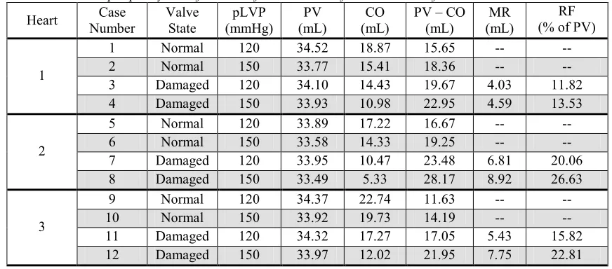
Table 1. Cardiac output per cycle before and after induction of mitral valve defects
Heart Case
Number Valve State pLVP (mmHg) PV (mL) CO (mL)
PV – CO (mL)
MR (mL)
RF (% of PV)
1 Normal 120 34.52 18.87 15.65 -- --
2 Normal 150 33.77 15.41 18.36 -- --
3 Damaged 120 34.10 14.43 19.67 4.03 11.82
1
4 Damaged 150 33.93 10.98 22.95 4.59 13.53
5 Normal 120 33.89 17.22 16.67 -- --
6 Normal 150 33.58 14.33 19.25 -- --
7 Damaged 120 33.95 10.47 23.48 6.81 20.06
2
8 Damaged 150 33.49 5.33 28.17 8.92 26.63
9 Normal 120 34.37 22.74 11.63 -- --
10 Normal 150 33.92 19.73 14.19 -- --
11 Damaged 120 34.32 17.27 17.05 5.43 15.82
3
12 Damaged 150 33.97 12.02 21.95 7.75 22.81
CO = Cardiac Output volume; MR = Mitral Regurgitation; pLVP = peak Left Ventricular Pressure; PV = Piston Stroke Volume; RF = Regurgitant Fraction
Malvern, PA) with both 7 MHz and 10 MHz cardiac probes (models 7V3 and 10V4,
respectively). For optimal imaging and velocity measurements, the echo probes were placed directly on the surface of the heart. At each experimental state, levels of mitral and aortic regurgitation were graded based on echocardiographic images.
3. Results
3.1 Changes in cardiac output
As shown in Table 1, cardiac output at a given pLVP was significantly reduced following induction of mitral valve defects, by as much as a 39.2% reduction at 120 mmHg pLVP and a 62.8% reduction at 150 mmHg pLVP with the second heart. Additionally, cardiac output for normal valve states reduced as pLVP increased from 120 to 150 mmHg due to ventricular wall compliance, by as much as 18.3% in the first heart. Mitral regurgitation volume was calculated based on Equation 3 (with normalized output values for 60 cardiac cycles) and was shown to increase with increasing pLVP, with an average increase of 29.3% as pLVP increased from 120 mmHg to 150 mmHg.
3.2 Pressure waveforms
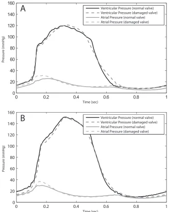
For each case, 10 cycles of measured pressure were averaged to compute mean left
Figure 4. Pressure waveform comparisons: A) normal and damaged valve with pLVP of 120mmHg; B) normal and damaged valve with pLVP of 150mmHg
3.3 Echocardiographic analysis
Analysis of echocardiographic images, acquired using a 10 MHz probe, revealed no visible mitral regurgitation in either of the normal valve cases (Figure 5A and Figure 5B). Following
0 0.2 0.4 0.6 0.8 1
0 20 40 60 80 100 120 140 160 Time (sec) P ressur e (mmHg)
B
Ventricular Pressure (normal valve)Ventricular Pressure (damaged valve) Atrial Pressure (normal valve) Atrial Pressure (damaged valve)
0 0.2 0.4 0.6 0.8 1 0 20 40 60 80 100 120 140 160 Time (sec) P ressur e (mmHg)
A
Ventricular Pressure (normal valve)Figure 5. Color doppler images of left atrium (LA) and left ventricle (LV) during peak systole: A) normal valve, 120 mmHg pLVP; B) normal valve, 150 mmHg pLVP;
C) failed valve, 120 mmHg pLVP; D) failed valve, 150 mmHg pLVP
Moderate-to-Severe, with an estimated jet area equal to 50% of the left atrial area. The Case 4 jet was described as being more visible and organized than in the previous low pressure case. Thus, with a 25% increase in pLVP, the MR echo severity increased by two grades. Echocardiographic images taken during peak systole in each case are presented in Figure 5.
4. Discussion
A positive relationship between left ventricular pressure and MR has been well established in the literature [14], and its demonstration in this study confirmed the capabilities of the Heart Cart to detect and quantify changes in MR. Echocardiographic imaging, the clinical standard for MR diagnosis, was used to verify the existence and severity of MR quantified using the Heart Cart. Echocardiography is an extremely user-dependent imaging modality and its limitations in measuring precise hemodynamic variables in systems such as the heart are widely documented [18]. For this reason, quantifying MR using echocardiography was not attempted; instead, a standard grading scale was adopted. While the Proximal Isovelocity Surface Area method (PISA) is currently seen as the best method for semi-quantitative analysis of MR [16, 19, 20], the required convergence flow field was not consistently visible in these experiments, thus PISA was not performed.
pump, and not by external activation of cardiac muscle. Future studies will examine the effects of modifying PD pump trajectories to recreate various pressure waveforms.
The Heart Cart was designed to be an effective and affordable precursor to animal and clinical trials. By keeping experimental costs low, numerous preliminary studies can be performed to evaluate newly emerging heart repair techniques and devices. Animal trials, which require lengthy approval by the Institutional Animal Care and Use Committee (IACUC), are less efficient for initial testing of surgical prototypes. Based on customary charges at NC State College of Veterinary Medicine, the experimental costs associated with an equivalent animal trial on a live 70 to 100 kg swine would cost approximately 2500 USD per specimen, which includes a standard 14 day acclimation time. Alternatively, porcine hearts are easily obtained from local sources, enabling emerging valve repair techniques to be tested within hours of the idea’s conception, thus maximizing use of available resources and eliminating the need for animal trials. The cost of materials needed to run these experiments on the Heart Cart system was under 25 USD per trial; the majority of this cost was attributed to the type and amount of suture used.
predetermined template prior to testing. While they offer the benefit of complete control over all hemodynamic parameters, they bypass many of the features of individual heart specimens. In an effort to negate all differences between samples, these systems fail to realistically reproduce the interaction of the atrium and ventricle with the mitral valve complex.
Other advancements have increased the viability of using isolated working heart models. Both Chinchoy et al [11] and Araki and colleagues [12] have had success in
maintaining isolated swine hearts for several hours. Recently, Hill et al [13] have managed to adapt this technique for use on isolated human hearts. These systems are ideal for many ex vivo studies, but they require complicated preparation and rigorous adherence to proper transplantation techniques. After the transition to working heart mode, there is a limited and variable window of time for consistent physiological operation of each heart. To date, these systems have been used for intracardiac anatomical imaging; attempts to stop and restart the heart after initial reanimation (required steps in evaluating changes in heart output) has not been reported. Further, these systems still require laboratory animals for heart
transplantation, which negates the benefit of using beating heart models as efficient precursors to animal trials.
because it provides repeatable control of hemodynamic variables for the duration of
experimental testing. Since mitral regurgitation can be easily and accurately quantified using the system, the Heart Cart can be used to demonstrate and test emerging heart repair
techniques, both surgical and device based.
5. Limitations
Proper operation of the Heart Cart relies on the use of heart specimens that are defect free, since comparisons must be made between normal functioning valves and induced defections. The open design of the Heart Cart allows easy detection of problems such as septal defects, both ventricular and atrial in nature, and other defects which result in system leakage. Proper valve function, both mitral and aortic, can be visually assessed using an endoscopic camera, or with the use of echocardiography. Throughout these experiments, endoscopic and
echocardiographic imaging showed full competency of the aortic valve. Checks were also done to ensure that the pressure catheters did not interfere with valve function.
corresponds to an increase in left ventricular pressure; however, unlike physiological
systems, the increased pressure causes the ventricular volume to increase during systole and
decrease during diastole. The basic functionality of the mitral valve remains the same, but the changes in annular shape during in vivo cardiac cycle [16] cannot be duplicated. Lack of muscle contraction also results in an absence of papillary muscle activation. This was visible in the echo imaging as a slight bulging of the mitral leaflets into the left atrium.
References
1. Mohr FW, Falk V, Diegeler A, Walther T, van Son JA, and Autschbach R. Minimally Invasive Port-Access Mitral Valve Surgery. The Journal of Thoracic and Cardiovascular Surgery 1998;115:567,74; discussion 574-6.
2. Casselman FP, Van Slycke S, Dom H, Lambrechts DL, Vermeulen Y, and Vanermen H. Endoscopic Mitral Valve Repair: Feasible, Reproducible, and Durable. The Journal of Thoracic and Cardiovascular Surgery 2003;125:273-82.
3. Nifong LW, Chitwood WR, Pappas PS et al. Robotic Mitral Valve Surgery: A United States Multicenter Trial. The Journal of Thoracic and Cardiovascular Surgery
2005;129:1395-404.
4. Maisano F, Torracca L, Oppizzi M et al. The Edge-to-Edge Technique: A Simplified Method to Correct Mitral Insufficiency. European Journal of Cardio-Thoracic Surgery : Official Journal of the European Association for Cardio-Thoracic Surgery 1998;13:240,5; discussion 245-6.
5. Fukamachi K, Inoue M, Popovic ZB et al. Off-Pump Mitral Valve Repair using the
6. von Oppell UO, and Mohr FW. Chordal Replacement for both Minimally Invasive and Conventional Mitral Valve Surgery using Premeasured Gore-Tex Loops. The Annals of Thoracic Surgery 2000;70:2166-8.
7. Katoh T, Ikeda N, Nishi K et al. A Newly Designed Adapter for Testing an Ex Vivo Mitral Valve Apparatus. Artificial Organs 1999;23:920-3.
8. Espino DM, Hukins DW, Shepherd DE, Watson MA, and Buchan K. Determination of the Pressure Required to Cause Mitral Valve Failure. Medical Engineering & Physics
2006;28:36-41.
9. Arita M, Tono S, Kasegawa H, and Umezu M. Multiple Purpose Simulator using a Natural Porcine Mitral Valve. Asian Cardiovascular & Thoracic Annals 2004;12:350-6.
10. He S, Fontaine AA, Schwammenthal E, Yoganathan AP, and Levine RA. Integrated Mechanism for Functional Mitral Regurgitation: Leaflet Restriction Versus Coapting Force: In Vitro Studies. Circulation 1997;96:1826-34.
11. Chinchoy E, Soule CL, Houlton AJ et al. Isolated Four-Chamber Working Swine Heart Model. The Annals of Thoracic Surgery 2000;70:1607-14.
of Thoracic Surgery : Official Journal of the European Association for Cardio-Thoracic Surgery 2005;28:435-42.
13. Hill AJ, Laske TG, Coles JA,Jr et al. In Vitro Studies of Human Hearts. The Annals of Thoracic Surgery 2005;79:168-77.
14. Braunwald E, Welch GH,Jr, and Sarnoff SJ. Hemodynamic Effects of Quantitatively Varied Experimental Mitral Regurgitation. Circulation Research 1957;5:539-45.
15. Rosenstrauch D, Akay HM, Bolukoglu H et al. Ex Vivo Resuscitation of Adult Pig Hearts. Texas Heart Institute Journal / from the Texas Heart Institute of St.Luke's Episcopal Hospital, Texas Children's Hospital 2003;30:121-7.
16. Kaplan SR, Bashein G, Sheehan FH et al. Three-Dimensional Echocardiographic Assessment of Annular Shape Changes in the Normal and Regurgitant Mitral Valve. American Heart Journal 2000;139:378-87.
17. Carpentier AF, Lessana A, Relland JY et al. The "Physio-Ring": An Advanced Concept in Mitral Valve Annuloplasty. The Annals of Thoracic Surgery 1995;60:1177,85; discussion 1185-6.
19. Enriquez-Sarano M, Miller FA,Jr, Hayes SN, Bailey KR, Tajik AJ, and Seward JB. Effective Mitral Regurgitant Orifice Area: Clinical use and Pitfalls of the Proximal Isovelocity Surface Area Method. Journal of the American College of Cardiology 1995;25:703-9.
20. Enriquez-Sarano M, Sinak LJ, Tajik AJ, Bailey KR, and Seward JB. Changes in Effective Regurgitant Orifice Throughout Systole in Patients with Mitral Valve Prolapse. A Clinical Study using the Proximal Isovelocity Surface Area Method. Circulation 1995;92:2951-8.
21. Moulopoulos SD, Cardiomechanics. Springfield, Illinois: Charles C. Thomas; 1963:193.