4(2): 660-670, 2014
SCIENCEDOMAIN international www.sciencedomain.org
Roles of CD5
+
, CD19
+
, CD41a
+
, CD55
+
and
CD59
+
in Chronic Immune Thrombocytopenic
Purpura (ITP)
Fatma Kaya
1, Ilhami Berber
2*, Mehmet Ali Erkurt
2, Ismet Aydogdu
3and Ismail Reisli
41
Department of Internal Medicine, Selcuk University, Meram Medical Faculty, Turkey. 2
Department of Hematology, Inonu University Medical Faculty, Turkey. 3
Department of Hematology, Selcuk University, Meram Medical Faculty, Turkey. 4
Department of Pediatric Immunology, Selcuk University, Meram Medical Faculty, Turkey.
Authors’ contributions
This report reflects the opinion of the authors and does not represent the official position of any institution or sponsor. Author FK was responsible for reviewing previous research, journal hand searching, and drafting the report. Authors IB, MAE and IR contributed to the final draft of the manuscript and analysis of relevant data. Author IA were responsible for project coordination. All authors read and approved the final manuscript.
Received 14th June 2013 Accepted 19th September 2013 Published 19th October 2013
ABSTRACT
Aims: The aim of this study was to investigate of the roles of CD5+ and CD19+ on lymphocytes, CD5+ on B lymphocytes, CD41a+ on platelets and CD55+ and CD59+ on erythrocytes in platelet destruction; and evaluate them according to the patient response status to steroid therapy and platelet counts in chronic immune thrombocytopenic purpura (ITP).
Study Design: This study included 20 chronic ITP patients and 20 healthy controls. We
investigated the roles of CD5+ and CD19+ expression on lymphocytes, CD5+ expression on B lymphocytes, CD41a+ expression on platelets, and CD55+ and CD59+ expression on erythrocytes, as well as the platelet counts in healthy and chronic ITP patients. Additionally, these markers were evaluated according to the patient response status to steroid therapy and platelet counts.
Place and Duration of Study: This study took place at the Department of Internal
between November, 2008 and July, 2009.
Methodology: A total of 40 patients (26 women, 14 men, age range: 19-79 years) were
studied. The study group included 20 chronic ITP patients (12 women and 8 men, age range: 19-78 years) and the control group included 20 healthy volunteers (14 women and 6 men, age range: 22-79 years). The platelet counts and expressions of CD5+ and CD19+ on lymphocytes, CD5+ on B lymphocytes, CD41a+ on platelets, and CD55+ and CD59+ on erythrocytes were analysed in the patients and control subjects. The chronic ITP patients were evaluated according to their requirements of treatment. Five patients whose platelet counts were above 50,000 mm–3 were observed without treatment. The other 15 patients whose platelet counts were under 50.000 mm–3 and had bleeding, or whose platelet counts were under 20,000 mm–3, were given methylprednisolone treatments (1 mg/kg/day orally). Three of the 15 patients discontinued treatment for various reasons. The twelve patients who continued the methylprednisolone treatment were divided into two subgroups according to their responder status of steroid treatment. The patients whose platelet counts slowly increased above 30,000 mm–3 within three months included the steroid treatment responder subgroups.
The chronic ITP patients were also divided into two subgroups according to the severity of their thrombocytopenia. The limit of the platelet count was 30,000 mm–3 for severe thrombocytopenia. These parameters were analysed according to the response status of the steroid treatment and platelet counts. The platelet counts, and the expressions of these markers, were compared between the subgroups.
Results: The level of CD5+ on B lymphocyte expression (2.19 ± 1.65) in peripheral blood lymphocytes was significantly higher in the immune thrombocytopenic purpura patients than in the controls (P = .05). The CD55+ + CD59+ expression on erythrocytes (98.03 ± 1.77) was significantly higher in the ITP patients than in the controls (P = .05). There was no significant relationship between the expression of CD5+, CD19+ or CD5+ on B lymphocytes, CD41a+ expression on platelets or CD55+ and CD59+ expression on erythrocytes, according to the response status to steroid therapy in the patient group (P > 0.05). Additionally, the patients were evaluated according to platelet counts, and there was a significantly positive correlation between the level of CD41a+ expression on the platelets and the platelet count (P = .05).
Conclusion: The level of CD5+ on B lymphocytes was significantly higher in the ITP patients than in the controls. A relationship between CD55+ plus CD59+ expression on erythrocytes and immune destruction of platelets was not observed in the chronic ITP patients.
Keywords: Chronic ITP; pathogenesis; CD5; CD19; CD55; CD59; CD41a.
1. INTRODUCTION
Immune thrombocytopenic purpura (ITP) is an autoimmune disease characterized by an increase in platelet destruction due to antibodies against circulating platelets [1]. In chronic ITP patients, thrombocytopenia occurs due to autoimmune destruction, complement-mediated thrombolysis and platelet structural changes. A resistant disease course, despite immunosuppressive treatment and splenectomy, indicates that other mechanisms are responsible for the pathogenesis of the disease [2].
CD5+ B lymphocytes against platelet glycoproteins may play an important role in the pathogenesis of chronic ITP [5]. Ming Hou et al. reported a study about the relationship between CD5+ B cells and chronic ITP. It was concluded that both splenic CD5+ B cells and CD5- B cells produced platelet IgG GP-specific autoantibodies, which may both play a role in the pathogenic process of ITP [6]. CD5+ B lymphocytes are known to be responsible for haemolytic anaemia and ITP formation in patients with lymphoma [7].
Mizutani et al. compared 30 ITP patients and healthy controls, and reported CD5+ B lymphocyte proliferation in the peripheral blood and the spleen [8]. Furthermore, elevated CD5+ B lymphocyte levels in the peripheral blood in some autoimmune diseases, such as rheumatoid arthritis and Sjögren’s Syndrome, was also reported [9].
CD41a+ is a calcium-linked Gp-IIb/IIIa complex and a surface antigen, which is only present on platelet and megakaryocyte surfaces [10]. Antibodies against platelet surfaces, especially against the Gp-IIb/IIIa complex, play an important role in the pathogenesis of ITP [11,12].
The complement system helps or “complements” the ability of antibodies and phagocytic cells to clear pathogens from an organism. Over 25 proteins and protein fragments make up the complement system, including serum proteins, serosal proteins and cell membrane receptors. Three biochemical pathways activate the complement system: the classical complement pathway, the alternative complement pathway and the lectin pathway. CD55+ and CD59+ are complementary regulatory proteins; additionally, CD55+ is a glycoprotein that is expressed in peripheral blood, vascular endothelial cells and extravascular epithelial cell surfaces. It inhibits the activity of the C3 convertase in the classical and alternative pathways. CD59+ is a phosphatidylinositol-linked membrane protein. It is expressed on erythrocytes, lymphocytes, monocytes, neutrophils, platelets, endothelial/epithelial cells, etc., and blocks C9 binding to C5b-8, preventing the membrane attack complex formation and lysis [13].
In patients with paroxysmal nocturnal haemoglobinuria (PNH) thrombocytopenia and thrombosis are commonly seen together. In such cases, deficient or insufficient decay accelerating factors (DAF, CD55) and membrane inhibitor reactive lysis factors (CD59), which are found on the surfaces of the cells, protecting them against complement-mediated lysis, are thought to be etiological factors for the disease [14].
In the present study, the role of the CD5+ and CD19+ on lymphocytes, CD5+ on B lymphocytes, CD41a+ on platelets and CD55+ and CD59+ on erythrocytes in platelet destruction, and their evaluation according to response status to steroid therapy, as well as platelet counts in chronic ITP patients, were investigated.
2. MATERIALS AND METHODS
This study included patients who presented to Selçuk University Meram Medical Faculty, Department of Internal Medicine and Haematology, between November, 2008 and July, 2009. Twenty patients (inpatients and outpatients) and 20 healthy controls were considered to be appropriate for this study. Inclusion criteria for patients were as follows: age ≥ 18 years and previously healthy, newly diagnosed ITP that was not previously treated, negative history of cancer, and isolated thrombocytopenia (platelet count < 100,000 mm–3
The platelet counts, CD5+ and CD19+ on lymphocytes, CD5+ expression on B lymphocytes, CD41a+ expression on platelets, and CD55+ plus CD59+ expression on erythrocytes were analysed in the ITP and control patients. The chronic ITP patients were evaluated according to their requirements of treatment. Five patients whose platelet counts were above 50,000 mm–3 were observed without treatment. The other 15 patients, whose platelet counts were under 50,000 mm–3 and had bleeding, or whose platelet counts were under 20,000 mm–3, were given methylprednisolone treatments (1 mg/kg/day orally). Three of the 15 patients discontinued treatment for various reasons. The other 12 patients who continued the methylprednisolone treatment were divided into two subgroups according to their responder (or non-responder) status of steroid treatment. The patients whose platelet counts slowly moved above 30,000 mm–3 within three months included the steroid treatment responder subgroups.
The chronic ITP patients were divided into two subgroups according to the severity of their thrombocytopenia. The limit of the platelet count was detected to be 30,000 mm–3 for severe thrombocytopenia; and a bone marrow aspiration was performed in these patients. Other causes of thrombocytopenia (such as malignancy, other haematological diseases) were excluded during the bone marrow examination.
Two millimetres of blood were drawn into EDTA tubes from the patients and control group, and the haemoglobin, platelet, leukocyte and mean platelet volume (MPV) levels were evaluated by means of a laser system Advia 21-20 device. Additionally, three millilitres of blood were drawn from the patients and control group, and the samples were analysed using flow cytometry with a Becton Dickinson (BD) Facs Calibur device. The platelet counts were also examined in the patient group after treatment.
The CD5+ study was performed as follows: 20 µL of a CD5+ fluorescein isothiocyenate monoclonal antibody was added to 100 µl of a venous blood sample drawn into an EDTA tube, inside a flow cytometry tube. The CD19+ study was performed similarly: 20 µL of a CD19+ pycoerythrin monoclonal antibody was added to 100 µl of a venous blood sample drawn into an EDTA tube, inside a flow cytometry tube.
The CD41a+ study was performed as follows: a venous blood sample was drawn into an
EDTA tube and centrifuged at 1800 g for 5 minutes, and 100 µl of the supernatant was drawn into a flow cytometry tube. Additionally, 20 µL of a CD41a+ fluorescein isothiocyenate monoclonal antibody was added to this. Cytometric immunotyping was performed using a Becton Dickinson FACSCalibur (BD) multipurpose flow cytometer.
The CD55+ study was performed as follows: 4 ml of BD Cell WASH was added to 2 µl of a venous blood sample drawn into an EDTA tube. This mixture was centrifuged at 1800 g for 5 minutes and the supernatant was thrown away; then, 200 µl of BD Cell WASH was added to the precipitate. Finally, 20 µL of a CD55+ pycoerythrin monoclonal antibody was added to 100 µl of this solution.
Statistical analyses were performed at the Selçuk University Meram Medical Faculty, Department of Medical Biostatistics, using Excel 2003 and SPSS v.15. The parameters are expressed as the median ± standard deviation and percentages. The parameters that were not distributed normally were assessed using the Mann-Whitney U test and Wilcoxon test, and the parameters with a normal distribution were assessed using the student’s t-test. A Pearson Chi-Square test was used to compare the steroid response group to the non-response group. P = .05 was considered to be statistically significant.
3. RESULTS
The patient group included 20 patients (12 female and 8 male), and the control group included 20 healthy volunteers (14 female and 6 male). The median age in the patient group was 44.3 ± 16.8 years (age range: 19-78 years), and it was 35.9 ± 14.29 years (age range: 22-79 years) in the control group.
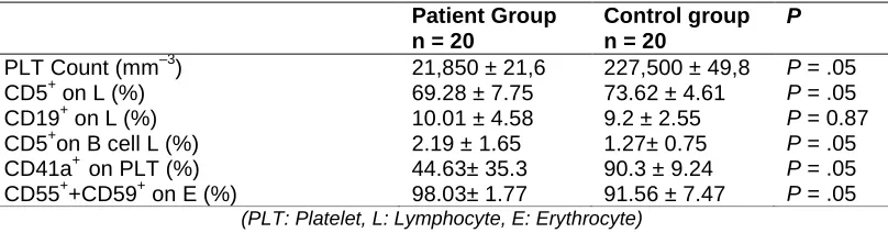
In the patient group, the level of CD5+ expression on B lymphocytes was significantly higher (P = .05). The CD5+ expression on lymphocytes in the patient group was significantly lower than in the control group (P = .05). There was no statistically significant difference in the CD19+ expression on lymphocytes between the groups (P = 0.87). The CD41a+ expression on platelets was significantly lower in the patient group, and the CD55+ + CD59+ expression on erythrocytes was significantly higher in the patient group (P = .05) (Table 1).
Table 1. Comparison of the platelet count and CD expression between the groups.
Patient Group n = 20
Control group n = 20
P
PLT Count (mm–3) 21,850 ± 21,6 227,500 ± 49,8 P = .05
CD5+ on L (%) 69.28 ± 7.75 73.62 ± 4.61 P = .05
CD19+ on L (%) 10.01 ± 4.58 9.2 ± 2.55 P = 0.87
CD5+on B cell L (%) 2.19 ± 1.65 1.27± 0.75 P = .05
CD41a+ on PLT (%) CD55++CD59+ on E (%)
44.63± 35.3 98.03± 1.77
90.3 ± 9.24 91.56 ± 7.47
P = .05 P = .05 (PLT: Platelet, L: Lymphocyte, E: Erythrocyte)
In all, 15 patients received steroid treatments and 5 patients were followed-up without steroid treatments. Among the patients, 4 responded to the steroid treatment (steroid treatment responder subgroup) and 8 did not (steroid treatment non-responder subgroup). Three patients dropped out of the study during follow-up, after they started steroid treatment. In the steroid treatment non-responder subgroup, 1 patient responded to splenectomy and 1 responded to azathiopurine treatment. The comparisons of CD5+ and CD19+ on lymphocytes, CD5+ on B lymphocytes, CD41a+ on platelets, and CD55+ + CD59+ expression on erythrocytes between the two groups showed that there weren’t any statistically significant differences (P > 0.05) (Table 2).
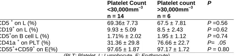
In chronic ITP patients, CD5+ and CD19+ on lymphocytes, CD5+ on B lymphocytes, CD41a+ on platelets, and CD55+ plus CD59+ expression on erythrocytes were compared according to the platelet counts. Any platelet count < 30,000 mm–3 was considered to be severe cytopenia. Patient subgroups were formed based on a platelet count < 30,000 mm–3
CD59+ expression between the 2 subgroups was not noted (P > 0.05); however, CD41a+ expression was significantly higher in the subgroup of patients with a platelet count > 30,000 mm–3 (P = .05) (Table 3).
Table 2. Comparison of CD expression between the steroid treatment responder and steroid treatment non-responder subgroups
Steroid Treatment Responders (n = 4)
Steroid Treatment Non-Responders (n =8)
P
CD5 + on L (%) CD19 + on L (%) CD5+on B cell L (%) CD41a + on PLT (%)
65.25 ± 11.0 9.0± 7.96 2.5± 3.79 18.75± 9.33
71.25 ± 5.88 9.88± 4.52 1.32± 0.71 48.0 ± 33.1
P =0,23 P =0.61 P =0.86 P =0.13 CD55++CD59+ on E (%) 96.75 ± 3.31 97.75 ± 1.04 P =0.86
(PLT: Platelet, L: Lymphocyte, E: Erythrocyte)
Table 3. Comparison of CD expression between the platelet count subgroups
Platelet Count <30,000mm–3 n = 14
Platelet count >30,000mm–3 n = 6
P
CD5 + on L (%) 69.36± 7.73 67.5 ± 7.81 P =0.56
P =0.62 CD19+ on L (%)
CD5+on B cell L (%) CD41a + on PLT (%) CD55++CD59+ on E(%)
9.93 ± 5.09 1.71% ± 2.02 31.36 ± 29.8 97.65 ± 1.87
8.5 ± 2.43 1.95 ± 1.12 76.66 ± 22.7 97.17 ± 1.72
P =0.74 P= .05 P = 0.80 (PLT: Platelet, L: Lymphocyte, E: Erythrocyte)
4. DISCUSSION
ITP is a common haematological disease, and elderly patients generally have a chronic form. In the present study, lymphocyte, erythrocyte and platelet surface marker expressions were examined. Van der Harst et al. reported that CD5+ expression was high on B lymphocytes in ITP patients [15]. Iyori et al. investigated CD5+ expression on lymphocytes and platelet-related autoantibodies in 29 paediatric chronic ITP patients (and a control group); and they reported that there was no statistically significant difference in CD5+ expression on B-lymphocytes between the 2 groups. However, a significant relationship between the level of CD5+ B lymphocytes and platelet-related IgM was observed [16]. Hou et
al. reported that CD5+ expression on B lymphocytes was higher in the patient group than in
the control group, but that the difference was not statistically significant [3]. Yu et al. reported that the level of CD5+ on B lymphocytes was markedly increased in active chronic ITP, and active hyperthyroid patients with Graves' disease, and it was shown that the platelet count is inversely correlated with CD5+ expression on B lymphocytes. The researchers concluded that CD5+ B lymphocytes are responsible for autoantibody formation in some autoimmune diseases. Additionally, a strong relationship between high rheumatoid factor titres and the level of CD5+ expression on B lymphocytes in rheumatoid arthritis (an autoimmune disease) was reported [9].
lymphocytes and clinical severity of the disease [8]. In the present study, CD5+ expression on B lymphocytes in the peripheral blood was significantly higher in the ITP patients than in the controls. In spite of the fact that the mechanism that triggers CD5+ B lymphocytes in ITP patients remains obscure based on currently available data, it could be concluded that CD5+ B lymphocytes are responsible for autoantibody production, and may induce an autoimmune response in such patients.
The relationship between CD5+ expression on B lymphocytes and disease severity was also investigated in the present study. The disease severity was assessed according to the platelet count. The CD5+ expression on B lymphocytes in the subgroups of patients with a platelet count < 30,000 mm–3 and > 30,000 mm–3 was compared; however, a statistically significant relationship was not observed between the disease severity and CD5+ expression on B lymphocytes, perhaps because the patient numbers were too low. Additional evidence is needed to more definitively prove the relationship between CD5+ expression on B lymphocytes and ITP severity.
Hua et al. reported that an increased number of CD5+ on B cells in which IL-10 is accumulated, with decreased IL-10 concentration in the supernatant, suggests that the ability of CD5+ on B cells to secret IL-10 is impaired in ITP patients. Both the aberrant number and ability of IL-10 secretion of CD5+ on B cells could be corrected by a high dose of dexamethasone [17]. A statistically significant relationship between CD5+ expression on B lymphocytes and response to steroid treatment was not observed in the present study.
Evidence of the role of CD5+ T lymphocytes in the immune response of ITP patients is lacking [18]. In the present study, the CD5+ expression on lymphocytes was significantly lower in the ITP patient group, and it is known that CD5+ is expressed on T and B lymphocytes in the peripheral blood [6]. Since the CD5+ expression on B lymphocytes was significantly higher in the patient group, the observed high level of CD5+ expression on lymphocytes in the control group was attributed to the T lymphocytes, which also express CD5+. Based on the present study’s findings, larger study groups should be investigated in the future.
CD19+ expression is observed on B lymphocytes in all stages of differentiation and development, keeping in mind that T lymphocytes, granulocytes, and monocytes don’t express CD19+ [4]. Zhu et al. showed that a high B lymphocyte activating factor titre plays a role in the pathogenesis of some autoimmune diseases. They also reported that the B lymphocyte activating factor prolongs the survival of CD19+ and CD8+ lymphocytes, and increases platelet apoptosis and IFNγ secretion [19]. In the present study, CD19 expression+ on B lymphocytes was higher in the patient group than in the control group; however, the difference was not statistically significant. Furthermore, we did not observe a statistically significant relationship between CD19+ on lymphocytes and the platelet count, or between CD19+ on lymphocytes and the response to steroid treatment.
CD41a+ plays an important role in the pathogenesis of ITP [11,12]. Kahng et al. compared CD41a+, CD41+, CD41b+ and CD61+ expression on thrombocytes in 20 ITP patients, 34 aplastic anaemia patients and several healthy controls. The CD41a+ and CD41+ expressions were significantly lower in the ITP patient group than in the control group [20]. In the present study, the CD41a+ expression, which is responsible (rather than the antibodies against CD41a+) for the pathogenesis of ITP, was investigated. The CD41a+ expression was significantly lower in the patient group than in the control group.
It has been suggested that in ITP patients, the Gp-IIb/IIIa complex on the platelets is covered by antibodies; therefore, the CD41a+ platelet count would be low. Moreover, due to the fact that the platelet count is low, low-level CD41a+ expression would also be expected. A statistically significant correlation was observed in the patient group between the CD41a+ expression and the platelet count. Among the present study’s patients with severe thrombocytopenia, CD41a+ expression was low in the platelet count < 30,000 mm–3 subgroup. Additionally, there was no significant difference in CD41a+ expression between the steroid treatment responder and non-responder subgroups.
Paroxysmal nocturnal hemoglobinuria (PNH) is a hereditary disease, characterized by the deficiency of CD55+ and CD59+ expressions, which causes the destruction of erythrocytes and leukocytes [21]. Recent studies have reported that the cause of haemolysis in PNH is CD59+ deficiency [22]. As with erythrocytes, platelets and leucocytes are also affected by complement-mediated destruction in CD55+ and CD59+ deficiencies [23]. Ruiz-Argüelles studied CD55+ and CD59+ expression deficiencyies in erythrocytes, lymphocytes and platelets in patients with 3 different types of cytopenia (haemolytic anaemia, autoimmune thrombocytopenia and SLE). They compared CD55+ and CD59+ in 30 ITP patients and 30 healthy controls, and observed a statistically significant relationship between CD59+ deficiency and the severity of thrombocytopenia; however, such a relationship has not been demonstrated in CD55+ expression deficiency. Additionally, there weren’t any relationships between CD55+ deficiency, CD59+ deficiency, or both, and the platelet antibodies.
It has been reported that CD55+ and/or CD59+ expression deficiency is related to haemolytic anaemia, autoimmune thrombocytopenia and systemic lupus erythematosus (SLE) lymphopaenia [24]. In the present study, a relationship between CD55+ + CD59+ expression on the surface of erythrocytes and ITP was observed. The CD55+ + CD59+ expression on erythrocytes was significantly higher in the patient group, when compared to that in the control group; however, CD55+ + CD59+ expression deficiency was not observed in any of the patients. In the previous study, the CD55+ + CD59+ expression on platelets was investigated [24], whereas in the present study, CD55+ + CD59+ expression on erythrocytes was investigated. According to the present study’s findings, there was no relationship between the autoimmune response and CD55+ + CD59+ expression on erythrocytes; in other words, CD55+ + CD59+ expression on erythrocytes is not affected by autoimmune response.
Studies with larger patient populations are needed in order to provide more definitive evidence of resistance in CD55+ + CD59+ expression (either alone or together) to the autoimmune response. A statistically significant relationship between CD55+ + CD59+ expression and the severity of thrombocytopenia was not observed in the present study. Furthermore, there was no correlation between CD55+ + CD59+ expression and response status to steroid treatment.
5. CONCLUSION
In conclusion, CD5+ expression on B lymphocytes was significantly higher in the present study’s patient group than in the control group, and these results were similar to the results found in the literature. The CD5+ expression on lymphocytes was significantly lower in the patient group; however, the role of CD5+ expression on lymphocytes in the pathogenesis of ITP is not clear. The CD19+ expression on B lymphocytes was also higher in the patient group, but not significantly. The CD41a+ expression was significantly lower in the patient group than in the control group, and the CD55+ + CD59+ expression on erythrocytes was significantly higher in the present study’s patient group. A relationship between the CD55+ + CD59+ expression on erythrocytes and the immune destruction of platelets was not observed.
Larger, multi-centre studies should be performed on the effects of CD5+ B lymphocytes and CD55+ + CD59+ expression in chronic ITP patients. If the pathology of chronic ITP is clarified, it would be easier to treat patients with treatment-resistance.
CONSENT
Written informed consent was obtained from the patient’s next of kin for publication of this manuscript and accompanying images.
ETHICAL APPROVAL
This study was approved by the Selçuk University Meram Medical Faculty Ethics Committee and was financed by the Selçuk University Meram Medical Faculty Research Project Coordination Department (BAP project no: 1350).
ACKNOWLEDGEMENTS
The authors are thankful to all the physicians of the Selçuk University Meram Medical Faculty and Turgut Ozal Medical Center and the patients who participated in the present study.
COMPETING INTERESTS
The authors declare that they have no competing interests.
REFERENCES
1. Neunert C, Lim W, Crowther M, Cohen A, Solberg L Jr, Crowther MA. The American Society of Hematology. Evidence-based practice guideline for immune thrombocytopenia. Blood. 2011;117(16):4190-207.
2. Johnsen J. Pathogenesis in immune thrombocytopenia: new insights. Hematology Am Soc Hematol Educ Program. 2012;2012:306-12. doi: 10.1182/asheducation-2012.1.306.
4. Loken MR, Shah VO, Dattilio KL, Civin Cl. Flow cytometric analysis of human bone marrow. II. Normal B lymphocyte development. Blood. 1987;70:1316-24.
5. Shaw PX, Hörkkö S, Chang MK, Curtiss LK, Palinski W, Silverman GJ, et al. Natural antibodies with the T15 idiotype may act in atherosclerosis, apoptotic clearance, and protective immunity. J Clin Invest. 2000;(12):1731-40.
6. Hou M, Lv B, He Q, Lu L, Shi Y, Ji X, et al. Both splenic CD5(+) B and CD5(-) B cells produce platelet glycoprotein-specific autoantibodies in chronic ITP. Thromb Res. 2003;110(1):1-5
7. Fink K, AI-Mondhiry H. ldiopathicthrombocytopenic purpura in lymphoma. Cancer 1976;37(4):1999-2004.
8. Mizutani H, Furubayashi T, Kashiwagi H, Honda S, Take H, Kurata Y, et al. B cells expressing CD5 antigen are markedly increased in peripheral blood and spleen lymphocytes from patients with immune thrombocytopenic purpura. Br J Hematol. 1991;78(4):474-9.
9. Yu JR, Qiu ZX, Yang H. CD5+B cells and autoimmune diseases. Zhonghua Nei Ke Za Zhi. 1994;33(9):590-2.
10. Debili N, Robin C, Schiavon V, Letestu R, Pflumio F, Mitjavila-Garcia MT, et al. Different expression of CD41 on human lymphoid and myeloid progenitors from adults and neonates. Blood. 2001;97(7):2023-30.
11. McMillan R. Autoantibodies and autoantigens in chronic immune thrombocytopenic purpura. Semin Hematol 2000;37:239-48.
12. Escher R, Muller D, Vogel M, Miescher S, Stadler BM, Berchtold P. Recombinant human natural autoantibodies Against GPIIb/IIIa inhibit binding of autoantibodies from patients with AITP. Br J Haematology, 1998;102(3):820-28.
13. Abbas AK, Lichtman AH, Pillai S, editors. Cellular and Molecular Immunology. 6th ed. Elsevier. ISBN 978-1-4160-3123-9;2010.
14. Parker CJ. Paroxysmal nocturnal hemoglobinuria. Curr Opin Hematol. 2012;19(3):141-8.
15. Van der Harst D, De Jong D, Limpens J, Kluin PM, Rozier Y, van Ommen GJ, et al. Clonal B-cell populations in patients with idiopathic thrombocytopenic purpura. Blood. 1990;76(11):2321-6.
16. Iyori H, Fujisawa K, Akatsuka J. Autoantibodies and CD5+ B cells in childhood onset immune thrombocytopenic purpura. Acta Paediatr Jpn. 1995;37(3):325-30.
17. Hua F, Ji L, Zhan Y, Li F, Zou S, Wang X, et al. Pulsed high-dose dexamethasone improves interleukin 10 secretion by CD5+ B cells in patients with primary immune thrombocytopenia. J Clin Immunol. 2012;32:1233-42.
18. Kuwana M, Ikeda Y. The role of autoreactive T-cells in the pathogenesis of idiopathic thrombocytopenic purpura. Int J Hematol. 2005;81(2):106-12.
19. Zhu XJ, Shi Y, Peng J, Guo CS, Shan NN, Qin P, et al. The effects of BAFF and BAFF-R-Fc fusion protein in immune thrombocytopenia. Blood. 2009;114(26):5362-7. 20. Kahng J, Park HH, Han K, Choi BY, Lee W. Quantitative comparisons of antibody-binding sites of platelet glycoprotein IIb/IIIa in aplastic anemia and idiopathic thrombocytopenic purpura. Ann Clin Lab Sci. 2008;38(1):6-11.
21. Bessler M, Mason PJ, Hillmen P, Miyata T, Yamada N, Takeda J, et al. Paroxysmal nocturnal haemoglobinuria (PNH) is caused by somatic mutations in the PIG-A gene. Embo J. 1994;13(1):110-7.
23. Devine DV, Siegel RS, Rosse WF. Interactions of the platelets in paroxysmal nocturnal hemoglobinuria with complement. Relationship to defects in the regulation of complement and to platelet survival in vivo. J Clin Invest. 1987;79(1):131-7.
24. Ruiz-Argüelles A, Llorente L. The role of complement regulatory proteins (CD55 and CD59) in the pathogenesis of autoimmune hemocytopenias, Autoimmun Rev. 2007;6(3):155-61.
25. Berchtold P, McMillan R. Therapy of chronic idiopathic thrombocytopenic purpura in adults. Blood. 1989;74(7):2309-17.
© 2014 Kaya et al.; This is an Open Access article distributed under the terms of the Creative Commons Attribution License (http://creativecommons.org/licenses/by/3.0), which permits unrestricted use, distribution, and reproduction in any medium, provided the original work is properly cited.
Peer-review history: