Subthalamic-pallidal interactions are critical in
deter-mining normal and abnormal functioning of the basal
ganglia
Andrew Gillies, David Willshaw and Zhaoping Li†
Institute for Adaptive & Neural Computation, 5 Forrest Hill, University of Edinburgh, Edinburgh EH1 2QL, UK.
†Department of Psychology, University College, London, UK.
Correspondence to:
Andrew Gillies
Institute for Adaptive&Neural Computation 5 Forrest Hill
The University of Edinburgh Edinburgh EH1 2QL
Scotland, UK
tel:+44 (0) 131 650 3096 fax:+44 (0) 131 650 4406 email:[email protected]
Subthalamic-pallidal interactions are critical in
deter-mining normal and abnormal functioning of the basal
ganglia
Andrew Gillies, David Willshaw and Zhaoping Li†
Institute for Adaptive & Neural Computation, 5 Forrest Hill, University of Edinburgh, Edinburgh EH1 2QL, UK.
†Department of Psychology, University College, London, UK.
The subthalamic nucleus (STN) and external globus pallidus (GP) form a recurrent
excitatory–inhibitory interaction within the basal ganglia. Through a computational
model of these interactions we show that, under the influence of appropriate
exter-nal input, the two nuclei can be switched between states of high and low activity or
can generate oscillations consisting of bursts of high frequency activity repeated
at a low rate. It is further demonstrated from the model that the generation of the
repetitive bursting behaviour is favoured by increased inhibition of GPwhich is a
condition indicated in Parkinson’s disease. Paradoxically, increased striatal
inhibi-tion ofGPis predicted to cause an increase rather than a decrease in its mean firing
rate. These behaviours, arising from a biologically inspired computational model
of theSTN–GPinteraction, have important consequences for basal ganglia function
and dysfunction.
Keywords: basal ganglia, subthalamic nucleus, globus pallidus, bifurcation analysis,
1. INTRODUCTION
The basal ganglia (Fig. 1a) have been implicated in movement control and motor
plan-ning (Houk & Wise, 1995), motor and cognitive sequence generation (Suri & Schultz,
1998), sequence encoding (Beiser & Houk, 1998), context detection via reinforcement
learn-ing (Houk & Wise, 1995; Domineyet al., 1995) and action selection (Redgraveet al., 1999).
There is growing realisation that processing within the basal ganglia is more complex than
is implied in the classic view of a direct and an indirect pathway involving neocortex, basal
ganglia and thalamus (Smithet al., 1998; Alexanderet al., 1990). In particular,
contempo-rary neuroanatomy of the basal ganglia reveals a prominent feedback system, involving
the excitatory subthalamic nucleus and the inhibitory globus pallidus (Smithet al., 1994;
Joel & Weiner, 1997; Shinket al., 1996; Wichmann & DeLong, 1999).STNprojection neurons
use glutamate as their primary neurotransmitter and exert a powerful excitatory influence
on their targets (Kita, 1992). GP projection neurons use GABA(γ-Aminobutyric acid) as
their primary neurotransmitter and exert an inhibitory influence on their targets (Smith &
Bolam, 1989). STNis also subject to excitatory influences from cortex and from thalamus,
whilstGPalso receives inhibition from striatum (see Fig. 1a). In addition, local collaterals,
suggesting significant interconnectivity within each nucleus, have been described (Kita
et al., 1983a; Iwahori, 1978; Kita & Kitai, 1994).
Recent analysis (Gillies & Willshaw, 1998) has suggested that the presence of even weak
interconnectivity will cause the subthalamic nucleus to exhibit bistable behaviour. This
means that the entire system can reside in a state in which all neurons fire at close to
maximum rate, close to500Hz, or in a state with a much lower firing rate. Short term
excitatory input can shift the system into the high state and it was suggested that GP
STNcan become uniformly and synchronously active as observed, for example, in ratSTN
after localised cortical stimulation (Fujimoto & Kita, 1993).
In culture preparations of the basal ganglia, Plenz & Kitai (1999) have recently reported
electrical oscillations. The generation of the oscillations depends on the interaction
be-tween theGPand theSTNwhich mirrors that observed in anatomical studies (Plenzet al.,
1998; Joel & Weiner, 1997). Rhythmic bursting oscillations have also been observed in
STNneurons of MPTP monkeys (Niniet al., 1995) and in human patients with Parkinson’s
disease (Levy et al., 2000; Magari ˜nos-Asconeet al., 2000). Some of the STNcells in these
patients have a frequency matching that observed in tremor (5–6 Hz) and are influenced
by tremor changes (Magari ˜nos-Ascone et al., 2000). Other STN cells oscillate at higher
frequencies (∼15 Hz) with evidence of phase locking across the nucleus.
Understanding the underlying dynamics betweenSTNandGPis essential to
understand-ing these behaviours and how they may be modified or controlled. We have analysed the
simplest representative circuit made up of an ensemble of excitatorySTNneurons and an
ensemble of inhibitory GP neurons (Fig. 1b). The dynamic behaviours observed in this
simple system are essentially similar to those in larger scale systems. In the model, the
STNneurons exciteGPand are subject to excitation through local feedback and from
cor-tex. Conversely, the GP neurons inhibit STN and are subject to inhibition through local
feedback and from striatum. Analysis of this model has enabled us to identify the types
of behaviour that this coupledSTN–GP dynamical system can generate; in particular the
2. METHODS
The STN–GP interaction is computed from coupled differential equations describing the
mean membrane potentialsx,yof populations ofSTNandGPneurons respectively:
τSTNdx/dt=−x+aσ(x)−cξ(y) +ICTX
τGPedy/dt=−y−bξ(y) +dσ(x) +ISTR
(1)
τSTN and τGPe are the time constants of the STN and GP neurons respectively. ICTX and
ISTR are external inputs from the cortex and striatum respectively. a, b, c andd specify
connection strengths as indicated in Fig. 1b.σ(x), andξ(y)are sigmoidal functions relating
theSTNandGPpotentialsx, yto their respective firing frequencies.
We present a mathematical analysis of this system along with accompanying simulation
results to demonstrate the behaviours identified. For a general analysis of an excitatory–
inhibitory oscillator system see Ermentrout & Cowan (1979). In our modified system, the
equations are formulated in terms of membrane potential to allow direct application of
experimental data (for example, derived voltage firing-rate curves, where the parameters
specifying the shapes of the curves are derived from experimental data, see Table 1).
Our system is analysed and discussed within the context ofSTN/GPinteractions and
pa-rameters. As the strengths of the external inputs and connections between and within
STNandGPare substantially unconstrained, we explore how different strengths of neural
connections can influence the types of behaviour the system may exhibit under
differ-ent strengths of external inputs from cortex and from striatum. The external inputs are
assumed to be constant for the period of analysis and we assume connection delays are
negligible. We focus on the general physiological properties that reveal the most salient
system behaviours. The model description and assumptions are at the level of general cell
system. The behaviour of the model is characterised in terms of the potentials (x0,y0) at the equilibrium point, wheredx/dt=dy/dt= 0, and the dynamic deviations from it. For
BOX 1 – MATHEMATICAL DETAILS
1. Formalisation. The coupled differential equations describing the membrane potentialsx, y of the
STN and GP neurons specify the ensemble average of a spike train model where impulses are con-volved with an alpha type response function (see Shamma, 1989). This allows us to model spike trains in a dynamic manner:
τSTNdx/dt=
−x+aσ(x)−cξ(y) +ICTX τGPedy/dt=
−y−bξ(y) +dσ(x) +ISTR (1)
wherea, b, c, dspecify the couplings within and between STN and GP;ICTX, ISTRrepresent cortical and
striatal influences. The STN and GP potentialsx, y are related to firing frequencyσ(x), ξ(y)via sig-moidal functions:
σ(x) = σmax
1 +e−κ(x−xth) ξ(y) =
ξmax
1 +e−η(y−yth) (2)
2. Equilibrium points.The equilibrium values of (x, y) in Equation 1 are found by setting
dx/dt=dy/dt= 0. The nature of these equilibria can be obtained by looking at the linear deviations
(ˆx,yˆ) of (x, y) from the equilibrium point (x0,y0) as expressed by the matrix equation
dx/dtˆ
dy/dtˆ =M ˆ x ˆ y
, where M= −A −C
D −B
!
,
A≡(1−aσ0(x0))/τSTN B≡(1 +bξ0(y0)))/τGPe C≡cξ0(y0)/τSTN D≡dσ0(x0)/τGPe
whereσ0(x0)andξ0(y0)are the derivatives ofσandξat (x0,y0). The deviations follow the solution
ˆ
x∝eλ1,2twhereλ
1,2are the eigenvalues ofM
λ1,2=− 1
2(A+B)± 1 2
p
(A+B)2−4(AB+CD) (3)
3. Nature of the equilibrium points.It is useful to distinguish the following three cases, which differ
with respect to the signs ofA+BandAB+CD. Note thatB, C, Dare restricted to positive or zero
values, whereasA, which depends ona, the amount of interconnectivity within STN, can have both
positive and negative values.
3.1.A+B >0andAB+CD >0.The eigenvaluesλ1,2are real or complex with negative real parts, giving a stable equilibrium point.
3.2. A+B < 0 andAB+CD > 0. Both eigenvalues have positive real parts, giving an unstable equilibrium point. When the system has a single equilibrium point which is of this type, limit cycles (oscillations) result, as specified by the Poincar´e-Bendixson theorem (Edelstein-Keshet, 1988).
3.3.AB+CD < 0. Both eigenvalues are real but with opposite signs, yielding an unstable saddle point from which deviations will reach one of the two separate basins.
4. Effect of changes in external inputs. Changing the cortical and striatal inputs byδICTX, δISTRchange
the equilibrium potentials (x0,y0) by
δxo =
B(δICTX/τSTN)−C(δISTR/τGPe)
AB+CD (4)
δyo =
D(δICTX/τSTN) +A(δISTR/τGPe)
3. RESULTS
Setting of parameter valuesThe parametersσmax,κ,xthinσ(x),ξmax,η,yth inξ(y),τSTN
andτGPeare assigned values obtained from the experimental literature as given in Table 1.
Sources Name Value
STN
Kitaet al.(1983b); Afsharpour (1985), τSTN 6mS
σmax 500Hz
Nakanishiet al.(1987). xth 15mV
κ 0.3mV−1 GP
Kita & Kitai (1991), τGPe 14mS
Nambu & Llin´as (1994). ξmax 100Hz
yth 10mV η 0.2mV−1
Table 1: Parameter values used in the model.
We now describe the effects of varying the parameter values that remain unspecified,
namelya, b, c, d, ICTX, ISTR. We summarise the mathematical and numerical results which
shed light on the following issues: (i) the function of the interconnectivity within STN;
(ii) the effects of cortical and striatal input; (iii) the conditions under which the system
is bistable; (iv) the conditions under which oscillations occur; (v) what determines the
frequency of oscillations. An important factor is the value ofawhich determines the
in-terconnectivity withinSTN.
The system can exist in three different types of state: single stable, bistable and
oscilla-tory states. We first give an informal explanation of these states. STNandGPare subject
to excitatory and inhibitory influences, mediated by external inputs and the connectivity
parametersa, b, c, d. We consider the effects of gradually increasing the value ofa,
self-excitation ofSTN, inhibition predominates, keeping the firing rates of both nuclei low
and stable; there is a single stable state. For a higher value ofa, the stable states of the
sys-tem reflect the balancing out of the excitatory and inhibitory influences on the two nuclei.
Stable states emerge at both high and low firing rates. For example, when the system is at
rest with low firing rates, short term excitation of theSTNfrom the cortex will increase the
activity of theSTN, pushing the entire system into a stable state with sustained high firing
rates. Conversely, increased excitation of theGPwhilst in this high state will increase the
inhibition of itself andSTN, ultimately driving both back to their low states (Fig. 2a). Both
the high and low activity states are stable, and separating them is a barrier which can be
crossed by external drive. This bistability causes the system to act in a “switch-like”
man-ner. The oscillatory state could be regarded as alternating jumps, around the barrier, and
between the high and low firing states which are not self-sustaining. The low frequency
bursting–like oscillations emerge in both nuclei. Finally, for very largeaand/or inputs
ICT X, excitation from
STNpredominates to keep both nuclei firing at a high rate.
We now give a more mathematically based description of the system states and their
prop-erties.
(i)Interconnectivity withinSTNIf there is no interconnectivity, theSTN-GPcomplex
sim-ply responds to its external input, by summing the afferent influences (from cortex and
striatum); without interconnectivity it simply follows its transient input. The system
re-sides at equilibrium and when disturbed will always return to this equilibrium. In this
casea = 0leading to A > 0(A ≡ (1−aσ0(x
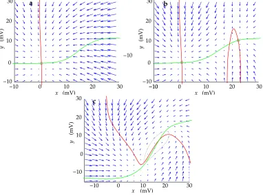
0))/τSTN). This is condition3.1 described in Box 1, where the state (x0,y0) is stable (see phase plot Fig. 3a).
(ii)Influences of cortical and striatal inputIn the presence of low levels of
in the equilibrium firing rates of theSTNandGP. Paradoxically, increased inhibition ofGP
can lead to an increase rather than a decrease in its mean firing rate (Fig. 2b). In particular,
ifais sufficiently small thatAis negativeyetA+B andAB+CD both remain positive,
all equilibrium points are stable (see case3.1and equations (4), (5)).
(iii)Conditions under which bistable behaviour occurs.
In previous work (Gillies & Willshaw, 1998) it was shown in a detailed model of theSTN
that it can display bistable behaviour. It can maintain activity at a high firing rate or at
a low firing rate and can be driven between the two states by transient external input.
A similar situation can occur in this coupled system. If ais sufficiently large so that A
is sufficiently negative to make AB+CD negative (case3.3 of Box 1), the state (x0, y0)
becomes an unstable saddle point. Two new stable states then appear, which will attract
any deviation from (x0, y0) (see phase plot Fig. 3b, Fig. 2a), one at a high firing rate, the
other at a low firing rate.
(iv)Conditions for the generation of oscillations. With the system in the bistable state, a
sufficient increase in the value ofacauses two (one stable and one unstable) of the three
equilibrium points to disappear, the remaining equilibrium point becoming unstable (case
3.2, Box 1). As the values of (x, y) are bounded, a limit cycle around this unstable point
results, giving rise to oscillations in the values ofxandy(Fig. 2c, Fig. 3c). The mathematics
relating to this condition are summarised in Box 2. An important result is the relationship
between external inputs and the onset of oscillations. As striatal inhibition is increased,
lesscortical input is needed for the system to enter the oscillatory state (Fig. 4).
(v)Frequency of oscillations. The frequency is proportional top
CD−(A−B)2
/4. This
suggests that an increased x0 (e.g. by increased cortical input) is likely to lead to lower
BOX 2 – MATHEMATICS OF OSCILLATIONS
1. Existence of oscillations. Box 1 shows that oscillation occurs around a particular type of
unsta-ble fixed point which hasA+B < 0andAB+CD >0. This unstable fixed point can arise out
of a stable fixed point only by decreasingAto a negative value (sinceB, C, D >0). This can be
achieved by increasing interconnectionawithin STN and/or increasingσ0(x
o)by shiftingxovia
external drivesICT X andIST R. In the case in Fig. 2c, the parent (stable) fixed point is the one in
Fig. 2b, and is the only fixed point of the system (Fig. 3a). In other cases, the parent fixed point can be one of the two stable fixed points among the 3 fixed points in the bistable condition (Fig. 3b). For instance, in our simulations, increasing a causes the unstable equilibrium point to coalesce
with the lower stable fixed point. Increasingafurther causes both to disappear, leaving just one
equilibrium. This happens at the point at which the two curvesdx/dt= 0, dy/dt= 0drawn in the (x, y) plane have identical gradients near the fixed points. In both these cases, the single remaining
equilibrium point (withAB+CD >0) is unstable, taking complex eigenvalues with positive real
values (case3.2, Box 1). According to the Poincar´e-Bendixson theorem (Edelstein-Keshet, 1988), this implies the existence of limit cycles if bothxandyare bounded. This is true in this case as
both(x, y)are bounded from above and below. For example, from Equation (1),xis bounded as
the sigmoidal functions are bounded0< σ(x)< σmax.
2. The onset of oscillationsAsais increased, which causes a decrease inA, the value ofA+Bfor
the stable fixed point moves from positive to zero, at which point oscillations occur:
A+B= (1−aσ0(x
o))/τST N+ (1 +bξ0(y0))/τGP e= 0 (6)
Under STN and GP resting conditions (rest firing rates ∼15Hz and ∼32Hz respectively(F´eger
et al., 1991; Kita & Kitai, 1991)),x0is at the base of its sigmoid functionσ(x0)giving a small value
of σ0(x0). Increasing (x
0, y0) by stronger cortical inputICTX or striatal inhibition (−ISTR) leads to increasedσ0(x0)eventually to giveA+B= 0(Equation (6)). This is demonstrated in the bifurcation
diagram of Fig. 4. Either increased cortical input to STN or increased striatal input to GP can help bring about oscillations.
3. Frequency of oscillations. The frequency of the oscillations is approximated by the imaginary
part of the eigenvaluesλ1,2. It is proportional to
p
CD−(A−B)2/4
observation that cutting the cortical input to STN results in higher oscillation
4. DISCUSSION
In our model of theSTN–GPinteraction, three types of behaviour have been identified: a
single stable state (which is the only possible state in the absence of interactions within
STN), a state in which the system acts as a bistable switch, being driven between the high
and the low states by brief cortical or striatal stimulation; a state comprising synchronised,
low frequency bursts of activity.
Assuming that the system has other functions than merely returning to a single stable
state when disturbed from it, the bistable state could be interpreted as the normal mode
of behaviour of theSTN–GPsystem by means of which signals are provided fromSTNto
the two output nuclei of the basal ganglia to provide control signals such as those for
ter-minating movement (Gillies & Willshaw, 1998). The other mode of behaviour comprises
synchronised, low frequency bursts of activity. These could be identified with such
move-ment disorders as Parkinsonism and could be interpreted either as one of the normally
occurring pacemaker activities that are unmasked by Parkinsonian conditions (Plenz &
Kitai, 1999) or as an abnormally functioning state of the system.
Our model of the interactions betweenGP andSTNcan be used to characterise the
oscil-lations generated by such a system under these abnormal conditions and to isolate what
parameters are important in their generation, as follows:
(1)Assuming interactions within theSTN, increased inhibition ofGPfrom striatum is
pre-dicted to lead to anincreasein firing rate inGP. This arises from the dynamic interaction
between theSTNandGP. Increased inhibition of theGPreduces its influence over theSTN,
ac-tivity in theGP(Fig. 2b). Increased cortical excitation ofSTNalso increases bothSTNand
GPfiring rate (Fig. 2d); see Results: (ii).
(2) The amount of excitatory cortical input to STN required to generate oscillations
de-pends on the amount of striatal input to GP. As inhibitory striatal input to theGP is
in-creased, the amount of excitatory drive required to generate oscillatory activity decreases
(Fig. 4, Fig. 2c; see Results: (iv)). This prediction has important consequences for
inter-preting disorders of the basal ganglia. Under a current conceptual model of Parkinson’s
disease, striatal input to theGPis enhanced (Albinet al., 1989). Our analysis demonstrates
how this can lead to increased oscillatory activity in theSTN–GPcomplex. In animal
mod-els of Parkinson’s disease significantly increased oscillatory activity is observed in theSTN
andGP(Niniet al., 1995; Bergmanet al., 1994). In human patients there is a significant
pop-ulation of rhythmicSTN cells (Levyet al., 2000; Magari ˜nos-Asconeet al., 2000). This is a
small population in comparison to observed phasic or tonicSTNcell firing patterns
(Ma-gari ˜nos-Asconeet al., 2000). Our model demonstrates the circuits that may underlie the
generation of the rhythmic patterns while not ruling out other circuits and inputs that will
allow cells to deviate from this rhythmic activity (e.g. via direct cortical input to theSTN).
(3) The frequency of repetition of the high frequency bursts can vary in the model and
it depends upon the strengths of connections between structures, time constants of the
neurons and the external inputs (see Results: (v)). The range of oscillation frequencies
ob-served within biologically realistic parameter regions in computer simulation is 3–25Hz.
This fits well with burst frequencies found inin vivomonkey recordings under
Parkinson-ism type conditions (Niniet al., 1995) and in recordings from human patients (Levyet al.,
2000; Magari ˜nos-Asconeet al., 2000). This differs from cell culture studies (Plenz & Kitai,
significantly from those found inin vivorecordings.
REFERENCES
Afsharpour, S. 1985 Light microscopic analysis of golgi–impregnated rat subthalamic neu-rons.J. Comp. Neurol.,236, 1–13.
Albin, R., Young, A., & Penny, J. 1989 Functional anatomy of basal ganglia disorders. Trends in Neurosci.,12, 366–375.
Alexander, G., Crutcher, M., & DeLong, M. 1990 Basal ganglia–thalamocortical cir-cuits: Parallel substrates for motor, oculomotor, prefrontal, and limbic functions. In H. Uylings, C. Van Eden, J. De Bruin, M. Corner, & M. Freenstra, editors, Progress in Brain Res., volume 85, pages 119–146. Elsevier Science, Oxford.
Beiser, D. & Houk, J. 1998 Model of cortical-basal ganglia ganglionic processing: encoding the serial order of sensory events. J. Neurophysiology,79, 3168–3188.
Bergman, H., Wichmann, T., Karmon, B., & DeLong, M. 1994 The primate subthalamic nucleus. ii. neuronal activity in the MPTP model of Parkinsonism. J. Neurophysiology,
72, 507–520.
Dominey, P., Arbib, M., & Joseph, J. 1995 A model of corticostriatal plasticity for learning oculomotor associations and sequences.J. Cognitive Neuroscience,7, 311–336.
Edelstein-Keshet, L. 1988Mathematical Models in Biology. McGraw–Hill Inc., New York.
Ermentrout, G. & Cowan, J. 1979 Temporal oscillations in neural nets. J. Math. Biology,7, 265–280.
F´eger, J., Robledo, P., & Renwart, N. 1991 The subthalamic nucleus: new data, new ques-tions. In G. Bernardi, M. Carpenter, G. Chiara, M. Morelli, & P. Stanzione, editors,The Basal Ganglia III, pages 99–108. Plenum Press, New York.
Fujimoto, K. & Kita, H. 1993 Response characteristics of subthalamic neurons to the stim-ulation of the sensorimotor cortex in rat.Brain Res.,609, 185–192.
Gillies, A. & Willshaw, D. 1998 A massively connected subthalamic nucleus leads to the generation of widespread pulses. Proc. R. Soc. Lond.B,265, 2101–2109.
Houk, J. & Wise, S. 1995 Distributed modular architectures linking basal ganglia, cerebel-lum, and cerebral cortex: their role in planning and controlling action. Cerebral Cortex,
Iwahori, N. 1978 A golgi study on the subthalamic nucleus of the cat.J. Comp. Neurol.,182, 383–398.
Joel, D. & Weiner, I. 1997 The connections of the primate subthalamic nucleus: indirect pathways and the open–interconnected scheme of basal ganglia–thalamocortical cir-cuitry.Brain Res. Reviews,23, 62–78.
Kita, H. 1992 Responses of globus pallidus neurons to cortical stimulation: intracellular study in the rat. Brain Res.,589, 84–90.
Kita, H. & Kitai, S. 1991 Intracellular study of rat globus pallidus neurons: membrane properties and response to neostriatal, subthalamic, and nigral stimulation. Brain Res.,
564, 296–305.
Kita, H. & Kitai, S. 1994 The morphology of globus pallidus projection neurons in the rat: an intracellular staining study.Brain Res.,636, 308–319.
Kita, H., Chang, H., & Kitai, S. 1983a The morphology of intracellularly labeled rat sub-thalamic neurons: a light microscope analysis.J. Comp. Neurol.,215, 245–257.
Kita, H., Chang, H., & Kitai, S. 1983b Pallidal inputs to the subthalamus: intracellular analysis. Brain Res.,264, 255–265.
Levy, R., Hutchison, W., Lozano, A., & Dostrovsky, J. 2000 High-frequency synchroniza-tion of neuronal activity in the subthalamic nucleus of Parkinsonian patients with limb tremor.J. Neuroscience,20, 7766–7775.
Magari ˜nos-Ascone, C., Figueiras-Mendez, R., Riva-Meana, C., & C ´ordoba-Fern´andez, A. 2000 Subthalamic neuron activity related to tremor and movement in Parkinson’s dis-ease.European Journal of Neuroscience,12, 2597–2607.
Nakanishi, H., Kita, H., & Kitai, S. 1987 Electrical membrane properties of rat subthalamic neurons in an in vitro slice preparation.Brain Res.,437, 35–44.
Nambu, A. & Llin´as, R. 1994 Electrophysiology of globus pallidus neurons in vitro. J. Neurophysiology,72, 1127–1139.
Nini, A., Feingold, A., Slovin, H., & Bergman, H. 1995 Neurons in the globus pallidus do not show correlated activity in the normal monkey, but phase–locked oscillations appear in the MPTP model of Parkinsonism.J. Neurophysiology,74, 1800–1805.
Plenz, D. & Kitai, S. 1999 A basal ganglia pacemaker formed by the subthalamic nucleus and external globus pallidus.Nature,400, 677–682.
Redgrave, P., Prescott, T., & Gurney, K. 1999 The basal ganglia: a vertebrate solution to the selection problem? Neuroscience,89, 1009–1023.
Shamma, S. 1989 Spatial and temporal processing in central auditory networks. In C. Koch & I. Segev, editors, Methods in Neuronal Modeling, pages 247–289. MIT Press, Mas-sachusetts.
Shink, E., Bevan, M., Bolam, J., & Smith, Y. 1996 The subthalamic nucleus and the exter-nal pallidum: two tightly interconnected structures that control the output of the basal ganglia in the monkey.Neuroscience,73, 335–357.
Smith, Y. & Bolam, J. 1989 Neurons of the substantia nigra reticulata receive a dense gaba-containing input from the globus pallidus in rat. Brain Res.,493, 160–167.
Smith, Y., Wichmann, T., & DeLong, M. 1994 The external pallidum and the subthala-mic nucleus send convergent synaptic inputs onto single neurons in the internal pal-lidal segment in monkey: Anatomical organization and functional significance. In G. Percheron, J. McKenzie, & J. F´eger, editors,The Basal Ganglia IV, pages 51–62. Plenum Press, New York.
Smith, Y., Bevan, M., Shink, E., & Bolam, J. 1998 Microcircuitry of the direct and indirect pathways of the basal ganglia.Neuroscience,86, 353–387.
Suri, R. & Schultz, W. 1998 Learning of sequential movements by neural network model with dopamine–like reinforcement signal.Exp. Brain Res.,121, 350–354.
a
c
d
STN GPe
STR CTX
b
CTX
STN ENT
SNr SNc
THAL STR
GPe
y
x
a b
CTX
firing rate (Hz)
a c b 400 0 100 200 300 time (ms) I 0 (mV) STR 200 −13 400 600
300 500 700 100
firing rate (Hz)
firing rate (Hz)
0
100 200 300 400 500 time (ms) 500 d 400 300 200 100 (mV) I I 8 0 CTX STR 0 100 time (ms)
200 300 400 500 150 100 50 0 −2 I (mV) STR
firing rate (Hz)
0 100 time (ms) 200 300 80 40 20 400 500 8 0 I (mV)
Figure 2: Example simulations of theSTN–GPsystem. In all graphs blue and yellow lines represent firing rate of the STN andGP respectively. a, Bistable Behaviour. Brief stimu-lation of the STN at t = 100ms pushes the system from a low state to a sustained high state.Excitationof theGPatt= 400ms returns the system to the low state (such excitation may arise fromGPafferents or from internal cell properties - see Gillies & Willshaw, 1998).
b, Increased inhibition of theGP at t = 200ms increases the mean activities of both the STN andGP.c, Oscillations. An increase in the inhibition (ISTR) at t = 200ms results in
system oscillation. d, increased excitation to theSTNincreases the mean activities of both theSTNandGP. Parameters in all simulations: σmax= 500Hz,κ= 0.3mV−1,
xth= 15mV, ξmax = 100Hz, η = 0.2mV−1, yth = 10mV, τSTN = 6ms, τGPe = 14ms, a = 50µVHz−1,
b = 100µVHz−1
, d = 80µVHz−1
, c = 120µVHz−1
. Variations are: a, b = 140µVHz−1 , d = 40µVHz−1,
c = 10µVHz−1. b,
ICTX = 8mV. c, ICTX = 9mV. a = 54µVHz−1. d,
10 20 30
10
0 20 30
a
(mV)
x
(mV)
y
0
0 10 20
30 b
10
0 20 30
(mV)
(mV)
y
x
y
x
0
10 30
20
c
0 10 20 30
(mV)
(mV)
9
20 10
0 -10
6
3 12
30
x^
-14
I
I
STR
CTX
-7 0
(mV)
Figure 4: Diagram illustrating the influence of cortical (ICTX) and striatal (ISTR) inputs on
oscillatory behaviour. The value of the fixed pointxˆis plotted againstICTXandISTR. Blue