The Action Potential,
Synaptic Transmission,
and Maintenance of
Nerve Function
Cynthia J. Forehand, Ph.D.
3
C H A P T E R
3
■PASSIVE MEMBRANE PROPERTIES, THE ACTION POTENTIAL, AND ELECTRICAL SIGNALING BY NEURONS
■SYNAPTIC TRANSMISSION
■NEUROCHEMICAL TRANSMISSION
■THE MAINTENANCE OF NERVE CELL FUNCTION C H A P T E R O U T L I N E
1. Nongated ion channels establish the resting membrane potential of neurons; voltage-gated ion channels are re-sponsible for the action potential and the release of neuro-transmitter.
2. Ligand-gated ion channels cause membrane depolariza-tion or hyperpolarizadepolariza-tion in response to neurotransmit-ter.
3. Nongated ion channels are distributed throughout the neu-ronal membrane; voltage-gated channels are largely re-stricted to the axon and its terminals, while ligand-gated channels predominate on the cell body (soma) and den-dritic membrane.
4. Membrane conductance and capacitance affect ion flow in neurons.
5. An action potential is a transient change in membrane po-tential characterized by a rapid depolarization followed by a repolarization; the depolarization phase is due to a rapid activation of voltage-gated sodium channels and the repo-larization phase to an inactivation of the sodium channels and the delayed activation of voltage-gated potassium channels.
6. Initiation of an action potential occurs when an axon hillock is depolarized to a threshold for rapid activation of a large number of voltage-gated sodium channels.
7. Propagation of an action potential depends on local cur-rent flow derived from the inward sodium curcur-rent depolar-izing adjacent regions of an axon to threshold.
8. Conduction velocity depends on the size of an axon and the thickness of its myelin sheath, if present.
9. Following an action potential in one region of an axon, that region is temporarily refractory to the generation of an-other action potential because of the inactivation of the voltage-gated sodium channels.
10. When an action potential invades the nerve terminal, volt-age-gated calcium channels open, allowing calcium to en-ter the en-terminal and start a cascade of events leading to the release of neurotransmitter.
11. Synaptic transmission involves a relatively small number of neurotransmitters that activate specific receptors on their postsynaptic target cells.
12. Most neurotransmitters are stored in synaptic vesicles and released upon nerve stimulation by a process of calcium-mediated exocytosis; once released, the neurotransmitter binds to and stimulates its receptors briefly before being rapidly removed from the synapse.
13. Metabolic maintenance of neurons requires specialized functions to match their specialized morphology and com-plex interconnections.
K E Y C O N C E P T S
37
T
he nervous system coordinates the activities of manyother organ systems. It activates muscles for move-ment, controls the secretion of hormones from glands, reg-ulates the rate and depth of breathing, and is involved in modulating and regulating a multitude of other physiolog-ical processes. To perform these functions, the nervous
sys-tem relies on neurons, which are designed for the rapid transmission of information from one cell to another by conducting electrical impulses and secreting chemical neu-rotransmitters. The electrical impulses propagate along the length of nerve fiber processes to their terminals, where they initiate a series of events that cause the release of
chemical neurotransmitters. The release of neurotransmit-ters occurs at sites of synaptic contact between two nerve cells. Released neurotransmitters bind with their receptors on the postsynaptic cell membrane. The activation of these receptors either excites or inhibits the postsynaptic neuron. The propagation of action potentials, the release of neu-rotransmitters, and the activation of receptors constitute the means whereby nerve cells communicate and transmit in-formation to one another and to nonneuronal tissues. In this chapter, we examine the specialized membrane properties of nerve cells that endow them with the ability to produce action potentials, explore the basic mechanisms of synaptic transmission, and discuss aspects of neuronal structure nec-essary for the maintenance of nerve cell function.
PASSIVE MEMBRANE PROPERTIES, THE ACTION POTENTIAL, AND ELECTRICAL SIGNALING BY NEURONS
Neurons communicate by a combination of electrical and chemical signaling. Generally, information is integrated and transmitted along the processes of a single neuron electri-cally and then transmitted to a target cell chemielectri-cally. The chemical signal then initiates an electrical change in the tar-get cell. Electrical signals that depend on the passive prop-erties of the neuronal cell membrane spread electrotonically over short distances. These potentials are initiated by local current flow and decay with distance from their site of initi-ation. Alternatively, an action potentialis an electrical sig-nal that propagates over a long distance without a change in amplitude. Action potentials depend on a regenerative wave of channel openings and closings in the membrane. Special Anatomic Features of Neurons Adapt Them for Communicating Information
The shape of a nerve cell is highly specialized for the re-ception and transmission of information. One region of the neuron is designed to receive and process incoming mation; another is designed to conduct and transmit infor-mation to other cells. The type of inforinfor-mation that is processed and transmitted by a neuron depends on its loca-tion in the nervous system. For example, nerve cells associ-ated with visual pathways convey information about the ex-ternal environment, such as light and dark, to the brain; neurons associated with motor pathways convey informa-tion to control the contracinforma-tion and relaxainforma-tion of muscles for walking. Regardless of the type of information trans-mitted by neurons, they transduce and transmit this infor-mation via similar mechanisms. The mechanisms depend mostly on the specialized structures of the neuron and the electrical properties of their membranes.
Emerging from the soma(cell body) of a neuron are processes called dendritesandaxons(Fig. 3.1). Many neu-rons in the central nervous system (CNS) also have knob-like structures called dendritic spinesthat extend from the dendrites. The dendritic spines, dendrites, and soma re-ceive information from other nerve cells. The axon con-ducts and transmits information and may also receive infor-mation. Some axons are coated with myelin, a lipid
structure formed by glial cells (oligodendrocytes in the CNS or Schwann cells in the peripheral nervous system, the PNS). Regular intermittent gaps in the myelin sheath are called nodes of Ranvier. The speed with which an axon conducts information is directly proportional to the size of the axon and the thickness of the myelin sheath. The end of the axon, the axon terminal, contains small vesicles packed with neurotransmittermolecules. The site of con-tact between a neuron and its target cell is called a synapse. Synapses are classified according to their site of contact as axospinous, axodendritic, axosomatic, or axoaxonic (Fig. 3.2). When a neuron is activated, an action potential is gen-erated in the axon hillock (or initial segment) and con-ducted along the axon. The action potential causes the re-lease of a neurotransmitter from the terminal. These neurotransmitter molecules bind to receptors located on target cells.
The binding of a neurotransmitter to its receptor typi-cally causes a flow of ions across the membrane of the post-synaptic cell. This temporary redistribution of ionic charge can lead to the generation of an action potential, which it-self is mediated by the flow of specific ions across the mem-brane. These electrical charges, critical for the transmission of information, are the result of ions moving through ion channels in the plasma membrane (see Chapter 2). Channels Allow Ions to Flow Through the Nerve Cell Membrane
Ions can flow across the nerve cell membrane through three types of ion channels: nongated (leakage), ligand-gated, and voltage-gated (Fig. 3.3). Nongated ion channelsare al-ways open. They are responsible for the influx of Na⫹and efflux of K⫹when the neuron is in its resting state. Ligand-gated ion channelsare directly or indirectly activated by chemical neurotransmitters binding to membrane recep-tors. In this type of channel, the receptor itself forms part of the ion channel or may be coupled to the channel via a G protein and a second messenger. When chemical trans-mitters bind to their receptors, the associated ion channels can either open or close to permit or block the movement of specific ions across the cell membrane. Voltage-gated ion channelsare sensitive to the voltage difference across the membrane. In their initial resting state, these channels are typically closed; they open when a critical voltage level is reached.
Each type of ion channel has a unique distribution on the nerve cell membrane. Nongated ion channels, important for the establishment of the resting membrane potential, are found throughout the neuron. Ligand-gated channels, lo-cated at sites of synaptic contact, are found predominantly on dendritic spines, dendrites, and somata. Voltage-gated channels, required for the initiation and propagation of ac-tion potentials or for neurotransmitter release, are found predominantly on axons and axon terminals.
In the unstimulated state, nerve cells exhibit a resting membrane potentialthat is approximately -60 mV relative to the extracellular fluid. The resting membrane potential reflects a steady state that can be described by the Goldman equation (see Chapter 2). One should remember that the extracellular concentration of Na⫹is much greater than the
where Iionis the ion current flow, Emis the membrane po-tential, Eionis the equilibrium (Nernst) potential for a spec-ified ion, and gionis the channel conductance for an ion. Notice that if Em⫽Eion, there is no net movement of the ion and Iion⫽0. The conductance for a nerve membrane is the summation of all of its single channel conductances.
Another electrical property of the nerve membrane that influences the movement of ions is capacitance, the mem-brane’s ability to store an electrical charge. A capacitor con-sists of two conductors separated by an insulator. Positive charge accumulates on one of the conductive plates while negative charge accumulates on the other plate. The bio-logical capacitor is the lipid bilayer of the plasma mem-brane, which separates two conductive regions, the extra-cellular and intraextra-cellular fluids. Positive charge accumulates on the extracellular side while negative charge accumulates
Dendrite
Dendritic spine
Axon hillock (initial segment)
Node of Ranvier Myelin
Axon terminal
Synapse
Soma (cell body)
Axon
A
B
The structure of a neuron. A,A light micro-graph.B,The structural components and a synapse.
FIGURE 3.1
intracellular concentration of Na⫹, while the opposite is true for K⫹. Moreover, the permeability of the membrane to potassium (PK) is much greater than the permeability to sodium (PNa) because there are many more leakage (non-gated) channels in the membrane for K⫹than in the mem-brane for Na⫹; therefore, the resting membrane potential is much closer to the equilibrium potential for potassium (EK) than it is for sodium (see Chapter 2). Typical values for equi-librium potentials in neurons are ⫹70 mV for sodium and ⫺100 mV for potassium. Because sodium is far from its equi-librium potential, there is a large driving force on sodium, so sodium ions move readily whenever a voltage-gated or lig-and-gated sodium channel opens in the membrane. Electrical Properties of the Neuronal Membrane Affect Ion Flow
The electrical properties of the neuronal membrane play important roles in the flow of ions through the membrane, the initiation and conduction of action potentials along the axon, and the integration of incoming information at the dendrites and the soma. These properties include mem-brane conductance and capacitance.
The movement of ions across the nerve membrane is driven by ionic concentration and electrical gradients (see Chapter 2). The ease with which ions flow across the mem-brane through their channels is a measure of the memmem-brane’s conductance;the greater the conductance, the greater the flow of ions. Conductance is the inverse of resistance, which is measured in ohms.The conductance (g) of a membrane or single channel is measured in siemens.For an individual ion channel and a given ionic solution, the conductance is a con-stant value, determined in part by such factors as the relative size of the ion with respect to that of the channel and the charge distribution within the channel. Ohm’s law describes the relationship between a single channel conductance, ionic current, and the membrane potential:
Iion⫽gion(Em⫺Eion) or
on the intracellular side. Membrane capacitance is meas-ured in units of farads (F).
One factor that contributes to the amount of charge a membrane can store is its surface area; the greater the sur-face area, the greater the storage capacity. Large-diameter dendrites can store more charge than small-diameter den-drites of the same length. The speed with which the charge accumulates when a current is applied depends on the re-sistance of the circuit. Charge is delivered more rapidly when resistance is low. The time required for the
mem-Axospinous
Axodendritic
Axosomatic
Axon
Axon terminal Axoaxonic
Soma (cell body) Dendritic
spine Dendrite
Types of synapses. The dendritic and somatic areas of the neuron, where most synapses oc-cur, integrate incoming information. Synapses can also occur on the axon, which conducts information in the form of electrical impulses.
FIGURE 3.2
Ion
Ligand
Ligand
Ion
-60 mV
Voltmeter
+ + + + +
--45 mV
Voltmeter
+ + + + +
-Closed channel
Ion
Open channel Closed channel
Open channel
A
B
C
The three types of ion channels. A,The nongated channel remains open, permitting the free movement of ions across the membrane. B,The ligand-gated channel remains closed (or open) until the binding of a neuro-transmitter.C,The voltage-gated channel remains closed until there is a change in membrane potential.
brane potential to change after a stimulus is applied is called thetime constantor, and its relationship to capacitance (C) and resistance (R) is defined by the following equation:
⫽RC (2)
In the absence of an action potential, a stimulus applied to the neuronal membrane results in a local potential change that decreases with distance away from the point of stimulation. The voltage change at any point is a function of current and resistance as defined by Ohm’s law. If a lig-and-gated channel opens briefly and allows positive ions to enter the neuron, the electrical potential derived from that current will be greatest near the channels that opened, and the voltage change will steadily decline with increasing dis-tance away from that point. The reason for the decline in voltage change with distance is that some of the ions back-leak out of the membrane because it is not a perfect insula-tor, and less charge reaches more distant sites. Since mem-brane resistance is a stable property of the memmem-brane, the diminished current with distance away from the source re-sults in a diminished voltage change. The distance at which the initial transmembrane voltage change has fallen to 37% of its peak value is defined as the space constantor. The value of the space constant depends on the internal axo-plasmic resistance (Ra) and on the transmembrane resist-ance (Rm) as defined by the following equation:
⫽兹R苶m苶苶a/R苶 (3) Rmis usually measured in ohm-cm and Rain ohm/cm. Ra decreases with increasing diameter of the axon or dendrite; thus, more current will flow farther along inside the cell, and the space constant is larger. Similarly, if Rmincreases, less current leaks out and the space constant is larger. The larger the space constant, the farther along the membrane a volt-age change is observed after a local stimulus is applied.
Membrane capacitance and resistance, and the resultant time and space constants, play an important role in both the propagation of the action potential and the integration of incoming information.
An Action Potential Is Generated at the Axon Hillock and Conducted Along the Axon
An action potential depends on the presence of voltage-gated sodium and potassium channels that open when the neuronal membrane is depolarized. These voltage-gated channels are restricted to the axon of most neurons. Thus, neuronal dendrites and cell bodies do not conduct action potentials. In most neurons, the axon hillock of the axon has a very high density of these voltage-gated channels. This region is also known as the trigger zonefor the action potential. In sensory neurons that convey information to the CNS from distant peripheral targets, the trigger zone is in the region of the axon close to the peripheral target.
When the axon is depolarized slightly, some voltage-gated sodium channels open; as Na⫹ions enter and cause more depolarization, more of these channels open. At a critical membrane potential called the threshold, incoming Na⫹exceeds outgoing K⫹(through leakage channels), and the resulting explosive opening of the remaining
voltage-gated sodium channels initiates an action potential. The ac-tion potential then propagates to the axon terminal, where the associated depolarization causes the release of neuro-transmitter. The initial depolarization to start this process derives from synaptic inputs causing ligand-gated channels to open on the dendrites and somata of most neurons. For peripheral sensory neurons, the initial depolarization re-sults from a generator potential initiated by a variety of sen-sory receptor mechanisms (see Chapter 4).
Characteristics of the Action Potential. Depolarization of the axon hillock to threshold results in the generation and propagation of an action potential. The action poten-tial is a transient change in the membrane potenpoten-tial charac-terized by a gradual depolarization to threshold, a rapid ris-ing phase, an overshoot, and a repolarization phase. The repolarization phase is followed by a brief afterhyperpolar-ization (undershoot)before the membrane potential again reaches resting level (Fig. 3.4A).
The phases of an action potential. A, Depo-larization to threshold, the rising phase, over-shoot, peak, repolarization, afterhyperpolarization, and return to the resting membrane potential. B,Changes in sodium (gNa) and
potassium (gK) conductances associated with an action potential.
The rising phase of the action potential is the result of an increase in sodium conductance, while the repolarization phase is a result of a decrease in sodium conductance and a delayed increase in potassium conductance.
The action potential may be recorded by placing a mi-croelectrode inside a nerve cell or its axon. The voltage measured is compared to that detected by a reference elec-trode placed outside the cell. The difference between the two measurements is a measure of the membrane potential. This technique is used to monitor the membrane potential at rest, as well as during an action potential.
Action Potential Gating Mechanisms. The depolarizing and repolarizing phases of the action potential can be ex-plained by relative changes in membrane conductance (permeability) to sodium and potassium. During the rising phase, the nerve cell membrane becomes more permeable to sodium; as a consequence, the membrane potential be-gins to shift more toward the equilibrium potential for sodium. However, before the membrane potential reaches ENa, sodium permeability begins to decrease and potassium permeability increases. This change in membrane conduc-tance again drives the membrane potential toward EK, ac-counting for repolarization of the membrane (Fig. 3.4B).
The action potential can also be viewed in terms of the flow of charged ions through selective ion channels. These voltage-gated channels are closed when the neuron is at rest (Fig. 3.5A). When the membrane is depolarized, these channels begin to open. The Na⫹channel quickly opens its activation gateand allows Na⫹ions to flow into the cell (Fig. 3.5B). The influx of positively charged Na⫹ ions causes the membrane to depolarize. In fact, the membrane potential actually reverses, with the inside becoming posi-tive; this is called the overshoot.In the initial stage of the action potential, more Na⫹than K⫹channels are opened because the K⫹channels open more slowly in response to depolarization. This increase in Na⫹ permeability com-pared to that of K⫹causes the membrane potential to move toward the equilibrium potential for Na⫹.
At the peak of the action potential, the sodium conduc-tance begins to fall as an inactivation gate closes. Also, more K⫹channels open, allowing more positively charged K⫹ions to leave the neuron. The net effect of inactivating Na⫹channels and opening additional K⫹ channels is the repolarization of the membrane (Fig. 3.5C).
As the membrane continues to repolarize, the membrane potential becomes more negative than its resting level. This afterhyperpolarizationis a result of K⫹channels remaining open, allowing the continued efflux of K⫹ions. Another way to think about afterhyperpolarization is that the mem-brane’s permeability to K⫹is higher than when the neuron is at rest. Consequently, the membrane potential is driven even more toward the K⫹equilibrium potential (Fig. 3.5D). The changes in membrane potential during an action potential result from selective alterations in membrane conductance (see Fig. 3.4B). These membrane conductance changes reflect the summated activity of individual volt-age-gated sodium and potassium ion channels. From the temporal relationship of the action potential and the mem-brane conductance changes, the depolarization and rising phase of the action potential can be attributed to the in-crease in sodium ion conductance, the repolarization phases to both the decrease in sodium conductance and the increase in potassium conductance, and afterhyperpolariza-tion to the sustained increase of potassium conductance.
Alterations in voltage-gated sodium and potassium nels, as well as in voltage-gated calcium and chloride chan-nels, are now known to be the basis of several diseases of nerve and muscle. These diseases are collectively known as channelopathies(see Clinical Focus Box 3.1).
Initiation of the Action Potential. In most neurons, the axon hillock (initial segment) is the trigger zone that gen-erates the action potential. The membrane of the initial segment contains a high density of voltage-gated sodium and potassium ion channels. When the membrane of the initial segment is depolarized, voltage-gated sodium chan-nels are opened, permitting an influx of sodium ions. The influx of these positively charged ions further depolarizes the membrane, leading to the opening of other voltage-gated sodium channels. This cycle of membrane depolar-ization, sodium channel activation, sodium ion influx, and membrane depolarization is an example of positive feed-back, a regenerative process (Fig. 1.3) that results in the ex-plosive activation of many sodium ion channels when the threshold membrane potential is reached. If the depolariza-tion of the initial segment does not reach threshold, then not enough sodium channels are activated to initiate the re-generative process. The initiation of an action potential is, therefore, an “all-or-none” event; it is generated completely or not at all.
Propagation and Speed of the Action Potential. After an action potential is generated, it propagates along the axon toward the axon terminal; it is conducted along the axon with no decrement in amplitude. The mode in which action potentials propagate and the speed with which they are conducted along an axon depend on whether the axon is myelinated. The diameter of the axon also influences the speed of action potential conduction: larger-diameter ax-ons have faster action potential conduction velocities than smaller-diameter axons.
In unmyelinated axons, voltage-gated Na⫹ and K⫹ channels are distributed uniformly along the length of the axonal membrane. An action potential is generated when the axon hillock is depolarized by the passive spread of synaptic potentials along the somatic and dendritic mem-brane (see below). The hillock acts as a “sink” where Na⫹ ions enter the cell. The “source” of these Na⫹ions is the ex-tracellular space along the length of the axon. The entry of Na⫹ions into the axon hillock causes the adjacent region of the axon to depolarize as the ions that entered the cell, during the peak of the action potential, flow away from the sink. This local spread of the current depolarizes the adja-cent region to threshold and causes an action potential in that region. By sequentially depolarizing adjacent segments of the axon, the action potential propagates or moves along the length of the axon from point to point, like a traveling wave (Fig. 3.6A).
Just as large-diameter tubes allow a greater flow of wa-ter than small-diamewa-ter tubes because of their decreased resistance, large-diameter axons have less cytoplasmic re-sistance, thereby permitting a greater flow of ions. This in-crease in ion flow in the cytoplasm causes greater lengths of the axon to be depolarized, decreasing the time needed for the action potential to travel along the axon. Recall
Na+
Inactive state
C
K+
Active state
Na+
Active state
B
K+
Resting state
Na+
Resting state
A
K+
Resting state
Na+
Closed and inactive state
D
K+
Active state
Voltage-gated Na+ Channel Voltage-gated K+ Channel +50
0
-50
-100
Time Depolarizing
phase
Repolarizing phase
Resting state Resting
state Afterhyper-polarization B
A
D A
C Em (mV)
The states of voltage-gated sodium and potassium channels correlated with the course of the action potential. A,At the resting membrane potential, both channels are in a closed, resting state. B, Dur-ing the depolarizDur-ing phase of the action potential the voltage-gated sodium channels are activated (open), but the potassium channels open more slowly and, therefore, have not yet responded to the depo-larization.C,During the repolariz-ing phase, sodium channels become inactivated, while the potassium channels become activated (open). D,During the afterhyperpolariza-tion, the sodium channels are both closed and inactivated, and the potassium channels remain in their active state. Eventually, the potas-sium channels close and the sodium channel inactivation is removed, so that both channels are in their rest-ing state and the membrane poten-tial returns to resting membrane po-tential. Note that the voltage-gated potassium channel does not have an inactivated state. (Modified from Matthews GG. Neurobiology: Mol-ecules, Cells and Systems. Malden, MA: Blackwell Science, 1998.)
FIGURE 3.5
that the space constant, , determines the length along the axon that a voltage change is observed after a local stimu-lus is applied. In this case, the local stimustimu-lus is the inward sodium current that accompanies the action potential. The larger the space constant, the farther along the membrane
a voltage change is observed after a local stimulus is ap-plied. The space constant increases with axon diameter be-cause the internal axoplasmic resistance, Ra, decreases, al-lowing the current to spread farther down the inside of the axon before leaking back across the membrane. Therefore,
when an action potential is generated in one region of the axon, more of the adjacent region that is depolarized by the inward current accompanying the action potential reaches the threshold for action potential generation. The result is that the speed at which action potentials are con-ducted, or conduction velocity, increases as a function of increasing axon diameter and concomitant increase in the space constant.
Several factors act to increase significantly the conduc-tion velocity of acconduc-tion potentials in myelinated axons. Schwann cells in the PNS and oligodendrocytes in the CNS wrap themselves around axons to form myelin, layers of lipid membrane that insulate the axon and prevent the passage of ions through the axonal membrane (Fig. 3.6B). Between the myelinated segments of the axon are the nodes of Ranvier,where action potentials are generated.
The signal that causes these glial cells to myelinate the axons apparently derives from the axon, and its potency is a function of axon size. In general, axons larger than ap-proximately 1 m in diameter are myelinated, and the thickness of the myelin increases as a function of axon di-ameter. Since the smallest myelinated axon is bigger than the largest unmyelinated axon, conduction velocity is faster for myelinated axons based on size alone. In addition, the myelin acts to increase the effective resistance of the axonal membrane, Rm, since ions that flow across the axonal mem-brane must also flow through the tightly wrapped layers of
myelin before they reach the extracellular fluid. This in-crease in Rmincreases the space constant. The layers of myelin also decrease the effective capacitance of the axonal membrane because the distance between the extracellular and intracellular conducting fluid compartments is in-creased. Because the capacitance is decreased, the time constant is decreased, increasing the conduction velocity.
While the effect of myelin on Rmand capacitance are important for increasing conduction velocity, there is an even greater factor at play—an alteration in the mode of conduction. In myelinated axons, voltage-gated Na⫹ channels are highly concentrated in the nodes of Ranvier, where the myelin sheath is absent, and are in low density beneath the segments of myelin. When an action potential is initiated at the axon hillock, the influx of Na⫹ ions causes the adjacent node of Ranvier to depolarize, result-ing in an action potential at the node. This, in turn, causes depolarization of the next node of Ranvier and the even-tual initiation of an action potential. Action potentials are successively generated at neighboring nodes of Ranvier; therefore, the action potential in a myelinated axon ap-pears to jump from one node to the next, a process called saltatory conduction(Fig. 3.6C). This process results in a faster conduction velocity for myelinated than unmyeli-nated axons. The conduction velocity in mammals ranges from 3 to 120 m/sec for myelinated axons and 0.5 to 2.0 m/sec for unmyelinated axons.
C L I N I C A L F O C U S B O X 3 . 1
Channelopathies
Voltage-gated channels for sodium, potassium, calcium, and chloride are intimately associated with excitability in neurons and muscle cells and in synaptic transmission. Until the early 1990s, most of our knowledge about chan-nel properties derived from biophysical studies of isolated cells or their membranes. The advent of molecular ap-proaches resulted in the cloning of the genes for a variety of channels and the subsequent expression of these genes in a large cell, such as the Xenopusoocyte, for further char-acterization.
This approach also allowed experimental manipulation of the channels by expressing genes that were altered in known ways. In this way, researchers could determine which parts of channel molecules were responsible for particular properties, including voltage sensitivity, ion specificity, activation, inactivation, kinetics, and interaction with other cellular components. This genetic understand-ing of the control of channel properties led to the realiza-tion that many unexplained diseases may be caused by al-terations in the genes for ion channels. Diseases based on altered ion channel function are now collectively called
channelopathies. These diseases affect neurons, skeletal muscle, cardiac muscle, and even nonexcitable cells, such as kidney tubular cells.
One of the best-known sets of channelopathies is a group of channel mutations that lead to the Long Q-T (LQT) syndrome in the heart. The QT interval on the elec-trocardiogram is the time between the beginning of ven-tricular depolarization and the end of venven-tricular repolar-ization. In patients with LQT, the QT interval is
abnormally long because of defective membrane repo-larization, which can lead to ventricular arrhythmia and sudden death. Affected individuals generally have no cardiovascular disease other than that associated with electrical abnormality. The defect in membrane repolar-ization could be a result of a prolonged inward sodium current or a reduced outward potassium current. In fact, mutations in potassium channels account for two differ-ent LQT syndromes, and a third derives from a sodium channel mutation.
Myotonia is a condition characterized by a delayed re-laxation of muscle following contraction. There are several types of myotonias, all related to abnormalities in muscle membrane. Some myotonias are associated with a skele-tal muscle sodium channel, and others are associated with a skeletal muscle chloride channel.
Channelopathies affecting neurons include episodic and spinocerebellar ataxias, some forms of epilepsy, and familial hemiplegic migraine. Ataxias are a disruption in gait mediated by abnormalities in the cerebellum and spinal motor neurons. One specific ataxia associated with an abnormal potassium channel is episodic ataxia with myokymia. In this disease, which is autosomal-dominant, cerebellar neurons have abnormal excitability and motor neurons are chronically hyperexcitable. This hyperex-citability causes indiscriminant firing of motor neurons, observed as the twitching of small groups of muscle fibers, akin to worms crawling under the skin (myokymia). It is likely that many other neuronal (and muscle) disorders of currently unknown pathology will be identified as chan-nelopathies.
ated no matter how much the membrane is depolarized. The importance of the absolute refractory period is that it limits the rate of firing of action potentials. The absolute re-fractory period also prevents action potentials from travel-ing in the wrong direction along the axon.
In the relative refractory period, the inactivation gate of a portion of the voltage-gated Na⫹channels is open. Since these channels have returned to their initial resting state, they can now respond to depolarizations of the membrane. Con-sequently, when the membrane is depolarized, many of the channels open their activation gates and permit the influx of Na⫹ions. However, because only a portion of the Na⫹ chan-nels have returned to the resting state, depolarization of the membrane to the original threshold level activates an insuffi-cient number of channels to initiate an action potential. With greater levels of depolarization, more channels are activated, until eventually an action potential is generated. The K⫹ channels are maintained in the open state during the relative refractory period, leading to membrane hyperpolarization. By these two mechanisms, the action potential threshold is in-creased during the relative refractory period.
SYNAPTIC TRANSMISSION
Neurons communicate at synapses. Two types of synapses have been identified: electrical and chemical. At electrical synapses, passageways known as gap junctionsconnect the cytoplasm of adjacent neurons (see Fig. 1.6) and permit the bidirectional passage of ions from one cell to another. Elec-trical synapses are uncommon in the adult mammalian nervous system. Typically, they are found at dendroden-dritic sites of contact; they are thought to synchronize the activity of neuronal populations. Gap junctions are more common in the embryonic nervous system, where they may act to aid the development of appropriate synaptic connec-tions based on synchronous firing of neuronal populaconnec-tions.
Axon + + ++ ++ Peak of action
potential here
Inward current
Depolarized region Direction of propagation
Axon 1
2 2
+
+ +
+ +
A
B
C
Glial cell Axon
Action potential
here
Depolarizes node here
Glial cell
Myelinated axons and saltatory conduction.
A,Propagation of an action potential in an un-myelinated axon. The initiation of an action potential in one seg-ment of the axon depolarizes the immediately adjacent section, bringing it to threshold and generating an action potential. B,A sheath of myelin surrounding an axon. C,The propagation of an action potential in a myelinated axon. The initiation of an action potential in one node of Ranvier depolarizes the next node. Jump-ing from one node to the next is called saltatory conduction. (Modified from Matthews GG. Neurobiology: Molecules, Cells and Systems. Malden, MA: Blackwell Science, 1998.)
FIGURE 3.6
Absolute and relative refractory periods.
Immediately after the start of an action poten-tial, a nerve cell is incapable of generating another impulse. This is the absolute refractory period. With time, the neuron can gen-erate another action potential, but only at higher levels of depo-larization. The period of increased threshold for impulse initia-tion is the relative refractory period. Note that acinitia-tion potentials initiated during the relative refractory period have lower-than-normal amplitude.
FIGURE 3.7 Refractory Periods. After the start of an action potential,
there are periods when the initiation of additional action potentials requires a greater degree of depolarization and when action potentials cannot be initiated at all. These are called the relativeandabsolute refractory periods, respec-tively (Fig. 3.7).
The inability of a neuronal membrane to generate an ac-tion potential during the absolute refractory period is pri-marily due to the state of the voltage-gated Na⫹channel. After the inactivation gate closes during the repolarization phase of an action potential, it remains closed for some time; therefore, another action potential cannot be
gener-Synaptic Transmission Usually Occurs via Chemical Neurotransmitters
Atchemical synapses, a space called the synaptic cleft sep-arates the presynaptic axon terminal from the postsynaptic cell (Fig. 3.8). The presynaptic terminal is packed with vesi-cles containing chemical neurotransmitters that are re-leased into the synaptic cleft when an action potential en-ters the terminal. Once released, the chemical neurotransmitter diffuses across the synaptic cleft and binds to receptors on the postsynaptic cell. The binding of the transmitter to its receptor leads to the opening (or closing) of specific ion channels, which, in turn, alter the membrane potential of the postsynaptic cell.
The release of neurotransmitters from the presynaptic terminal begins with the invasion of the action potential into the axon terminal (Fig. 3.9). The depolarization of the terminal by the action potential causes the activation of voltage-gated Ca2⫹channels. The electrochemical gra-dients for Ca2⫹result in forces that drive Ca2⫹into the terminal. This increase in intracellular ionized calcium causes a fusion of vesicles, containing neurotransmitters, with the presynaptic membrane at active zones. The neu-rotransmitters are then released into the cleft by exocyto-sis.Increasing the amount of Ca2⫹that enters the terminal increases the amount of transmitter released into the synap-tic cleft. The number of transmitter molecules released by any one exocytosed vesicle is called a quantum,and the to-tal number of quanta released when the synapse is activated is called the quantum content. Under normal conditions, quanta are fixed in size but quantum content varies, partic-ularly with the amount of Ca2⫹that enters the terminal.
*
sc
sc
sv
sv
A
B
A chemical synapse. A,This electron micro-graph shows a presynaptic terminal (asterisk) with synaptic vesicles (SV) and synaptic cleft (SC) separating presynaptic and postsynaptic membranes (magnification 60,000⫻) (Courtesy of Dr. Lazaros Triarhou, Indiana University School of Medicine.) B,The main components of a chemical synapse.
FIGURE 3.8
The release of neurotransmitter. Depolariza-tion of the nerve terminal by the acDepolariza-tion poten-tial opens voltage-gated calcium channels. Increased intracellular Ca2⫹initiates fusion of synaptic vesicles with the presynaptic
membrane, resulting in the release of neurotransmitter molecules into the synaptic cleft and binding with postsynaptic receptors.
The way in which the entry of Ca2⫹leads to the fusion of the vesicles with the presynaptic membrane is still being elucidated. It is clear that there are several proteins in-volved in this process. One hypothesis is that the vesicles are anchored to cytoskeletal components in the terminal by synapsin, a protein surrounding the vesicle. The entry of Ca2⫹ ions into the terminal is thought to result in phos-phorylation of this protein and a decrease in its binding to the cytoskeleton, releasing the vesicles so they may move to the synaptic release sites.
Other proteins (rab GTP-binding proteins) are involved in targeting synaptic vesicles to specific docking sites in the presynaptic terminal. Still other proteins cause the vesicles to dock and bind to the presynaptic terminal membrane; these proteins are called SNARESand are found on both the vesi-cle and the nerve terminal membrane (called v-SNARES or t-SNARES, respectively). Tetanus toxin and botulinum toxin exert their devastating effects on the nervous system by dis-rupting the function of SNARES, preventing synaptic trans-mission. Exposure to these toxins can be fatal because the failure of neurotransmission between neurons and the mus-cles involved in breathing results in respiratory failure. To complete the process begun by Ca2⫹ entry into the nerve terminal, the docked and bound vesicles must fuse with the membrane and create a pore through which the transmitter may be released into the synaptic cleft. The vesicle mem-brane is then removed from the terminal memmem-brane and re-cycled within the nerve terminal.
Once released into the synaptic cleft, neurotransmitter molecules exert their actions by binding to receptors in the postsynaptic membrane. These receptors are of two types. In some, the receptor forms part of an ion channel; in oth-ers, the receptor is coupled to an ion channel via a G pro-tein and a second messenger system. In receptors associated with a specific G protein, a series of enzyme steps is initi-ated by binding of a transmitter to its receptor, producing a second messenger that alters intracellular functions over a longer time than for direct ion channel opening. These membrane-bound enzymes and the second messengers they produce inside the target cells include adenylyl cy-clase,which produces cAMP; guanylyl cyclase,which pro-duces cGMP; and phospholipase C,which leads to the for-mation of two second messengers, diacylglycerol and inositol trisphosphate (see Chapter 1).
When a transmitter binds to its receptor, membrane conductance changes occur, leading to depolarization or hyperpolarization. An increase in membrane conductance to Na⫹ depolarizes the membrane. An increase in mem-brane conductance that permits the efflux of K⫹or the in-flux of Cl⫺ hyperpolarizes the membrane. In some cases, membrane hyperpolarization can occur when a decrease in membrane conductance reduces the influx of Na⫹. Each of these effects results from specific alterations in ion channel function, and there are many different ligand-gated and voltage-gated channels.
Integration of Postsynaptic Potentials Occurs in the Dendrites and Soma
The transduction of information between neurons in the nervous system is mediated by changes in the membrane
po-tential of the postsynaptic cell. These membrane depolariza-tions and hyperpolarizadepolariza-tions are integrated or summated and can result in activation or inhibition of the postsynaptic neu-ron. Alterations in the membrane potential that occur in the postsynaptic neuron initially take place in the dendrites and the soma as a result of the activation of afferent inputs.
Since depolarizations can lead to the excitation and ac-tivation of a neuron, they are commonly called excitatory postsynaptic potentials (EPSPs). In contrast, hyperpolar-izations of the membrane prevent the cell from becoming activated and are called inhibitory postsynaptic potentials (IPSPs). These membrane potential changes are caused by the influx or efflux of specific ions (Fig. 3.10).
The rate at which the membrane potential of a postsy-naptic neuron is altered can greatly influence the efficiency of transducing information from one neuron to the next. If the activation of a synapse leads to the influx of positively charged ions, the postsynaptic membrane will depolarize. When the influx of these ions is stopped, the membrane will repolarize back to the resting level. The rate at which it re-polarizes depends on the membrane time constant, , which is a function of membrane resistance and capacitance and represents the time required for the membrane potential to decay to 37% of its initial peak value (Fig. 3.11).
The decay rate for repolarization is slower for longer time constants because the increase in membrane resistance and/or capacitance results in a slower discharge of the membrane. The slow decay of the repolarization allows ad-ditional time for the synapse to be reactivated and depolar-ize the membrane. A second depolarization of the
mem-EPSP
IPSP
A
B
Excitatory and inhibitory postsynaptic po-tentials.A,The depolarization of the mem-brane (arrow) brings a nerve cell closer to the threshold for the initiation of an action potential and produces an excitatory post-synaptic potential (EPSP). B,The hyperpolarization of the mem-brane produces an inhibitory postsynaptic potential (IPSP).
τm2 Em
Ι
τm1
Time
Time
Membrane potential decay rate and time constant.The rate of decay of membrane po-tential (Em) varies with a given neuron’s membrane time constant.
The responses of two neurons to a brief application of depolariz-ing current (I) are shown here. Each neuron depolarizes to the same degree, but the time for return to the baseline membrane po-tential differs for each. Neuron 2 takes longer to return to baseline than neuron 1 because its time constant is longer (m2⬎ m1).
FIGURE 3.11
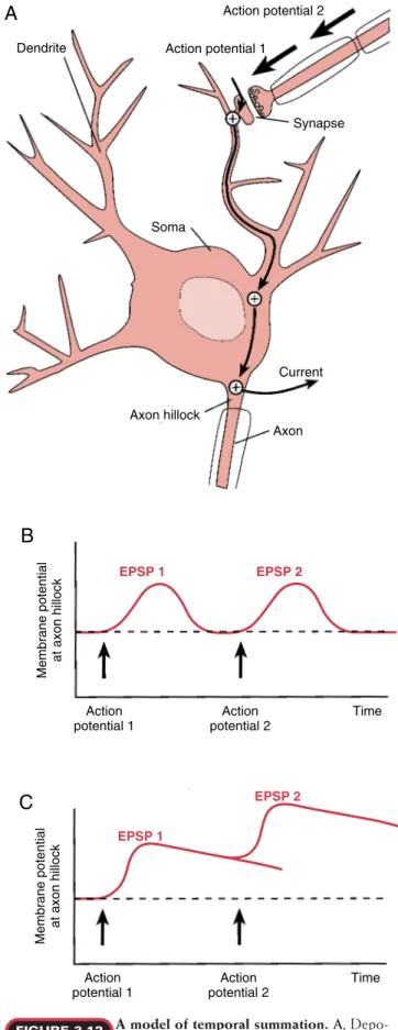
Current Synapse
Axon Action potential 2 Action potential 1
Dendrite
Soma
Axon hillock
Membrane potential
at axon hillock
Action potential 1
Action potential 2
Time
EPSP 2 EPSP 1
B
A
Membrane potential
at axon hillock
Action potential 1
Action potential 2
Time
EPSP 2
EPSP 1
C
A model of temporal summation. A, Depo-larization of a dendrite by two sequential ac-tion potentials. B,A dendritic membrane with a short time con-stant is unable to summate postsynaptic potentials. C,A dendritic membrane with a long time constant is able to summate mem-brane potential changes.
FIGURE 3.12 brane can be added to that of the first depolarization.
Con-sequently, longer periods of depolarization increase the likelihood of summating two postsynaptic potentials. The process in which postsynaptic membrane potentials are added with time is called temporal summation (Fig. 3.12). If the magnitude of the summated depolarizations is above a threshold value, as detected at the axon hillock, it will generate an action potential.
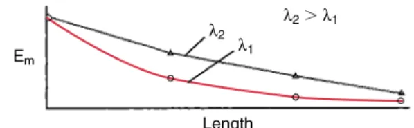
The summation of postsynaptic potentials also occurs with the activation of several synapses located at differ-ent sites of contact. This process is called spatial summa-tion. When a synapse is activated, causing an influx of positively charged ions, a depolarizing electrotonic po-tentialdevelops, with maximal depolarization occurring at the site of synaptic activation. The electrotonic poten-tial is due to the passive spread of ions in the dendritic cytoplasm and across the membrane. The amplitude of the electrotonic potential decays with distance from the synapse activation site (Fig. 3.13). The decay of the elec-trotonic potential per unit length along the dendrite is determined by the length or space constant, , which represents the length required for the membrane poten-tial depolarization to decay to 37% of its maximal value. The larger the space constant value, the smaller the de-cay per unit length; thus, more charge is delivered to more distant membrane patches.
By depolarizing distal patches of membrane, other electrotonic potentials that occur by activating synaptic inputs at other sites can summate to produce even greater depolarization, and the resulting postsynaptic potentials
are added along the length of the dendrite. As with tem-poral summation, if the depolarizations resulting from spatial summation are sufficient to cause the membrane potential in the region of axon hillock to reach threshold, the postsynaptic neuron will generate an action potential (Fig. 3.14).
Because of the spatial decay of the electrotonic poten-tial, the location of the synaptic contact strongly influ-ences whether a synapse can activate a postsynaptic neu-ron. For example, axodendritic synapses, located in distal segments of the dendritic tree, are far removed from the axon hillock, and their activation has little impact on the membrane potential near this trigger zone. In contrast, axosomatic synapses have a greater effect in altering the membrane potential at the axon hillock because of their proximal location.
NEUROCHEMICAL TRANSMISSION
Neurons communicate with other cells by the release of chemical neurotransmitters, which act transiently on post-synaptic receptors and then must be removed from the synaptic cleft (Fig. 3.15). Transmitter is stored in synaptic vesicles and released on nerve stimulation by the process of exocytosis, following the opening of voltage-gated calcium ion channels in the nerve terminal. Once released, the neu-rotransmitter binds to and stimulates its receptors briefly before being rapidly removed from the synapse, thereby al-lowing the transmission of a new neuronal message. The most common mode of removal of the neurotransmitter fol-lowing release is called high-affinity reuptakeby the presy-naptic terminal. This is a carrier-mediated, sodium-depend-ent, secondary active transport that uses energy from the Na⫹/K⫹- ATPase pump. Other removal mechanisms in-clude enzymatic degradation into a nonactive metabolite in the synapse or diffusion away from the synapse into the ex-tracellular space.
The details of synaptic events in chemical transmission were originally described for PNS synapses. CNS synapses appear to use similar mechanisms, with the important dif-ference that muscle and gland cells are the targets of trans-mission in peripheral nerves, whereas neurons make up the postsynaptic elements at central synapses. In the central nervous system, glial cells also play a crucial role in
remov-Em
Length
λ2 λ2⬎λ1
λ1
A profile of the electrotonic membrane po-tential produced along the length of a den-drite.The decay of the membrane potential, Em, as it proceeds
along the length of the dendrite is affected by the space constant, m. Long space constants cause the electrotonic potential to
de-cay more gradually. Profiles are shown for two dendrites with dif-ferent space constants, 1and2. The electrotonic potential of
dendrite 2 decays less steeply than that of dendrite 1 because its space constant is longer.
FIGURE 3.13
Synapse 1 Synapse 2 Dendrite
length
Membrane potential
along dendrite
C
A model of spatial summation. A,The depo-larization of a dendrite at two spatially sepa-rated synapses. B,A dendritic membrane with a short space con-stant is unable to summate postsynaptic potentials. C,A dendritic membrane with a long space constant is able to summate mem-brane potential changes.
FIGURE 3.14
Current Action potential Action potential
Synapse 1 Dendrite
Synapse 2
Axon Axon hillock
A
Synapse 1 Synapse 2 Dendrite
length
Membrane potential
along dendrite
and substance P. The best known membrane-soluble neu-rotransmitters are nitric oxide and arachidonic acid.
The human nervous system has some 100 billion neu-rons, each of which communicates with postsynaptic tar-gets via chemical neurotransmission. As noted above, there are essentially only a handful of neurotransmitters. Even counting all the peptides known to act as transmitters, the number is well less than 50. Peptide transmitters can be colocalized, in a variety of combinations, with nonpeptide and other peptide transmitters, increasing the number of different types of chemical synapses. However, the specific neuronal signaling that allows the enormous complexity of function in the nervous system is due largely to the speci-ficity of neuronal connections made during development.
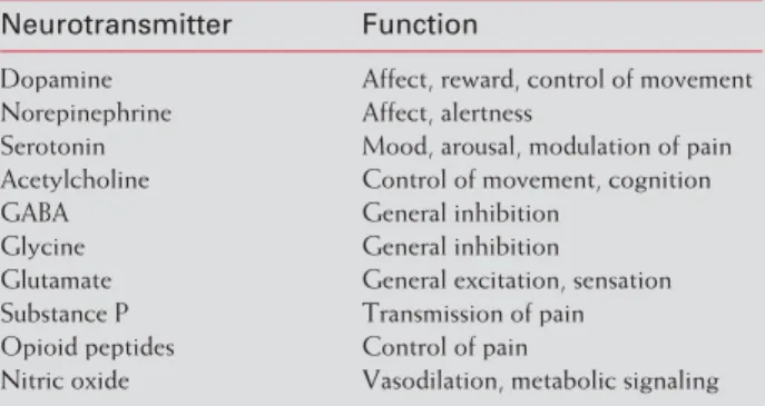
There is a pattern to neurotransmitter distribution. Par-ticular sets of pathways use the same neurotransmitter; some functions are performed by the same neurotransmit-ter in many places (Table 3.1). This redundant use of neu-rotransmitters is problematic in pathological conditions af-fecting one anatomic pathway or one neurotransmitter type. A classic example is Parkinson’s disease, in which a particular set of dopaminergic neurons in the brain degen-erates, resulting in a specific movement disorder. Therapies for Parkinson’s disease, such as L-DOPA, that increase
dopamine signaling do so globally, so other dopaminergic pathways become overly active. In some cases, patients re-ceiving L-DOPA develop psychotic reactions because of
excess dopamine signaling in limbic system pathways. Conversely, antipsychotic medications designed to de-crease dopamine signaling in the limbic system may cause parkinsonian side effects. One strategy for decreasing the adverse effects of medications that affect neurotransmission is to target the therapies to specific types of receptors that may be preferentially distributed in one of the pathways that use the same neurotransmitter.
Acetylcholine. Neurons that use acetylcholine(ACh) as their neurotransmitter are known as cholinergic neurons. Acetylcholine is synthesized in the cholinergic neuron from choline and acetate, under the influence of the en-zymecholine acetyltransferaseor choline acetylase. This enzyme is localized in the cytoplasm of cholinergic neu-rons, especially in the vicinity of storage vesicles, and it is an identifying marker of the cholinergic neuron.
T
T T
T T
Presynaptic terminal
Metabolite
Reuptake Enzyme
Receptor
Postsynaptic cell T
1
2 3 4
5 Diffusion
The basic steps in neurochemical transmis-sion.Neurotransmitter molecules (T) are re-leased into the synaptic cleft (1), reversibly bind to receptors on the postsynaptic cell (2), and are removed from the cleft by enzy-matic degradation (3), reuptake into the presynaptic nerve termi-nal (4), or diffusion (5).
FIGURE 3.15
ing some neurotransmitters from the synaptic cleft via high-affinity reuptake.
There Are Several Classes of Neurotransmitters The first neurotransmitters described were acetylcholine and norepinephrine, identified at synapses in the peripheral nervous system. Many others have since been identified, and they fall into three main classes: amino acids, monoamines, and polypeptides. Amino acids and monoamines are collectively termed small-molecule trans-mitters. The monoamines (or biogenic amines) are so named because they are synthesized from a single, readily available amino acid precursor. The polypeptide transmit-ters (or neuropeptides) consist of an amino acid chain, varying in length from three to several dozen. Recently, a novel set of neurotransmitters has been identified; these are membrane-soluble molecules that may act as both antero-grade and retroantero-grade signaling molecules between neurons. Examples of amino acid transmitters include the excita-tory amino acids glutamate and aspartate and the inhibiexcita-tory amino acids glycine and ␥-aminobutyric. (Note that ␥ -aminobutyric is biosynthetically a monoamine, but it has the features of an amino acid transmitter, not a monoamin-ergic one.) Examples of monoaminmonoamin-ergic neurotransmitters are acetylcholine, derived from choline; the catecholamine transmitters dopamine, norepinephrine, and epinephrine, derived from the amino acid tyrosine; and an indoleamine, serotonin or 5-hydroxytryptamine, derived from trypto-phan. Examples of polypeptide transmitters are the opioids
TABLE 3.1 General Functions of Neurotransmitters
Neurotransmitter Function
Dopamine Affect, reward, control of movement Norepinephrine Affect, alertness
Serotonin Mood, arousal, modulation of pain Acetylcholine Control of movement, cognition GABA General inhibition
Glycine General inhibition Glutamate General excitation, sensation Substance P Transmission of pain Opioid peptides Control of pain
All the components for the synthesis, storage, and re-lease of ACh are localized in the terminal region of the cholinergic neuron (Fig. 3.16). The storage vesicles and choline acetyltransferase are produced in the soma and are transported to the axon terminals. The rate-limiting step in ACh synthesis in the nerve terminals is the availability of choline, of which specialized mechanisms ensure a contin-uous supply. Acetylcholine is stored in vesicles in the axon terminals, where it is protected from enzymatic degrada-tion and packaged appropriately for release upon nerve stimulation.
The enzyme acetylcholinesterase (AChE) hydrolyzes ACh back to choline and acetate after the release of ACh. This enzyme is found in both presynaptic and postsynaptic cell membranes, allowing rapid and efficient hydrolysis of extracellular ACh. This enzymatic mechanism is so effi-cient that normally no ACh spills over from the synapse into the general circulation. The choline generated from ACh hydrolysis is taken back up by the cholinergic neuron by a high-affinity, sodium-dependent uptake mechanism, which ensures a steady supply of the precursor for ACh synthesis. An additional source of choline is the low-affin-ity transport used by all cells to take up choline from the ex-tracellular fluid for use in the synthesis of phospholipids.
The receptors for ACh, known as cholinergic receptors, fall into two categories, based on the drugs that mimic or antagonize the actions of ACh on its many target cell types. In classical studies dating to the early twentieth century, the drugs muscarine,isolated from poisonous mushrooms, andnicotine,isolated from tobacco, were used to distin-guish two separate receptors for ACh. Muscarine stimulates some of the receptors and nicotine stimulates all the others, so receptors were designated as either muscarinicor nico-tinic.It should be noted that ACh has the actions of both muscarine and nicotine at cholinergic receptors (Fig. 3.16); however, these two drugs cause fundamental differences that ACh cannot distinguish.
The nicotinic acetylcholine receptor is composed of five components: two ␣subunits and a ,␥, and ␦subunit (Fig. 3.17). The two ␣subunits are binding sites for ACh. When ACh molecules bind to both ␣subunits, a confor-mational change occurs in the receptor, which results in an increase in channel conductance for Na⫹and K⫹, leading to depolarization of the postsynaptic membrane. This de-polarization is due to the strong inward electrical and chemical gradient for Na⫹, which predominates over the outward gradient for K⫹ ions and results in a net inward flux of positively charged ions.
Glucose
Acetyl-CoA
⫹
Choline
Presynaptic terminal
ACh
ACh
ACh Choline
N
Nicotinic receptor Acetylcholinesterase
enzyme
Choline acetyltransferase
ACh
Postsynaptic cell ACh
M
Muscarinic receptor
Cholinergic neurotransmission. When an ac-tion potential invades the presynaptic terminal, ACh is released into the synaptic cleft and binds to receptors on the postsynaptic cell to activate either nicotinic or muscarinic re-ceptors. ACh is also hydrolyzed in the cleft by the enzyme acetylcholinesterase (AChE) to produce the metabolites choline and acetate. Choline is transported back into the presynaptic ter-minal by a high-affinity transport process to be reused in ACh resynthesis.
FIGURE 3.16
Ion channel
Cross section
Top view
ACh ACh
Extracellular
Intracellular α
α
β
γ δ
The structure of a nicotinic acetylcholine receptor.The nicotinic receptor is composed of five subunits: two ␣subunits and ,␥, and ␦subunits. The two ␣subunits serve as binding sites for ACh. Both binding sites must be occupied to open the channel, permitting sodium ion influx and potassium ion efflux.
The structure and the function of the muscarinic acetyl-choline receptorare different. Five subtypes of muscarinic receptors have been identified. The M1and M2receptors are composed of seven membrane-spanning domains, with each exerting action through a G protein. The activation of M1receptors results in a decrease in K⫹conductance via phospholipase C, and activation of M2receptors causes an increase in K⫹conductance by inhibiting adenylyl cyclase. As a consequence, when ACh binds to an M1receptor, it results in membrane depolarization; when ACh binds to an M2receptor, it causes hyperpolarization.
Catecholamines. The catecholamines are so named be-cause they consist of a catechol moiety (a phenyl ring with two attached hydroxyl groups) and an ethylamine side chain. The catecholamines dopamine(DA),norepinephrine(NE), andepinephrine(EPI) share a common pathway for enzy-matic biosynthesis (Fig. 3.18). Three of the enzymes in-volved—tyrosine hydroxylase (TH), dopamine  -hydroxy-lase (DBH), and phenylethanolamine N-methyl transferase (PNMT)—are unique to catecholamine-secreting cells and all are derived from a common ancestral gene. Dopaminer-gic neurons express only TH, noradrenergic neurons press both TH and DBH, and epinephrine-secreting cells ex-press all three. Epinephrine-secreting cells include a small population of CNS neurons, as well as the hormonal cells of the adrenal medulla, chromaffin cells,which secrete EPI dur-ing the fight-or-flight response (see Chapter 6).
The rate-limiting enzyme in catecholamine biosynthesis istyrosine hydroxylase, which converts L-tyrosine to L
-3,4-dihydroxyphenylalanine (L-DOPA). Tyrosine hydroxylase
is regulated by short-term activation and long-term induc-tion. Short-term excitation of dopaminergic neurons results in an increase in the conversion of tyrosine to DA. This phenomenon is mediated by the phosphorylation of TH via a cAMP-dependent protein kinase, which results in an increase in functional TH activity. Long-term induction is mediated by the synthesis of new TH.
A nonspecific cytoplasmic enzyme, aromatic L-amino acid decarboxylase, catalyzes the formation of dopamine fromL-DOPA. Dopamine is then taken up in storage
vesi-cles and protected from enzymatic attack. In NE- and EPI-synthesizing neurons, DBH, which converts DA to NE, is found within vesicles, unlike the other synthetic enzymes, which are in the cytoplasm. In EPI-secreting cells, PNMT is localized in the cytoplasm. The PNMT adds a methyl group to the amine in NE to form EPI.
Two enzymes are involved in degrading the cate-cholamines following vesicle exocytosis. Monoamine oxi-dase (MAO) removes the amine group, and catechol-O -methyltransferase (COMT) methylates the 3-OH group on the catechol ring. As shown in Figure 3.19, MAO is lo-calized in mitochondria, present in both presynaptic and postsynaptic cells, whereas COMT is localized in the cyto-plasm and only postsynaptically. At synapses of noradren-ergic neurons in the PNS (i.e., postganglionic sympathetic neurons of the autonomic nervous system) (see Chapter 6), the postsynaptic COMT-containing cells are the muscle and gland cells and other nonneuronal tissues that receive sympathetic stimulation. In the CNS, on the other hand, most of the COMT is localized in glial cells (especially as-trocytes) rather than in postsynaptic target neurons.
The synthesis of catecholamines. The cate-cholamine neurotransmitters are synthesized by
FIGURE 3.18 way of a chain of enzymatic reactions to produce L-DOPA,
Most of the catecholamine released into the synapse (up to 80%) is rapidly removed by uptake into the presynaptic neuron. Once inside the presynaptic neuron, the transmit-ter entransmit-ters the synaptic vesicles and is made available for re-cycling. In peripheral noradrenergic synapses (the sympa-thetic nervous system), the neuronal uptake process described above is referred to as uptake 1,to distinguish it from a second uptake mechanism, uptake 2,localized in the target cells (smooth muscle, cardiac muscle, and gland cells) (Fig. 3.19B). In contrast with uptake 1, an active transport, uptake 2 is a facilitated diffusion mechanism, which takes up the sympathetic transmitter NE, as well as the circulating hormone EPI, and degrades them enzymat-ically by MAO and COMT localized in the target cells. In the CNS, there is little evidence of an uptake 2 of NE, but
glia serve a comparable role by taking up catecholamines and degrading them enzymatically by glial MAO and COMT. Unlike uptake 2 in the PNS, glial uptake of cate-cholamines has many characteristics of uptake 1.
The catecholamines differ substantially in their interac-tions with receptors; DA interacts with DA receptors and NE and EPI interact with adrenergic receptors. Up to five sub-types of DA receptors have been described in the CNS. Of these five, two have been well characterized. D1receptors
are coupled to stimulatory G proteins (Gs), which activate adenylyl cyclase, and D2receptorsare coupled to inhibitory
G proteins (Gi), which inhibit adenylyl cyclase. Activation of D2receptors hyperpolarizes the postsynaptic membrane by increasing potassium conductance. A third subtype of DA receptor postulated to modulate the release of DA is
local-L
Catecholaminergic neurotransmission. A,In dopamine-producing nerve terminals, dopamine is enzymatically synthesized from tyrosine and taken up and stored in vesicles. The fusion of DA-containing vesicles with the terminal membrane results in the release of DA into the synaptic cleft and permits DA to bind to dopamine receptors (D1and D2)
in the postsynaptic cell. The termination of DA neurotransmis-sion occurs when DA is transported back into the presynaptic ter-minal via a high-affinity mechanism. B,In norepinephrine (NE)-producing nerve terminals, DA is transported into synaptic
FIGURE 3.19 vesicles and converted into NE by the enzyme dopamine  -hy-droxylase (DBH). On release into the synaptic cleft, NE can bind to postsynaptic ␣- or -adrenergic receptors and presynaptic ␣2
-adrenergic receptors. Uptake of NE into the presynaptic terminal (uptake 1) is responsible for the termination of synaptic transmis-sion. In the presynaptic terminal, NE is repackaged into vesicles or deaminated by mitochondrial MAO. NE can also be transported into the postsynaptic cell by a low-affinity process (uptake 2), in which it is deaminated by MAO and O-methylated by
ized on the cell membrane of the nerve terminal that releases DA; accordingly, it is called an autoreceptor.
Adrenergic receptors, stimulated by EPI and NE, are lo-cated on cells throughout the body, including the CNS and the peripheral target organs of the sympathetic nervous system (see Chapter 6). Adrenergic receptors are classified as either ␣or, based on the rank order of potency of cat-echolamines and related analogs in stimulating each type. The analogs used originally in distinguishing ␣- from  -adrenergic receptors are NE, EPI, and the two synthetic compounds isoproterenol (ISO) and phenylephrine (PE). Ahlquist, in 1948, designated ␣as those receptors in which EPI was highest in potency and ISO was least potent (EPI ⬎NE⬎ ⬎ISO).-Receptors exhibited a different rank or-der: ISO was most potent and EPI either more potent or equal in potency to NE. Studies with PE further distin-guished these two classes of receptors: ␣-receptors were stimulated by PE, whereas -receptors were not.
Serotonin. Serotoninor5-hydroxytryptamine(5-HT) is the transmitter in serotonergic neurons.Chemical trans-mission in these neurons is similar in several ways to that described for catecholaminergic neurons. Tryptophan hy-droxylase, a marker of serotonergic neurons, converts tryp-tophan to 5-hydroxytryptryp-tophan (5-HTP), which is then converted to 5-HT by decarboxylation (Fig. 3.20).
5-Hydroxytryptamine is stored in vesicles and is re-leased by exocytosis upon nerve depolarization. The major mode of removal of released 5-HT is by a high-affinity, sodium-dependent, active uptake mechanism. There are several receptor subtypes for serotonin. The 5-HT-3 re-ceptor contains an ion channel. Activation results in an in-crease in sodium and potassium ion conductances, leading to EPSPs. The remaining well-characterized receptor sub-types appear to operate through second messenger sys-tems. The 5-HT-1A receptor, for example, uses cAMP. Ac-tivation of this receptor results in an increase in K⫹ ion conductance, producing IPSPs.
Glutamate and Aspartate. Both glutamate (GLU) and aspartate (ASP) serve as excitatory transmitters of the CNS. These dicarboxylic amino acids are important sub-strates for transaminations in all cells; but, in certain neu-rons, they also serve as neurotransmitters—that is, they are sequestered in high concentration in synaptic vesicles, re-leased by exocytosis, stimulate specific receptors in the synapse, and are removed by high-affinity uptake. Since GLU and ASP are readily interconvertible in transamina-tion reactransamina-tions in cells, including neurons, it has been diffi-cult to distinguish neurons that use glutamate as a
transmit-Serotonergic neurotransmission. Serotonin (5-HT) is synthesized by the hydroxylation of tryptophan to form 5-hydroxytryptophan (5-HTP) and the de-carboxylation of 5-HTP to form 5-HT. On release into the synaptic cleft, 5-HT can bind to a variety of serotonergic recep-tors on the postsynaptic cell. Synaptic transmission is terminated when 5-HT is transported back into the presynaptic terminal for repackaging into vesicles.
FIGURE 3.20 Glutamatergic neurotransmission. Glutamate
(GLU) is synthesized from ␣-ketoglutarate by enzymatic amination. Upon release into the synaptic cleft, GLU can bind to a variety of receptors. The removal of GLU is prima-rily by transport into glial cells, where it is converted into gluta-mine. Glutamine, in turn, is transported from glial cells to the nerve terminal, where it is converted to glutamate by the enzyme glutaminase.