© 2019 by the Serbian Biological Society How to cite this article: Dragičević S, Kovačević D, Divac Rankov A, Nikolić A, 409 Radojković D, Radović S. Evaluation of toxicity and antioxidative effects of Tussilago farfara and Verbascum thapsus water extracts in zebrafish and in bronchial epithelial cells. Arch Biol Sci. 2019;71(3):409-16.
Evaluation of toxicity and antioxidative effects of
Tussilago farfara
and
Verbascum
thapsus
water extracts in zebrafish and in bronchial epithelial cells
Sandra Dragičević1, Draginja Kovačević1,2, Aleksandra Divac Rankov1,*, Aleksandra Nikolić1, Dragica
Radojković1 and Svetlana Radović3
1Institute of Molecular Genetics and Genetic Engineering, University of Belgrade, Belgrade, Serbia 2Research Center Borstel, Leibniz Lung Center, Borstel, Germany
3Faculty of Biology, University of Belgrade, Belgrade, Serbia
*Corresponding author: [email protected]
Received: December 13, 2018; Revised: March 1, 2019; Accepted: March 26, 2019; Published online: April 4, 2019
Abstract: Tussilago farfara (coltsfoot) and Verbascum thapsus (mullein) have been used as folk remedies for treating respi-ratory disorders. The aim of this study was to test the toxicity of the water extracts of T. farfara and V. thapsusin vivo in zebrafish and in vitro in BEAS 2B epithelial bronchial cells. To the best of our knowledge, this is the first study to investigate the antioxidative properties of T. farfara and V. thapsus extracts in cell culture. Our results show that the T. farfara leaf extract does not produce toxic effects on zebrafish embryos or BEAS 2B cells, and that it has a protective effect in BEAS 2B after induction of oxidative stress. The water extract from V. thapsus displayed pronounced toxic effects on zebrafish embryos and BEAS 2B cells and did not exhibit a significant antioxidative effect on BEAS 2B cells exposed to oxidative stress. Our results suggest that the use of T. farfara water leaf extract is potentially safe and effective in treating respiratory disorders, whereas the use of V. thapsus needs further investigation.
Keywords:Tussilago farfara (coltsfoot); Verbascum thapsus (mullein); BEAS 2B cells; zebrafish; asthma treatment
INTRODUCTION
The use of herbs for medicinal and pharmaceutical purposes is a longstanding practice and has increased in the last few years [1,2]. Since plants are a source of numerous bioactive compounds, the toxicological effects and potential beneficial properties of plant ex-tracts are intensively investigated [3]. Among the many plants that have been used in traditional medicine are
Tussilago farfara and Verbascum thapsus (Linn). Both plants are usually used in the form of tea, oil and crude drugs. T. farfara, known also as coltsfoot, has long been used as a supplement in the treatment of respiratory disorders such as cough, bronchitis and asthma [4]. It prevents inflammation in airways and some com-pounds isolated from the flowers have been shown to exert antioxidant activity[5]. Flower and leaf extracts of V. thapsus, commonly known as mullein, exhibit demulcent properties and are used in the treatment of various respiratory and gastrointestinal problems [6]. Various extracts of V. thapsus have antibacterial,
antiviral and antifungal activities [6,7]. The antioxidant activities of extracts prepared from different parts of
V. thapsus were reported in previous studies [8-10]. Although, V. thapsus is suggested to be a rich source of antioxidants, it is not clear which compounds from this plant possess antioxidant activity [11].
mac-romolecules [14] and consequently, the generated secondary radicals can lead to cell damage and death [12,14]. Enzymatic and nonenzymatic antioxidants are included in the protection of lungs from oxidative stress [15-17]. Supplementation with antioxidants might be useful for maintaining redox homeostasis and disease prevention [12].
We used two model systems to investigate the potential toxicity of the water extracts of T. farfara and
V. thapsus on zebrafish embryos and human bronchial epithelial cell line (BEAS 2B) cells. Zebrafish is one of the standard in vivo models for toxicity analysis. Since both plants have been used in the treatment of respiratory disorders, we chose the BEAS 2B as our in vitro model. These cells were also used to assess the potential antioxidant properties of the water extracts of T. farfara and V. thapsus.
MATERIALS AND METHODS
Plant material
T. farfara leaf water extract was prepared from the commercial product “Farfarae folium” obtained from the Herbal Pharmacy of the Institute for the Study of Medicinal Herbs “Josif Pančić”, Belgrade, Serbia. Dried leaves were treated with hot water for 15 min at a ratio of 1:10 (w/v), allowed to cool to room temperature and the extract was dried in an oven at 45°C. The dried extract was resuspended in sterile distilled water to bring to a concentration of 11 µg/mL, and filtered through a filter membrane (pore size 0.22 µm). The obtained filtrate was used in the experiments as the coltsfoot water extract. Fresh flowers of V. thapsus
were collected in August 2014 on the premises of the Institute of Molecular Genetics and Genetic Engineer-ing, Belgrade, Serbia (GPS coordinates: 44.7484° N, 20.4934° E). The flowers were rinsed with distilled water, dried on sterile filter paper and transferred to a tube with sterile water. Flowers were homogenized with pestle and extracted for 2 h at room temperature with gentle shaking at a 1:10 plant material to water ratio. The crude extract was centrifuged for 5 min at 6000 x
g and the obtained supernatant was dried in a rotary evaporator. The dried supernatant was resuspended in sterile distilled water to a concentration of 36 µg/
mL and filtered through a filter membrane (pore size 0.22 µm). The filtrate was used in the experiments as the mullein water extract.
Zebrafish toxicity assay
Zebrafish, Tübingen wild-type strain, were maintained and bred according to the Zebrafish Book [18]. The obtained embryos were treated with T. farfara and V. thapsus water extracts from 6 h post fertilization (hpf), observed at 24 hpf, 48 hpf and 72 hpf, and the mortality and malformations were recorded, according to OECD guidelines [19]. The embryos were treated in 24-well plates, 12 embryos per well in 1 mL of solution or embryo water (5.5 mg/L KCl, 294 mg/L CaCl2·2H2O, 123 mg/L MgSO4·7H2O and 63 mg/L NaHCO3). Three replicates were prepared for each dilution (20x, 100x and 1000x (where x denotes fold dilution)), and the experiments were repeated three times.
Cultivation and treatment of BEAS 2B cells
BEAS 2B cells were cultivated in Dulbecco’s Modified Eagle’s Medium (DMEM) (Thermo Fisher Scientific, USA) supplemented with 5% fetal bovine serum (FBS), 10 µL penicillin and 10 ng/mL streptomycin. The cells were incubated in a humidified atmosphere at 37°C with 5% CO2 and were passaged after reaching 70-80% confluence. The cells were seeded the day before treatment into a 6-well plate at a density of 0.2x10⁶.
The next day, the medium was aspired and replaced with fresh medium containing 100x, 500x and 1000x diluted extracts of T. farfara or V. thapsus. After 24 h, the cells were exposed to 440 μM and 880 μM hydrogen peroxide dilutions for 30 min at 37°C. The experiment was performed in triplicate and it included untreated control cells, cells pretreated with plant extracts with and without exposure to hydrogen peroxide and cells treated only with hydrogen peroxide.
MTT assay
were treated for 24 h with various dilutions of plant extracts in medium (100x, 500x and 1000x). We did not use a 20x diluted extract since it showed increased mortality and toxicity in the in vivo system. MTT pow-der was dissolved in phosphate buffered saline (PBS) and diluted in the medium. After the treatment, the medium from each well was replaced with 100 μL of MTT solution and the cells were incubated for 45-60 min at 37°C. The mixture was removed and 100 μL of dimethyl sulfoxide (DMSO) was added into the wells in order to solubilize the formazan crystals. Cell vi-ability was quantified by measuring the absorbance at 550 nm on an automatic Infinite 200 PRO multimode reader (Tecan, Switzerland), and the data were analyzed using Magellan software (Tecan, Switzerland). The percentage of growth inhibition was calculated using the following formula:
percentage of cell survival=(As/Ac)x100, where As is the absorbance value of the sample and Ac is the absorbance value of the control [20].
ROS detection
Dihydroethidium (DHE; Life Technologies, USA), an oxidant-sensitive fluorescent probe, was used for detection of the intracellular superoxide anion by flow cytometry. After pretreatment with plant extracts and induction of oxidative stress, the cells were incu-bated for 45 min at 37°C in the dark with DHE (10 µM) diluted in medium without FBS. The cells were trypsinized, washed twice and resuspended in PBS. The fluorescence intensity was quantified by CyFlow Space flow cytometer (Partec, Germany) equipped with a blue 488-nm laser for excitation and band pass filter 590/50 for emission detection. A minimum of 10000 events was assayed for each sample. Untreated cells served as the control for assessing the basal level of oxidative stress and stress induced by sample prepara-tion. Analysis was performed on live cells using FloMax software (Partec, Germany).
Statistical analysis
Statistical analysis was performed using the Statistical Package for Social Sciences 20.0 (SPSS Inc., USA). Data were expressed as percentages and mean±standard
deviation (SD) for continuous variables, and as percent-ages for categorical variables. To test the normality of parameters, one sample Kolmogorov-Smirnov test was used. Differences between the groups were analyzed by the independent samples Mann-Whitney U test, Kruskal-Wallis test and one-way analysis of variance (ANOVA) with post hoc Tukey’s test. A P value less than 0.05 was considered statistically significant.
RESULTS
V. thapsus extract displays extensive zebrafish
embryo toxicity
To determine the in vivo toxicity of the plant extracts, we used several dilutions (20x, 100x and 1000x) of the water extracts and exposed zebrafish embryos to them. Exposure to the lowest dilution (20x) of T. farfara
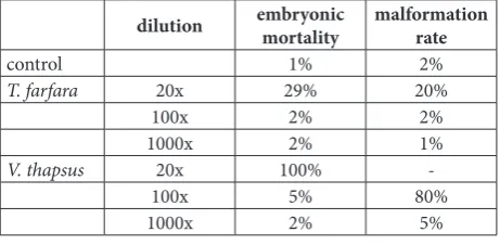
extract caused an increase in embryo mortality after 72 h, with about 30% of the embryos not surviving the treatment (Table 1). Additionally, there were some malformations observed in about 20% of the surviving embryos, such as no tail detachment after 24 h, peri-cardial edema and bradycardia (Supplementary Fig. S1). However, at the other two dilutions tested (100x and 1000x), there were no changes in embryo survival nor were any malformations observed as compared to the untreated control.
Table 1. Effects of T. farfara and V. thapsus water extracts on zebrafish embryo mortality and malformations.
dilution embryonic mortality malformation rate
control 1% 2%
T. farfara 20x 29% 20%
100x 2% 2%
1000x 2% 1%
V. thapsus 20x 100%
-100x 5% 80%
1000x 2% 5%
dilution used (1000x) showed no significant devel-opmental malformations compared to the untreated control (Fig. 1).
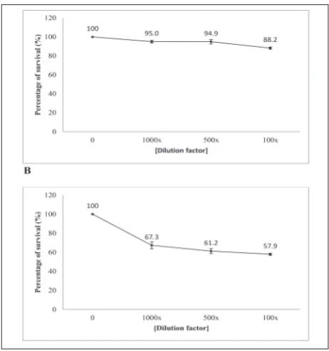
T. farfara extract does not affect cell viability, V.
thapsus extract causes a decrease in cell viability
The influence of water extracts of T. farfara and V. thapsus on BEAS 2B cell viability was determined by the MTT assay. After treatment with T. farfara extracts, a decrease in cell viability of about 10% was observed at the lowest dilution (100x) (Fig. 2A). After the expo-sure of cells to V. thapsus extracts, the viability of cells was inhibited in a dose-dependent manner (Fig. 2B).
T. farfara extract exhibits a protective effect on
cells after induction of oxidative stress, V. thapsus
extract does not
We investigated the antioxidant activity of T. farfara
and V. thapsus water extracts in BEAS 2B cells. The superoxide anion level was detected by DHE staining and assessed by flow cytometry. Treatment of cells with prepared plant extracts did not cause significant changes in the production of the superoxide anion compared to untreated cells (p=0.940 for T. farfara and p=0.833 for V. thapsus). Intracellular accumulation of the superoxide anion was induced by treatment with two different hydrogen peroxide concentrations (440 µM and 880 µM) previously reported in literature [21]. The superoxide anion level was slightly increased in cells treated with 440 µM of hydrogen peroxide in comparison with untreated cells, but the difference was not statistically significant (p=0.126). The intensity of DHE fluorescence was significantly increased after treatment with 880 µM of hydrogen peroxide compared to treatment with 440 µM hydrogen peroxide (p=0.002) and compared to untreated cells (p=0.004), which indicated that this concentration induced a significant accumulation of superoxide anion.
Next, we examined if pretreatment with plant extracts altered the production of superoxide anion induced by hydrogen peroxide treatments. After ex-posure to 440 µM hydrogen peroxide, similar levels of the superoxide anion were observed in cells previously treated with different dilutions of T. farfara extract and in cells treated only with hydrogen peroxide (p=0.678).
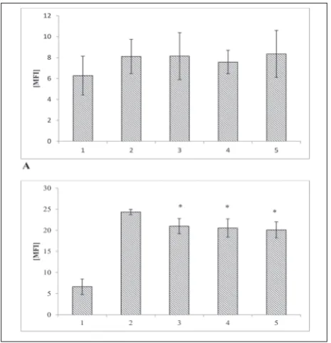
When oxidative stress was induced by hydrogen perox-ide at the higher concentration (880 µM), a decrease in the production of the superoxide anion was observed in cells pretreated with different dilutions of T. farfara
extract (p=0.004), pointing to the protective effect of this extract (Fig. 3).
A higher intensity of DHE fluorescence was de-tected in cells pretreated with the V. thapsus extract when compared to cells treated with 440 µM of
hydro-Fig. 1. V. thapsus water extracts show toxic effects on zebrafish embryos at 72 hpf. A – untreated embryo; B – embryo treated with a 100x dilution of V. thapsus extract. Malformations are indicated by arrows.
gen peroxide, but the difference was not statistically significant (p=0.251), while for the higher hydrogen peroxide concentration, a correlation was not estab-lished (p=0.6), indicating no significant effect of this extract (Fig. 4).
DISCUSSION
T. farfara and V. thapsus extracts prepared in differ-ent ways and with differdiffer-ent solvdiffer-ents were shown to exhibit antibacterial, antiviral and antifungal properties [6,11,22,23]. Antioxidant and free radical scavenging activities of these plants were also shown using mostly biochemical assays [5,24-26]. The present study was designed to examine the potential toxic and antioxi-dative effects of the water extracts of T. farfara and
V. thapsus in two model systems, in vitro in BEAS 2B epithelial bronchial cells and in vivo in zebrafish
embryos. The obtained results demonstrated in both model systems that T. farfara leaf water extract had low acute toxicity and a significant antioxidative ef-fect when oxidative stress was induced by hydrogen peroxide. V. thapsus flower water extract showed high acute toxicity and no antioxidative effects. To the best of our knowledge, this is the first study investigating the toxicity of the water extracts of T. farfara and V. thapsus in zebrafish embryo model system and their antioxidative properties in cell culture.
T. farfara leaf water extract showed limited toxicity in our experimental setup. Additionally, cytotoxicity was not observed in BEAS 2B cells. These results are in agreement with previous results [26,27]. In the latter paper it was shown that the methanol leaf extract of T. farfara produced no cytotoxic effect on the human liver cell line Huh7. In addition, the ethyl-acetate fraction of T. farfara flower did not exhibit a cytotoxic effects on primary rat cortical cells [28].
Fig. 3. T. farfara water extract displays a protective effect on cells after induction of oxidative stress. Superoxide anion was detected in BEAS 2B cells pretreated with the water extracts of T. farfara
after exposure to 440 µM (A) and 880 µM (B) hydrogen peroxide. Data are the mean of fluorescence intensity (MFI)±SD measured by flow cytometry. 1 – untreated BEAS 2B cells; 2 –BEAS 2B cells treated with hydrogen peroxide; 3-5 – BEAS 2B cells pretreated with 1000x, 500x and 100x diluted water extracts, respectively, and treated with hydrogen peroxide; * – statistically significant difference compared to cells treated only with hydrogen peroxide (one-way ANOVA; p<0.05).
Dose-dependent toxic effects of V. thapsus water extractwere observed in both zebrafish and BEAS 2B cells. The water extract of V. thapsus prepared from fresh flowers induced malformations in zebrafish em-bryos at higher dilutions (100x and 1000x), while at the lower dilution (20x) it had a lethal effect. The toxic ef-fects of leaf and flower extracts or compounds isolated from them have not been investigated thus far on the zebrafish model, but it is known that V. thapsus seed can be used as a fish poison [6]. The result described from our study warrants further investigation in order to identify toxic compounds and to further elucidate their potential toxicity in humans. The cytotoxic ef-fects of V. thapsus have been reported [7,29,30]. The water extract ofthe crude plant produced toxic effects in three human liver tumor cell lines at a concentra-tion of 2000 µg/mL, which is much higher than the concentration used in our experiments (0.36 µg/mL). Cell viability was reduced by 31.0% and 69.9% in two hepatitis B virus genome-containing cell lines, HepG2/ C3A and HA22T/VGH, respectively, while in the non-hepatitis B virus genome-containing cell line PLC/ PRF/5 inhibition of cell growth was by 11.6% [29]. The antiproliferative activity of V. thapsus on tumor lung cells A549 was investigated [30]. The authors described significant pro-apoptotic effects of luteolin and 3-O-fucopyranosylsaikogenin F isolated from
V. thapsus at concentrations of 30 µg/mL and 3 µg/ mL, respectively [30]. In our study, cell viability was reduced by 32.4% at the lowest dilution used (0.36 µg/ mL). This finding revealed higher sensitivity to the cytotoxic effect of the extract in normal, non-tumor cells originating from the bronchial epithelium, and a possible synergistic effect of the compounds in the extract on cell viability. This result also emphasizes the importance of using different cell types to assess the toxicity of compounds and extracts.
Oxidative stress plays a major role in the develop-ment of chronic respiratory diseases such as asthma, which are treated with extracts of T. farfara and V. thap-sus. It was shown that T. farfara prevents inflammation in airways by inhibiting the metabolism of arachidonic acid and the generation of various inflammatory me-diators, including prostaglandins, leukotrienes and reactive oxygen radicals [28]. Also, the extract inhibits the synthesis of nitric oxide in lipopolysaccharide-stimulated macrophages [31].
Extracts from different parts of V. thapsus have been shown to possess an antioxidative potential [9-11]. Previous studies mostly used the 2,2-diphenyl-1-picrylhydrazil (DPHH) free radical scavenging as-say to study the antioxidative properties of different compounds isolated from T. farfara and V. thapsus. In our study the influence of T. farfara and V. thap-sus water extracts on the oxidative stress in BEAS 2B cells was examined by flow cytometry. Our results showed no effects of either T. farfara or V. thapsus
water extracts on superoxide anion production. We observed a protective effect of T. farfara against oxida-tive stress induced by hydrogen peroxide. This finding is consistent with previous studies [5,24,32]. Differ-ent compounds isolated from flowers showed cyto-protective activities against glucose oxidase-induced oxidative stress in rat fibroblast cells (NIH3T3) and human keratinocyte cells (HaCaT) [33]. Ravipati et al.
[32] reported higher free radical scavenging activity of T. farfara flower water extracts in comparison to the ethanol extract, which was probably due to the higher content of total phenols and flavonoids in the aqueous extracts. Quercetin and kaempferol are the most common flavonols present in flavonoid fractions from T. farfara flowers and leaves [34]. Kim et al. [5] found that among flavonoids, quercetin glycosides have higher free radical scavenging activity in com-parison to quercetin, thus, the antioxidant activity of leaf water extract observed in our study might be due to the presence of these flavonoids.On the other hand, we found no antioxidant activity of V. thapsus flower water extracts. The methanol extract of V. thapsus
The present study was the first study to test the toxicity of T. farfara and V. thapsus extracts in the zebrafish experimental model. We showed that T. farfara leaf water extract caused neither increased mortality of zebrafish embryos nor significant changes in BEAS 2B proliferation, which is important and increases our knowledge on the safety of this plant for use in traditional medicine. Our findings further elucidate the action of T. farfara function as a natural medicine for treating respiratory disorders, such as asthma, and suggest that its actions are the result of its antioxidative effects on epithelial bronchial cells. On the other hand, water extracts from V. thapsus
showed pronounced toxic effects on zebrafish embryos and caused decreased proliferation of BEAS 2B cells in our experimental setup. This extract did not show a significant antioxidative effect on BEAS 2B cells exposed to oxidative stress. Identification of the toxic and potentially beneficial compounds in V. thapsus
extracts and elucidation of their mechanism of action would greatly benefit its safer use as a natural medicine.
Funding: This work was supported by the Ministry of Education, Science and Technological Development of the Republic of Serbia, Grant No. ON173008.
Author contributions: SD, ADR and SR designed the study. SD, DK and ADR performed the experiments. SD and ADR wrote the manuscript with support of DK. AN, DR and SR critically reviewed the manuscript. All authors read and approved the final manuscript. Conflict of interest disclosure: The authors declare no conflict of interest.
REFERENCES
1. Lee KH. Research and future trends in the pharmaceutical development of medicinal herbs from Chinese medicine. Public Health Nutr. 2000;3(4A):515-22.
2. World Health Organization. WHO guidelines on safety mon-itoring of herbal medicines in pharmacovigilance systems. Geneva: World Health Organization; 2004. 82 p.
3. Grieve M. A modern herbal. New York: Dover Publications; 1981. 902 p.
4. Uzun E, Sariyar G, Adsersen A, Karakoc B, Otük G, Oktayo-glu E, Pirildar S. Traditional medicine in Sakarya province (Turkey) and antimicrobial activities of selected species. J Ethnopharmacol. 2004;95(2-3):287-96.
5. Kim MR, Lee JY, Lee HH, Aryal DK, Kim YG, Kim SK, Woo ER, Kang KW. Antioxidative effects of quercetin-glycosides isolated from the flower buds of Tussilago farfara L. Food and chemical toxicology : an international journal published
for the British Industrial Biological Research Association. 2006;44(8):1299-307.
6. Turker AU, Gurel E. Common mullein (Verbascum thapsus L.): recent advances in research. Phytother Res. 2005;19(9):733-9.
7. Escobar FM, Sabini MC, Zanon SM, Tonn CE, Sabini LI. Antiviral effect and mode of action of methanolic extract of Verbascum thapsus L. on pseudorabies virus (strain RC/79). Nat Prod Res. 2012;26(17):1621-5.
8. Kogje K, Jagdale V, Dudhe S, Phanikumar G, Badere R. Anti-oxidant property and phenolic compounds of few important plants from trans-himalayan regions of north India. Journal of Herbal Medicine and Toxicology. 2010;4(2):145-51. 9. Pal H, Kumar T, Karki H. In vitro antioxidant and
renopro-tective potential of methanolic extract of Verbascum thapsus leaf in rats. Der Pharmacia Sinica. 2013;4(1):113-22. 10. Narayanaswamy N, Balakrishnan KP. Evaluation of some
Medicinal Plants for their Antioxidant Properties. Int J Pharm Tech Res. 2011;3:381–385.
11. Riaz M, Zia-Ul-Haq M, Jaafar HZE. Common mullein, pharmacological and chemical aspects. Braz J Pharmacogn. 2013;23(6):948-59.
12. Bhattacharya S. Reactive Oxygen Species and Cellular Defense System. In: Rani V, Yadav U, editors. Free Radicals in Human Health and Disease. New Delhi: Springer New Delhi; 2015. p. 17-29.
13. van der Toorn M, Smit-de Vries MP, Slebos DJ, de Bruin HG, Abello N, van Oosterhout AJ, Bischoff R, Kauffman HF. Cigarette smoke irreversibly modifies glutathione in airway epithelial cells. Am J Physiol Lung Cell Mol Physiol. 2007;293(5):L1156-62.
14. Forman HJ, Torres M, Fukuto J. Redox signaling. Mol Cell Biochem. 2002;234-235(1-2):49-62.
15. Cross CE, Valacchi G, Schock B, Wilson M, Weber S, Eiserich J, van der Vliet A. Environmental oxidant pollutant effects on biologic systems: a focus on micronutrient antioxidant-oxidant interactions. Am J Respir Crit Care Med. 2002;166(12 Pt 2):S44-50.
16. Christofidou-Solomidou M, Muzykantov VR. Antioxi-dant strategies in respiratory medicine. Treat Respir Med. 2006;5(1):47-78.
17. Kinnula VL, Crapo JD. Superoxide dismutases in the lung and human lung diseases. Am J Respir Crit Care Med. 2003;167(12):1600-19.
18. Westerfield M. The zebrafish book: a guide for the labora-tory use of zebrafish (Danio rerio). 4th ed. [Interent] Eugene: Institute of Neuroscience, University of Oregon; 2000. [cited 2018 Dec 1] Available from: https://zfin.org/zf_info/zfbook/ zfbk.html
19. Organisation for Economic Co-operation and Development. Test No. 236: Fish Embryo Acute Toxicity (FET) test OECD Guidelines for the Testing of Chemicals; Section 2) [Interent]. Paris: OECD Publishing; 2013. [cited 2018 Dec 1] 22 p. Avail-able from: https://www.oecd-ilibrary.org/environment/test-no-236-fish-embryo-acute-toxicity-fet-test_9789264203709-en 20. Patel S, Gheewala N, Suthar A, Patel S. In vitro Cytotoxicity
21. Wages PA, Silbajoris R, Speen A, Brighton L, Henriquez A, Tong H, Bromberg PA, Simmons SO, Samet JM. Role of H2O2 in the oxidative effects of zinc exposure in human air-way epithelial cells. Redox Biol. 2014;3:47-55.
22. Kokoska L, Polesny Z, Rada V, Nepovim A, Vanek T. Screen-ing of some Siberian medicinal plants for antimicrobial activ-ity. J Ethnopharmacol. 2002;82(1):51-3.
23. Zhang L, Ravipati AS, Koyyalamudi SR, Jeong SC, Reddy N, Bartlett J, Smith PT, Shanmugam K, Münch G, Wu MJ, Satyanarayanan M, Vysetti B. Anti-fungal and anti-bacterial activities of ethanol extracts of selected traditional Chinese medicinal herbs. Asian Pac J Trop Med. 2013;6(9):673-81. 24. Song FL, Gan RY, Zhang Y, Xiao Q, Kuang L, Li HB. Total
phenolic contents and antioxidant capacities of selected chi-nese medicinal plants. Int J Mol Sci. 2010;11(6):2362-72. 25. Dobravalskyte D, Petras Rimantas Venskutonis P, Talou T,
Zebib B, Merah O, Ragažinskienė O. Antioxidant Properties and Composition of Deodorized Extracts of Tussilago farfara L. Rec Nat Prod. 2013;7(3):201-9.
26. Li CT, Liu YP, He FC, Li Y. In vitro antioxidant activities of
Tussilago farfara, a new record species to Chanbai Mountain. Chin J Nat Med. 2012;10(4):260-2.
27. Lee HJ, Cho HS, Jun SY, Lee JJ, Yoon JY, Lee JH, Song HH, Choi SH, Kim SY, Saloura V, Park CG, Kim NS. Tussilago far-fara L. augments TRAIL-induced apoptosis through MKK7/ JNK activation by inhibition of MKK7-TIPRL in human hepa-tocellular carcinoma cells. Oncol Rep. 2014;32(3):1117-23. 28. Cho J, Kim HM, Ryu JH, Jeong YS, Lee YS, Jin C.
Neuro-protective and antioxidant effects of the ethyl acetate frac-tion prepared from Tussilago farfara L. Biol Pharm Bull. 2005;28(3):455-60.
29. Lin LT, Liu LT, Chiang LC, Lin CC. In vitro anti-hepatoma activity of fifteen natural medicines from Canada. Phytother Res. 2002;16(5):440-4.
30. Zhao YL, Wang SF, Li Y, He QX, Liu KC, Yang YP, Li XL. Iso-lation of chemical constituents from the aerial parts of Ver-bascum thapsus and their antiangiogenic and antiproliferative activities. Arch Pharm Res. 2011;34(5):703-7.
31. Ryu JH, Jeong YS, Sohn DH. A new bisabolene epoxide from Tussilago farfara, and inhibition of nitric oxide synthesis in LPS-activated macrophages. J Nat Prod. 1999;62(10):1437-8. 32. Ravipati AS, Zhang L, Koyyalamudi SR, Jeong SC, Reddy N,
Bartlett J, Smith PT, Shanmugam K, Münch G, Wu MJ, Saty-anarayanan M, Vysetti B. Antioxidant and anti-inflammatory activities of selected Chinese medicinal plants and their rela-tion with antioxidant content. BMC Complement Altern Med. 2012;12:173.
33. Kang U, Park J, Han AR, Woo MH, Lee JH, Lee SK, Chang TS, Woo HA, Seo EK. Identification of cytoprotective constitu-ents of the flower buds of Tussilago farfara against glucose oxidase-induced oxidative stress in mouse fibroblast NIH3T3 cells and human keratinocyte HaCaT cells. Arch Pharm Res. 2016;39(4):474-80.
34. Chanaj-Kaczmarek J, Wojcinska M, Matlawska I. Phenolics in the Tussilago farfara leaves. Herba Polonica. 2013;59(1):35-43.
Supplementary Data