639
© 2018 by the Serbian Biological Society How to cite this article: Abbaspour Hossein, Safipour Afshar A. Curcumin inhibits the expression of ornithine decarboxylase and adenosine deaminase genes in MCF-7 human breast cancer cells. Arch Biol Sci. 2018;70(4):639-45.
Curcumin inhibits the expression of ornithine decarboxylase and adenosine deaminase
genes in MCF-7 human breast cancer cells
Hossein Abbaspour1,* and Akbar Safipour Afshar2
1Department of Biology, Damghan Branch, Islamic Azad University, Damghan, Iran 2Department of Biology, Neyshabur Branch, Islamic Azad University, Neyshabur, Iran
*Corresponding author: [email protected]
Received: February 9, 2018; Revised: May 21, 2018; Accepted: May 29, 2018; Published online: May 31, 2018
Abstract: Curcumin is the active ingredient of Curcumalonga, which inhibits the development of malignant cells. Preven-tion and treatment of cancer by natural compounds, especially curcumin, and understanding the mechanism of acPreven-tion, is an area of interest in cancer research. In this study, we evaluated the effects of curcumin on cell proliferation, ornithine decarboxylase 1 (ODC1) and adenosine deaminase (ADA) gene expression in human breast cancer cell line (MCF-7) as compared to the non-cancer line (MCF-10A). Both cell lines were subjected to increasing doses of curcumin, ranging from 0 to 30 μg/mL. Cell viability was quantified by the MTT assay. In vitro clonogenic survival assay was performed on MCF-7 cells. Expression of ADA and ODC1 were analyzed by Western blotting and qRT-PCR. Curcumininhibited the growth of malignant cells in a time- and dose-dependent manner. The calculated IC50 value for MCF-7 cells in 48 h was 12 μg/mL. Forty-five to 70% decreases in colony formation were observed in MCF-7 cells treated with 30-60 μg/mL curcumin, respec-tively. Our data revealed a dose-dependent downregulation of ODC1 and ADA expression and respective enzyme activities by curcumin, which correlated with decreased proliferation in the MCF-7 breast cancer cell line.These data suggest that curcumin represses the proliferation of breast cancer cells through downregulation of ODC1 and ADA gene expression, which might be another mechanism of curcumin-mediated tumor growth inhibition.
Key words: antitumor; breast cancer; cell proliferation; adenosine deaminase (ADA); ornithine decarboxylase 1 (ODC1)
INTRODUCTION
Breast cancer is the most common cancer in devel-oped and developing countries and is in fifth place in all types of cancers. In women, breast cancer is the second leading cause of death after lung cancer. In America, one out of every eight women has the po-tential to develop breast cancer [1]. In Asia, there is an increase in the incidence of breast cancer due to lifestyle changes. Mortality rates are rising due to the lack of screening programs and therapeutic facilities for breast cancer in developing countries. [2]. There-fore, providing counseling for preventing this type of cancer is of great importance in these communi-ties. In this regard, the use of diets containing natural compounds that are commonly found in traditional medicine in these societies and have general accept-ance could be a suitable approach in preventing and treating breast cancer.
Curcumin, as a natural compound derived from
Curcuma longa, is used in traditional medicine and
diet in Asian countries, including Iran [3]. Curcumin is effective in the treatment of cardiovascular, pulmo-nary and metabolic diseases as well as diseases of the nervous system [4]. Several laboratories and clinical studies have indicated the role of curcumin in the prevention and treatment of various types of cancer [5]. In studies of the antitumor effects of curcumin, several mechanisms have been proposed and proven, including apoptosis [6] and cell cycle arrest [7].
concentrations in breast cancer tissue as compared to normal breast tissue, and that polyamine biosyn-thesis inhibitors decreased the growth of cancerous tumors. This enzyme was investigated in curcumin-related studies at the level of enzymatic activity. ADA, which assumes a role in purine metabolism, converts the molecule of adenosine into inosine [12]. Accord-ing to previous studies, the enzyme activity of ADA is increased in human breast cancer tumors in contrast with normal tissues [8,13]. In cancer cells and tissues, high ADA activity is responsible for the reduction of adenosine, a molecule that inhibits the growth of can-cer cells by antiinflammatory responses [8]. Therefore, ADA can be a new target for curcumin. In the present study, the antiproliferative activity of curcumin as a result of reduced expression of ODC1 and ADA genes in human breast cancer cells is evaluated.
MATeRIAls AND MeThODs Materials
MCF-7 and MCF-10A cells were obtained from the Pasteur Institute (Tehran, Iran). The cells were cul-tured in Dulbecco’s modified Eagle medium (DMEM) supplemented with 10% fetal bovine serum, and incu-bated at 37ºC in a humidified incubator containing 5% CO2. Curcumin was purchased from Sigma (St. Louis, MO, USA), dissolved in dimethyl sulfoxide (DMSO) to a 10-mM stock solution, and stored at -20°C.
Cell proliferation assay
The cytotoxic activity of curcumin was determined by the MTT assay. Breast and normal (4×10³) cells in
200 μL DMEM were seeded in a sterile 96-well plate and incubated for 24 h at 37°C. The cells were treated with different concentrations of curcumin (0, 10, 20 and 30 μg/mL) and were incubated for 24, 48 and 72 h. A 2-mM MTT solution was added to each well, followed by 4 h of incubation. After removing the su-pernatant, 100 mL of DMSO was added and the plate was shaken for 10 min. Finally, the optical density was determined at 540 nm using an ELISA microplate reader (Awareness, Palm City, FL, USA). In this assay Paclitaxel (Sigma), a known cytotoxic agent, was used as a positive control.
Colony formation assay
Cells (500 cells/mL) were plated in 60-mm Petri dishes and allowed to adhere for 24 h. The next day, the cells were treated for 48 h with curcumin (10, 20 and 30 µg/mL), or 0.1% DMSO. The colonies were fixed with a solution of acetic acid and methanol (1:3) for 15 min, stained with 0.5% crystal violet for 30 min and counted under the stereomicroscope [14].
Ornithine decarboxylase (eC 4.1.1.17) and adenosine deaminase (eC 3.5.4.4) activity assays
ODC1 activity was assayed by determination of 14CO 2
formation as previously described [15]. ADA activity was determined by the colorimetric method of Giusti [16]. The results are presented as percent inhibition ob-tained after comparison to the activity of the controls.
Western blot analysis
Total proteins extraction and sodium dodecyl sulfate polyacrylamide gel electrophoresis (SDS-PAGE) were performed as described previously [17]. Briefly, the cells were lysed with a cell lysis buffer for 30 min to 1 h on ice. The cell lysates were centrifuged at 7,000xg
for 10 min at 4°C and the concentration of proteins was determined by the Bio-Rad Protein Assay Rea-gent. The proteins were subjected to 12% SDS-PAGE. Following electrophoresis, the gels were transferred to polyvinylidene difluoride membrane. After the blocking step, blots were exposed to the primary and secondary antibodies. Detection was performed us-ing chemiluminescence detection reagents, and im-aging was carried out using a chemiluminescence analyzer (Bio-Rad Laboratories). Quantification was performed by ImageJ software.
qRT-PCR
was then amplified and quantified using the SYBR Green PCR kit (Takara Bio, Japan) with the ABI Step-One system (Applied Biosystems, Foster City, CA, USA). Primers for ODC1, ADA and βACT were designed using Oligo 7.5 software (Molecular Biology Insights Inc., Colorado Springs, USA). The primer sequences for ODC1 were: FW: 5´GTGGGTGATT-GGATGCTCTTTG 3´ RV: 5´AGGCCCTGACAT-CACATAGTAG 3´. The primer sequences for ADA were: FW: 5´ACCAGGCTAACTACTCGCTCAA3´ RV: 5´TCAGTAAAGCCCATGTCCCGTT3´. Prim-er sequences for βACT wPrim-ere: FW: 5´TCCATCAT-GAAGTGTGACGT3´ RV: 5´GAGCAATGATCTT-GATCTTCAT3´.
A PCR reaction mixture of 20 μL contained 10 μL SYBR Green Master Mix, 0.4 μL reverse primer and 0.4 μl forward primer, 6.2 μl dH2O (RNase free) and 3 μl cDNA (4 ng). Three pairs of primers were used separately. The thermal cycling conditions were as follow: denaturation cycle at 95°C for 10 min, 40 cycles at 95°C for 15 s, and at 60°C for 60 s (annealing and extension temperature). The qRT-PCR data were analyzed using the relative gene expression (ΔΔCt) method. Briefly, the results are presented as the fold change in gene expression normalized to the reference gene (β-actin) and were determined using the equa-tion fold change=2-ΔΔCt [18]. For each single reaction,
melting curve analysis was performed to exclude am-plification of nonspecific products. Each valid ampli-fication reaction showed a single peak at the desired melting temperature.
statistical analysis
The data are presented as the mean±SEM. Student’s t-test was used and P<0.05 was taken as the level of significance. All results were analyzed by statistical software SAS (USA).
ResUlTs
Cytotoxicity of curcumin
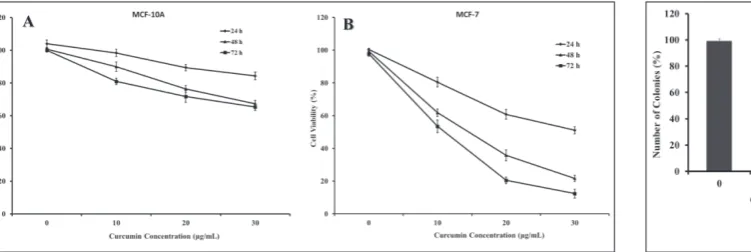
The MTT and clonogenic assays were conducted to evaluate the potential cytotoxic effect of curcumin on human breast cancer cells (MCF-7) and human mam-mary epithelial cells (MCF-10A). The cell lines were treated with different concentrations of curcumin for 24, 48 and 72 h, and cell viability was measured by the MTT assay. The results demonstrated that curcumin significantly decreased the viability of malignant cells in a time- and dose-dependent manner. This antipro-liferation effect was observed within a 24 h period, and continued to increase over the next 72 h. The survival rates of MCF-7 cells were 62, 36 and 22% after exposure to 10, 20 and 30 μg/mL curcumin for 48 h, respectively (Fig. 1B); they were lower than those for MCF-10A cells (Fig. 1A). Curcumin IC50 values of MCF-7 cells were 12 and 7.5 μg/mL for 48 and 72 h, respectively. Therefore, nontoxic concentrations (<10 μg/mL for 48 h) of cur-cumin were used in subsequent experiments. Colony formation in MCF-7 cells after curcumin treatments significantly decreased in a dose-dependent manner.
Fig. 1. Cytotoxic effects of curcumin on MCF-10A (A) and MCF-7 (B) human breast cell lines. Cells were treated for 24, 48 and 72 h with different concentrations of curcumin. Cytotoxicity was determined by the MTT assay. Values represent means±SEM.
Thirty μg/mL and 60 μg/mL curcumin caused 45 and 80% decreases in colony formation in MCF-7 cells, re-spectively (Fig. 2). Cytotoxicity results were correlated with morphological alterations (loss of cell volume, cell shrinkage and nuclear condensation). Cells treated with high concentration of curcumin became spherical and shrunken, while untreated cells remained normal in size and shape (Fig. 3). Paclitaxel as a positive control at a 700 nM concentration decreased the viability of MCF-7 cells by 6.8±0.3% after 48 h in comparison with untreated control cells.
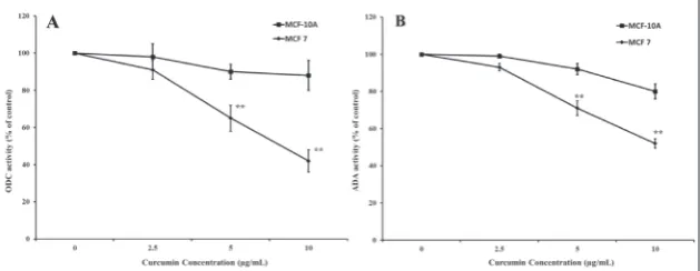
ODC1 and ADA activities
To determine how regulation of ODC1 and ADA en-zyme activities was involved in the antiproliferative effect of curcumin, MCF-7 and MCF-10A cells were treated with concentrations of curcumin ranging from 0 to 10 μg/mL for 48 h. The effect of the treatment of cells with curcumin on ODC1 and ADA activity are shown in Fig. 4. Treatment with 10 μg/mL curcumin
resulted in 5.5- and 4-fold decreas-es in ODC1 and ADA activitidecreas-es as compared to control cells, respec-tively. The treatment of cells with curcumin resulted in significant dose-dependent inhibition of en-zyme activities.
Changes in ODC1 and ADA expression
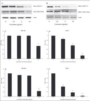
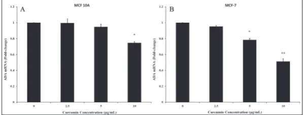
In order to investigate the level of ODC1 and ADA regulation (translational or transcriptional) by curcumin, immunoblotting and qRT-PCR assays were performed to assess the expression of ODC1 and ADA in MCF-7 and MCF-10A cells. The results revealed that cur-cumin significantly downregulated the expression of ODC1 and ADA proteins (Fig. 5) and genes (Figs. 6 and 7) in a dose-dependent man-ner in the MCF-7 cancer cell line, but not in non-cancer MCF-10A cells. Curcumin at a concentration of 10 μg/mL significantly decreased the expression of ODC1 and ADA genes by 3.5- and 1.9-fold, respectively, in MCF-7 cells. However, in MCF-10A cells, the mRNA levels of ODC1 and ADA were reduced by only 1.3-fold at 10 μg/mL concentration. (Fig. 6-7).
DIsCUssION
Curcumin is an effective and multitarget natural product for the treatment and prevention of many cancers, including breast cancer [19]. Different stud-ies have examined the mechanism of its activity, and it appears to influence different proteins and genes, such as p53, NF-κB, PI3K-AKT and COX2 [20]. In the present study, we examined a less considered role of curcumin, its impact on two ODC1 and ADA enzymes. The former enzyme is involved in the tabolism of polyamines and the latter in purine me-tabolism, and the activities of both are significantly increased in malignant cells [8,11]. In this research,
Fig. 3. Morphological changes of MCF-7 cells treated with different concentrations of curcumin for 48 h viewed under the inverted phase-contrast microscope (×200). A reduc-tion in cell populareduc-tion was noted with the increase in the concentrareduc-tion of the treatment as compared to control (untreated cells).
the antiproliferative effect of cur-cumin on the human breast can-cer cell line MCF7 was dose- and time-dependent. Likewise, the CFU assay indicated that breast cancer cells after prolonged exposure to curcumin significantly lost the abil-ity to form colonies. Many reports have shown that curcumin and its derivatives have growth inhibitory or antiproliferative effects on can-cer cells. It was demonstrated [21] that curcumin inhibits the growth of HL-60 cells by inducing apopto-sis. The antiproliferative activity of curcumin was also demonstrated by arresting the cell cycle at the G2/M phase in MCF7 cells [22]. To determine the antiproliferative mechanism of curcumin, the enzy-matic activity of ODC1 in normal and malignant cells was measured after exposure to nonlethal con-centrations of curcumin, reveal-ing a decline in the activity of this enzyme. ODC1 catalyzes the first and rate-limiting step in the bio-synthesis of polyamines and has a very short half-life [23]. Regulation of the activity of this enzyme can also be at the protein level through degradation in the proteasome by an antizyme, or at the transcript level by reducing mRNA produc-tion [24]. According to our results, the decrease at the levels of ODC1 protein and mRNA after treatment with curcumin suggests regulation of ODC1 at the transcript level. In parallel with our findings, Berrak et al. [7] showed in curcumin-treated MCF-7 cells that ODC1 expression was downregulated. Reducing the amount of ODC1 protein dimin-ishes the amount of polyamines, which are important molecules that scavenge excess reactive oxy-gen species (ROS) [25]. Increasing
Fig. 5. Western blot analysis (A, B) and a histogram presenting the expression of adenosine deaminase (C, D) and ornithine decarboxylase (e, F) in MCF-10A and MCF-7 cells after treatment with 0-10 μg/mL curcumin for 48 h. Values are means±SEM, *p<0.05 and ** p<0.01 compared to control.
free radicals leads to the destruction of cellular struc-tures and molecules and ultimately causes apoptosis [26]. Thus, studies have demonstrated that curcumin specifically disables ROS [27]. This is a paradox that curcumin, itself a free-radical scavenger, causes the accumulation of free radicals. Curcumin promotes apoptosis via other pathways, mediated by NF-κB, STAT-3 and PI3-kinase/AKT [28].
Our results showed that the activity and expres-sion levels (protein and mRNA) of ADA in MCF7 cells treated with curcumin declined. The correlation be-tween results of Western blots and qRT-PCR showed that the regulation of ADA protein by curcumin was at the level of transcription. ADA is a protein that, in addition to its importance in the metabolism of pu-rines in the cell, also affects adenosine receptor activ-ity outside the cell, which plays an important role in the physiological activity of the nervous system [29]. Therefore, ADA is important both as an enzyme and allosteric modulator [30]. Cancer cells require purine precursor molecules because of their increased prolif-eration activity. In addition, reducing the amount of adenosine molecule that plays an important role in an-tiinflammatory responses is an added advantage [31]. Thus, curcumin diminishes purine precursors by less-ening ADA gene expression, and on the other hand, in the absence of adenosine catabolism, the pathway for antiinflammatory responses becomes more active and cells lose their ability to proliferate [32].
In this study, we examined the effects of curcu-min on the estrogen receptor-positive cell line (MCF7) (ER+/PR+/HER2-), and our findings regarding the
cytotoxicity and enzymatic activities are similar to other works on the ER/PR/ HER2 triple negative breast cancer cell line (MDA-MB231) [33]. Therefore, the antiproliferative effect of curcumin and its inhibitory effect on ODC1 and ADA activities is not estrogen-dependent.
In conclusion, the results of this study indicate that curcumin reduces the expression of ODC1 and ADA genes at the transcriptional level in the human breast cancer MCF7 cell line. Curcu-min, in addition to the usual pathways for growth inhibition and apoptosis, seems to trigger antitumor activity by regulating the expression of other genes that are involved in the formation of cancer, including ODC1 and ADA. Further studies on the upstream reg-ulatory elements of these genes will provide a better understanding of the mechanism of curcumin action.
Funding: This work was financially supported by Damghan Branch, Islamic Azad University, Damghan, Iran.
Author contributions: HA designed and supervised the experi-ments, analyzed the data and drafted the paper; ASA designed and performed the experiments, analyzed data and drafted the paper. All the authors read the final manuscript and approved the submission.
Conflicts of interest disclosure: The authors declare no conflicts of interest.
ReFeReNCes
1. Siegel RL, Miller KD, Jemal A. Cancer statistics, 2016. CA Cancer J Clin. 2016;66:7-30.
2. Ferlay J, Héry C, Autier P, Sankaranarayanan R. Global Bur-den of Breast Cancer. In: Li C, editor. Breast Cancer Epide-miology. New York, NY: Springer New York; 2010. p.1-19. 3. Kocaadam B, Sanlier N. Curcumin, an active component of
turmeric (Curcumalonga), and its effects on health. Crit Rev Food Sci Nutr. 2017;57:2889-95.
4. Campbell MS, Fleenor BS. The emerging role of curcumin for improving vascular dysfunction: A review. Crit Rev Food Sci Nutr. 2017:1-10.
5. Cui T, Zhang S, Sun H. Co-delivery of doxorubicin and pH-sensitive curcumin prodrug by transferrin-targeted nanopar-ticles for breast cancer treatment. Oncol Rep. 2017;37:1253-60. 6. Elmegeed GA, Yahya SM, Abd-Elhalim MM, Mohamed MS, Mohareb RM, Elsayed GH. Evaluation of heterocyclic ste-roids and curcumin derivatives as anti-breast cancer agents:
Studying the effect on apoptosis in MCF-7 breast cancer cells. Steroids. 2016;115:80-9.
7. Berrak O, Akkoc Y, Arisan ED, Coker-Gurkan A, Obakan-Yerlikaya P, Palavan-Unsal N. The inhibition of PI3K and NFkappaB promoted curcumin-induced cell cycle arrest at G2/M via altering polyamine metabolism in Bcl-2 overex-pressing MCF-7 breast cancer cells. Biomed Pharmacother. 2016;77:150-60.
8. Mahajan M, Tiwari N, Sharma R, Kaur S, Singh N. Oxidative stress and its relationship with adenosine deaminase activ-ity in various stages of breast cancer. Indian J Clin Biochem. 2013;28:51-4.
9. Afshar AS, Nematpour FS, Meshkani M, Khafi A. Growth inhibition of human breast cancer cells and down-regulation of ODC1 and ADA genes by Nepeta binaloudensis. Rev Bras Farmacogn. 2017;27:84-90.
10. Murray-Stewart TR, Woster PM, Casero RA. Targeting poly-amine metabolism for cancer therapy and prevention. Bio-chem J. 2016;473:2937-53.
11. Zhu Q, Jin L, Casero RA, Davidson NE, Huang Y. Role of ornithine decarboxylase in regulation of estrogen receptor alpha expression and growth in human breast cancer cells. Breast Cancer Res Treat. 2012;136:57-66.
12. Cortés A, Gracia E, Moreno E, Mallol J, Lluís C, Canela EI, Casadó V. Moonlighting adenosine deaminase: a target pro-tein for drug development. Med Res Rev. 2015;35:85-125. 13. Aghaei M, Karami-Tehrani F, Salami S, Atri M. Diagnostic
value of adeno-sine deaminase activity in benign and malig-nant breast tumors. Arch Med Res 2010;41:14–8.
14. Franken NA, Rodermond HM, Stap J, Haveman J, van Bree C. Clonogenic assay of cells in vitro. Nat Protoc. 2006;1:2315-9. 15. Ostrowski J, Woszczynski M, Kowalczyk P, Wocial T, Hennig
E, Trzeciak L, Janik P, Bomsztyk K. Increased activity of MAP, p70S6 and p90rs kinases is associated with AP-1 activation in spontaneous liver tumours, but not in adjacent tissue in mice. Br J Cancer. 2000;82:1041.
16. Gupta BK, Bharat A, Debapriya B, Baruah H. Adenosine deaminase levels in CSF of tuberculous meningitis patients. J Clin Med Res. 2010;2:220.
17. Wang X, Hang Y, Liu J, Hou Y, Wang N, Wang M. Antican-cer effect of curcumin inhibits cell growth through miR-21/ PTEN/Akt pathway in breast cancer cell. Oncology Lett. 2017;13(6):4825-31.
18. Livak KJ, Schmittgen TD. Analysis of relative gene expres-sion data using real-time quantitative PCR and the 2− ΔΔCT method. Methods. 2001;25:402-8.
19. Devassy JG, Nwachukwu ID, Jones PJ. Curcumin and cancer: barriers to obtaining a health claim. Nutr Rev. 2015;73:155-65. 20. Ravindran J, Prasad S, Aggarwal BB. Curcumin and cancer
cells: how many ways can curry kill tumor cells selectively?. AAPS J. 2009;11(3):495-510.
21. Liao YF, Hung HC, Hour TC, Hsu PC, Kao MC, Tsay GJ, Liu GY. Curcumin induces apoptosis through an ornithine decarboxylase-dependent pathway in human promyelocytic leukemia HL-60 cells. Life Sci. 2008;82:367-75.
22. Jiang M, Huang O, Zhang X, Xie Z, Shen A, Liu H, Geng M, Shen K. Curcumin induces cell death and restores tamoxifen sensitivity in the antiestrogen-resistant breast cancer cell lines MCF-7/LCC2 and MCF-7/LCC9. Molecules. 2013;18:701-20. 23. Battaglia V, DeStefano Shields C, Murray-Stewart T, Casero RA Jr. Polyamine catabolism in carcinogenesis: potential tar-gets for chemotherapy and chemoprevention. Amino Acids. 2014;46:511-9.
24. Nowotarski SL, Origanti S, Shantz LM. Posttranscriptional regulation of ornithine decarboxylase. Methods Mol Biol. 2011;720:279-92.
25. Murray-Stewart TR, Woster PM, Casero RA Jr. Targeting polyamine metabolism for cancer therapy and prevention. Biochem J. 2016;473:2937-53.
26. Moloney JN, Cotter TG. ROS signalling in the biology of cancer. Semin Cell Dev Biol. 2017;https://doi.org/10.1016/j. semcdb.2017.05.023.
27. Dai F, Chen WF, Zhou B, Yang L, Liu ZL. Antioxidative effects of curcumin and its analogues against the free-radical-induced peroxidation of linoleic acid in micelles. Phytother Res. 2009;23:1220-8.
28. Qiao Q, Jiang Y, Li G. Inhibition of the PI3K/AKT-NF-kap-paB pathway with curcumin enhanced radiation-induced apoptosis in human Burkitt’s lymphoma. J Pharmacol Sci. 2013;121:247-56.
29. Gracia E, Pérez-Capote K, Moreno E, Barkešová J, Mallol J, Lluís C, Franco R, Cortés A, Casadó V, Canela EI. A2A adenosine receptor ligand binding and signalling is allo-sterically modulated by adenosine deaminase. Biochem J. 2011;435:701-9.
30. Gracia E, Farré D, Cortés A, Ferrer-Costa C, Orozco M, Mal-lol J, Lluís C, Canela EI, McCormick PJ, Franco R, Fanelli F, Casadó V. The catalytic site structural gate of adenosine deaminase allosterically modulates ligand binding to adenos-ine receptors. Faseb J. 2013;27:1048-61.
31. Ye JH, Rajendran VM. Adenosine: an immune modula-tor of inflammamodula-tory bowel diseases. World J Gastroenterol. 2009;15:4491-8.
32. Ohta A. A Metabolic Immune Checkpoint: Adenosine in Tumor Microenvironment. Front Immunol. 2016;7:109. 33. Pereira MC, Mohammed R, Van Otterlo WA, De Koning CB,