ISSN: 2252-8938, DOI: 10.11591/ijai.v9.i2.pp349-355 349
Evaluation of psoriasis skin disease classification using
convolutional neural network
Rosniza Roslan1, Iman Najwa Mohd Razly2, Nurbaity Sabri3, Zaidah Ibrahim4
1,2,3Faculty of Computer and Mathematical Sciences, Universiti Teknologi MARA Melaka Kampus Jasin, 77300 Merlimau, Melaka, Malaysia
4Faculty of Computer and Mathematical Sciences, UniversitiTeknologi MARA, 40450 Shah Alam, Selangor, Malaysia
Article Info ABSTRACT
Article history: Received Dec 22, 2019 Revised Feb 27, 2020 Accepted Mar 12, 2020
Skin disease has lower impact on mortality compared to others but instead it has greater effect on quality of life because it involves symptoms such as pain, stinging and itchiness. Psoriasis is one of the ordinary skin diseases which are relapsing, chronic and immune-mediated inflammatory disease. It is estimated about 125 million people worldwide being infected with various types of skin infection. Challenges arise when patients only predict the skin type disease they had without being accurately and precisely examined. This is because as human being, they only observe and look at the diseases on the surface of the skin with their naked eye, where there are some limits, for example, human vision lacks of accuracy, reproducibility and quantification in the collection of image information. As Plaque and Guttate are the most common Psoriasis skin disease happened among people, this paper presents an evaluation of Psoriasis skin disease classification using Convolutional Neural Network. A total of 187 images which consist of 82 images for Plaque Psoriasis and 105 images for Guttate Psoriasis has been used which are retrieved from Psoriasis Image Library, International Psoriasis Council (IPC) and DermNet NZ. Convolutional Neural Network (CNN) is applied in extracting features and analysing the classification of Psoriasis skin disease. This paper showed the promising used of CNN with the accuracy rate of 82.9% and 72.4% for Plaque and Guttate Psoriasis skin disease, respectively. Keywords:
Classification
Convolutional neural network Deep learning
Psoriasis Skin disease
This is an open access article under the CC BY-SA license.
Corresponding Author: Rosniza Roslan,
Faculty of Computer and Mathematical Sciences, Universiti Teknologi MARA Melaka Kampus Jasin, 77300 Merlimau, Melaka, Malaysia.
Email: [email protected]
1. INTRODUCTION
Human skin is the biggest body organ. The mass of skin lies between six to nine pounds and estimated about two square yards of surface area. The inner part of body is separated by skin. Skin also provides protection against fungal infection, bacteria, allergy, viruses and controls temperature of body [1]. There are many of people that are suffered from the skin disease which affected by bacteria or viruses as many people neglect and take care of their own skin hygiene [1]. There are many types
The statistics showed that 125 million people worldwide are being infected with various types of skin infection. It is estimated about 296 patients with Psoriasis skin disease, Malays are the most common with 175 (59.1%), followed by Indians 82 (27.7%), Chinese 37 (12.5%) and others 2 (0.6%). The ratio for male and female is 1.2:1. There are more than half of the patients (age 40 or less) which is 54.7% had an early disease of Psoriasis skin disease. The most common clinical disease are chronic Plaque Psoriasis (89.9%), followed by Erythrodermic Psoriasis (4.7%), Guttate Psoriasis (3.0%) and Pustular Psoriasis (1.7%).
Psoriasis is a type of disorder characterized by red scaling papules that form a round-to-oval plaque and can be seen from the normal skin around them [3]. Psoriasis occurs most of the time on the scalp, elbows, knees and lower back, which can grow further to all parts of the body and have various dangerous effects on the skin until it affects the physical appearance of somebody. Psoriasis lead to thickening of the epidermal layer, expansion of blood vessels, and infiltration of huge number of immune cells into the dermis and often into epidermal compartments [4].
Psoriasis skin disease are divided into several kind with different characteristics which are Plaque, Guttate, Inverse, Pustular and Erythrodermic [5-6]. Plaque Psoriasis is the most common type of psoriasis [7]. In Malaysia, it is about 2 to 6 percent of the population with Psoriasis was registered in the Dermatological Society of Malaysia. Plaque Psoriasis shows the highest percentage of 85.1% which represents the most common and popular skin disease among the others skin disease [7]. Nowadays, there are many skin diseases with the same appearance that make it difficult for people to identify the type of abnormalities. Previous research has shown that there are many applications and technologies in medical image analysis applied in medical domain such as image processing [8-14], fuzzy logic [15-16], machine learning [17], artificial neural network [18], Convolutional Neural Network [19-26] and many more [27].
Image processing refers to the process of converting image into digital for further use in image research such as in medical field and signal processing. Image processing has been used to obtain useful information from an image that has been converted into digital form with certain operation apply on it. Image processing manages image enhancement, image segmentation and extraction of image [17]. Image enhancement is defined as the operation that boosts the appearance to a human view or convert an image to an arrangement better that suited to machine processing [28]. Image segmentation is the process of dividing an image into several parts, such as dividing and identifying an image of the human body or any other part of an object and combining the entire segmented image into an object [29]. Some features in an image such as color can help to identify pixel relationships within the segmented image. In recent years, deep Convolutional Neural Networks (CNN) which also known as ConvNet becomes very popular in feature learning and object classification. The high-performance of GPU used, makes it possible to train a network on a large-scale datasets as to obtain a better performance. CNN is one kind of Artificial Neural Networks (ANN) that are known to be extremely powerful in the part of identification and classification of images [19-21]. However, it is challenges and very hard to detect between eczema and Psoriasis because both look similar visually. Furthermore, diagnosis is usually very difficult to obtain an accurate result for the first time.
Therefore, this paper presents an evaluation of Psoriasis skin disease classification using Convolutional Neural Network (CNN). It is shows that CNN works in analysing the Psoriasis skin disease images which produced the promising classification results. This paper is comprised as follows. Section 1 clarifies the motivation and existing research on Psoriasis skin disease research. Section 2 discuss on the process flow that been conducted during the research. This section is also describes the details on the employed methods and experimental techniques. Section 3 analyses the experimental results and evaluation that have done in the experimental process. The conclusion and recommendation for future works are explain in the Section 4.
2. RESEARCH METHOD
The objective of this paper is to classify and identify the type of Plaque and Guttate Psoriasis skin disease which to acquire a better performance. In this research, there are four phases that are data acquisition, pre-processing, processing (feature extraction and Psoriasis skin disease classification) and post-processing (i.e test and evaluation). Figure 1 shows the process flow of the proposed algorithm.
Figure 1. Flowchart of the proposed psoriasis classification
2.1. Image dataset
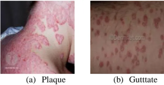
The image datasets of Psoriasis skin disease has been collected from the Psoriasis Image Library, International Psoriasis Council (IPC) and DermNet NZ website [30]. This paper covers for two type of Psoriasis that are Plaque Psoriasis and Guttate Psoriasis. There are 187 total images of Psoriasis skin disease utilized as the test images which are 82 images of Plaque Psoriasis and 105 images of Guttate Psoriasis. Figure 2 shows a sample of Plaque and Guttate Psoriasis skin disease.
(a) Plaque (b) Gutttate
Figure 2. Sample of psoriasis skin disease [30]
2.2. Pre-processing
The input image of Plaque and Guttate Psoriasis skin disease image is pre-processed to resize the image size input to ensure all image dataset in a set of uniform data. Input image of Plaque and Guttate Psoriasis is resizing from original input image into 160 x 160 pixels in order to get a precise classification.
Start
Psoriasis skin disease image
Pre-processing
Convolutional Neural Network
Convolutional Layer
Pooling Layer
Fully Connected Layer
Plaque or Guttate Psoriasis skin disease
End Convolutional Layer
Pooling Layer
2.3. Convolutional neural network
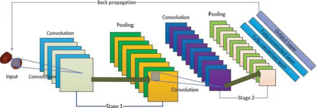
Convolutional Neural Network (CNN) is one of the robust method for image identification and classification in deep learning domain. CNN is a part of method in Artificial Neural Networks (ANN) [21]. It is able to train a network on a large dataset to obtain a better performance. This method is composed of convolution layer, pooling layer and fully connected layer as illustrated in Figure 3 [22].
Figure 3. Basic structure of CNN
2.3.1. Convolutional layer
The CNN core element is the Convolutional layer (CL), which consist local connections and weights of shared characteristics [26]. Each channel has CL for its own filter. CL has a set of trained kernels which are convolved crosswise the width and the height of the input features during the forward pass producing a two-dimensional activation map of the kernel [20]. The CL is aim to learn feature representation of the input features. CL maintains the pixels spatial interrelation [21]. Figure 4 shows the sample of convolution with 2x2 kernel [22].
Figure 4. Convolution with 2x2 Kernel Sample Result
2.3.2. Pooling layer
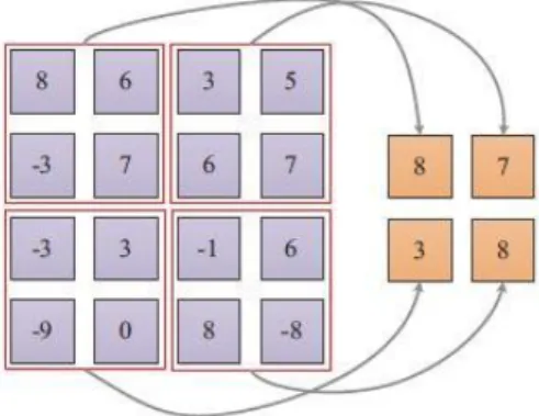
Pooling layer (PL) usually placed between two CL. PL or called as sub-sampling is a nonlinear compression of the feature maps that pass through a non-linear transformation, with a group of pixels (usually 2×2 size) to one pixel [20]. The size of feature maps in PL is determined according to the moving step of kernels [26]. PL has its own benefit which can reduce feature maps dimensions and also can improve the effectiveness of feature extraction [22]. If some features have already been detected in the previous folding operation, a detailed image is not needed for further processing, and it is condensed to less detail [20]. Average pooling and max pooling are the common pooling operation or method [26]. The process of max pooling with size 2x2 has been illustrated as shown in Figure 5 [22].
Figure 5. Max pooling with Size of 2x2
2.3.3. Fully connected layer
Typically, one or more Fully Connected Layers (FCL) is a classifier of CNN [26]. Each node in a FCL is straight connected to each node in both of the previous and in the next layer [27]. The output from CL and PL operations delivers new features which are extracted from the image. Then, features are utilized by FCL for transferring the input image into different classes predicated on the training dataset [21]. There is no spatial information preserved in FCL [26]. Softmax regression is frequently used because of it generating a well-performed probability distribution of the outputs. AlexNet is eight-layer Convolution Neural Network, the first five layers are convolutions, the last three are all connected layers while the last layer is classified by softmax. Model uses Rectified Linear Units (ReLU) to replace the traditional Sigmoid and tanh functions as neuron's non-linear activation functions, and proposes the dropout method to reduce the over-fitting problem. 2.4. Test and evaluation
The purpose of quantitative evaluation is to evaluate the statistical classification in machine learning domain. The accuracy evaluation are verified based on the following confusion matrix of True Positive (TP) and False Negative (FN) [10]. The definition is as follows:
True Positive (TP): To compute the predicted result of Plaque and Guttate Psoriasis skin disease as actual result of Plaque and Guttate Psoriasis skin disease which are true classified. The TP accuracy rate can be computed as follows.
𝑇𝑃 =𝐴
𝐵× 100
where
𝐴 represents the number of predicted image of Plaque and Guttate Psoriasis which are classified as actual image of Plaque and Guttate Psoriasis.
𝐵 represents the total number of Plaque and Guttate Psoriasis image
False Negative (FN): To compute the predicted result of Plaque and Guttate Psoriasis skin disease as false classified result of Plaque and Guttate Psoriasis skin disease. The FN accuracy rate can be computed as follows.
𝐹𝑁 =𝐴
𝐵× 100
where
𝐴 represents the number of predicted image of Plaque and Guttate Psoriasis which are not classified as actual image of Plaque and Guttate Psoriasis.
3. RESULTS AND ANALYSIS
This paper experimented on the Plaque and GuttatePsoriasis skin disease classification using CNN. The proposed method has been evaluated for 187 images which are 82 images for Plaque Psoriasis and 105 images for Guttate Psoriasis. The results of Psoriasis image classification have been evaluated by using confusion matrix as illustrated in Table 1.
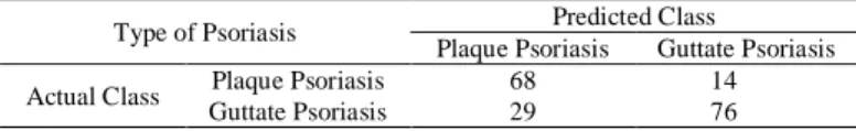
Table 1. The result of confusion matrix
Type of Psoriasis Predicted Class
Plaque Psoriasis Guttate Psoriasis
Actual Class Plaque Psoriasis 68 14
Guttate Psoriasis 29 76
Table 1 shows the result of confusion matrix of Plaque and Guttate Psoriasis skin disease classification. As can be seen from Table 1, it showed that 68 number of predicted Plaque images which are classified as actual Plaque images while 14 number of predicted Plaque Psoriasis images which are not classified as actual Plaque Psoriasis image. Thus, the accuracy rate of TP for Plaque Psoriasis skin disease is 82.9% and accuracy rate of FN is 17.1%. For the Guttate Psoriasis, it is tabulated that 76 out of 105 number of images are classified as true with the actual image while 29 from 105 number of images are not classified as Guttate Psoriasis skin disease. Hence, the accuracy rate for TP of Guttate Psoriasis skin disease is 72.4% are classified as true whereas FN for Guttate Psoriasis is 27.6% are false classified. Therefore, this research conclude that CNN produced a better performance in Plaque and Guttate Psoriasis skin disease classification which shows the higher accuracy rate of TP for the addressed problem.
4. CONCLUSION
This paper presented a non-invasive Psoriasis skin disease classification using CNN which been utilized on 187 images, consist of 82 images from Plaque Psoriasis and 105 images for Guttate Psoriasis. It is aims to obtain a better performance when classifying the type of Plaque and Guttate Psoriasis skin disease by using CNN. Based on Table 1, it is demonstrated that the accuracy rate of TP is 82.9% for Plaque Psoriasis skin disease whereas 72.4% of TP are true classified for Guttate Psoriasis skin disease. Overall, CNN is the best deep learning technique in Psoriasis skin disease classification which the percentage of true classified are more than 80%. This research and the findings may also be used in other medical image analysis. This is because CNN are capable of processing data directly from the raw pixels. Thus, this research is also beneficial and useful in medical dermatology domain especially in Psoriasis skin disease field. For futurework, further research on experimenting the parameters in pooling and fully connected layers of CNN in order to produce the robust performance and better accuracy results. And it is hope that this research would be valuable and advantageous to medical field in the near future as a medical tool which will assist the dermatologist to classify Psoriasis skin disease.
ACKNOWLEDGEMENTS
The authors would like to thank the Ministry of Higher Education, Malaysia and Universiti Teknologi MARA for the research funding and support via grant number FRGS/1/2017/ICT05/UITM/03/1.
REFERENCES
[1] Yadav N, Yadav N, Narang VK. Skin diseases detection models using image processing: A survey. International Journal of Computer Applications. 137(12):0034-9, 2016.
[2] Dhandra B, Soma S, Reddy S, Mukarambi G. Color Histogram Approach for Analysis of Psoriasis Skin Disease. InInt. Conf. on Multimedia Processing 2013.
[3] Juang LH, Wu MN. Psoriasis image identification using k-means clustering with morphological processing. Measurement. 44(5):895-905, 2011.
[4] Pal A, Garain U, Chandra A, Chatterjee R, Senapati S. Psoriasis skin biopsy image segmentation using Deep Convolutional Neural Network. Computer methods and programs in biomedicine. 159:59-69, 2018.
[5] Shrivastava VK, Londhe ND, Sonawane RS, Suri JS. Reliable and accurate psoriasis disease classification in dermatology images using comprehensive feature space in machine learning paradigm. Expert Systems with Applications. 42(15-16):6184-95, 2015.
[6] Shrivastava VK, Londhe ND, Sonawane RS, Suri JS. Computer-aided diagnosis of psoriasis skin images with HOS, texture and color features: a first comparative study of its kind. Computer methods and programs in biomedicine. 126:98-109,2016.
[7] MohdAffandi A, Khan I, NgahSaaya N. Epidemiology and Clinical Features of Adult Patients with Psoriasis in Malaysia: 10-Year Review from the Malaysian Psoriasis Registry (2007–2016). Dermatology research and practice. 2018.
[8] Roslan R, Jamil N, Mahmud R. Skull stripping magnetic resonance images brain images: region growing versus mathematical morphology. International Journal of Computer Information Systems and Industrial Management Applications.3:150-8, 2011.
[9] Roslan R, Jamil N, Mahmud R. Skull stripping of MRI brain images using mathematical morphology. In Biomedical Engineering and Sciences (IECBES), 2010 IEEE EMBS Conference on 2010 Nov 30 (pp. 26-31). IEEE. [10] Roslan R, Jamil N, Za’ba N. Spectral Texture Segmentation for Glioma Brain Tumour Detection. Journal of Next
Generation Information Technology (JNIT), Vol4, No 6, 2013.
[11] Mokhtar F, Ngadiran R, Basheer T, Rahim AN. Analysis of wavelet-based full reference image quality assessment algorithm. Bulletin of Electrical Engineering and Informatics. 8(2):527-32, 2019.
[12] Withana U, Fernando P. Differential diagnosis of eczema and psoriasis using categorical data in image processing. In2017 Seventeenth International Conference on Advances in ICT for Emerging Regions (ICTer) 2017 Sep 6 (pp. 1-6). IEEE.
[13] Kahya MA. Classification enhancement of breast cancer histopathological image using penalized logistic regression. Indonesian Journal of Electrical Engineering and Computer Science. 13(1):405-10, 2019.
[14] Lu J, Kazmierczak E, Manton JH, Sinclair R. Automatic segmentation of scaling in 2-d psoriasis skin images. IEEE transactions on medical imaging. 32(4):719-30, 2012.
[15] Kim KB, Song DH. Colored facial image restoration by similarity enhanced implicative fuzzy association memory. Indonesian Journal of Electrical Engineering and Computer Science (IJEECS). 13:199-204,2019.
[16] Tawfeeq FN, Alwan NA, Khashman BM. Optimization of Digital Histopathology Image Quality. International Journal of Artificial Intelligence (IJ-AI). Vol 7. Issue 2 pp 71-77, 2018.
[17] Isa NM, Amir A, Ilyas MZ, Razalli MS. Motor imagery classification in Brain computer interface (BCI) based on EEG signal by using machine learning technique. Bulletin of Electrical Engineering and Informatics. 8(1):269-75, 2019.
[18] Rawat AS, Rana A, Kumar A, Bagwari A. Application of Multi-Layer Artificial Neural Network in the Diagnosis System: A Systematic Review. 7:138-42,2018.
[19] Al-Saffar AA, Tao H, Talab MA. Review of deep convolution neural network in image classification. In2017 International Conference on Radar, Antenna, Microwave, Electronics, and Telecommunications (ICRAMET) 2017 Oct 23 (pp. 26-31). IEEE.
[20] Vaityshyn V, Chekhovych M, Poreva A. Convolutional Neural Networks for the Classification of Bronchopulmonary System Diseases with the Use of Lung Sounds. In2018 IEEE 38th International Conference on Electronics and Nanotechnology (ELNANO) 2018 Apr 24 (pp. 383-386). IEEE.
[21] Rathod J, Wazhmode V, Sodha A, Bhavathankar P. Diagnosis of skin diseases using Convolutional Neural Networks. In2018 Second International Conference on Electronics, Communication and Aerospace Technology (ICECA) 2018 Mar 29 (pp. 1048-1051). IEEE.
[22] Begum A, Fatima F, Sabahath A. Implementation of Deep Learning Algorithm with Perceptron using TenzorFlow Library. In2019 International Conference on Communication and Signal Processing (ICCSP) 2019 Apr 4 (pp. 0172-0175). IEEE.
[23] Bengio Y, Courville A, Vincent P. Representation learning: A review and new perspectives. IEEE transactions on pattern analysis and machine intelligence. 35(8):1798-828, 2013.
[24] Guo T, Dong J, Li H, Gao Y. Simple convolutional neural network on image classification. In2017 IEEE 2nd International Conference on Big Data Analysis (ICBDA), (2017 Mar 10 (pp. 721-724). IEEE.
[25] Lu S, Lu Z, Aok S, Graham L. Fruit Classification Based on Six Layer Convolutional Neural Network. In2018 IEEE 23rd International Conference on Digital Signal Processing (DSP) 2018 Nov 19 (pp. 1-5). IEEE.
[26] Bala R. Survey on texture feature extraction methods. International Journal of Engineering Science. 10375,2017. [27] Zhou Y, Shi C, Lai B, Jimenez G. Contrast enhancement of medical images using a new version of the World Cup
Optimization algorithm. Quantitative imaging in medicine and surgery. 9(9):1528, 2019.
[28] Taur JS, Lee GH, Tao CW, Chen CC, Yang CW. Segmentation of psoriasis vulgaris images using multiresolution-based orthogonal subspace techniques. IEEE Transactions on Systems, Man, and Cybernetics, Part B (Cybernetics). 36(2):390-402, 2006.
[29] DermNet NZ. Cysts. DermNet NZ website.
[30] Albawi S, Mohammed TA, Al-Zawi S. Understanding of a convolutional neural network. In2017 International Conference on Engineering and Technology (ICET) 2017 Aug 21 (pp. 1-6). IEEE.