ULTRAFINE PARTICLES ALTER ENDOTHELIAL PHENOTYPE THROUGH OXIDANT SIGNALING
Samantha Jean Snow
A dissertation submitted to the faculty of the University of North Carolina at Chapel Hill in partial fulfillment of the requirements for the degree of Doctor of Philosophy in the
Curriculum in Toxicology
Chapel Hill 2012
Approved by:
ii ABSTRACT
SAMANTHA JEAN SNOW: Ultrafine particles alter endothelial phenotype through oxidant signaling
(Under the direction of Dr. Martha Sue Carraway and Dr. David Diaz-Sanchez)
iii
iv
ACKNOWLEDGMENTS
I would like to express my appreciation and gratitude for Dr. Martha Sue Carraway for her guidance, mentorship, and support during my doctorial training. I would also like to thank my committee members Drs. David Diaz-Sanchez, Alisa Wolberg, Ilona Jaspers, and Frank Church for their time, expertise, and advice they so gratefully provided during my studies.
I could not have completed my doctorate degree without the generous support I received from my coworkers and scientific colleagues over the years. My deepest appreciations go to members of the Clinical Research Branch for their sincere care and encouragement, particularly Lisa Dailey, Joleen Soukup, Dr. WanYun Cheng, Eugene Gibbs, Candice Bailey, Rob Silbajoris, and Dr. Andy Ghio. Several individuals of the Curriculum in Toxicology were instrumental in guiding me through this process, and I would especially like to acknowledge Dr. Marila Cordeiro-Stone, Julie Cannefax, Dr. Jonathon Shannahan, and Jessica Sorrentino for their advice and support.
v
TABLE OF CONTENTS
LIST OF TABLES ... viii
LIST OF FIGURES ... ix
CHAPTER 1. INTRODUCTION ... 1
CHAPTER 2. SOLUBLE COMPONENTS OF ULTRAFINE PARTICLES STIMULATE H2O2 PRODUCTION IN ENDOTHELIAL CELLS ... 21
2.1 Introduction ... 21
2.2 Materials and Methods ... 25
2.2.1 Reagents and Chemicals ... 25
2.2.2 Cell Culture ... 25
2.2.3 Ultrafine Particles ... 25
2.2.4 Extracellular H2O2 Measurement using the Amplex Red Assay ... 26
2.2.5 Measurement of Cytotoxicity ... 27
2.2.6 Statistical Analysis ... 27
2.3 Results ... 28
2.3.1 Coarse, fine, and UF PM exposure increases extracellular H2O2 generation in endothelial cells ... 28
2.3.2 Soluble UF exposure causes a dose- and time-dependent increase in extracellular H2O2 generation in endothelial cells ... 31
2.3.3 Soluble UF-induced ROS production is from NOX enzymes ... 34
2.3.4 Soluble UF-induced extracellular H2O2 generation in HCAEC is dependent on transition metals ... 36
CHAPTER 3. SOLUBLE COMPONENTS OF ULTRAFINE PARTICLES INDUCE ENDOTHELIAL PROCOAGULANT ACTIVITY THROUGH OXIDANT SIGNALING ... 43
3.1 Introduction ... 43
3.2 Materials and Methods ... 46
3.2.1 Reagents and Chemicals ... 46
vi
3.2.3 Ultrafine Particles ... 46
3.2.4 Measurement of Cytotoxicity ... 47
3.2.5 Calibrated Automated Thrombography (CAT) Assay ... 47
3.2.6 Turbidity Assay ... 48
3.2.7 Real-Time Quantitative PCR ... 48
3.2.8 Immunofluorescence ... 49
3.2.9 Extracellular H2O2 Measurement using the Amplex Red Assay ... 50
3.2.10 Intracellular H2O2 Detection using PG1 ... 50
3.2.11 Western Blot Analysis ... 51
3.2.12 siRNA Transfection ... 52
3.2.13 Statistical Analysis ... 52
3.3 Results ... 53
3.3.1 Soluble UF cause HCAEC to promote faster onset of TF-dependent thrombin generation and fibrin thrombus formation ... 53
3.3.2 Soluble UF increases TF mRNA expression in HCAEC ... 57
3.3.3 Soluble UF-induced TF upregulation is ROS dependent ... 60
3.3.4 Soluble UF exposure causes H2O2 production in HCAEC... 62
3.3.5 ROS production from NOX-4 enzyme leads to upregulation of TF mRNA expression ... 65
3.4 Discussion ... 69
CHAPTER 4. SOLUBLE COMPONENTS OF ULTRAFINE PARTICLES INDUCE PROINFLAMMATORY RESPONSES IN ENDOTHELIAL CELLS ... 74
4.1 Introduction ... 74
4.2 Materials and Methods ... 77
4.2.1 Reagents and Chemicals ... 77
4.2.2 Cell Culture ... 77
4.2.3 Ultrafine Particles ... 77
4.2.4 Real-Time Quantitative PCR ... 78
4.2.5 siRNA Transfection ... 79
4.2.6 Statistical Analysis ... 79
4.3 Results ... 80
vii
4.3.2 Soluble UF induces mRNA expression of proinflammatory
cytokines in HCAEC ... 82
4.3.3 Soluble UF induces mRNA expression of cell adhesion molecules in HCAEC .. 85
4.3.4 Soluble UF increases HO-1 expression in HCAEC ... 87
4.3.5 ROS production from NOX enzymes lead to upregulation of IL-1β and HO-1 mRNA expression ... 89
4.4 Discussion ... 92
CHAPTER 5. OVERALL CONCLUSIONS AND SIGNIFICANCE ... 96
viii
LIST OF TABLES
ix
LIST OF FIGURES
Figure 1.1 Broad biological pathways whereby PM may cause CV events. ... 4
Figure 1.2 Schematic diagram illustrating how exposure to soluble UF can lead to adverse CV events. ... 8
Figure 1.3 Schematic diagram illustrating the extrinsic pathway of the coagulation cascade. 9 Figure 1.4 Pathways of ROS production. ... 14
Figure 1.5 Schematic diagram illustrating activation of the Amplex Red reagent and PG1 by H2O2. ... 16
Figure 1.6 Schematic diagram illustrating endothelial activation following exposure to soluble UF. ... 20
Figure 2.1 Exposure to soluble UF induces rapid H2O2 generation by EA and HCAEC. ... 30
Figure 2.2 Non-cytotoxic doses of soluble UF increase H2O2 production in a dose- and time-dependent manner in EA and HCAEC... 32
Figure 2.3 Soluble UF-induced ROS production is dependent on NOX enzymes. ... 35
Figure 2.4 Soluble UF-induced H2O2 production is dependent on transition metals. ... 38
Figure 3.1 Exposure to soluble UF leads to faster onset of TF-dependent thrombin generation and fibrin thrombus formation. ... 55
Figure 3.2 Exposure to soluble UF leads to upregulation of TF. ... 59
Figure 3.3 Soluble UF-induced upregulation of TF is ROS dependent. ... 61
Figure 3.4 Exposure to soluble UF induces rapid H2O2 generation by HCAEC. ... 63
Figure 3.5 TF mRNA expression following exposure to soluble UF requires ROS production and NOX-4. ... 67
Figure 4.1 Exposure to soluble UF leads to upregulation of IL-8 in EA cells. ... 81
x
Figure 4.3 Exposure to soluble UF leads to upregulation of cell adhesion molecules in HCAEC. ... 86 Figure 4.4 Exposure to soluble UF leads to upregulation of HO-1 in HCAEC. ... 88 Figure 4.5 IL-1β and HO-1 mRNA expression following exposure to soluble
xi
LIST OF ABBREVIATIONS
99m
Tc 99mTechnetium
A549 Human Lung Adenocarcinoma Epithelial Cell Line ANOVA Analysis of Variance
AP-1 Activating Protein-1 APC Activated Protein C β-actin Beta-Actin
BSA Bovine Serum Albumin
CAT Calibrated Automated Thrombography cDNA Complementary Deoxyribonucleic Acid COPD Chronic Obstructive Pulmonary Disease
CV Cardiovascular
DAPI 4', 6-diamidino-2-phenylindole DEP Diesel Exhaust Particles
DMEM Dulbecco’s Modified Eagle’s Medium DNA Deoxyribonucleic Acid
DPI Diphenyleneiodonium DUOX Dual Oxidase
xii EPCR Endothelial Protein C Receptor ETP Endogenous Thrombin Potential FBS Fetal Bovine Serum
FV Factor V
FVa Activated Factor V FVIIa Activated Factor VII FVIII Factor VIII
FVIIIa Activated Factor VIII
FX Factor X
FXa Activated Factor X
FXI Factor XI
GAPDH Glyceraldehyde 3-Phosphate Dehydrogenase
GM-CSF Granulocyte Macrophage Colony Stimulating Factor HCAEC Human Coronary Artery Endothelial Cells
HCL Hydrochloric Acid
HO Heme Oxygenase
HO-1 Heme Oxygenase-1 H2O2 Hydrogen Peroxide HRP Horseradish Peroxidase
HUVECs Human Umbilical Vein Endothelial Cells ICAM-1 Intercellular Adhesion Molecule-1
IgG Immunoglobulin G
xiii KCN Potassium Cyanide
LDH Lactate Dehydrogenase
MCP-1 Monocyte Chemoattractant Protein-1 mRNA Messenger Ribonucleic Acid
NaCl Sodium Cloride
NADPH Nicotinamide Adenine Dinucleotide Phosphate Nrf2 Nuclear Factor (Erythroid-Derived 2)-Like 2 NF-κB Nuclear Factor-Kappa B
NO Nitric Oxide
NOX Nicotinamide Adenine Dinucleotide Phosphate Oxidase NOX-1 Nicotinamide Adenine Dinucleotide Phosphate Oxidase-1 NOX-2 Nicotinamide Adenine Dinucleotide Phosphate Oxidase-2 NOX-4 Nicotinamide Adenine Dinucleotide Phosphate Oxidase-4
O2 Oxygen
·O2 Superoxide Anion
·OH Hydroxyl Radical
p53 Protein 53
PAI-1 Plasminogen Activator Inhibitor-1 PARs Protein Activated Receptors PBS Phosphate Buffer Saline
PC Protein C
PCR Polymerase Chain Reaction PEG Polyethylene Glycol
PG1 Peroxy Green 1
xiv PM Particulate Matter
PM10 Coarse Particulate Matter PM2.5 Fine Particulate Matter PM0.1 Ultrafine Particulate Matter R3-IGF-1 R3-Insulin-like Growth Factor-1 RFU Relative Fluorescence Units
rhEGF Recombinant Human Epidermal Growth Factor rhFGF-B Recombinant Human Fibroblast Growth Factor-Basic RNA Ribonucleic Acid
ROS Reactive Oxygen Species
RT-PCR Reverse Transcriptase Polymerase Chain Reaction SDS Sodium Dodecyl Sulfate
SDS-PAGE Sodium Dodecyl Sulfate Polyacrylamide Gel Electrophoresis siRNA Small Interfering Ribonucleic Acid
SOD Superoxide Dismutase
Soluble UF Soluble Components of Ultrafine Particles SP1 Specificity Protein 1
TF Tissue Factor
TFPI Tissue Factor Pathway Inhibitor TiO2 Titanium Dioxide
TNF-α Tumor Necrosis Factor-Alpha
TM Thrombomodulin
UF Ultrafine
CHAPTER 1 INTRODUCTION
In the early 20th century, a series of air pollution incidents in Meuse Valley, Belgium (1930), Donora, Pennsylvania (1948), and London, England (1952) illustrated the adverse effects individuals experience during severe air pollution episodes. These events occurred in heavily industrialized cities during a meteorological inversion that concentrated the already severe air pollution and resulted in increased hospitalizations and elevated rates of mortality (Stanek et al., 2011). These incidents raised public awareness about the severity of air pollution, led to new regulations across the developed world, and spurred investigators to conduct epidemiological and toxicological studies to establish the relationship and mechanisms behind air pollution exposure and adverse health effects.
2
systemic oxidative stress in mice (Araujo et al., 2008), accelerated atherosclerotic progression in susceptible mouse (Sun et al., 2005) and rabbit models (Suwa et al., 2002), and enhanced experimental arterial and venous platelet rich-thrombus formation in hamsters (Nemmar et al., 2003).
The enactment of the Clean Air Act in 1970 required the Environmental Protection Agency (EPA) to set National Ambient Air Quality Standards for six criteria air pollutants including particulate matter (PM), ozone, carbon monoxide, sulfur dioxide, nitrogen oxides, and lead. These air pollutants were deemed to be ubiquitous in the United States and were suspected or known to induce adverse health effects in humans or the environment (Suh et al., 2000). Of these six criteria air pollutants, PM and ground level ozone present the most widespread threat to human health and are known to commonly exceed the federal standards set by the EPA (Laumbach, 2010).
3
The EPA currently monitors and regulates coarse and fine PM according to the Clean Air Act, but increasing evidence suggests that UF particles, which are not currently regulated, are of particular importance in CV effects of air pollution (Nel, 2005). UF particles make up only a small portion of ambient PM by mass concentration; however, they constitute the majority of particle number per unit mass and have a significantly high surface area-to-mass ratio as compared to coarse and fine particles. This property suggests that UF particles are capable of adsorbing large amounts of toxic substances on the surface. In addition, the small diameter of the particle makes it more likely to deposit in the alveolar region of the lung due to Brownian motion and diffusion transportation (Kreyling et al., 2004). UF particles are also able to stay in the lung longer than the larger fine and coarse particles because their small diameter makes them more likely to avoid being recognized or phagocytized by alveolar macrophages, a critical lung defense mechanism (Gonzalez et al., 1996).
4 Figure 1.1
5
It remains unclear as to whether UF sized particles can cross from the lung into the vasculature. A study by Nemmar et al. exposed humans to an aerosol of radioactive 99m
technetium (99mTc) labeled UF carbon particles followed by imaging to determine particle distribution following inhalation (Nemmar et al., 2002). They found a majority of the radioactivity remained in the lungs, but also recorded levels of radioactivity in the blood, urine, bladder, and liver, suggesting extrapulmonary translocation of 99mTc labeled UF carbon particles. These researchers have reported similar results using animal models following exposure to radioactive labeled UF particles (Nemmar et al., 2001). However, there are several caveats to these studies. For instance, the investigators were unable to detect the radioactive labeled UF particles in the blood using electron microscopy. In addition, a study conducted by Mills et al., which also exposed humans to 99mTc labeled UF carbon particles, found no evidence of extrapulmonary translocation of the radioactive labeled particles (Mills et al., 2006). They instead explain the findings of Nemmar et al. by suggesting leaching of the radiolabel from the UF particle and/or contamination of free radiolabeled 99mTc-pertechnetate in their aerosol, which is formed using the same generator but with minimal oxygen contamination. These findings are supported by an additional study in which COPD patients were exposed to 99mTc labeled UF carbon particles with no evidence of extrapulmonary translocation of the radioactive label particles (Brown et al., 2002).
6
(Wallenborn et al., 2009; Wallenborn et al., 2007). Additionally, the ability of these metals to translocate from the lung into the systemic circulation was dependent on their solubility in water (Wallenborn et al., 2007). These water-soluble transition metals are capable of leaching off PM particles in the lung lining fluid and translocating into the circulation, possibly via metal transporters or other mechanisms, before being cleared by the liver.
Transition metals, such as vanadium, nickel, iron, copper, chromium, and zinc, are major constituents of the water-soluble fraction of UF particles and may play an important role in the adverse cardiopulmonary effects associated with air pollution exposure (Costa and Dreher, 1997). Transition metals can increase oxidative DNA damage, induce ROS production, activate inflammatory pathways, and induce procoagulant events in both in vitro
7
pollution particles are not known, but one possibility is that the components have direct effects on cells of the vasculature including inducing endothelial activation.
Endothelial cell activation leads to increased vascular coagulation and inflammation, and it plays an important role in the pathogenesis of certain cardiovascular diseases including atherosclerosis and hypertension (Lwaleed et al., 2007; Sprague and Khalil, 2009). Endothelial activation is typically defined by five principal changes that occur including loss of vascular integrity, upregulation of human leukocyte antigen molecules, induction of inflammatory cytokines, expression of leukocyte adhesion molecules, and a change from an antithrombotic to prothrombotic phenotype (Hunt and Jurd, 1998). Endothelial cells are critical components of the vasculature as they line the blood vessels and are involved in hemostatic balance, vasomotor tone, blood cell trafficking, permeability, and immunity. In addition, endothelial cells have a critical role in the coagulation system and are able to express procoagulant factors, anti-coagulant factors, and fibrinolytic proteins (Aird, 2007).
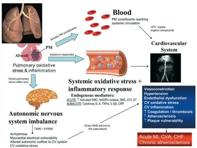
8 Figure 1.2
Figure 1.2 Schematic diagram illustrating how exposure to soluble UF can lead to
adverse CV events.
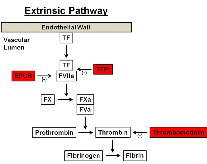
9 Figure 1.3
Figure 1.3 Schematic diagram illustrating the extrinsic pathway of the coagulation
cascade.
10
studies, this assay has been utilized to determine the effects of air pollution on thrombin generation in animal and human models (Emmerechts et al., 2012; Kilinc et al., 2011; Rudez et al., 2009). For example, Emmerechts et al. demonstrated that elevated levels of coarse PM shortened the lag time of thrombin generation in microparticle-rich plasma obtained from diabetic individuals exposed to ambient air pollution (Emmerechts et al., 2012). In addition, fibrin thrombus formation can be measured as a way to assess the functional coagulation balance following exposure to air pollution. For instance, Metassan et al. have shown increased polymerization of purified human fibrinogen in the presence of UF PM (Metassan et al., 2010).
11
indication of endothelial cell activation and are known to play a role in thrombosis and atherosclerosis (Chironi et al., 2009).
p53-12
mediated apoptosis (Anastasiou et al., 2012; Cheng et al., 2003). The balance of these factors is critical, and enhanced procoagulant activity or inhibition of anticoagulant activity could indicate an activated endothelial cell phenotype, resulting in a prothrombotic state (Gilmour et al., 2005).
There is strong cross-talk between the coagulation and inflammatory pathways that involves inflammatory mediators activating the coagulation cascade or suppressing anticoagulant proteins, and coagulation proteins inducing inflammation (Levi et al., 2004a). Inflammation predominately induces coagulation by activating TF and the extrinsic coagulation pathway leading to thrombin generation (van der Poll et al., 2011). For example, proinflammatory cytokines, namely TNF-α and IL-1β, have been shown to induce TF expression and increase procoagulant activity in endothelial cells (Dinarello, 1991). Inflammation is also capable of impairing several anticoagulant pathways (Levi and van der Poll, 2005). Proinflammatory cytokines have been shown to down-regulate the anticoagulant protein TM, which in turn prevents the formation of the thrombomodulin-thrombin complex that is necessary for the activation of PC (Nawroth and Stern, 1986). Inflammation also reduces levels of the anticoagulant protein antithrombin, the main inhibitor of thrombin and FXa (Levi et al., 2004a), and down-regulates TFPI, the main inhibitor of TF (Delvaeye and Conway, 2009).
13
der Poll, 2005). The TF/FVIIa complex can bind protease activated receptors (PARs), leading to the induction of cell signaling pathways that result in increased expression of proinflammatory cytokines (Cunningham et al., 1999). Thrombin and FXa are also capable of binding to PARs and activating these signaling pathways (Levi et al., 2004b). In addition, it was shown in baboons that inhibition of TF activity prevents inflammation-induced thrombin generation (Taylor et al., 1991) and genetically altered mice with low levels of TF had attenuated levels of proinflammatory cytokines following exposure to endotoxin (Pawlinski et al., 2004).
Aberrant induction of TF has been associated with multiple CV disease pathologies including venous thrombosis, atherosclerosis, and diabetes (Lwaleed et al., 2007; Manly et al., 2010). TF can be induced by a variety of stimuli that primarily activate phospholipase C, resulting in a cascade of signaling events that eventually cumulate in the upregulation of TF expression (Herkert and Gorlach, 2002). TF is also a redox-regulated protein, and TF gene expression and protein levels have been shown to be modulated by ROS in vascular cells (Herkert et al., 2004). Additionally, TF mRNA expression is attenuated in the presence of antioxidants following exposure to ionizing radiation and inflammatory cytokines, further illustrating the critical role ROS plays in TF regulation (Szotowski et al., 2007). ROS is thought to induce TF through activation of transcription factors resulting in induction of TF mRNA, and the TF gene in primary endothelial cells has been shown to contain binding sites for the redox-sensitive transcription factors NF-κB, AP-1, and SP1 (Herkert and Gorlach, 2002; Moll et al., 1995).
14
production of the superoxide anion (·O2) from molecular oxygen (O2). Superoxide is transformed into a subsequent ROS, hydrogen peroxide (H2O2), a reaction that is facilitated by the enzyme superoxide dismutase (SOD) (Mills et al., 2007). H2O2 can then be cleaved by catalase into water and oxygen. In the presence of transition metals, H2O2 can also be converted into the very reactive hydroxyl radical (·OH) (Droge, 2002) (Figure 1.4).
Figure 1.4
Figure 1.4 Pathways of ROS production.
Cellular generation of ROS is important physiologically because these molecules can be used as both intra- and intercellular signaling molecules (D'Autreaux and Toledano, 2007). Superoxide is relatively unstable in aqueous conditions and is rapidly transformed into H2O2 either by SOD or spontaneously, so it is generally thought that signaling events occur through the H2O2 molecule (Li and Shah, 2004). H2O2 is capable of oxidizing cysteine residues in proteins to cysteine sulfenic acid or disulfide, which can lead to protein phosphorylation and initiation of signaling cascades (Rhee et al., 2000). Additionally, H2O2 has been shown to activate transcription factors, such as NF-κB (Schmidt et al., 1995), AP-1 (Karin and Shaulian, 2001) and p53 (Thomas et al., 2006) , leading to induction of redox-sensitive genes.
15
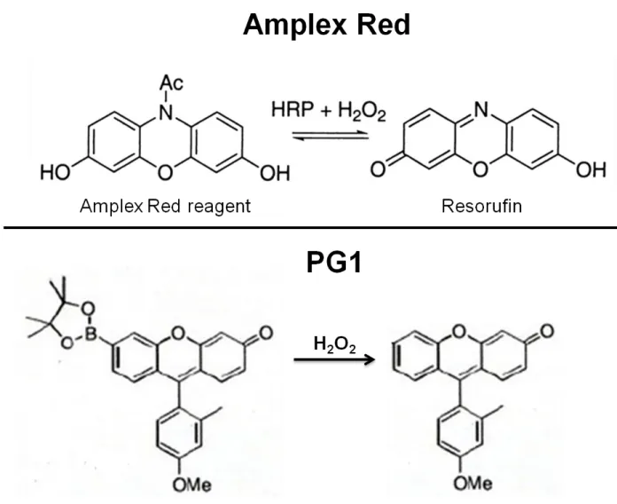
16 Figure 1.5
Figure 1.5 Schematic diagram illustrating activation of the Amplex Red reagent and
PG1 by H2O2. Images were modified from Wentworth et al. and Miller et al. (Miller et
al., 2007; Wentworth et al., 2000).
17
H2O2 have been shown to induce cellular injury by causing damage to key cellular molecules such as DNA (Imlay et al., 1988), proteins (Knock and Ward, 2011), and lipids (Kellogg and Fridovich, 1975). Furthermore, elevated levels of ROS are implicated in the pathogenesis and progression of several CV diseases including atherosclerosis, hypertension, and diabetes (Madamanchi et al., 2005).
Endothelial cells can generate ROS in a variety of enzymatic and non-enzymatic ways, such as the mitochondrial electron transport chain, xanthine oxidases, and NADPH oxidase (NOX) enzymes (Droge, 2002; Li and Shah, 2004). In addition, transition metals, such as iron, copper, and chromium are capable of undergoing redox cycling and producing intracellular ROS through Fenton reactions (Jomova and Valko, 2011). Of these potential sources, NOX enzymes are proposed as the key generator of ROS production in the vasculature (Babior, 2000; Mohazzab et al., 1994).
NADPH oxidases are a family of enzymes comprised of 7 major members, NOX1-5 and Duox1-2 (Frey et al., 2008). NOX-2 was the first to be characterized, where it was discovered on the plasma membrane of phagocytic cells and shown to produce a respiratory burst of oxidants for microbial defense (Babior, 1984). Since that discovery, the NOX family of enzymes have been reported in an assortment of non-phagocytic cells including fibroblasts, vascular smooth muscle cells, and endothelial cells among others (Brown and Griendling, 2009). Endothelial cells express mainly the NOX-2 and NOX-4 isoforms (Li and Shah, 2004), but expression levels of the NOX isoforms varies among different endothelial cell types (Guzik et al., 2004).
18
be found in various subcellular locations and expression of these enzymes can dictate participation in distinct signaling pathways (Gough and Cotter, 2011). For example, the intracellular distribution of NOX-4 is broad and has been shown to be variably located in the perinuclear space, endoplasmic reticulum, mitochondria, and nucleus of endothelial cells (Lassegue and Griendling, 2010; Lassegue et al., 2012) and has been reported to be involved in several cellular processes including vasodilation, cell migration, and proliferation (Pendyala et al., 2009; Petry et al., 2006; Ray et al., 2011).
19
20 Figure 1.6
CHAPTER 2
SOLUBLE COMPONENTS OF ULTRAFINE PARTICLES STIMULATE H2O2
PRODUCTION IN ENDOTHELIAL CELLS 2.1 Introduction
A growing body of evidence shows a strong association between exposure to air pollution and cardiovascular (CV) morbidity and mortality. Exposure to air pollution leads to increased hospitalizations for circulatory disorders (Poloniecki et al., 1997), accelerates the progression of atherosclerosis (Suwa et al., 2002), and increases the risk of myocardial infarctions (Peters et al., 2001). In addition, elevated levels of air pollution are associated with increased risk of CV-related mortality (Pope et al., 2004). The World Health Organization estimates that exposure to ambient air pollution causes several million premature deaths worldwide each year, with particulate matter (PM) exposure accounting for approximately 800,000 of those deaths (Anderson et al., 2012).
22
and are comprised of volatile organic compounds, polycyclic aromatic hydrocarbons, transition metals, and other constituents adsorbed on a carbonaceous core (Polichetti et al., 2009).
23
the circulation and directly interact with cells of the vasculature such as endothelial cells (Wallenborn et al., 2007).
Transition metals are a major component of the soluble fraction and may play an important role in the adverse CV effects associated with air pollution exposure (Costa and Dreher, 1997). Transition metals can increase oxidative DNA damage in lymphocyte DNA and induce reactive oxygen species (ROS) production in endothelial cells, indicating that soluble components of PM are capable of inducing oxidative stress following exposure to air pollution (Montiel-Davalos et al., 2012; Sorensen et al., 2005).
Elevated levels of ROS under pathological conditions have been linked to the development of several CV diseases (Madamanchi et al., 2005). Endothelial cells can produce ROS in response to an array of both physiological and pathological stimuli from a variety of both enzymatic and non-enzymatic sources (Droge, 2002). The major sources of ROS production in endothelial cells include the mitochondrial electron transport chain, xanthine oxidases, and NADPH oxidases (NOX), and of these, NOX enzymes are proposed to be the key generator of ROS production following exposure to toxic substances such as UF particles (Li and Shah, 2004; Mo et al., 2009). Furthermore, NOX-induced oxidative stress in endothelial cells is implicated in the progression of various CV disorders including hypertension, atherosclerosis, and diabetes (Bengtsson et al., 2003)
24
25 2.2 Materials and Methods
2.2.1 Reagents and Chemicals
EA.hy926 (EA cells), are an immortalized endothelial cell line derived by fusing human umbilical vein endothelial cells with A549 cells, a human lung adenocarcinoma epithelial cell line (Edgell et al., 1983). EA cells were obtained from University of North Carolina’s Tissue Culture Facility (Chapel Hill, NC). Dulbecco’s modified Eagle’s medium (DMEM)-high glucose, fetal bovine serum (FBS), and antibiotic-antimycotic (100X) were obtained from Gibco (Grand Island, NY). Human coronary artery endothelial cells (HCAEC), endothelial growth medium (EGM-2), and EGM-2 Bullet Kit were obtained from Lonza (Walkersville, MD). All other chemicals and reagents were from Sigma Chemical Company (St. Louis, MO) unless otherwise stated.
2.2.2 Cell Culture
EA cells were cultured in DMEM-high glucose medium supplemented with 10% FBS and 1% antibiotic-antimycotic mix. HCAEC were cultured in EGM-2 media supplemented with the EGM-2 Bullet Kit (2% FBS, 0.4% rhFGF-B, 0.1% gentamicin sulfate amphotericin-B, 0.1% rhEGF, 0.1% heparin, 0.1% ascorbic acid, 0.1% R3-IGF-1, 0.1% VEGF, and 0.04% hydrocortisone). HCAEC were obtained from two adult donors with no known history of CV disease. Cells were grown to confluence and used between passages 5-8.
2.2.3 Ultrafine Particles
26
2.1). The soluble fraction of these particles was acquired by suspending the particles in PBS at the desired concentration and centrifuging the resultant suspension for 30 minutes at 20,000 x g. The supernatant from the pelleted particles was collected and used as the soluble fraction. The pellet was then re-suspended in the same volume of PBS and used as the insoluble fraction.
2.2.4 Extracellular H2O2 Measurement using the Amplex Red Assay
27 2.2.5 Measurement of Cytotoxicity
Cytotoxicity was determined by measuring supernatant lactate dehydrogenase (LDH) levels using the colorimetric CytoTox 96 Non-Radioactive Cytotoxicity Assay (Promega Corporation, Madison, WI) according to the manufacturer’s protocol. EA cells and HCAEC were exposed to soluble UF (0, 10, 50, and 100 µg/mL) for 6 or 24 hours. Supernatant from cells treated with saponin (0.5%) to disrupt the cellular membrane was a positive control for the assay.
2.2.6 Statistical Analysis
28 2.3 Results
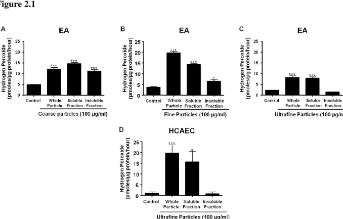
2.3.1 Coarse, fine, and UF PM exposure increases extracellular H2O2 generation in endothelial cells
29
30 Figure 2.1
Figure 2.1 Exposure to soluble UF induces rapid H2O2 generation by EA and HCAEC.
31
2.3.2 Soluble UF exposure causes a dose- and time-dependent increase in extracellular H2O2 generation in endothelial cells
32 Figure 2.2
Figure 2.2 Non-cytotoxic doses of soluble UF increase H2O2 production in a dose- and
33
34
2.3.3 Soluble UF-induced ROS production is from NOX enzymes
35 Figure 2.3
36
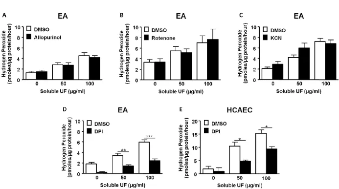
2.3.4 Soluble UF-induced extracellular H2O2 generation in HCAEC is dependent on transition metals
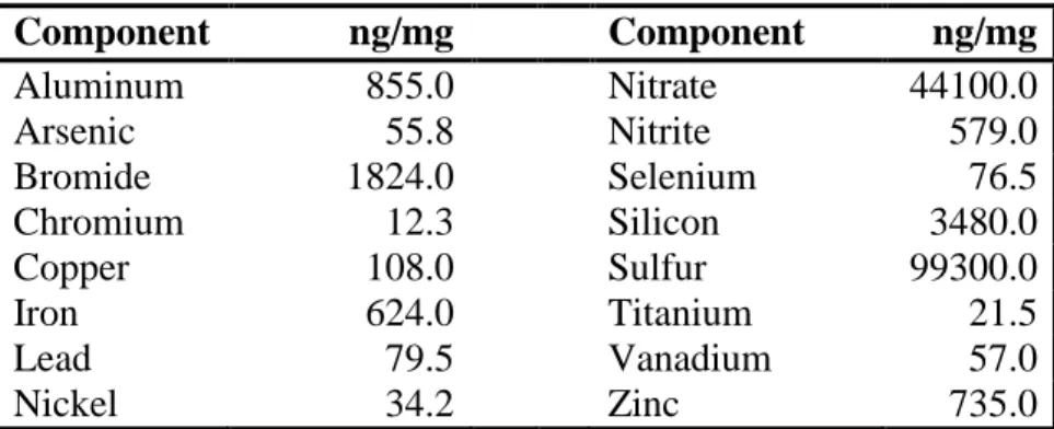
We next wanted to measure the UF particle-associated components to provide information on compounds present in the soluble fraction that could lead to increased ROS production. The UF particles were analyzed at the Research Triangle Institute and components are expressed relative to UF particle mass (ng/mg; Table 2.1). The measured levels of these elements and ions are comparable to those found in Chapel Hill UF particles previously collected and analyzed in 2001 and 2002 (Becker et al., 2005). Transition metals, such as chromium, copper, iron, nickel, vanadium, and zinc, were present in the UF particles. To determine if these transition metals have a role inducing extracellular H2O2 production, we measured the extracellular H2O2 release from HCAEC following exposure to 50 μg/mL soluble UF that had been pretreated with the metal chelator deferoxamine for 15 min. Pretreatment with deferoxamine (100 µM) reduced extracellular H2O2 production in soluble UF-exposed HCAEC by 45% (p<0.001; Fig. 2.4). These data indicate that transition metals play a partial role in the adverse effects induced in endothelial cells following exposure to soluble UF and suggest that other soluble components are also involved.
37 Table 2.1
Component ng/mg Component ng/mg
Aluminum 855.0 Nitrate 44100.0
Arsenic 55.8 Nitrite 579.0
Bromide 1824.0 Selenium 76.5
Chromium 12.3 Silicon 3480.0
Copper 108.0 Sulfur 99300.0
Iron 624.0 Titanium 21.5
Lead 79.5 Vanadium 57.0
Nickel 34.2 Zinc 735.0
38 Figure 2.4
Figure 2.4 Soluble UF-induced H2O2 production is dependent on transition metals.
39 2.4 Discussion
Increasing evidence suggests a strong association between exposure to air pollution and CV morbidity and mortality (Brook, 2008; Polichetti et al., 2009; Pope et al., 2004); however, further investigation needs to be conducted on the adverse vascular effects associated with exposure to components of PM. Here we report that exposure to non-cytotoxic doses of soluble components of UF particles increase extracellular H2O2 release in immortalized and primary endothelial cells. We further show that this induced H2O2 production is dependent on NOX enzymes and transition metals. These novel findings suggest that particle-induced ROS production in endothelial cells is a plausible mechanism for adverse CV events associated with exposure to air pollution.
40
traverse the alveolar-capillary barrier to enter the vasculature (Wallenborn et al., 2007), we designed our experiments to evaluate endothelial cell responses to PM exposure using the soluble fraction of UF particles.
Elevated levels of ROShave been shown to induce cellular injury by causing damage to key cellular molecules and have been implicated in the pathogenesis and progression of several CV diseases (Imlay et al., 1988; Kellogg and Fridovich, 1975; Knock and Ward, 2011; Madamanchi et al., 2005). We show here that exposure to soluble UF induces immediate ROS production in endothelial cells; suggesting vascular oxidative stress may occur following air pollution exposure. While there are several major sources of ROS production in endothelial cells, a recent study has shown that mouse pulmonary microvascular endothelial cells exposed to whole UF particles induced ROS production by NOX enzymes (Mo et al., 2009). Our data extend this finding by illustrating that NOX enzymes are the likely source of H2O2 production following exposure to water-soluble components of UF particles in human endothelial cells. Inhibition of NOX enzymes is an emerging strategy to combat oxidative stress-mediated diseases (Jaquet et al., 2009) and our data further illustrate the prospect of targeting these enzymes when trying to combat adverse CV effects due to air pollution exposure.
41
al., 2001), increase pulmonary inflammation in healthy human volunteers (Ghio and Devlin, 2001), and increase rates of respiratory related hospital admissions (Pope, 1989). In addition,
in vitro studies have shown that transition metals induce inflammatory responses and increase ROS production in pulmonary and vascular cells (Gojova et al., 2007; Montiel-Davalos et al., 2012; Shafer et al., 2010). For instance, Shafer et al. demonstrated that pretreatment with metal chelators attenuated particle-induced ROS production in rat alveolar macrophages, indicating that transition metals in the water-soluble fraction of coarse and fine PM are responsible for the increased ROS production (Shafer et al., 2010). Our results extend these observations by illustrating that transition metals associated with the soluble fraction of UF particles cause adverse vascular effects by inducing H2O2 production in endothelial cells.
42
maintain most of the characteristics of endothelial cells, are fast growing, and ideal for establishing protocols, but the effects produced in these cells may not fully represent effects in primary endothelial cells. Therefore, we also used HCAEC as a primary endothelial cellular model. Endothelial cells from the coronary artery are among the first vascular cells to receive fresh blood from the lung and will accordingly be exposed to high concentrations of any soluble components that cross into the circulation from the alveoli.
CHAPTER 3
SOLUBLE COMPONENTS OF ULTRAFINE PARTICLES INDUCE ENDOTHELIAL PROCOAGULANT ACTIVITY THROUGH OXIDANT
SIGNALING 3.1 Introduction
44
activity in animal and human models (Araujo et al., 2008; Sorensen et al., 2005); however, the mechanisms behind these responses have not been delineated.
A causal link between vascular effects of PM exposure and procoagulant responses is suggested by the finding that exposure to soluble UF increases tissue factor (TF) mRNA expression in human pulmonary artery endothelial cells (Karoly et al., 2007). Under normal physiological conditions, TF is not highly expressed on endothelial cells, but surface expression of TF can be induced by a variety of stimuli including fibrin, endotoxin, and the proinflammatory cytokines TNF-α and IL-1β (Colucci et al., 1983; Contrino et al., 1997; Dinarello, 1991; Levi and van der Poll, 2005). Altered expression and activity of TF is of particular interest because, once activated, this membrane-bound protein is the primary initiator of the extrinsic coagulation pathway resulting in thrombin generation and fibrin thrombus formation (Lwaleed et al., 2007). Elevated levels of TF have been associated with adverse CV effects including venous thrombosis and atherosclerosis (Lwaleed et al., 2007; Manly et al., 2010).
45
expression in platelet-activated vascular smooth muscle cells (Gorlach et al., 2000). In addition, NOX-induced oxidative stress in endothelial cells plays an important role in the pathogenesis of CV disorders (Madamanchi et al., 2005), perhaps through these and other mechanisms.
46 3.2 Materials and Methods
3.2.1 Reagents and Chemicals
Human coronary artery endothelial cells (HCAEC), endothelial growth medium (EGM-2), and EGM-2 Bullet Kit were obtained from Lonza (Walkersville, MD). All other chemicals and reagents were from Sigma Chemical Company (St. Louis, MO) unless otherwise stated.
3.2.2 Cell Culture
HCAEC were cultured in EGM-2 media supplemented with the EGM-2 Bullet Kit (2% FBS, 0.4% rhFGF-B, 0.1% gentamicin sulfate amphotericin-B, 0.1% rhEGF, 0.1% heparin, 0.1% ascorbic acid, 0.1% R3-IGF-1, 0.1% VEGF, and 0.04% hydrocortisone). HCAEC were obtained from two donors with no known history of CV disease. Cells were grown to confluence and used between passages 5-8.
3.2.3 Ultrafine Particles
47 3.2.4 Measurement of Cytotoxicity
Cytotoxicity was determined by measuring supernatant LDH levels using the colorimetric CytoTox 96 Non-Radioactive Cytotoxicity Assay (Promega Corporation, Madison, WI) according to the manufacturer’s protocol. Supernatant from cells treated with saponin (0.5%) to disrupt the cellular membrane was a positive control for the assay.
3.2.5 Calibrated Automated Thrombography (CAT) Assay
48
(time to reach the maximum peak height), and endogenous thrombin potential (ETP; total amount of thrombin generated during the test).
3.2.6 Turbidity Assay
HCAEC were cultured and exposed to soluble UF as described above. Recalcified (20mM, final) PFP and phospholipids (125 μM, 41% phosphatidylcholine/44% phosphatidylethanolamine/15% phosphatidylserine; Avanti Polar Lipids, Alabaster, AL) were added to the cells at a final volume of 100 μl to initiate thrombus formation (Gray et al., 2011). Fibrin thrombus formation was analyzed by an increase in turbidity at 405 nm with a SpectraMax 340PC plate reader (Molecular Devices, Sunnyvale, CA). Softmax® Pro Software version 1.21 (Molecular Devices, Sunnyvale, CA) was used to calculate thrombus formation onset (time to reach inflection point before turbidity increase) and Vmax (slope of the line fitted to maximum rate of turbidity increase using 10 points to determine the line). 3.2.7 Real-Time Quantitative PCR
Relative gene expression in HCAEC was obtained using quantitative RT-PCR. Total RNA was isolated from HCAEC using an RNeasy kit (Qiagen, Valencia, CA) according to the manufacturer’s protocol. RNA was quantified using a Nanodrop™ 1000 Spectrophotometer (Thermo Scientific, Wilmington, DE). cDNA was generated as previously described (Karoly et al., 2007). Oligonucleotide primer pairs and fluorescent probes for β-actin (forward, 5’-CCTGGCACCCAGCACAAT-3’; reverse, 5’-GCCGATCCACACGGAGTACT-3’; probe, 5’-ATCAAGATCATTGCTCCTCCTGAGCGC-3’) and TF (forward,
49
Express (Applied Biosystems) and obtained from Integrated DNA Technologies (Coralville, IA). Thrombomodulin, endothelial protein C receptor (EPCR), tissue factor pathway inhibitor (TFPI), IL-1β, TNF-α, GAPDH, and NOX-4 primer pairs and fluorescent probe sets were obtained as Taqman pre-developed assay reagents from Applied Biosystems (Foster City, CA). Quantitative fluorogenic amplification of cDNA was performed using the ABI Prism 7500 Sequence Detection System (Applied Biosystems, Foster City, CA), primer and probe sets of interest, and Taqman Universal PCR Master Mix (Applied Biosystems, Foster City, CA). Standard curves generated from a serially diluted standard pool of cDNA prepared from cultured human endothelial cells exposed to 100 ng/mL TNF-α for 6 hrs were used to determine the relative abundance of mRNA levels. The relative abundance of β-actin mRNA was used to normalize levels in genes of interest.
3.2.8 Immunofluorescence
50
3.2.9 Extracellular H2O2 Measurement using the Amplex Red Assay
Extracellular H2O2 release was measured using the Amplex Red reagent (10-acetyl-3,7-dihydroxyphenoxazine; Invitrogen, Carlsbad, CA), which reacts with H2O2 in the presence of horseradish peroxidase to produce resorufin, a highly fluorescent molecule. HCAEC were cultured on 12-well plates and exposed to soluble UF (0, 50, and 100 µg/mL). Immediately following exposure, Amplex Red (150 μM) and HRP (0.8 U/ml) were added, and plates were analyzed at 5 min intervals for 30 min on a Bioassay HTS7000 plate reader (Perkin-Elmer, Wellesley, MA) with HTSoft version 1.0 software (PE Applied Biosystems, Weiterstadt, Germany). Excitation wavelength was 535 nm and emission fluorescence was 590 nm. H2O2 was quantified by subtracting the baseline fluorescence from the final fluorescence, and normalizing these relative fluorescence units (RFU) to a standard curve of H2O2 (0-1 nm). Following analysis, the endothelial cells were washed with PBS and cellular protein was collected with lysis buffer (20 mM Tris, 150 mM NaCl, 1 mM EDTA, 1 mM EGTA, 1% Triton-X-100, pH 7.4) containing 1:100 Protease Inhibitor Cocktail Set 1 (Calbiochem, La Jolla, CA). Protein concentrations of cell lysates were measured using the Bio-Rad protein reagent (Bio-Rad, Richmond, CA) according to the manufacturer’s protocol. 3.2.10 Intracellular H2O2 Detection using PG1
51
Durham, NC) with an excitation filter of 485/12 nm and emission fluorescence was read with a 525/30 nm filter. Change in RFU was calculated for each dose by subtracting the baseline fluorescence from the final fluorescence. Live cell imaging was conducted using a Nikon Eclipse C1Si confocal microscope (Nikon Instruments Inc., Melville, NY, USA) and instrument settings as previously described (Cheng et al., 2010). Excitation was provided at 488 nm and emission fluorescence was read with a 525/50 nm filter. Imaging data was collected using Nikon EZ-C1 software and quantified by Nikon Elements (Nikon Instruments Inc., Melville, NY, USA) as previously described (Cheng et al., 2010).
3.2.11 Western Blot Analysis
52
(Sigma, St. Louis, MO) with HRP-conjugated goat anti-mouse IgG (Santa Cruz Biotechnology, Santa Cruz, CA) used as the secondary antibody.
3.2.12 siRNA Transfection
HCAEC were transfected with 90 nM concentration of NOX-4 or scrambled #5 siRNA (Ambion, Austin, TX) using siPORT Amine transfection agent (Ambion, Austin, TX) according to the manufacturer’s protocol. Cells were used for experiments 48 hrs post-transfection.
3.2.13 Statistical Analysis
53 3.3 Results
3.3.1 Soluble UF cause HCAEC to promote faster onset of TF-dependent thrombin generation and fibrin thrombus formation
55
56
57
3.3.2 Soluble UF increases TF mRNA expression in HCAEC
59
60
3.3.3 Soluble UF-induced TF upregulation is ROS dependent
To characterize the initiating events leading to increased TF production, we first measured the mRNA expression of the proinflammatory cytokines IL-1β and TNF-α which are able to induce TF mRNA expression in endothelial cells (Dinarello, 1991). Fast upregulation of these proinflammatory cytokines preceding an increase in TF would suggest that increases in TF mRNA expression are likely dependent on cytokine regulation following exposure to soluble UF. We exposed HCAEC to 50 μg/mL soluble UF for 2, 4, 6, and 24 hrs and measured the mRNA expression of IL-1β, TNF-α, and TF. There was an early and sustained significant increase in IL-1β (Fig. 3.3A) and TF (Fig. 3.3C) mRNA expression. Conversely, TNF-α mRNA levels (Fig. 3.3B) were not significantly induced following exposure to soluble UF. These data indicate that early induction of proinflammatory cytokines following exposure to soluble UF is not driving TF mRNA expression, suggesting parallel, and possibly independent mechanisms, upregulate proinflammatory and procoagulant proteins in this case.
61 Figure 3.3
62
3.3.4 Soluble UF exposure causes H2O2 production in HCAEC
63 Figure 3.4
Figure 3.4 Exposure to soluble UF induces rapid H2O2 generation by HCAEC. (A)
64
65
3.3.5 ROS production from NOX-4 enzyme leads to upregulation of TF mRNA expression To determine the cellular source of soluble UF-induced H2O2 production, we measured extracellular H2O2 production by HCAEC in the presence of chemical inhibitors of the major sources of ROS production in endothelial cells. We found that DPI, a NOX inhibitor, reduced extracellular H2O2 production in both the EA cells and the primary HCAEC exposed to soluble UF (Fig. 2.4D-E). Similar to our findings in the EA cells, soluble UF-induced extracellular H2O2 levels were not affected by inhibitors of xanthine oxidases (allopurinol) or mitochondrial sources (KCN, rotenone), which are important additional sources of endothelial ROS production (data not shown), indicating that NOX enzymes are likely a major source of H2O2 production following exposure to soluble UF. To determine if NOX enzymes mediate the increase in TF mRNA, we pretreated HCAEC with DPI followed by a 6 hr exposure to 50 μg/mL soluble UF. Pretreatment with the NOX inhibitor led to a 50% attenuation (p<0.01) of the increased TF mRNA levels in exposed cells (Fig. 3.5A).
66
67 Figure 3.5
68
69 3.4 Discussion
There is a strong epidemiological evidence supporting a casual relationship between air particle pollution exposure and adverse CV health effects in humans that include clinical events mediated by thrombosis, such as acute coronary syndrome (Peters et al., 2001; Pope et al., 2006), stroke (Kettunen et al., 2007; Wellenius et al., 2012), deep venous thrombosis and thromboembolism (Baccarelli et al., 2008; Martinelli et al., 2012). An important but understudied area in this field is the effect of inhaled air pollutants on vascular cells via soluble components that can translocate into the vasculature. Here we report that the soluble components of UF PM induce a procoagulant phenotype in endothelial cells, supporting faster onset of thrombin generation and fibrin thrombus formation, and that these activities are driven by upregulation of TF. We further show that this increase in TF mRNA is regulated by increased ROS production from NOX-4, and can be attenuated by treatment of cells with antioxidants or by inhibiting NOX-4. These novel findings provide mechanistic insight into the enhanced thrombosis and endothelial dysfunction that underly increased risk for CV morbidity and mortality associated with air pollution exposure.
70
decrease in the lag time, which has been previously shown to be a sensitive reflection of TF activity (Ollivier et al., 2010). We did not find a significant difference in ETP or peak thrombin following HCAEC exposure to soluble UF; however, this is not surprising since these parameters are typically more associated with alterations in the levels of soluble clotting factors (Machlus et al., 2009). In vivo, we anticipate that these observed changes in plasma (Emmerechts et al., 2012; Kilinc et al., 2011; Rudez et al., 2009) and cellular procoagulant activity may be additive or even synergistic, ultimately dysregulating multiple thrombin generation parameters simultaneously.
TF expression is generally suppressed in endothelial cells under normal physiological conditions but is induced by inflammation (Levi and van der Poll, 2005), endotoxin (Colucci et al., 1983), and exposure to air pollution (Karoly et al., 2007; Sun et al., 2008) in vitro and possibly in vivo (Mackman et al., 2007). Activation of TF from injury or pathological conditions is typically balanced by expression of anticoagulant proteins such as TFPI (Crawley and Lane, 2008); however, our findings show lack of parallel upregulation of these anticoagulant proteins resulting in a procoagulant environment that favors thrombin generation. The TF gene in primary endothelial cells contains binding sites for redox-sensitive transcription factors (Herkert and Gorlach, 2002; Moll et al., 1995). Accordingly, we found that in HCAEC, TF mRNA levels following exposure to soluble UF required both H2O2 and superoxide. This finding supports previous studies showing that TF mRNA expression is attenuated in the presence of antioxidants following exposure to ionizing radiation and inflammatory cytokines (Szotowski et al., 2007).
71
concentrations of 7-hydro-8-oxo-2’-deoxyguanosine in lymphocyte DNA was shown to be correlated with elevated levels of water-soluble transition metals in fine PM in humans exposed to ambient air pollution (Sorensen et al., 2005). This measurement has been used as a biomarker to assess oxidative damage caused by increased ROS production and suggests oxidative stress following air pollution exposure. We showed that soluble UF components lead to a significant increase in both extra- and intracellular endothelial H2O2 production immediately following exposure. Our data strongly suggest that the ROS is made intracellularly as exposure to soluble UF caused an increase in PG1 fluorescence intensity, a chemical indicator specific for H2O2 with the ability to cross the cellular membrane (Miller et al., 2007). This premise is further supported by confocal microscopy, which showed a cytoplasmic distribution of H2O2.
72
Soluble UF-induction of NOX-4 increased TF mRNA expression in HCAEC. Others have shown that after agonist stimulation by thrombin or platelet-derived products from activated human platelets, ROS from NOX enzymes induce TF mRNA expression in vascular smooth muscle cells (Gorlach et al., 2000; Herkert et al., 2002). Our data extend this finding to endothelial cells after a more subtle stimulation with soluble UF. NOX enzymes are important therapeutic targets for strategies trying to limit overproduction of ROS (Schramm et al., 2012) and our data reinforce the concept that inhibiting these enzymes is a potential approach to combat adverse CV effects from air pollution exposure.
73
exposure to soluble UF was due to endothelial activation rather than cellular death. Consequently, our findings suggest the observed effects are likely to be associated with chronic activation of coagulation and inflammation, with substantial potential for long-term pathologic effects. Furthermore, the specific doses of PM experienced by HCAEC are not known. However, individuals exposed to smoke or cooking fumes indoors can be exposed to elevated concentrations of UF particles similar to the doses used in this study(Karoly et al., 2007). These concentrations apply to amounts that would be experienced by alveolar epithelial cells and, by extension, the levels of the water-soluble fraction of UF PM experienced by cells in the vasculature.
CHAPTER 4
SOLUBLE COMPONENTS OF ULTRAFINE PARTICLES INDUCE PROINFLAMMATORY RESPONSES IN ENDOTHELIAL CELLS 4.1 Introduction
Cardiovascular (CV) disease is the leading cause of death and disability worldwide (Lauer, 2012), and a growing body of evidence suggests a strong association between exposure to air pollution and CV morbidity and mortality (Brook et al., 2004; Pope, 2009; Simkhovich et al., 2008). Particulate matter (PM) is a major component of air pollution and has been shown to contribute to these adverse CV health effects in both epidemiological and toxicological studies. For instance, individuals living in the greater Boston area had an increased risk of myocardial infarctions when PM concentrations were elevated (Peters et al., 2001). In addition, it was found that exposure to PM accelerated atherosclerotic progression in susceptible mouse (Sun et al., 2005) and rabbit models (Suwa et al., 2002). Although these epidemiological and toxicological studies show a strong relationship between PM exposure and adverse CV health effects, the mechanisms behind these associations are currently not known.
75
hospitalizations for heart failure in Spain (Dominguez-Rodriguez et al., 2011) and cardiopulmonary and total mortality in Germany (Stolzel et al., 2007). Soluble components of UF particles (soluble UF) can traverse the thin alveolar-capillary membrane to enter the circulation (Wallenborn et al., 2007) and interact with cells of the vasculature including endothelial cells. These soluble components can lead to the formation of reactive oxygen species (ROS; Chapter 2), increase endothelial procoagulant activity (Chapter 3), and potentially cause proinflammatory responses.
Inflammation has been linked to numerous CV disorders including atherosclerosis (Libby, 2002), diabetes (Navarro and Mora, 2006), and chronic heart failure (Yndestad et al., 2006). Several studies have illustrated that exposure to PM leads to upregulation of various mediators of inflammation, such as proinflammatory cytokines and cell adhesion molecules, in lung bronchial epithelial cells (Fujii et al., 2001), alveolar macrophages (Sawyer et al., 2009), and endothelial cells (Aung et al., 2011; Yatera et al., 2008). Many of these inflammatory mediators are regulated by ROS and can be induced following activation of redox-sensitive transcription factors (Lavrovsky et al., 2000). For example, PM exposure induces ROS production from NADPH oxidase (NOX) enzymes, an important source of endothelial ROS production, leading to the upregulation of the proinflammatory cytokines (Mo et al., 2009). Furthermore, there is a strong cross-talk between the coagulation and inflammatory pathways (Levi and van der Poll, 2005) as tissue factor (TF) and thrombin are able to increase expression of proinflammatory cytokines (Cunningham et al., 1999; Levi et al., 2004b).
76
macrophages and bronchial epithelial cells (Li et al., 2003). HO-1 catalyzes the degradation of heme to carbon monoxide, biliverdin, and iron. Biliverdin is further reduced to bilirubin, both of which are known to have antioxidant properties (Wu et al., 2011). Furthermore, HO-1 is an inducible protein in response to oxidative stress and has been shown to be regulated by ROS generated from NOX enzymes in murine macrophages exposed to a lipid metabolite derived from prostaglandin D2 (Hong et al., 2008).
77 4.2 Materials and Methods
4.2.1 Reagents and Chemicals
The immortalized endothelial cell line, EA.hy926 (EA cells), are derived by fusing human umbilical vein endothelial cells with A549 cells, a human lung adenocarcinoma epithelial cell line (Edgell et al., 1983). EA cells were obtained from University of North Carolina’s Tissue Culture Facility (Chapel Hill, NC). Dulbecco’s modified Eagle’s medium (DMEM)-high glucose, fetal bovine serum (FBS), and antibiotic-antimycotic (100X) were obtained from Gibco (Grand Island, NY). Human coronary artery endothelial cells (HCAEC), endothelial growth medium (EGM-2), and EGM-2 Bullet Kit were obtained from Lonza (Walkersville, MD). All other chemicals and reagents were from Sigma Chemical Company (St. Louis, MO) unless otherwise stated.
4.2.2 Cell Culture
EA cells were cultured in DMEM-high glucose medium supplemented with 10% FBS and 1% antibiotic-antimycotic mix. HCAEC were cultured in EGM-2 media supplemented with the EGM-2 Bullet Kit (2% FBS, 0.4% rhFGF-B, 0.1% gentamicin sulfate amphotericin-B, 0.1% rhEGF, 0.1% heparin, 0.1% ascorbic acid, 0.1% R3-IGF-1, 0.1% VEGF, and 0.04% hydrocortisone). HCAEC were obtained from two donors with no known history of CV disease. Cells were grown to confluence and used between passages 5-8.
4.2.3 Ultrafine Particles
78
The soluble fraction of these particles was acquired by suspending the particles in PBS at the desired concentration and centrifuging the resultant suspension for 30 minutes at 20,000 x g. The supernatant from the pelleted particles was collected and used as the soluble fraction. 4.2.4 Real-Time Quantitative PCR
Relative gene expression in EA and HCAEC were obtained using quantitative RT-PCR. Total RNA was isolated from the endothelial cells using an RNeasy kit (Qiagen, Valencia, CA) according to the manufacturer’s protocol. RNA was quantified using a Nanodrop™ 1000 Spectrophotometer (Thermo Scientific, Wilmington, DE). cDNA was generated as previously described (Karoly et al., 2007). Oligonucleotide primer pairs and fluorescent probes for β-actin (forward, 5’-CCTGGCACCCAGCACAAT-3’; reverse, 5’-GCCGATCCACACGGAGTACT-3’; probe, 5’-ATCAAGATCATTGCTCCTCCTGAGCGC-3’) and TF (forward,
79
relative abundance of mRNA levels. The relative abundance of β-actin mRNA was used to normalize levels in genes of interest.
4.2.5 siRNA Transfection
HCAEC were transfected with 90 nM concentration of NOX-4 or scrambled #5 siRNA (Ambion, Austin, TX) using siPORT Amine transfection agent (Ambion, Austin, TX) according to the manufacturer’s protocol. Cells were used for experimentations 48 hrs post-transfection.
4.2.6 Statistical Analysis
80 4.3 Results
4.3.1 Soluble UF induces IL-8 mRNA expression in EA cells
81 Figure 4.1
Figure 4.1 Exposure to soluble UF leads to upregulation of IL-8 in EA cells. EA cells
82
83 Figure 4.2
84
85
4.3.3 Soluble UF induces mRNA expression of cell adhesion molecules in HCAEC
86 Figure 4.3
87
4.3.4 Soluble UF increases HO-1 expression in HCAEC
88 Figure 4.4
89
4.3.5 ROS production from NOX enzymes lead to upregulation of IL-1β and HO-1 mRNA expression
ROS are key intermediates in inducing gene expression of certain proinflammatory cytokines, adhesion molecules, and stress response genes like HO-1 (Lee and Yang, 2012). In Chapter 3, we showed that HCAEC exposed to soluble UF induce procoagulant responses that are dependent upon increased ROS production and NOX-4. To determine if ROS from NOX enzymes mediate the increases in mRNA expression of the key proinflammatory cytokines, cell adhesion molecules, and HO-1 by soluble UF (Fig. 4.2, 4.3, and 4.4), we pretreated HCAEC with DPI followed by a 6 hr exposure to 50 μg/mL soluble UF. Pretreatment with the NOX inhibitor attenuated the increase in IL-1β (Fig. 4.5A) and HO-1 (Fig. 4.5B) mRNA levels by 62% and 57%, respectively, in HCAEC exposed to soluble UF. Conversely, we found no significant changes in the soluble UF-induced mRNA expression of the other proinflammatory markers from Figure 4.2 in HCAEC pretreated with DPI (data not shown).
90
91 Figure 4.5
Figure 4.5 IL-1β and HO-1 mRNA expression following exposure to soluble UF requires ROS production from NOX enzymes. (A-B) HCAEC were pretreated with DPI
92 4.4 Discussion
Increasing evidence shows a strong association between exposure to air pollution and CV morbidity and mortality (Baccarelli et al., 2008; Mann et al., 2002; Pope et al., 2004) but the mechanisms behind this relationship remain unknown. An important area of research that needs to be further investigated is determining if components of PM lead to endothelial activation, which could link inhaled air pollutants to vascular dysfunction. Here we report that exposure to the soluble components of UF particles increase the expression of proinflammatory cytokines and cell adhesion molecules in endothelial cells. We further show that soluble UF exposure induces the expression of the oxidant stress-inducible protein HO-1. These responses, which indicate endothelial activation, were dependent upon increased ROS production and NOX enzymes. These novel findings provide mechanistic insight into the proinflammatory responses and endothelial dysfunction that may contribute to the pathophysiology of CV diseases associated with exposure to air pollution.
93
chance of entering the vasculature, increase the gene expression of proinflammatory cytokines and cell adhesion molecules in endothelial cells.
The elevated expression of these proinflammatory mediators and increased endothelial procoagulant activity we demonstrated in Chapter 3 indicate that exposure to soluble UF leads to endothelial activation in cultured primary endothelial cells (Hunt and Jurd, 1998). Endothelial cell activation increases vascular coagulation and inflammation in vivo, and is involved in the pathogenesis of several CV diseases including atherosclerosis and hypertension (Lwaleed et al., 2007; Sprague and Khalil, 2009). In addition, endothelial dysfunction contributes to the formation of venous and arterial thrombosis, which upon rupture may lead to myocardial infarctions or strokes (Lowe, 2008). Recent studies have demonstrated that exposure to elevated levels of PM can increase the risk of deep vein thrombosis, myocardial infarctions, and strokes (Baccarelli et al., 2008; Kettunen et al., 2007; Murakami and Ono, 2006), supporting the concept that endothelial activation may contribute to vascular dysfunction following air particle pollution exposure (Figure 1.2).
94
endothelial dysfunction caused by exposure to oxidized low-density lipoprotein and TNF-α (Kawamura et al., 2005). HO-1 has emerged as therapeutic target for the treatment of CV disorders (Durante, 2010). Our studies demonstrate that exposure to soluble UF induces oxidant production (Chapter 2), increases endothelial procoagulant activity (Chapter 3), and elevates the expression of proinflammatory mediators (Chapter 4) in the presence of increased HO-1 expression; however, targeting this antioxidant protein may still be a viable therapeutic option when trying to combat adverse CV effects associated with air pollution exposure.
95
targeting NOX enzymes is a viable therapeutic option to address adverse CV effects caused by exposure to air pollution.