REPETITIVE HEAD TRAUMA IN EARLY COGNITIVE AND BEHAVIORAL DECLINE AND FRONTOLOIMBIC DYSFUNCTION.
Michael David Clark
A dissertation submitted to the faculty at the University of North Carolina at Chapel Hill in partial fulfillment of the requirements for the degree of Doctor of Philosophy in the Human Movement
Science Curriculum in the Department of Allied Health Sciences in the School of Medicine.
Chapel Hill 2017
Approved by:
Kevin Guskiewicz
Kelly Giovanello
Daniel Kaufer
Aysenil Belger
Hongtu Zhu
iii ABSTRACT
Michael David Clark: Repetitive Head Trauma in Early Cognitive and Behavioral Decline and Frontolimbic Dysfunction.
(Under the direction of Kevin Guskiewicz)
Converging evidence suggests an association between exposure to recurrent concussion and detriments to cognitive and behavioral health later in life. Many believe the clinical symptoms present in those with a history of recurrent trauma may be related to underlying onset or progression of neurodegeneration, though the relationship remains incompletely understood. Chronic traumatic encephalopathy (CTE), a neurodegenerative disease characterized by an accumulation of hyperphosphorylated tau deposits, has been posited as the disease most likely in individuals with clinical symptoms who have been exposed to repetitive head trauma. This thesis focuses on the phenotype of early cognitive and
behavioral decline in former professional football players with a history of recurrent concussions and high-volume exposure to subconcussive impacts. This population is believed to be at an especially high risk of developing CTE. We identify 18 cases of mild cognitive and behavioral impairments in this highly exposed group and compare them to 15 healthy controls – former professional football players with a similar playing background.
iv
group coinciding with a loss of white matter integrity in the bilateral uncinate fasciculi. The functional consequences of such neuroanatomical changes are evidenced by enhanced interference of emotionally valent distractors on working memory task performance and
corresponding over-activation of the bilateral temporal poles when such distractors are present.
This work furthers our understanding of the neural correlates to mild clinical impairments in a group at high-risk for developing neurodegeneration. Such information provides critical insight into pathophysiology and may contribute to early diagnosis, provide targets for pharmacological or psychotherapy, and improve prognosis of future decline.
v
vi
ACKNOWLEDGMENTS
Many favors have been paid to me throughout the course of this project and I would like to express my sincere appreciation to everyone who helped me along the way. This work would not be possible without the efforts of many people to whom I am deeply indebted.
I am very grateful to the participants in this project. Many of them traveled from across the country to engage in research that they believe will benefit the broader population of football players, knowing that they will not reap the benefits of this work themselves. Seeing the impact of neurodegenerative disease on their lives and the lives of their loved ones was a poignant reminder of the importance and urgency of this area of research. In many cases, participants went out of their way to refer other people to the study and promote our work in former player community. Their patience and selflessness cannot be understated.
I would like to thank Amy Matthews for her efforts in recruiting participants and assisting with the numerous chores that arise in launching a clinical research study. Her experience and dedication to the project in the face of the many logistical complications we encountered was admirable and I’m extremely grateful for her help. I’d also like to thank Shannon Leith for her efforts both in recruitment and in administering the neuropsychological testing battery. Both of these women are much beloved by the study participants, and myself, for their warm approach and clear and effective communication.
vii
I would like to thank Dr. Cecile Ladouceur at the University of Pittsburgh for providing an E-Prime script for the fMRI paradigm used in the study. I’d also like to thank Chris Foster for his assistance in modifying this script to fit our specific protocol. Thank you to Dr. Martin Styner, who provided guidance on the diffusion-weighted imaging analysis and helped troubleshoot problems along the way.
I would like to recognize the funding agencies that made this work possible: The National Institute of Neurological Disorders and Stroke (F30-NS090816), the North Carolina Translational and Clinical Research Institute (#550KR121512), the Matthew Gfeller Foundation Trust, and the University of North Carolina Medical Scientist Program (NIH T32 GM008719).
viii
TABLE OF CONTENTS
LIST OF TABLES ...x
LIST OF FIGURES ... xii
LIST OF ABBREVIATIONS ... xiv
CHAPTER 1: INTRODUCTION ... 1
Overview and Background ... 1
Specific Aims and Hypotheses ... 4
Clinical Application and Limitations ... 5
CHAPTER 2: LITERATURE REVIEW ... 9
Sport-related Concussions ... 9
Recurrent Concussions ... 10
Neurodegeneration ... 13
Aim 1: Neuropsychiatric Symptomatology ... 15
The Frontolimbic Neural Network ... 21
Neuroimaging ... 24
Aim 2: Diffusion-Weighted Imaging ... 25
Aim 3: Working Memory N-back with Emotional Face Distractors fMRI Paradigm ... 28
Summary ... 33
CHAPTER 3: METHODS ... 34
Overview ... 34
ix
Participants ... 36
Classification of Participants ... 37
Aim 1: Neuropsychological and Neuropsychiatric Assessment. ... 38
Aims 2 and 3: NeuroimagingOverview ... 40
Aim 2: Diffusion-Weighted Imaging Acquisition, Processing, and Analysis ... 45
Aim 3: Functional MR Image Acquisition, Processing, and Analysis ... 54
Data Analysis ... 59
CHAPTER 4: RESULTS ... 60
Overview ... 60
White Matter integrity in Former Football Players with Mild Cognitive and Behavioral Impairments ... 61
Frontolimbic Neural Recruitment in Former Football Players with Mild Cognitive and Behavioral Impairments ... 92
x
LIST OF TABLES
Table 2.1: Symptoms reported by informants of NFL retirees with 2+ previous concussions .... 19
Table 3.1: Overview of instruments and methods. Bolded items are primary outcomes ... 39
Table 3.2: Available data by aim ... 44
Table 4.1: Demographics of sample comparing Asymptomatic to Impaired (MCI or MBI) ...67
Table 4.2: Demographics of sample comparing Asymptomatic, MCI, and MCI+MBI ... 68
Table 4.3: Summary of CDR domains and NPIQ symptoms across study cohort ... 69
Table 4.4: Paper and pencil cognitive assessments comparing Asymptomatic to Impaired (MCI or MBI ... 71
Table 4.5: Paper and pencil cognitive assessments comparing Asymptomatic, MCI, and MCI+MBI ...72
Table 4.6: Psychiatric symptom surveys comparing Asymptomatic to Impaired (MCI or MBI) ...73
Table 4.7: Psychiatric symptom surveys comparing Asymptomatic, MCI, and MCI+MBI ... 74
Table 4.8: NIH Cognition Toolbox uncorrected standard scores comparing Asymptomatic to Impaired (MCI or MBI) ...76
Table 4.9: NIH Cognition Toolbox uncorrected standard scores comparing Asymptomatic, MCI, and MCI+MBI ...77
Table 4.10: Weighted fractional anisotropy for each region of interest comparing Asymptomatic to Impaired (MCI or MBI) ... 79
Table 4.11: Weighted mean NODDI metrics for each region of interest comparing Asymptomatic to Impaired (MCI or MBI) ... 85
Table 4.12: Summary of models assessing clinical correlates to fractional anisotropy (FA) ... 87
Table 4.13: N-back task performance by n-back level and distractor condition comparing Asymptomatic and Impaired (MCI or MBI) ... 101
Table 4.14: N-back task performance by n-back level and distractor condition comparing Asymptomatic, MCI, and MCI+MBI ... 102
xi
Table 4.16: Analysis of Variance of N-back task performance including
Impairment status and interactions. Type III sums of squares with Satterthwaite
xii
LIST OF FIGURES
Figure 2.1: Cognitive complaints of retired NFL players with history of concussion ... 12
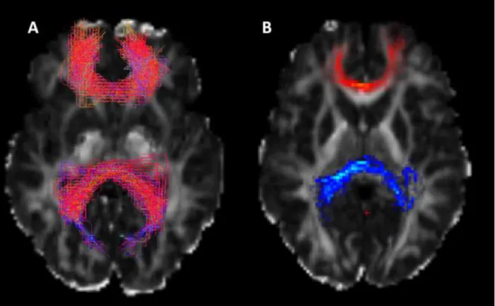
Figure 2.2: Tract-based analysis of the relationship between fractional anisotropy (FA) and concussion history and playing position... 27
Figure 2.3: Comparison of BOLD response to a relational memory task in former NFL players with variable concussion history... 31
Figure 3.1: Summary of recruitment outcomes ... 35
Figure 3.2: Representative example of FreeSurfer surface reconstruction and segmentation ... 42
Figure 3.3: Subject with global atrophy whose data was discarded from imaging analyses ... 43
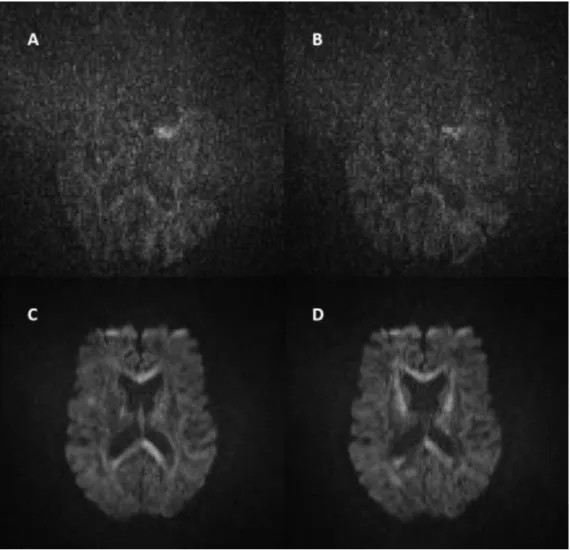
Figure 3.4: Raw diffusion images from a subject with poor signal-to-noise ... 47
Figure 3.5: Example of crossing fiber modelling using BEDPOSTx ... 49
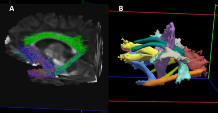
Figure 3.6: Example of reconstructed white matter pathways in TRACULA ... 50
Figure 3.7: Example of streamlines with underlying probability function associated with path reconstruction ... 51
Figure 3.8: DTI and NODDI images from a single subject. A) Color FA map with color-labeled orientation directions, B) Fractional Anisotropy, C) Orientation Dispersion Index, D) Intracellular Volume Fraction, and E) Isotropic volume fraction ... 53
Figure 3.9: Example of functional registration to FreeSurfer reconstructed white matter surface ... 57
Figure 4.1: Mean weighted fractional anisotropy in the left uncinate fasciculus comparing Asymptomatic and Impaired ... 80
Figure 4.2: Mean weighted fractional anisotropy in the right uncinate fasciculus comparing Asymptomatic and Impaired ...81
Figure 4.3: Mean weighted fractional anisotropy in the left uncinate fasciculus comparing Asymptomatic, MCI, and MCI+MBI ... 82
xiii
Figure 4.5: Weighted fractional anisotropy in the forceps minor plotted
against age for all subjects ... 84 Figure 4.7: Mean task accuracy across n-back levels plotted separately
for each distractor condition comparing Normal(Asymptomatic) and
Impaired (MCI or MBI). Standard error bars shown ... 105 Figure 4.8: Mean task accuracy across n-back levels plotted separately
for each distractor condition comparing Normal(Asymptomatic), MCI and MCI+MBI. Standard error bars shown. Note: all subjects in the MCI group
completed the 0-back, blank condition with 100% accuracy ... 106 Figure 4.9: Mean task accuracy across distractor conditions is plotted
separately for each n-back level comparing Normal(Asymptomatic) and
Impaired (MCI or MBI). Standard error bars shown ... 107 Figure 4.10: Mean task accuracy across distractor conditions is plotted
separately for each n-back level comparing Normal(Asymptomatic), MCI and MCI+MBI. Standard error bars shown. Note: all subjects in
the MCI group completed the 0-back, blank condition with 100% accuracy ... 108 Figure 4.11: Corrected statistical parametric maps showing clusters that
are significantly activated (red-yellow) or deactivated (blue-light blue)
in response to the task for the cohort as a whole. Cluster-wise P<0.05 ... 112 Figure 4.12: A) Cluster of voxels in the left precuneus with significantly
different activation between the groups (cluster-wise P<0.05). B) Mean percent signal change was extracted from voxels within the cluster and
plotted by group ... 113 Figure 4.13: A) Significant clusters in the 2-back condition, Fearful > Blank
contrast omnibus F-test of difference between impairment groups. B) Mean BOLD-PSC from voxels within the two clusters in the superior frontal lobe,
and C) Mean BOLD-PSC from voxels in the temporal pole cluster... 114 Figure 4.14: BOLD-PSC from the voxel clusters in the superior frontal region
plotted against total Beck depression inventory score... 115 Figure 4.15: BOLD-PSC from the voxel cluster in the temporal pole plotted
xiv
LIST OF ABBREVIATIONS
fMRI Functional magnetic resonance imaging
CTE Chronic traumatic encephalopathy
MCI Mild cognitive impairment
NFL National Football League
DWI Diffusion weighted imaging
DTI Diffusion tensor imaging
AD Alzheimer’s disease
NPI-Q Neuropsychiatric inventory – questionnaire
mTICS Modified telephone interview of cognitive status
CDR Clinical dementia rating
1
CHAPTER 1: INTRODUCTION Overview and Background
This project concerns the association between large-volume exposure to concussive and subconcussive head impacts and late-life, neurological health of former professional football players. The overarching goal of this project is to identify and characterize early cognitive and behavioral impairments in former professional football players and to better understand the neural underpinning of such impairments. In this thesis, I introduce these clinical problems and discuss their public health significance. In doing so, relevant observational and epidemiological evidence are presented along with current gaps in our understanding of the relationship
between head trauma and neurological health. This background will provide context to the project. The specific aims will then be presented with the underlying hypotheses that motivate their focus. Finally, this chapter concludes with an abbreviated discussion of core concepts integral to the project; this discussion is furthered in the review of literature constituting Chapter Two.
While relatively few studies have examined the long-term effects of recurrent, sport-related concussion, several trends have emerged, including: increased risk of depression,1 cognitive impairment,2,3 Alzheimer’s disease (AD) and other causes of dementia,4 and
2
Postmortem brain histopathology studies of former contact athletes and former military veterans, populations with well-described exposure to head trauma, have revealed a consistent pattern of neurodegeneration referred to as chronic traumatic encephalopathy (CTE).6,7 The clinical presentation of CTE is believed to consist of behavioral disturbances, primarily depression, aggression and agitation, anxiety, and cognitive impairments, such as memory problems and executive dysfunction.8 Additionally, Parkinsonism has also been noted as feature of the disease.6,9,10 In light of these findings, and despite some overlap of symptoms, CTE is believed to be neuropathologically and clinically distinct from other forms of dementia, particularly AD.11-14 However, in vivo evidence of CTE is limited and there is contention concerning its status as a distinct clinical syndrome as no diagnostic criteria have been
established. Consequently, there is an absence of an evidence-based framework for diagnosis and prognosis of CTE. Nevertheless, striking neuropathological studies of CTE make urgent the need to better characterize CTE in vivo.
This study examines a group at high risk of CTE, former professional football players aged 56 to 76 with nine or more years of football experience and a history of recurrent
3
On the other hand, if we fail to support our hypotheses, we will still derive useful information from this work. Failing to reject the null hypotheses may suggest that we cannot detect subtle deficits in the early stages of disease using the methodology within this study. The variability between subjects may be too great to discern smaller effect sizes between the groups in our modest sample size. In either case, we will have generated a rich and complex dataset that will allow us to examine the relationships between multiple imaging modalities, measures of postural control, and a thorough complement of neuropsychological data.
The base population from which we will draw our clinical sample is former professional football players with at least three years of experience at the professional level and who report experiencing at least three concussions in their lifetime. Cases must have a clinical dementia rating (CDR) of 0.5, suggestive of mild cognitive impairment, while controls must be free of cognitive impairments. By holding head impact exposure constant, we will be able to discern neuropsychiatric and neurophysiological differences between the former athletes who appear to be developing underlying neurodegenerative disease and those who are not. Using a focused battery of neuropsychological tests and psychiatric symptom surveys, we will further
characterize the cognitive and behavioral function of the groups. Using diffusion-weighted and functional imaging, we will probe the underlying neural substrate of these domains of function.
Based on previous CTE literature and observational studies of former athletes with recurrent concussion, we hypothesize that in comparison to non-athletes, the former
4 Specific Aims and Hypotheses
Aim 1: To specify differences in cognitive and neuropsychiatric function between former athletes
with and without cognitive impairments.
Hypothesis: Previous investigations on athletes with recurrent concussion converge on marked limbic dysfunction as a feature of decline unique to those with repetitive head trauma. We predict that the former professional football players with mild cognitive impairment (MCI) will have greater behavioral and psychiatric symptomatology than former NFL players without MCI. Specifically, we expect the MCI group to have greater symptoms related to anxiety, depression, and impulsive aggression.
Aim 2: To specify differences in white matter integrity of major white matter tracts within the
frontolimbic network between former athletes with and without cognitive impairments.
Hypothesis: Trauma caused by recurrent concussion reduces the white matter integrity of association fibers within the frontolimbic neural network. Previous studies have shown a reduction in integrity within the uncinate fasciculus and cingulate bundle in those with a history of recurrent concussion compared to healthy controls. Accordingly, we expect the impaired group will have reduced fractional anisotropy within these tracts. We also expect mean diffusivity and orientation dispersion index of these tracts will be increased with a corresponding decrease in the intracellular volume.
Aim 3: To specify differences in functional neural recruitment in response to a working memory,
emotional faces distractor N-back task between former athletes with and without cognitive
impairments.
5
worse performance on the N-back task when facial distractors are present, and that they will show an increased signal in task irrelevant regions (dedifferentiation) when the cognitive load of the task is maximal.
Clinical Application and Limitations
Mounting evidence suggests recurrent concussions are associated with CTE
neurodegeneration. However, specific criteria of a clinical syndrome associated with underlying CTE have not been elucidated, in large part due to a lack of in vivo studies. Informant interview studies of autopsy confirmed CTE in former professional football players suggest a clinical syndrome consisting of multi-domain cognitive impairments and behavioral disturbances. Interestingly, literature concerning late life cognition in those with a history of moderate or severe TBI suggest these injuries are risk factors for Alzheimer’s disease (AD) and that those with AD and a prior history of moderate or severe TBI are more likely to have higher scores on the Neuropsychiatric Inventory – Questionnaire (NPIQ).15 Specifically, they are more likely to report depression, aggression, and irritability and they have a lower odds ratio of reporting issues with memory, the most commonly and severely affected cognitive domain in typical AD. This may be taken as evidence that these patients were possibly misdiagnosed; erroneous diagnoses of AD have been reported previously for cases of confirmed CTE.6 Regardless of its clinical label, the pattern of cognitive impairments in those with a history of repetitive head trauma may not follow the typical progression of AD in which episodic memory is the prominent cognitive impairment.
6
possible that patients would be able to retain independence for a longer period of time. Identifying early cognitive and behavioral changes in those with a history of concussive and subconcussive impact exposure may identify those who are most likely to go on to have progressive neurodegeneration.
Beyond differences in cognitive functioning, we believe there will be important
differences in neuropsychiatric status between our impaired and asymptomatic subjects, and that these symptoms will be related to changes in the frontolimbic network caused by repetitive stress to the neurons composing the network. Thus, the current model of MCI as a clinically observable prodrome for a broad range of distinct neurodegenerative diseases is non-specific and poorly constructed for non-AD related decline. Behavioral domains are not currently
covered by the criteria of MCI, which reduces the clinical utility of such a label in the former NFL player population. The recent construct of mild behavioral impairment (MBI), may distinguish typical AD from other types of neurodegeneration (namely, frontotemporal lobar degeneration), and may be more appropriate in the context of CTE. In such cases, cognitive function may not be the primary manifestation, but rather behavioral impairments. This aligns with the postulation of Stern et al., who suggests two variants of CTE, one of which is more behaviorally dominant while the other presents with largely cognitive impairments.8,14 By studying those with a high exposure to head impacts (both concussive and subconcussive), we can begin to appreciate the unique symptoms and characteristics of early decline in those believed to be at highest risk of progressing to a clinical syndrome corresponding to an underlying CTE neuropathology.
It is unknown if repetitive head trauma is sufficient for the development of this phenotype of cognitive decline, or if such trauma only modifies the expression of an underlying
neurodegenerative process through damage to specific neural networks. This question of
7
history of concussions and subconcussive impact exposure.
Furthermore, this work does not address the underlying neuropathology of CTE.
Considerable strides have been made in characterizing the patterns of tau neurofibrillary tangle (NFT) deposition in those with autopsy-confirmed CTE. The neuropathological consensus criteria are adequately specific to distinguish CTE from other forms to neurodegeneration, though there is overlap between the diseases, and the presence of one does not exclude the presence of the others. In a subset of our study sample, we have augmented our imaging protocol with positron emission tomography with the ligand [18F]-THK-5351, which binds to intracellular tau aggregates. This may provide complementary evidence for the presence of CTE pathology in the early stages of cognitive decline. These data are outside the scope of this project as funding limits our ability to get a PET scan for all subjects. In the absence of PET imaging, we will be unable to determine the tau deposition burden in our subjects.
8
open to another insult that may again go unresolved. Quantifying the clinical course following each reported concussion is subject to recall bias and poor recognition of concussion symptoms in the past.
Because of the variety of concussion mechanisms and our inability to access information about the specific mechanism and clinical course for each self-reported concussion, we will only be able to discern the common elements of concussions. In other words, the specific character of exposure to concussive and subconcussive impacts may add significant heterogeneity to the imaging results. However, the frontolimbic regions and the white matter tracts connecting them are ventral, caudal, and near midline, making this network susceptible to shear forces in a variety of concussive mechanisms. Thus, we expect the heterogeneity of imaging findings to be minimal within the network of interest. This hypothesis predicts the symptom profile with respect to neuropsychiatric functioning will also be common in the group with impairments, though it is acknowledged this assumption may not be correct.
9
CHAPTER 2: LITERATURE REVIEW Sport-related Concussions
An estimated 1.6 to 3.8 million sport-related traumatic brain injuries (TBIs) occur in the United States annually,16 with an estimated 10 million all-cause TBIs occurring globally each year.17 Concussions, or mild TBIs, are the most common form, accounting for an estimated 80% of all severities of TBI. At least 20% of emergency department visits for concussions with loss of consciousness are attributed to sports and recreational activities.18 Organized team sports account for approximately half of emergency department visits for concussion in the 14-18 year old age range.19
Concussion is a concern at every competitive level in contact sports.20-23 Of particular concern is American football, which has one of the highest incidences of sport-related
concussion across all levels of competition.24-26 More than 7 million U.S. high school students compete in interscholastic sports every year27 with over one million high school athletes playing football.28 Furthermore, over 250,000 children ages 5-14 participate in Pop Warner football every year.
In addition to athletics, concussions are a concern for military service-members as well; as many as 20% of Iraq War veterans suffered a mild TBI while deployed.29 TBI has been described as the “signature injury” of the Iraq and Afghanistan Wars, partly due to the emphasis on explosive devices in modern combat. Blast mechanisms of concussive injury are
10
effects of multiple concussions, as will be discussed in the next subsection.
While great strides have been made in recognizing concussion symptoms and promoting proper management, as many as 50% of sport-related concussions are believed to be
unreported.30 The factors contributing to the underreporting of concussive injury are
multifactorial and include gaps in knowledge concerning concussion symptoms, beliefs that the injury is not serious, and unwillingness to be removed from competition.30,31 A study of
confidential symptom reporting by concussed collegiate athletes found that they were more forthcoming in reporting symptoms when it would not be considered in return-to-play decisions.32 These lines of converging evidence underscore the unwillingness of athletes to report their injuries and their symptoms, particularly when it affects their ability to continue playing their sport, and thus, determining the true incidence of concussive injury is difficult. This is likely especially true in the military population, though it is a fundamentally difficult problem to assess.
Recurrent Concussions
Athletes with a history of concussion are at increased risk of additional concussions in the future.33-35 In the National Football League, 29.4% of athletes had a repeat concussion in the period of 1996-2007 and 51% of concussions during this period were repeat concussions. This rate did not decrease in the period from 2002-2007, despite more conservative management practices.36 In a 2007 study of 2,552 former NFL players, over 60% reported one or more concussions in their playing career with 24% reporting three or more concussions.1 As more information is disseminated through the lay press about the signs and symptoms of concussion, the self-reported number of concussions in this population is expected to increase as more players become aware that they likely failed to recognize concussions they experienced.
11
largely unknown. Athletes tend to report a greater number of symptoms after repeat injury 37 and a history of multiple previous concussions is associated with prolonged recovery. 38 High school athletes with three or more concussions tend to develop more acute symptoms upon
subsequent injury. 39 These findings suggest that there is a cumulative effect of recurrent concussions in the acute setting. These findings have been a focus in the military population as well, with increased concussive symptoms40 and sleep disturbances 41 being associated with a history of repeated mild TBI.
Long-term effects of recurrent concussions in former professional football players have been shown to include increased risk of depression and cognitive impairment (figure 2.11,2). Athletes reporting three or more concussions had five times the prevalence of clinically-diagnosed mild cognitive impairment (MCI) and three times the prevalence of significant memory problems, compared to players reporting no concussions.2 Furthermore, in the former NFL player population, there is evidence of increased prevalence of mild cognitive impairment, 42 dementia from any cause,4 and possibly earlier onset Alzheimer’s disease.2 The overall age-adjusted prevalence ratio for AD was 1.37 (95% CI: 0.98 — 1.56), indicating the football retirees tended to have higher prevalence than other U.S. males of the same age.2 Some have argued, however, that AD may be a misdiagnosis of those who actually have neuropathological
evidence of CTE. In addition, the 9-year risk of depression increases with more self-reported concussions (risk ratio of 2.2 – 5.8 depending on number of concussions reported) 43 and athletes with three or more concussions have a three times greater prevalence of depression than those without history of concussion.1
12
13 Neurodegeneration
While a single moderate or severe traumatic brain injury (TBI) is a strong risk factor for developing Alzheimer’s disease (AD), 44 the connection between a single concussion and late-life cognition is less clear. In the case of recurrent concussions, however, there is mounting evidence to suggest the injuries have a cumulative and lasting effect. Chronic traumatic encephalopathy (CTE) has been posited as a pathologically and clinically distinct neurodegenerative disease secondary to repetitive head trauma.
The mechanism underlying the link between recurrent concussion and the development of cognitive and psychiatric symptoms later in life is understudied. An association between repetitive head trauma and Chronic Traumatic Encephalopathy (CTE) has been proposed.6,8,10,45 CTE is described as a neurodegenerative disease, distinguishable from Alzheimer’s disease and other forms of dementia on the basis of post-mortem neuropathology. Histological studies on the brains of deceased contact athletes and military veterans have shown a distinct pattern of hyper-phosphorylated tau deposition.11-13,45,46
There is controversy regarding CTE’s classification as a distinct neuropathology secondary to head trauma.3,47-49 Thus far, in vivo evidence of CTE is lacking, and no clear clinical syndrome has been identified and studied prospectively. The lack of diagnostic criteria is a major criticism of the classification of CTE as a distinct neurological disease. Furthermore, there is contention over the neuropathological findings associated with the CTE phenotype. Tau and amyloid deposition has been observed in several neurodegenerative disease states,50 and has even been observed in subjects who had no previous history of cognitive or behavioral impairments.51,52
In 2015, The National Institutes of Health convened a consensus conference to define neuropathological criteria for the diagnosis of CTE.53 This consensus laid out the
14
diagnosing CTE. The primary feature was noted to be “abnormal perivascular accumulation of tau in neurons, astrocytes, and cell processes in an irregular pattern at the depths of the cortical
sulci.” Spatially, it was noted that tau distribution was variable, but that hippocampal and
neocortical involvement were common. However, involvement in CA1 of the hippocampus, in association with co-localized amyloid plaques, was more likely to be found in Alzheimer’s disease. Furthermore, the depth of tau deposition within the cytoarchitecture of the cortex is variable between AD and CTE. NFTs in CTE tend to be found in the deeper layers, whereas in AD, the deposits are more superficial.54
The clinical presentation of CTE is poorly understood. There have been attempts to describe the phenotype related to confirmed cases of CTE. Interviews with family members and other close informants of deceased athletes with confirmed CTE post mortem reveal a
syndrome comprising memory problems, executive dysfunction, behavioral and personality changes, depression, and aggression.8 Preliminary results of a recent studies demonstrate the presence of tau deposits in symptomatic former football players distributed in a pattern
consistent with previous autopsy studies.55-57 Specifically, in these studies, tau ligand signal was localized to frontolimbic regions, which underlies the clinical expression of behavioral
disturbance. Several lines of investigation provide converging evidence that the nature of neurodegenerative disease is unique in those with a history of concussive and subconcussive exposure. Thus, it is likely that the expressions of early, prodromal stages of the
15 Aim 1: Neuropsychiatric Symptomatology
A major focus of the neuropsychiatric domain of cognitive function in this study will be on impulsive aggression, anxiety, and depression. These symptoms are often present in those with confirmed CTE on autopsy through post-mortem informant interviews.8,9,58 While the interviews are likely biased by recall, especially in the case where the cause of death was a suicide, there remains a concern over the function of the frontolimbic neural network in those with a history of head trauma. In this study, these symptoms of frontolimbic dysfunction will be defined
dimensionally rather than categorically. The symptomatology in the subjects may not warrant a clinical label of, say, generalized anxiety disorder or unipolar depression (or whatever labels correspond to the categorical classification system one is using), but it is affecting their lives enough to report it on a survey or in an interview. Rather than detecting the presence or
absence of a categorical label, we will seek to quantify the three chosen dimensions of emotion, while using categorical labels for their overall cognitive and behavioral function. In the MCI population as a whole (i.e. not restricting to MCI-AD), the presence of one or more of apathy, aggression, irritability, anxiety, or depressive symptoms is approximately 50% prevalent.59 However, much of the previous literature has used a categorical approach, as in report of the symptoms or not, as reflected herein.
The selection of the specific symptoms of anxiety, depression, and aggression is based on the literature concerning neuropsychiatric outcomes following TBI. Many of these studies examine single episodes of TBI ranging the spectrum from mild to severe. Outcomes following repetitive trauma are far less studied and prone to strong selection biases. It is hypothesized that the outcomes of repetitive concussions/mild TBI are more similar to the moderate or severe TBI forms. However, where concussion and mild TBI literature is available, it is overviewed here.
16
studied phenomenon.60,61 Previous concussion has been shown to be a strong risk factor for depression in adolescents.62 The incidence rate of depression after TBI is between 15.3 to 33% and the prevalence ranges of 18.5 to 61%.63 These wide variations reflect considerable
heterogeneity in definitions, length of follow-up, and diagnostic criteria and instruments used in the studies included in the systematic review. Concussed athletes are more likely to report depressive symptoms following their injury, even after being returned to play.64 The relationship between TBI and depression is evident not only in the acute setting post-injury, but also years later. In former NFL players, the 9-year risk of depression increased with more self-reported concussions (risk ratio of 2.2 -- 5.8 depending on number of concussions reported)43 and athletes with three or more concussions have a three times greater prevalence of depression than those without history of concussion.1 Anxiety symptoms are rarely studied in the absence of depression as these are so often comorbid. However, one meta-analysis of 41 studies in adult, non-penetrating TBI observed 11% were diagnosed with generalized anxiety disorder (GAD) and 37% reported clinically significant levels of anxiety.65 The rates of GAD increased with injury severity (11% for mild and 15% for severe) and the diagnoses most prevalent 2-5 years after injury.
The construct of aggression is complex and heterogeneous; the variant under study is that of an impulsive, violent response to a perceived provocation, particularly with anger or hostile affect.66 This is often called impulsive aggression, to distinguish it from the more
premeditated, goal-oriented form associated with borderline or antisocial personality disorders. Here we will consider the term “aggression” to mean the impulsive variant. Aggression is
commonly observed following TBI with as many as 25% of those with moderate to severe injury being classified as aggressive following injury.67 Depending on definitions and study
17
less research specific to mild TBI and the development of aggression.
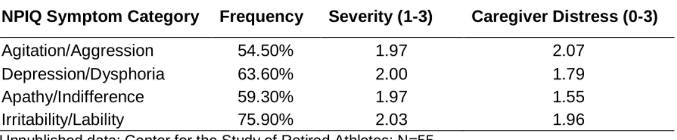
Our preliminary data collected in the Brain and Body Health Program for NFL retirees suggest neuropsychiatric symptoms, as assessed by the NPIQ are highly prevalent in this population (Table 2.1). Data from 55 participants in the program with two or more self-reported, lifetime concussions show elevated distress scores for both the retirees and caregivers. This population is a mix of those with cognitive impairments and those who are cognitively normal; we expect the prevalence is greater in those retirees with MCI. The connection between these domains of neuropsychiatric functioning cannot be ignored. The frontolimbic neural network has been associated with each of these symptoms, as will be discussed in the succeeding
subsections.
To assess neuropsychiatric symptomatology in this group, we will use a focused set of easily implement clinical measures. While we expect that mean symptom scores will be elevated in the MCI group, it may be the case where just a subset of those with MCI express neuropsychiatric symptoms. Accordingly, we will conduct a secondary analysis in which we define subjects as being the in “exposed, mild behavioral impairments” (MBI) group using the Neuropsychiatric Inventory Questionnaire (NPIQ). The NPIQ was originally developed and widely used for assessment of psychopathology in dementia. It is now used to investigate neuropsychiatric manifestations of other brain disorders and has demonstrated sensitivity to changes in neuropsychiatric status following a traumatic brain injury. The NPIQ evaluates 12 neuropsychiatric disturbances (apathy, irritability, euphoria, disinhibition, delusions,
hallucinations, agitation, dysphoria, anxiety, aberrant motor behavior, night-time behavior disturbances, and appetite and eating abnormalities). The NPIQ is administered to an informant of the patient, typically a spouse, relative, or caregiver. The NPIQ, however, has not been validated in subjects with remote history of head trauma, and thus, it may not be sensitive to
18
“mild behavioral impairment” differ in their clinical criteria and method of assessment. Our
definition is limited to the cut-offs described in the methodology in Chapter 3. To reduce analytical bias, the analyses presented herein are using the a priori cut-offs. Future directions
19
Table 2.1: Symptoms reported by informants of NFL retirees with 2+ previous concussions
*Unpublished data; Center for the Study of Retired Athletes; N=55
NPIQ Symptom Category Frequency Severity (1-3) Caregiver Distress (0-3)
Agitation/Aggression 54.50% 1.97 2.07
Depression/Dysphoria 63.60% 2.00 1.79
Apathy/Indifference 59.30% 1.97 1.55
20
Depression symptoms were measured using the Beck Depression Inventory (BDI), a 21-question multiple-choice, self-report inventory and one of the most widely used instruments for measuring the severity of depression. Depression is the most commonly associated
neuropsychiatric symptom with aggression at all time-points post-TBI.67 We use the total inventory score to assess depressive symptoms. A suicide action plan is in place if the subject reports thoughts of suicidality. The patient health questionnaire 9-item scale (PHQ-9) was used as a supplement to the BDI as this instrument is rapidly administered and asks questions that are complementary.
Aggression was assessed with the Buss-Perry Aggressiveness Questionnaire (BPAQ). The BPAQ has been found to be valid across a multitude of populations to evaluate
aggressiveness. The long form version has a high score of 60 with 29 total questions. It is capable of breaking aggressive behavior into four subscales, physical aggression (nine items), verbal aggression (five items), anger (seven items), and hostility (eight items). The BPAQ is given as a seven point-Likert scale ranging from “extremely uncharacteristic” to “extremely characteristic.”
Anxiety symptoms were assessed using the Generalized Anxiety Disorder 7-item scale (GAD-7),70 which has been validated in multiple populations in its ability to detect generalized anxiety, panic, social anxiety, and post-traumatic stress disorder (PTSD). This behavioral measure assesses the subject's anxiety levels over the past two weeks using seven questions. Answers range from "not at all" to "nearly every day" with total scores ranging from 0-21. The accepted cut-point for generalized anxiety is a score of 10. Alternate cut-points are 0-4 (minimal), 5-9 (mild), 10-14 (moderate), and 15-21 (severe).
21
between CTE and suicide are fallacious of the form “post hoc, ergo propter hoc (after TBI, therefore because of TBI) or cum hoc, ergo propter hoc (with TBI, therefore because of TBI).” 71 Suicidality was not noted to be part of the phenotype by McKee et al. in their review of all CTE cases up to 2009. 6 Despite the fallacious treatment of suicidality and suicidal ideation as part of the clinical syndrome, these problems remain of grave concern in those status post TBI. There is evidence to suggest TBI is a risk factor for later life suicide and that, alarmingly, there is no time period within 15 years post-injury in which the suicides tend to occur; some suicides occurred over 25 years post injury.72
Severe TBI appears to have a greater risk ratio than mild TBI, though this is clouded by the vague definitions common in mild TBI literature. It is often that such injuries are attributed to non-injury related factors, not the injury per se, and that this attribution to the injury is more common in severe TBI.73 In this same publication, Oquedo et al. noted that those with mild TBI were more likely to be aggressive than non-mild TBI subjects, that they were more likely to be suicide attempters, and that suicide attempts were most predicted by aggression and hostility, not mild TBI status. They concluded that suicide and mild TBI share antecedent risk factors of aggression and hostility, though this was a cross-sectional design with retrospective recall of psychiatric traits, and thus, strictly delimited in its causal inference. Depression remains the single greatest risk factor for suicide74 and there is considerable overlap between suicide risk factors and post-TBI sequelae, including depression.
The Frontolimbic Neural Network
22
the cingulate gyrus ending and the angular bundle) and uncinate fasciculus. This network and regions comprising the frontolimbic network have been shown to be dysfunctional after
concussive injury,60,75,76 particularly in patients who go on to have symptoms of anxiety. The frontolimbic network has yet to be explored in a population with recurrent concussion and long duration exposure to head impacts.
The regions composing the frontolimbic network are associated with a wide variety of emotional and cognitive functions. The amygdala plays a central role in the experience and generation of negative emotions.77-79 The balance of connectivity between the frontal and limbic regions is critical for regulating response to emotional stimuli.80 The associated regions in the frontal cortex appear to inhibit or suppression an emotional reaction to stimuli.81 The activity of the amygdala and ventromedial prefrontal cortex are inversely correlated during regulation of negative emotions.82 In addition to mediating a psychological response to emotion, the network has a close physiological connection to the hypothalamic-pituitary-adrenal axis.82,83 Young adults with a family history of depression showed lower activation of the dorsolateral prefrontal cortex in response to the Hariri task involving presentation of emotional faces (see succeeding section for more information on this task).84
Countless studies have been conducted on the regions that are comprised by the
frontolimbic network, particularly in relation to emotional processing, cognitive function, and TBI; very few examine the interaction of these three factors. Mayberg 1999, proposed the limbic – cortical network connections were critical in mediating symptoms of major depression.85 In fact, this hypothesis has spawned several clinical trials examining the efficacy of transcranial
magnetic or deep-brain stimulation as a treatment for depression.86 The orbitofrontal cortex has been identified as a key region in the expression of anxiety, spanning several distinct subtypes of anxiety disorders.87,88 Lastly, aggression and violence are believed to be caused by
23 prefrontal cortex.66
Two key studies have direct relevance to the proposed project; one was conducted in the population of interest, former athletes with history of concussion, the other in those with typical MCI leading to AD. Goswami et al. 2015, in a hypothesis-naïve machine learning study, observed that axial diffusivity of the uncinate fasciculus and thickness of the OFC negatively correlated with scores of aggression in athletes with a history of concussion.89 In Trzepacz et al. 2013, volume and thickness of several regions within the frontolimbic network (amygdala, anterior cingulate, orbitofrontal, among others) were shown to correlate with NPI-Q scores of agitation and aggression in those with early Alzheimer’s disease.90
24
a task-based fMRI paradigm, we can assess the functional communication between frontal and limbic regions. The specific task employed will be described in the succeeding section. The involvement of the frontolimbic network in early cognitive decline due to CTE may be a target for the development of tailored therapeutic drugs. Furthermore, the extent of disease involvement in the frontolimbic network may be a marker for the progression of CTE.
Neuroimaging
Neural network dysfunction is a hallmark of TBI; this is true even for mild TBI in which there is no focal damage to neurological structures apparent on cranial tomography or MRI.94 The confluence of shear, tensile, and compressive forces on neuronal cell bodies and their axons causes microscopic, diffuse axonal injury and acute impairment of synchronous neuronal firing.95 Thus, the nature of network dysfunction is apparent not only structurally, but functionally as well. Following acute injury, the extent and timeline of recovery for the neural networks affected by the injury are poorly understood. Some evidence suggests persistent changes to neurophysiology and neuroanatomy may be noticeable years after injury. Acute damage to microtubules from the acute injury results in hyper-phosphorylation of tau with subsequent aggregation within the neurons. Such damage may serve as a nidus for continued deposition of NFTs, becoming sufficiently extensive to impact neuronal function. Some posit that these NFTs may even be passed trans-synaptically to other neurons within the network.94
Because the nature of network dysfunction in TBI is believed to be structural, functional, and pathological, a multi-modal approach to neuroimaging is appropriate to best elucidate the neural mechanisms of CTE neurodegeneration. Accordingly, we will collect a variety of
25
as funds allow, we will also simultaneously collect PET data using a ligand specific to tau NFTs, [18F]THK-5351. In the next subsections, I will provide a justification for the inclusion of such modalities, with emphasis on diffusion tensor imaging (DTI) and functional MRI data collected by our research group. As the complexity of the data is such that analysis is likely to extend well beyond my tenure with the Matthew Gfeller Center, the focus of aims 2 and 3 is on the
hypothesis of the frontolimbic network dysfunction.
Aim 2: Diffusion-Weighted Imaging
Diffusion-weighted imaging exploits the magnetic properties of water diffusion and the differential diffusion properties of neurological tissues. In bundled, well-organized white matter, diffusion is more ordered and predictable; more net diffusion occurs along the length of axons compared to along the transverse plane of the axon. When damage occurs to white matter, the diffusion-weighted signal is often altered. In TBI, damage to white matter can affect the
connections between cortical and subcortical regions in a differential manner depending on the mechanism of the injury. However, there are consistencies in the literature regarding the location of damage in groups with variable mechanisms of injury. Specifically, the corpus callosum has been identified as the most commonly reported major white matter tract affected by TBI.96 The fornix, uncinate fasciculus, cingulum bundle, and hippocampus have all been observed to have lower integrity measures after TBI using DTI.97
26
Interestingly, the forceps minor was correlated to depression scores.99 In their asymptomatic collegiate athletes, Tremblay et al. did not observe significant clinical correlates to FA, however, they did observe significant relationships between a test of visual memory and other diffusivity metrics, including axial, radial, and mean diffusivity.100
27
28
While DTI can provide valuable information regarding the strength and integrity of white matter connections, it cannot provide information on the function of the gray matter regions that are connected by the white matter tracts. The function of the neurons composing a neural network gives rise to cognitive processes and behavior. In some cases, such cognitive functions and behaviors can be observed outwardly and are what constitute the phenotype of TBI and neurodegenerative disease. This is the grounding motivation for including fMRI in this project; that the neural substrate of the clinical phenotype of CTE may be examined using fMRI. To understand the nature of the fMRI signal and how it can be used in this project, an overview of key concepts in MRI are needed.
Aim 3: Working Memory N-back with Emotional Face Distractors fMRI Paradigm Functional MRI relies on changes in MR signal produced by the change in the
oxygenation of hemoglobin; this is called the blood oxygenation level dependent (BOLD) signal. Because the brain does not have a significant source of stored energy, neurons rely on a constant supply of oxygen and glucose. Without discussing action potentials in depth, the major rectifying ion pump is the Na+/K+ pump, which requires adenosine triphosphate (ATP, the major energy currency of cells). When action potentials occur in a brain region, there is a rise in demand for ATP, which is largely produced through glycolysis, citric acid cycle, and electron transport chain. These processes require oxygen and glucose; the former is carried by hemoglobin in red blood cells. Because of the increased demand for ATP, and therefore, oxygen and glucose, there is a need for increased blood flow to the region.
How the brain regulates blood flow in response to neuronal activity is not fully
29
mechanism, there is a well-documented phenomenon of increased blood flow to areas of synaptic activity.
Functional MRI gives information about neuronal activity indirectly through the BOLD response. This change in MR signal is due to the ratio of diamagnetic oxyhemoglobin to paramagnetic deoxyhemoglobin; the former increases signal, while the latter decreases signal. The BOLD signal is affected by changes in cerebral blood flow, cerebral blood volume, and/or the cerebral metabolic rate of oxygen consumption (CMRO2). When neurons fire, there is an initial spike in CMRO2, leading to a rise in deoxyhemoglobin. As this occurs, blood vessels dilate in response to neuronal activity through the release of vasoactive mediators, as discussed in the previous session. The subsequent increase in CBF far exceeds the rise in CMRO2 and the net result is an increase in the ratio of oxyhemoglobin to deoxyhemoglobin. The resultant BOLD signal is the “activation” seen in fMRI.
The advantages of fMRI lie in its spatial resolution (on the order of millimeters), temporal resolution (on the order of seconds; physiological change is rate-limiting), non-invasiveness, and signal-to-noise ratio. These qualities give fMRI excellent functional resolution, or the ability to map physiological variation to mental processes or behavior.
Our group has shown that in an fMRI-based working memory task (without emotionally valent content), former athletes with ≥3 previous concussions had broader functional
recruitment of neural circuits than those with 0-1 previous concussions. Despite equivalent performance on the task, the high concussion group (3+) required activation of several
additional cognitive resources than the group with 0-1 previous concussions, indicating neural compensation. Furthermore, there were differences in intact white-matter fibers between several regions of interest across both groups, appreciable with DTI, including several within the
30
Interestingly, differences in white matter tracts within the limbic system were noted, though this was incidental to the fMRI task as it was not intended to activate limbic regions. These results complement those of another previous study by our group examining players in the same age range who reported subjective recent memory problems, but did not satisfy criteria of mild cognitive impairment. In this study, players with ≥3 previous concussions
31
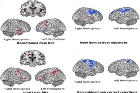
Figure 2.3: Comparison of BOLD response to a relational memory task in former NFL players with variable concussion history.Regions of activation (at p<0.005, k≥10) preferentially recruited by persons with increased
32
The paradigm used in the task-based fMRI in this study was an emotional working memory task previously published by Ladouceur et al.102,103 It is designed to “examine attentional control processes involved in resisting interference from emotionally salient distracters while performing a visual N-back task.”103. This task has not been used to study
former athletes, but it is an appropriate task for this population given the hypothesis of frontolimbic involvement in CTE. Ladouceur et al. observed that young (<18yo) offspring of parents with bipolar disorder had greater right ventrolateral prefrontal cortex activation that healthy controls when facial distractors were present.108 In another study, they observed that the presentation of fearful faces reduced task performance in children and adolescents with anxiety, suggesting a diminished ability to resist interference from threat-related stimuli as attentional resources are needing to be directed towards task completion (during the 2-back condition).102 Finally, neutral faces were found to reduce performance in depressed adolescents;
interesetingly, this reduction in performance was independent of memory load.104
Few studies have examined the frontolimbic network using fMRI in the context of traumatic brain injury. As discussed earlier in this chapter, the frontolimbic network is active during cognitive tasks involving faces expressing emotions and pictures depicting emotionally provocative scenes. Specifically, two major sets of cues have been used in fMRI paradigms previously, the International Affective Picture System (IAPS)105 and emotional faces.106 Hariri et al., 2002 demonstrated that the amygdala tends to be more highly activated in response to emotional faces than the IAPS set of cues.107 Moreover, the amygdala is thought to play a role in providing emotional bias signals in facial processing.108 This suggests that the amygdala is partly responsible for the interpretation of the emotional valence and content for facial features. In particular, fearful faces have been shown to be more activating than angry faces.109
33
study. In Aim 2 we will use an fMRI task of working memory with emotionally salient content. The design of the paradigm will follow Ladouceur et al, 2009 and 2013102,103 and is explained in detail in Chapter 3. This paradigm enables examination of the frontoparietal and frontolimbic networks, two distinct neural networks involved in attentional control, working memory, and emotional processing. The task requires subjects to engage prefrontal regions underlying executive control to maintain attention on the task in the presence of emotional distractors. We postulate that recurrent concussions and repetitive head impacts result in frontolimbic
neurodegeneration which will impair attentional control. We expect the frontoparietal network, which is activated in working memory tasks,110-112 to be similar between groups when distractors are not present, but that there will be greater activation of task irrelevant regions will in the impaired group in the presence of the distractors, reflecting a lack of inhibitory control of the limbic system by the prefrontal cortical areas.
Summary
34
CHAPTER 3: METHODS Overview
The purpose of this case-control study was to examine early cognitive and behavioral
impairments in former professional football players with a history of recurrent concussion. This
group has been shown to be at higher risk than the general population for depression, mild
cognitive impairment, and CTE. In vivo evidence of the onset and progression of CTE is lacking.
Identifying objective measures of impairment in the critical prodromal period of MCI or MBI (or
both) will inform diagnostic and prognostic criteria for the clinical assessment of such early
impairments.
Recruitment
From January 2016 to June 2017 we recruited a sample of 35 former NFL players from a registry maintained by the Center for the Study of Retired Athletes (CSRA). This registry was initially created for the purposes of the General Health Study, a mailed survey study of former NFL players covering aspects of physical, mental, and psychological health. The total
36 Participants
All participants gave both verbal and written informed consent in accordance with the requirements of the Institutional Review Board. The Modified Telephone Interview of Cognitive Status (mTICS) was administered to all subjects. Twelve subjects with a score of <20 were excluded – this likely represents more than mild impairment. Four subjects either self-reported being diagnosed with dementia or a spouse, caregiver, or guardian reported such diagnosis or equivalent advanced impairment preventing travel (e.g. lives in assisted care home).
Subjects must have played a minimum of three seasons at each of the following levels of football: high school, college, and professional (minimum of nine years of football exposure). Furthermore, they must have reported at least three concussions in their lifetime. A
standardized definition of concussion114 was provided to each subject before asking how many concussions they experienced over the course of their lifetimes. The definition provided was:
A concussion is a blow to the head followed by a variety of symptoms that may include any of the following: headache, dizziness, loss of balance, blurred vision, “seeing stars,” feeling in a fog or slowed down, memory problems, poor concentration, nausea, or throwing-up. Getting “knocked out” or being unconscious does not always occur with a concussion.
In two cases, subjects reported fewer than three concussions, but reported three or more events in which concussion symptoms were experienced (e.g. headache, “feeling not right,” “seeing stars,” etc.), stating that they did not consider these events to be “true”
concussions. Based on the criteria in the provided definition of concussion, such events were included in their total self-report and they were considered eligible for the study.
For all subjects, inclusion criteria were male sex and age between 55 and 80. For all subjects, exclusion criteria include any diagnosis of dementia including probable Alzheimer’s disease, frontotemporal dementia, vascular dementia, dementia with Lewy bodies, or
Creutzfeldt-Jakob disease; any contraindications for magnetic resonance imaging including,
claustrophobia, pacemaker, surgical clips, pins, plates, screws, metal sutures, wire mesh, or
37
severe psychiatric disease such as bipolar or schizophrenia; diagnosis of amyotrophic lateral
sclerosis, multiple sclerosis, or history of a major stroke. In this sequence of telephone
screeners, if any exclusion criteria were violated, the remaining instruments were not
administered. Once inclusion/exclusion criteria were met, enrolled subjects were brought to the University of North Carolina at Chapel Hill for assessment.
Classification of Participants
Classification of impairment was determined during the in-person visit. Subjects were classified as having either mild cognitive impairment (MCI), mild behavioral impairment (MBI), or both, or neither. To assess MCI, a trained research assistant administered the Clinical Dementia Rating (CDR). The CDR is a structured interview with the subject and an informant and covers both subjective and objective assessments of cognition and daily function. The benefit to using the CDR is the qualitative approach to assessing the presence and impact of cognitive
impairments on daily function. The CDR is scored from 0 to 3 with a score of 0.5 representing probable mild impairment and scores of 1-3 representing various stages of dementia. Subjects with a CDR score of 0.5 were classified as having MCI. Two subjects scored a 1 on the CDR and were excluded from the analyses as this reflects greater than mild impairment with subsequent loss of independent function A limitation of the CDR is that it is biased towards detecting functional impairments in those with Alzheimer’s disease. We classified MBI using the NPIQ. Subjects with two or more symptoms of greater than mild severity and which cause more than mild distress in the informant were classified as having MBI.
38
at least 78.6% sensitive and 85.5% specific in identifying MCI from those with asymptomatic cognition.115,116 However, other studies have shown the mTICS to be sensitive to age and educational attainment.117,118 As such, we decided against using a hard cut-off for defining probable memory impairment given the high educational attainment and variable age of our cohort. Instead, we relied on the in-person visit for classification, as described above.
Aim 1: Neuropsychological and Neuropsychiatric Assessment.
39
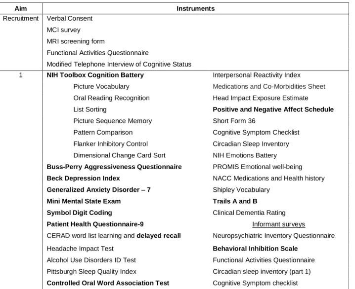
Table 3.1: Overview of instruments and methods. Bolded items are primary outcomes.
Aim Instruments
Recruitment Verbal Consent
MCI survey
MRI screening form
Functional Activities Questionnaire
Modified Telephone Interview of Cognitive Status
1 NIH Toolbox Cognition Battery Interpersonal Reactivity Index
Picture Vocabulary Medications and Co-Morbidities Sheet
Oral Reading Recognition Head Impact Exposure Estimate
List Sorting Positive and Negative Affect Schedule
Picture Sequence Memory Short Form 36
Pattern Comparison Cognitive Symptom Checklist
Flanker Inhibitory Control Circadian Sleep Inventory
Dimensional Change Card Sort NIH Emotions Battery
Buss-Perry Aggressiveness Questionnaire PROMIS Emotional well-being
Beck Depression Index NACC Medications and Health history
Generalized Anxiety Disorder – 7 Shipley Vocabulary
Mini Mental State Exam Trails A and B
Symbol Digit Coding Clinical Dementia Rating
Patient Health Questionnaire-9 Informant surveys
CERAD word list learning and delayed recall Neuropsychiatric Inventory Questionnaire
Headache Impact Test Behavioral Inhibition Scale
Alcohol Use Disorders ID Test Functional Activities Questionnaire
Pittsburgh Sleep Quality Index Circadian sleep inventory (part 1)
40
Aims 2 and 3: Neuroimaging Overview
All imaging was conducted in the Biomedical Research Imaging Center at the University of North Carolina at Chapel Hill on a Siemens Biograph mMR 3T MR-PET scanner. The
following sequences were collected for all subjects: high resolution structural (T1 and T2), diffusion-weighted imaging, resting-state functional, and emotional working memory functional. A subset of subjects was injected with a tau PET ligand and steady state uptake was collected for 30 minutes at the start of the scan while the structural and resting state scans were acquired. A sagittal T1-weighted magnetization prepared rapid gradient echo (MPRAGE) anatomical sequence was acquired with a voxel size of 1x1x1mm3 over 192 slices with TR/TE =
1900ms/2.26ms. This anatomical scan was used for registration of the diffusion-weighted and functional MR volumes. Additionally, a T2-weighted sequence was acquired with the same voxel size and number of slices with TR/TE=3200ms/402ms. This sequence was used to improve the FreeSurfer parcellation and segmentation described below.
The T1- and T2-weighted anatomical DICOM data were converted to NIfTI file format by the MRIcron tool, dcm2nii. These data were processed using the FreeSurfer recon-all
command, with the T2-weighted image used to improve definition of the pial surface. The FreeSurfer segmentation and parcellation processing stream includes the pre-processing steps of removing non-brain voxels (skull-stripping), registration of the T1- and T2-weighted images, cortical surface reconstruction, cortical and subcortical segmentation, and volume, cortical thickness, and surface area estimation.
Quality control of automated segmentation and parcellation was performed though visual inspection of the estimated pial and grey-white matter interface. Representative images of these surfaces are shown in Figure 3.2. Based on previous literature119,120 showing strong agreement between the estimated volumes of hand-drawn regions of interest (traced by trained
41
region-of-interest boundaries that were approximately correct. Instead, images with obviously incorrect boundaries (e.g. a pial surface drawn through a ventricle) were corrected per the FreeSurfer tutorial on quality assurance.
Missing Data
One subject in the MCI group could not complete any imaging sequence due to
42
Figure 3.2: Representative example of FreeSurfer surface reconstruction and
43
44 Table 3.2: Available data by aim.
Aim1 Aim 2 Aim 3
Asymptomatic 15 14 13
Impaired 18 17 15

![Table 4.2: Demographics of sample comparing Asymptomatic, MCI, and MCI+MBI. Mean (SD) [min, max]](https://thumb-us.123doks.com/thumbv2/123dok_us/8327868.2208517/83.1188.92.1103.147.381/table-demographics-sample-comparing-asymptomatic-mci-mci-mean.webp)

