SYMPOSIUM
A Hypothesis for the Composition of the Tardigrade Brain
and its Implications for Panarthropod Brain Evolution
Frank W. Smith,
1,*Paul J. Bartels
†and Bob Goldstein
**Department of Biology, University of North Carolina at Chapel Hill, Chapel Hill, NC 27599, USA; †Department of Biology, Warren Wilson College, PO Box 28815, Asheville, NC 9000, USA
From the symposium “The Evolution of Arthropod Body Plans–Integrating Phylogeny, Fossils and Development” presented at the annual meeting of the Society for Integrative and Comparative Biology, January 4–8, 2017 at New Orleans, Louisiana.
1
E-mail: [email protected]
Synopsis Incredibly disparate brain types are found in Metazoa, which raises the question of how this disparity evolved. Ecdysozoa includes representatives that exhibit ring-like brains—the Cycloneuralia—and representatives that exhibit ganglionic brains—the Panarthropoda (Euarthropoda, Onychophora, and Tardigrada). The evolutionary steps leading to these distinct brain types are unclear. Phylogenomic analyses suggest that the enigmatic Tardigrada is a closely related outgroup of a EuarthropodaþOnychophora clade; as such, the brains of tardigrades may provide insight into the evolution of ecdysozoan brains. Recently, evolutionarily salient questions have arisen regarding the composition of the tardigrade brain. To address these questions, we investigated brain anatomy in four tardigrade species—Hypsibius dujardini, Milnesium n. sp., Echiniscus n. sp., andBatillipes n. sp.—that together span Tardigrada. Our results suggest that general brain morphology is conserved across Tardigrada. Based on our results we present a hypothesis that proposes direct parallels between the tardigrade brain and the segmental trunk ganglia of the tardigrade ventral nervous system. In this hypothesis, brain neuropil nearly circumscribes the tardigrade foregut. We suggest that the tardigrade brain retains aspects of an ancestral cycloneuralian brain, while exhibiting ganglionic structure characteristic of euar-thropods and onychophorans.
Introduction
The evolutionary steps that connect disparate meta-zoan brains remain unclear (Schmidt-Rhaesa 2007; Hejnol and Lowe 2015). Ecdysozoa includes lineages characterized by ring-like brains—the Cycloneuralia (Nematoda, Nematomorpha, Priapulida, Kinorhyncha, and Loricifera)—and lineages character-ized by ganglionic brains—the Panarthropoda (Euarthropoda, Onychophora, and Tardigrada). The cycloneuralian brain typically consists of anterior and posterior clusters of neuronal somata with a ring of circumesophageal neuropil positioned between them (Schmidt-Rhaesa 1997/1998; Richter et al. 2010; Rothe and Schmidt-Rhaesa 2010;Martın-Duran et al. 2016;Henne et al. 2017b; reviewed inHejnol and Lowe 2015; Schafer 2016). The brains characteristic of Nematomorpha have diverged from this architecture (Schmidt-Rhaesa 1996), but retain evidence of
cycloneuralian ancestry (Henne et al. 2017a). Although often referred to as ganglia, the brain clusters of cycloneuralians are not true ganglia, because they do not contain neuropil (Richter et al. 2010;Schafer 2016). In contrast to cycloneuralian brains, panarthropods ex-hibit ganglionic brains (Scholtz and Edgecombe 2006; Mayer 2016; Mayer et al. 2013a). Intriguingly, Panarthropoda appears to be nested within Cycloneuralia (Campbell et al. 2011; Rota-Stabelli et al. 2013;Borner et al. 2014), suggesting that the gan-glionic brains of panarthropods evolved from a ring-like cycloneuralian brain (Hejnol and Lowe 2015).
The relatively simple tardigrade brain may hold im-portant clues regarding the transition from a cycloneura-lian brain to a ganglionic brain in the panarthropod stem lineage. Unlike the brains of euarthropods and onycho-phorans, which are composed of multiple segmental gan-glia (Scholtz and Edgecombe 2006;Strausfeld et al. 2006a;
ßThe Author 2017. Published by Oxford University Press on behalf of the Society for Integrative and Comparative Biology. All rights reserved. For permissions please email: [email protected].
Integrative and Comparative Biology
Integrative and Comparative Biology, pp. 1–14Mayer et al. 2010;Whitington and Mayer 2011;Martin and Mayer 2015), the tardigrade brain is composed of a single segmental ganglion (Hejnol and Schnabel 2005; Gabriel et al. 2007;Mayer et al. 2013a;Gross and Mayer 2015;Smith et al. 2016;Gross et al. 2017; seeZantke et al. 2008 and Persson et al. 2012 for alternative views)—a condition that is thought to be plesiomor-phic for Panarthropoda (reviewed in Strausfeld 2012; Smith and Goldstein 2017; Ortega-Hernandez et al. 2017). Furthermore, Tardigrada is most likely the sister group of a EuarthropodaþOnychophora clade (Campbell et al. 2011;Rota-Stabelli et al. 2013;Dunn et al. 2014; Giribet 2016; for an alternative view, see Borner et al. 2014andLaumer et al. 2015; reviewed in Edgecombe and Giribet, this volume). Under this hy-pothesis, the most recent common ancestor of Panarthropoda exhibited a brain composed of a single segmental ganglion, referred to as the protocerebrum; tardigrades retain a protocerebral brain, while more posterior segmental ganglia were incorporated into the brains of euarthropods and onychophorans after the euarthropodþonychophoran lineage diverged from Tardigrada (Martin and Mayer 2015; reviewed in Strausfeld 2012; Hejnol and Lowe 2015; Scholtz 2016; Strausfeld et al. 2016; Ortega-Hernandez et al. 2017; Smith and Goldstein 2017). Additionally, in terms of its position along the body axis, the entire brain of tardigrades is most likely homologous to the entire brain of non-panarthropod invertebrate animals (Steinmetz et al. 2010;Smith et al. 2016).
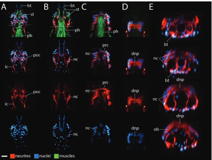
Unlike the circumesophageal brains of cycloneura-lians, the protocerebrum of euarthropods and ony-chophorans is restricted to a supraesophageal position (Eriksson and Budd 2000; Strausfeld et al. 2006a, 2006b; Martin and Mayer 2014; Ortega-Hernandez et al. 2017), which raises the question of when during panarthropod evolution the proto-cerebrum became restricted to a supraesophageal position. The position of the tardigrade brain relative to the foregut clearly has important implications for addressing this question, but recent investigations of tardigrade brain anatomy have led to several oppos-ing views regardoppos-ing the composition and position of the tardigrade brain (Fig. 1A; Zantke et al. 2008; Persson et al. 2012; Mayer et al. 2013a, 2013b; Schulze and Schmidt-Rhaesa 2013; Persson et al. 2014; Schulze et al. 2014; Smith and Jockusch 2014). In one model, tardigrades exhibit a circum-esophageal brain (Zantke et al. 2008); the circum-esophageal component of the tardigrade brain—the dorsal commissure (dco)þthe circumbuccal connec-tives (cco)þthe post-oral commissure (poc)—is suggested to be directly homologous to the ring of neuropil characteristic of cycloneuralian brains
(Fig. 1B, top panel). Tardigrades also exhibit a cir-cumesophageal brain in a second model (Fig. 1B, middle panel), but in this model the tardigrade brain is composed of three paired brain lobes—the outer lobes (ol), inner lobes (il), and ventrolateral lobes (vll)—that are not clearly relatable to cycloneuralian brains (Persson et al. 2012). In a third model, tardi-grades exhibit a supraesophageal brain (Mayer et al. 2013a). This model includes a circumesophageal ner-vous system component—the nerve ring (nr), but the nerve ring is predicted to be part of the stomo-deal nervous system, rather than part of the brain (Fig. 1B, bottom panel).
Distinguishing between different models of tardi-grade brain anatomy is difficult. Different tarditardi-grade species were investigated in different studies (Fig. 1A), raising the possibility that reported differ-ences represent true taxonomic variation. Furthermore, different immunohistochemical and imaging approaches were used in different studies. Both of these factors may have contributed to different results, and ultimately, different conclusions. Here, we investigate brain morphology in four tardigrade spe-cies, using identical immunohistochemical and imag-ing approaches. This includes the first study of a member of the tardigrade lineage Apochela using these techniques. Our results suggest that brain neuropil wraps around the tardigrade foregut. Based on our results, we present a hypothesis for the evolution of the ganglionic brains of panarthropods from an ances-trally cycloneuralian state.
Materials and methods
Specimen collectionSpecimen preparation
We prepared specimens to visualize neurites, nuclei, and muscles. Specimens were stretched in carbonated water. They were then transferred to 4% formaldehyde in 0.5 PB-Triton (0.5 phosphate-buffered saline, 0.1% Triton X-100, pH 7.4). They were stored in this solution at 4C for between 1 day and 1 week. Specimens were then washed five times with 0.5
PB-Triton. To permeabilize specimens, we bisected them transversely with a 25-gauge needle. Specimens were then washed two times for 1 h in 0.2% bovine serum albumin in 0.5X PB-Triton, followed by a 1.5 h wash in 5% normal goal serum (NGS in 0.5 PB-Triton). Specimens were incubated overnight in a 1:100 dilution of a b-tubulin antibody (E7, Developmental Studies Hybridoma Bank), which stains the tardigrade nervous system (Smith and Jockusch 2014). They were then washed three times
for 5 min and two times for 1 h in 0.5 PB-Triton. This was followed by two 1=2 h washes in NGS. Specimens were then incubated overnight at 4C in a
1:200 dilution of a goat anti-mouse Cy3-conjugated secondary antibody (Jackson ImmunoResearch) in NGS. Next, specimens were washed two times for 1 h and three times quickly in 0.5PB-Triton. They were then incubated in a 1:40 dilution of Oregon Green 488 phalloidin (Molecular Probes), which stains tardigrade muscles (Smith and Jockusch 2014), for at least 16 h at 4C. We then washed the specimens three times
quickly in 0.5 PB-Triton. We mounted specimens on slides in DAPI Fluoromount-G (SouthernBiotech) to visualize nuclei.
Imaging and analysis
Z-series of specimens were collected on a Zeiss 710 laser scanning confocal microscope. To help visualize
Fig. 1 Summary of recent studies that investigated tardigrade brain morphology. (A) Diversity of tardigrade species investigated with a combination of immunohistochemistry and laser scanning confocal microscopy. The numbers in parentheses refer to the models in (B) that the results of the studies most closely match, in terms of neuronal connectivity and the regions in the head that were considered to be part of the brain. *The model forEchiniscus testudonervous system anatomy was shown inSchulze et al. (2014), based on data fromSchulze and Schmidt-Rhaesa (2013). The phylogeny is based on Jørgensen et al. (2010),Guil and Giribet (2012), and Bertolani et al. (2014). (B) Models of tardigrade brain anatomy, based onZantke et al. (2008; top panel),Persson et al. (2012; middle panel), and Mayer et al. (2013a; bottom panel). Gray structures are considered to be part of the brain; black structures are not considered to be part of the brain. The dashed line demarcates the boundary between the head and the trunk. Heads are modeled in cross section; trunks are modeled in frontal view. For simplicity, the outer region of the brain that connects to the first trunk ganglion via the outer connectives—the outer lobe in the middle panel—is not diagrammed in the top and bottom panels. Abbreviations: cco, circumbuccal connective; co, commissure; dco, dorsal commissure; g0, subesophageal ganglion; g1, first trunk ganglion; ic, inner connective; il, inner lobe; mo, mouth; ne, neurites; np, neuropil; oc, outer connective; ol, outer lobe; nr, nerve ring; poc, post-oral commissure; vll, ventrolateral lobe.
brain morphology, we show the Cy3 excitation chan-nel (neurons) using the Glow look up table (LUT) and the DAPI excitation channel (nuclei) using the Cyan Hot LUT available in ImageJ. Maximum pro-jections were produced using the Zprojection tool in ImageJ. Virtual slices were produced using the Volume Viewer plugin in ImageJ. Levels were adjusted in either ImageJ or Adobe Photoshop CS4.
Results
We investigated brain morphology in a suite of species that span tardigrade phylogeny (Jørgensen et al. 2010; Guil and Giribet 2012;Bertolani et al. 2014).Milnesium n. sp. is similar to undescribed Milnesiumspecimens from the Great Smoky Mountains National Park with a [3-3]–[3-3] claw structure, smooth cuticle, and cylin-drical buccal tube.Echiniscusn. sp. is most similar to members of theEchiniscus bigranulatusspecies group, but differs from these species by lacking basal spurs on claws.Batillipesn. sp. is most similar toBatillipes mirus, but has smaller body size and lacks lateral processes between legs III and IV. We follow the guidelines set by Richter et al. (2010) for labeling nervous system morphology.
Hypsibius dujardinibrain morphology
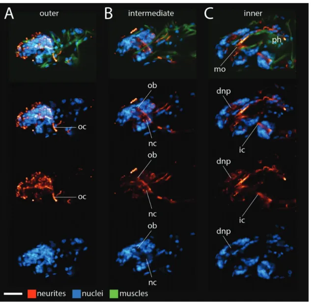
We started this project by focusing onH. dujardini hatch-lings because hatchhatch-lings are small and do not contain autofluorescent algae that H. dujardini eat in culture; both traits facilitate investigations with laser scanning confocal microscopy. InH. dujardini, inner connectives (ic) extended between the first trunk ganglion and the inner head (Figs. 2A and 3C). At a ventral position in the head, each inner connective met a neurite cluster (nc;Figs. 2A and 3B). Neurite clusters were in regions of low nucleus density (lower panels of Figs. 2A and 3B). A post-oral commissure (poc) extended between the neurite clusters at this position (Fig. 2A). From this position, the neurite clusters traveled dorsally along the internal head cavity around the foregut; we identified the foregut based on the positions of neurites extending posteriorly from the mouth (mo), that is, the stomodeal nervous system (Dewel et al. 1999), stytlet muscles (st)— which lie directly above, below, and along the buccal tube—and the muscular pharynx (ph) (top panels of Fig. 2B–D). Nuclei were still absent within the neurite clusters in this region (lower panels of Figs. 2B,C and 3B). Near the top of the mouth, a preoral commissure (prc) extended between the neurite clusters (Fig. 2C). Immediately dorsal to this position, a thick band of neu-rites extended across the dorsal mid-line of the brain (Figs. 2Dand3C); this band of neurites was located in a region that lacked cell nuclei (lower panels ofFigs. 2Dand3C).
Therefore, we refer to this structure as the dorsal neuropil (dnp). In virtual cross sections made using the Volume Viewer plugin in ImageJ, part of the dorsal neuropil appeared to be a continuation of the neurite clusters (Fig. 2E). Additional neurites extending from the outer brain region (ob)—the region that connects to the first trunk ganglion via the outer connective (oc)— contributed to the dorsal neuropil (Figs. 2E and 3A,B).
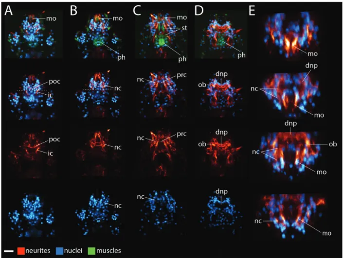
Brain morphology in other tardigrade species Next, we analyzed brain morphology in the other species of this study (Figs. 4–6). Given the large size of the Milnesium n. sp. specimens that we col-lected, we opted to analyze morphology of hatchlings that came from embryos that adult Milnesium n. sp. laid after we collected them. As in H. dujardini, the inner connectives met neurite clusters in the ventral head of Milnesium n. sp. specimens (Fig. 4A,B), Echiniscus n. sp. (Fig. 5A,B), and Batillipes n. sp. (Fig. 6B). A post-oral commissure extended between the neurite clusters at this position in these species (Figs. 4A, 5A, and 6B). The neurite clusters extended dorsally around either side of the internal head cav-ity in regions of low nucleus denscav-ity (lower panels of Figs. 4B,C, 5B,C, and 6B,C). A preoral commissure extended between the neurite clusters above the fore-gut (Figs. 4C, 5C, and 6B). From here, the neurite clusters continued to extend dorsally, ultimately reaching the dorsal neuropil (Figs. 4D,E, 5D,E, and 6C, E). As with H. dujardini, neurites emanating from the outer brain region also contributed to the dorsal neuropil in the other species of this study (Figs. 4E, 5D,E, and 6D,E). We identified anterior (ac) and posterior (pc) neurite clusters in Milnesium n. sp. in positions similar to those seen in other eutardigrades (Supplementary Fig. S1).
Trunk ganglion neuropil
Discussion
General brain anatomy is conserved across Tardigrada
We have investigated anterior nervous system morphology in members of Parachela, Apochela, Echiniscoidea, and Arthrotardigrada. These lineages span Tardigrada, allowing us to make inferences about anterior nervous system anatomy in the most recent common ancestor of Tardigrada. Across species, the inner connectives meet the head at ventral neurite clusters. From this position, the neurite clusters wrap around the foregut. Pre- and post-oral commissures connect the neurite clusters. Part of the dorsal brain neuropil appears to be a continuation of the neurite clusters in confocal Z-series that we analyzed and in virtual cross sections
that we produced; the remaining part of the dorsal neuropil appears to emanate from the outer brain region (Figs. 2–6)—the region that connects to the anteriormost trunk ganglion via the outer connective (Zantke et al. 2008;Persson et al. 2012;Mayer et al. 2013a, 2013b; Schulze and Schmidt-Rhaesa 2013; Persson et al. 2014; Schulze et al. 2014; Smith and Jockusch 2014). Based on our comparisons, we pre-dict that this general nervous system architecture was present in the most recent common ancestor of Tardigrada (Fig. 8A).
A new hypothesis for the composition of the tardigrade brain
Here we present a hypothesis for the composition of the tardigrade brain that relates the anterior nervous
Fig. 2 Frontal views ofHypsibius dujardinibrain morphology. (A–D) ConfocalZseries slices from ventral (left) to dorsal (right). Anterior is to the top. Neurites are stained with ab-tubulin antibody (red), muscles are stained with phalloidin (green), and nuclei are stained with DAPI (blue). The top panels show neurites, nuclei, and muscles. Lower panels show a subset of these structures. We use the top panels to label structures associated with the mouth and foregut. The scale bar in the lower left hand corner equals 10lm. (A) Dashed line represents the boundary between the head and the trunk. (E) Virtual cross-sections through the brain of the specimen shown in A–D, from anterior (top) to posterior (bottom). Neurons are rendered red and nuclei are rendered blue. Images are not to scale with each other in E. Abbreviations: dnp, dorsal neuropil; ic, inner connective; mo, mouth, that is, the stomodeal nervous system; nc, neurite clusters; ob; outer brain; ph, pharynx; poc, post-oral commissure; prc, preoral commissure; st, stylet muscles.
system to the trunk ganglia. In our hypothesis, the neurite clusters that wrap around the foregut and give rise to part of the dorsal brain neuropil are serially homologous to the neuropil of trunk gan-glia (Fig. 8A). Our hypothesis is based on structural and positional similarities between trunk ganglion neuropil and the neurite clusters (Fig. 7). The morphology of trunk ganglion neuropil and the neurite clusters appears to covary between species. Trunk ganglion neuropil and the neurite clusters are in regions of low nucleus density, a defining characteristic of ganglionic neuropil (Richter et al. 2010). Trunk ganglion neuropil and the ventral part of the neurite clusters are wider than the
paired connectives that they give rise to. Furthermore, paired connectives extend directly be-tween the ventral parts of the neurite clusters and the first trunk ganglion (the inner connectives in Figs. 2–6); paired connectives also extend directly between adjacent trunk ganglia (Zantke et al. 2008; Persson et al. 2012; Mayer et al. 2013a, 2013b; Schulze and Schmidt-Rhaesa 2013; Persson et al. 2014; Schulze et al. 2014; Smith and Jockusch 2014). Based on their structural and positional sim-ilarities to neuropil of trunk ganglia and their ap-parent direct connection to the dorsal brain neuropil (Figs. 2–6), we predict that the neurite clusters represent neuropil of the brain; therefore
in our model, the tardigrade brain nearly circum-scribes the foregut (Fig. 8A).
Our hypothesis could be tested by investigating the expression patterns of genes that regulate trunk gan-glion development during embryogenesis in H. dujardini. The conserved set of genes that regulates neurogenesis in euarthropods and other metazoans provides good candidates for this study (Stollewerk 2016). We predict that genes that pattern trunk gan-glia in H. dujardini will also be expressed in the re-gion of the developing head where neurite clusters will ultimately be positioned. Intriguingly, during H. dujardiniembryogenesis, a Paired box antibody marks nuclei in presumptive trunk ganglia and nuclei near where neurite clusters will be positioned (Gabriel and Goldstein 2007; Smith and Goldstein 2017), which
supports our hypothesis. Alternatively, the neurite clusters we identified might not represent ganglionic neuropil. In this case, expression of genes that pattern ganglia in the trunk may be restricted to a dorsal region in the head, if the brain is restricted to a supraesophageal position, which has been suggested (Mayer et al. 2013a;Schulze et al. 2014). We consider this alternative to be unlikely, given that our results suggest that the structure of the neurite clusters and the structure of trunk ganglion neuropil appear to be evolving in concert in Tardigrada (Fig. 7), in accord-ance with serial homology.
Comparison with previous reports
Our results support the conclusions of previous studies, while expanding on them in interesting
Fig. 4 Milnesiumn. sp. brain morphology. (A–D) Ventral (left) to dorsal (right). Anterior is to the top. (A–C) Virtual slices. (D) Maximum projection. (A, B) Dashed line represents the boundary between the head and the trunk. Neurites are stained with a b-tubulin antibody (red), muscles are stained with phalloidin (green), and nuclei are stained with DAPI (blue). The top panels show neurites, nuclei, and muscles. Lower panels show a subset of these structures. The buccal tube was also visible in the blue channel. We use the top panels to label structures associated with the mouth and the foregut. The scale bar in the lower left hand corner equals 10lm. (E) Virtual cross-section renderings of the specimen shown in A–D, from anterior (top) to posterior (bottom). Neurons are rendered red and nuclei are rendered blue. Images are not to scale with each other in E. Abbreviations: bt, buccal tube; dnp, dorsal neuropil; ic, inner connective; nc, neurite clusters; ob; outer brain; ph, pharynx; poc, post-oral commissure; prc, preoral commissure; st, stylet muscles.
ways. Zantke et al. (2008) suggested that the tardi-grade brain exhibits circumesophageal morphology. Results of our study support this conclusion. The circumesophageal component of the brain in the model of Zantke et al. (2008) consists of paired cir-cumbuccal connectives (cco) that are connected by a dorsal commissure (dco) above and a post-oral com-missure (poc) below the foregut (modeled inFig. 1B, top panel). We predict that the dorsal commissure, circumbuccal connectives, and post-oral commissure in the model presented by Zantke et al. (2008) cor-respond to the dorsal neuropil, neurite clusters, and post-oral commissure, respectively, of our model (compare Fig. 1B, top panel to Fig. 8A). In our model, we refer to the dorsal most part of the brain as the dorsal neuropil because it includes neurites
extending from two different positions—the outer and inner brain, that is, it represents outerandinner brain commissures that are not easily distinguish-able. We refer to the circumbuccal connectives iden-tified by Zantke et al. (2008) as neurite clusters because we predict that they represent a part of a ganglion—the neuropil; connectives refer to neurite bundles that connect ganglia, rather than referring to parts of ganglia (Richter et al. 2010). Unlike the model ofZantke et al. (2008), we identified a preoral commissure that directly connects to the neurite clusters in all species of our study. The model of Zantke et al. (2008) includes a preoral commissure, but it is not directly connected to the circumbuccal connectives in their model (the neurite clusters in our model).
Persson et al. (2012)predicted that the inner part of the tardigrade brain—the part adjacent to the foregut in their model—was composed of several lobes (modeled in Fig. 1B, middle panel). By con-trast, we did not identify independent lobes in the presumptive inner brain region. We predict that the subesophageal ganglion (g0), ventrolateral lobes (vll), and the inner lobes (il) of Persson et al. (2012) represent neuronal somata that lie adjacent to the neurite clusters in our model (compare Fig. 1B, middle panel to Fig. 8A). Furthermore, we predict that the commissures connecting the paired inner lobes and paired outer lobes in the model of Persson et al. (2012) represent the dorsal neuropil of our model. It is unclear what parts of their model correspond to the pre- and post-oral
commissures identified in our study. In agreement with the model of Persson et al. (2012, 2014), early descriptions of tardigrade nervous systems typically included a subesophageal ganglion (Marcus 1929; Kristensen 1983; Dewel and Dewel 1996; Dewel et al. 1999; Nielsen 2001). However, several recent studies that utilized immunohistochemical methods and confocal laser scanning microscopy could not identify a subesophageal ganglion (Zantke et al. 2008; Mayer et al. 2013a, 2013b; Schulze and Schmidt-Rhaesa 2013;Schulze et al. 2014;Smith and Jockusch 2014). Our model reconciles these different interpretations by suggesting that the subesophageal ganglion identified in earlier studies is actually paired ventrolateral extensions of the brain, as previously proposed (Mayer et al. 2013a).
Fig. 6 Batillipesn. sp. brain morphology. The mouth faces ventrally in this species. (A–D) Maximum projections of ventral (left) to dorsal (right)Z-series slices. Anterior is to the top. In the upper panels, neurites are stained with ab-tubulin antibody (red), muscles are stained with phalloidin (green), and nuclei are stained with DAPI (blue). The top panels show neurites, nuclei, and muscles. Lower panels show a subset of these structures. We use the top panels to label structures associated with the mouth and the foregut. The scale bar in the lower left hand corner equals 10lm. (B) Dashed line demarcates posterior boundary of the head. (E) Virtual cross-section renderings of the specimen shown in A–D, from anterior (top) to posterior (bottom). Neurons are rendered red and nuclei are rendered blue. Images are not to scale with each other in E. Abbreviations: dnp, dorsal commissure; ic, inner connective; mo, mouth, that is, the stomodeal nervous system; nc, neurite clusters; ob; outer brain; ph, pharynx; poc, post-oral commissure; prc, preoral commissure; st, stylet muscles.
Mayer et al. (2013a) suggested that the circumeso-phageal component of the tardigrade nervous system— referred to as the nerve ring (nr) in their model—is part of the stomodeal nervous system, rather than part of the brain (Fig. 1B, bottom panel). In their model, the tar-digrade brain is restricted to a dorsal position. We sug-gest that the nerve ring identified by Mayer et al. corresponds to the preoral commissure, neurite clus-ters, and post-oral commissure of our model (compare Fig. 1B, bottom panel toFig. 8A). We agree withMayer
et al. (2013a) that these components innervate the sto-modeal nervous system. However, in our model, the neurite clusters and the neuronal somata that give rise to them are part of the brain. This is based on our interpretation of the neurite clusters as ganglionic neuropil and the fact that part of the dorsal brain neuropil appears to be a direct extension of the neurite clusters. Therefore, we conclude that the tardigrade brain nearly circumscribes the foregut, rather than being restricted to a dorsal position.
Fig. 7 Comparison between the ventral parts of the inner neurite clusters (top panels) to trunk ganglion neuropil (bottom panels). (A–D)b-Tubulin antibody stained neurites. All panels show 1.5lm thick maximum projections besides the upper panel of B, which is a virtual slice. Neuropil is outlined in the bottom panel and predicted neuropil is outlined in the top panel. Within columns, images are to scale. (A) Hypsibius dujardini.Second trunk ganglion. (B) Milnesiumn. sp. Second trunk ganglion. (C)Echiniscus n. sp. First trunk ganglion. (D)Batillipesn. sp. First trunk ganglion. Abbreviation: co, commissure.
Our model expands on the models of previous workers by suggesting that the neurite clusters within the head represent ganglionic neuropil. Other studies have not recognized the ganglion-like morphology of the neurite clusters in the head. It is possible that our combination of b-tubulin antibody and Cy3 labeled secondary antibody is more effective for labeling ganglion-like structures compared with the combinations ofa-tubulin antibodies and secondary antibodies used in other studies. It also seems likely that the general morphology of the tardigrade brain is easiest to interpret in smaller specimens. For ex-ample, the boundary between the presumptive inner brain and the stomodeal nervous system is much clearer in H. dujardini hatchlings (Fig. 2) than it is at later stages (Smith and Jockusch 2014).
A model for the evolution of the panarthropod protocerebrum
In our interpretation of tardigrade brain morphology, brain neuropil nearly wraps around the foregut (Fig. 8A). Based on our interpretation and recent hypotheses regarding ecdysozoan phylogeny (Campbell et al. 2011;Rota-Stabelli et al. 2013;Dunn et al. 2014;Giribet 2016), we propose a model for the evolution of the protocerebral brain ganglion of Panarthropoda (Fig. 8B). In this model, the most recent common ancestor of Ecdysozoa exhibited a cycloneuralian-like brain (Fig. 8B, transition 1). A gan-glionated protocerebrum evolved after the panarthro-pods diverged from their cycloneuralian relatives (Fig. 8B, transition 2). This step would have required two major changes in nervous system morphology. First, the clusters of neuronal somata that typically sit in anterior and posterior positions relative to the neuropil ring in cycloneuralian brains would have to have moved to primarily lateral positions immediately adjacent to the neuropil ring. Second, the ventromedial nerve cord that extends from the nerve ring in cyclo-neuralians would have to have split into two independ-ent nerve cords that sit in vindepend-entrolateral positions. Interestingly, Schmidt-Rhaesa (1997/1998) suggested that the ventromedial nerve cord characteristic of Nematoda and Nematomorpha—which together form the proposed sister lineage of Panarthropoda (Campbell et al. 2011;Rota-Stabelli et al. 2013)—has a paired origin. Therefore, the evolution of paired ventrolateral nerve cords in Panarthropoda could have simply involved the repositioning of ventromedial nerve cords that were already paired in the most recent common ancestor of Panarthropoda and NematodaþNematomorpha. If the ventrolateral paired nerve cords of tardigrades were forced to sit
immediately adjacent to each other in a ventromedial position, neuropil would completely circumscribe the foregut in our model of the tardigrade brain, as it does in cycloneuralians. Lastly, in our model, dorsal restric-tion of protocerebral neuropil evolved in the euarthro-podþonychophoran lineage, after it split from Tardigrada (Fig. 8B, transition 3). In this sense, a dor-sally restricted protocerebral ganglion can be viewed as a synapomorphy of the euarthropod/onychophoran clade.
Our model is contingent on two conditions. First, the neurite clusters we identified must be part of the brain. Evidence supporting or refuting this condition could be collected via additional studies of brain development in tardigrades (see above). Second, our model is contingent on a particular view of ecdysozoan phylogeny. In this view, Tardigrada forms the sister group of a euarthro-podþonychophoran clade (Campbell et al. 2011; Rota-Stabelli et al. 2013;Dunn et al. 2014;Giribet 2016). By contrast, some recent molecular analyses recover Tardigrada as a sister group to either Nematoda or NematodaþNematomorpha (Borner et al. 2014; Laumer et al. 2015; reviewed in Edgecombe and Giribet, this volume). Additionally, recent morphological analyses recover Tardigrada as the sister group of Euarthropoda (Smith and Ortega-Hernandez 2014;Smith and Caron 2015;Murdock et al. 2016;Yang et al. 2016). Under these alternative phylogenetic hypotheses, when and how many times brains evolved ganglionated architecture or shifted to a supraesophageal position in ecdysozoan phylogeny would be less clear. However, the grouping of Tardigrada with Nematoda has been suggested to result from system-atic error due to long branch attraction (Dunn et al. 2008; Campbell et al. 2011; Giribet and Edgecombe 2012). Furthermore, the TardigradaþEuarthropoda clade recovered by recent morphological analyses is based on only a small number of similarities in nervous system architecture between these two lineages (Edgecombe and Giribet, this volume). Additional analyses are required to resolve ecdysozoan phylogeny.
Conclusions
While our hypothesis requires additional tests (see previous section), we find it interesting to speculate on its ramifications. Our hypothesis predicts that the most recent common ancestor of Panarthropoda exhibited a circumesophageal brain. If the tardigrade brain is homologous to the protocerebrum of euar-thropods and onychophorans, as evidence suggests (Mayer et al. 2013a; Smith et al. 2016), then it may be possible to identify the regions of the euar-thropod and onychophoran protocerebrum that evolved from the ancestral circumesophageal
component. The protocerebrum of euarthropods and onychophorans is composed of two regions—the archicerebrum, which includes the optic lobes, and the prosocerebrum, which includes neurosecretory centers (Siewing 1963; Scholtz and Edgecombe 2006; Steinmetz et al. 2010; Strausfeld 2012; Ortega-Hernandez et al. 2017). It is possible that the circumesophageal component of the tardigrade brain includes cells from either or both of these regions. By contrast, the tardigrade brain may not be divided into regions with clear homology to either the archicerebrum or prosocerebrum. Any of these possibilities would be informative with regards to the evolution of the panarthropod protocerebrum. These possibilities could be tested by investigating the em-bryonic expression patterns of orthodenticle and six3—markers of the archi- and prosocerebrum, re-spectively (Steinmetz et al. 2010; Ortega-Hernandez et al. 2017)—during tardigrade brain development. The evolutionary origins of the protocerebrum of Panarthropoda remain mysterious ( Ortega-Hernandez et al. 2017). Future studies of the tardi-grade brain may contribute to unlocking this mystery.
Acknowledgments
We would like to thank Ariel Chipman and Doug Erwin for organizing the Evolution of Arthropod Body Plans Symposium and inviting us to partici-pate. We thank Paulo Fontoura, Lukasz Kaczmarek, and Diane Nelson for help in identifying tardigrades. We thank Elizabeth L. Jockusch and two anonymous reviewers for helpful comments on our manuscript. Goldstein laboratory members Kira Glynn, Jenny Heppert, Ari Pani, and Mark Slabodnick helped to collect wild tardigrades. Thomas Boothby provided advice on collecting Batillipes n. sp. tardigrades. Jackie Meier helped collect Batillipes n. sp. tardi-grades and provided helpful comments on the manuscript.
Funding
This work was supported by an National Science Foundation grant [1557432 to B.G.].
Supplementary data
Supplementary Data available at ICB online.
References
Bertolani R, Guidetti R, Marchioro T, Altiero T, Rebecchi L, Cesari M. 2014. Phylogeny of Eutardigrada: new molecular data and their morphological support lead to the
identification of new evolutionary lineages. Mol Phylogenet Evol 76:110–26.
Borner J, Rehm P, Schill RO, Ebersberger I, Burmester T. 2014. A transcriptome approach to ecdysozoan phylogeny. Mol Phylogenet Evol 80:79–87.
Campbell LI, Rota-Stabelli O, Edgecombe GD, Marchioro T, Longhorn SJ, Telford MJ, Philippe H, Rebecchi L, Peterson KJ, Pisani D. 2011. MicroRNAs and phylogenomics resolve the relationships of Tardigrada and suggest that velvet worms are the sister group of Arthropoda. Proc Natl Acad Sci U S A 108:15920–4.
Dewel RA, Dewel WC. 1996. The brain of Echiniscus viridis-simus Peterfi, 1956 (Heterotardigrada): a key to under-standing the phylogenetic position of tardigrades and the evolution of the arthropod head. Zool J Linn Soc 116:35–49.
Dewel R, Budd G, Castano D, Dewel W. 1999. The organiza-tion of the subesophageal nervous system in tardigrades: insights into the evolution of the arthropod hypostome and tritocerebrum. Zool Anz 238:191–203.
Dunn CW, Hejnol A, Matus DQ, Pang K, Browne WE, Smith SA, Seaver E, Rouse GW, Obst M, Edgecombe GD, et al. 2008. Broad phylogenomic sampling improves resolution of the animal tree of life. Nature 452:745–9.
Dunn CW, Giribet G, Edgecombe GD, Hejnol A. 2014. Animal phylogeny and its evolutionary implications. Annu Rev Ecol Evol Syst 45:371–95.
Eriksson BJ, Budd G. 2000. Onychophoran cephalic nerves and their bearing on our understanding of head segmen-tation and stem-group evolution of Arthropoda. Arthropod Struct Dev 29:197–209.
Gabriel WN, Goldstein B. 2007. Segmental expression of Pax3/7 and engrailed homologs in tardigrade development. Dev Genes Evol 217:421–33.
Gabriel WN, McNuff R, Patel SK, Gregory TR, Jeck WR, Jones CD, Goldstein B. 2007. The tardigrade Hypsibius dujardini, a new model for studying the evolution of de-velopment. Dev Biol 312:545–59.
Giribet G, Edgecombe GD. 2012. Reevaluating the arthropod tree of life. Annu Rev Entomol 57:167–86.
Giribet G. 2016. Genomics and the animal tree of life: con-flicts and future prospects. Zool Scr 45:14–21.
Gross V, Mayer G. 2015. Neural development in the tardi-gradeHypsibius dujardini based on anti-acetylated a-tubu-lin immunolabea-tubu-ling. Evodevo 6:12.
Gross V, Minich I, Mayer G. 2017. External morphogenesis of the tardigrade Hypsibius dujardinias revealed by scanning electron microscopy. J Morphol 278:563–73.
Guil N, Giribet G. 2012. A comprehensive molecular phylogeny of tardigrades—adding genes and taxa to a poorly resolved phylum-level phylogeny. Cladistics 28:21–49.
Hejnol A, Schnabel R. 2005. The eutardigrade Thulinia ste-phaniaehas an indeterminate development and the poten-tial to regulate early blastomere ablations. Development 132:1349–61.
Henne S, Friedrich F, Hammel JU, Sombke A, Schmidt-Rhaesa A. 2017a. Reconstructing the anterior part of the nervous system of Gordius aquaticus (Nematomorpha, Cycloneuralia) by a multimethodological approach. J Morphol 278:106–18.
Henne S, Sombke A, Schmidt-Rhaesa A. 2017b. Immunohistochemical analysis of the anterior nervous sys-tem of the free-living nematode Plectus spp. (Nematoda, Plectidae). Zoomorphology 2017:1–16 published online (doi:10.1007/s00435-017-0347-x).
Jørgensen A, Faurby S, Hansen JG, Møbjerg N, Kristensen RM. 2010. Molecular phylogeny of Arthrotardigrada (Tardigrada). Mol Phylogenet Evol 54:1006–15.
Kristensen RM. 1983. The first record of cyclomorphosis in Tardigrada based on a new genus and species from arctic meiobenthos. J Zool Syst Evol Res 20:249–70.
Laumer CE, Bekkouche N, Kerbl A, Goetz F, Neves RC, Sørensen MV, Kristensen RM, Hejnol A, Dunn CW, Giribet G, et al. 2015. Spiralian phylogeny informs the evo-lution of microscopic lineages. Curr Biol 25:2000–6. Marcus E. 1929. Tardigrada. In: Bronn HG, editor. Klassen
und Ordnungen des Tier-reichs. Vol. 5, Section 4. Leipzig: Akademische Verlagsgesellschaft. Part 3: 1–609.
Martin C, Mayer G. 2014. Neuronal tracing of oral nerves in a velvet worm—implications for the evolution of the ecdy-sozoan brain. Front Neuroanat 8:7 published online (doi:10.3389/fnana.2014.00007).
Martin C, Mayer G. 2015. Insights into the segmental identity of post-oral commissures and pharyngeal nerves in Onychophora based on retrograde fills. BMC Neurosci 16:53 published online (doi:10.1186/s12868-015-0191-1). Martın-Duran JM, Wolff GH, Strausfeld NJ, Hejnol A. 2016.
The larval nervous system of the penis worm Priapulus caudatus (Ecdysozoa). Philos Trans R Soc Lond B Biol Sci 371:20150050.
Mayer G, Whitington PM, Sunnucks P, Pflu¨ger H. 2010. A revision of brain composition in Onychophora (velvet worms) suggests that the tritocerebrum evolved in arthro-pods. BMC Evol Biol 10:1 published online (doi:10.1186/ 1471-2148-10-255).
Mayer G, Kauschke S, Rudiger J, Stevenson PA. 2013a. Neural markers reveal a one-segmented head in tardigrades (water bears). PLoS One 8:e59090.
Mayer G, Martin C, Rudiger J, Kauschke S, Stevenson PA, Poprawa I, Hohberg K, Schill RO, Pfluger HJ, Schlegel M. 2013b. Selective neuronal staining in tardigrades and onychophorans provides insights into the evolution of segmental ganglia in panarthropods. BMC Evol Biol 13:230.
Mayer G. 2016. Onychophora. In: Schmidt-Rhaesa A, Harzsch S, Purschke G, editors. Structure and evolution of invertebrate nervous systems. London: Oxford University Press. p. 390–401. Murdock DJ, Gabbott SE, Purnell MA. 2016. The impact of
taphonomic data on phylogenetic resolution: Helenodora inopinata (carboniferous, mazon creek lagerst€atte) and the onychophoran stem lineage. BMC Evol Biol 16:22–19. Nielsen C. 2001. Animal evolution: interrelationships of the
living phyla. London: Oxford University Press.
Ortega-Hernandez J, Janssen R, Budd GE. 2017. Origin and evolution of the panarthropod head—a palaeobiological
and developmental perspective. Arthropod Struct Dev 46:354–79.
Persson DK, Halberg KA, Jørgensen A, Mobjerg N, Kristensen RM. 2012. Neuroanatomy of Halobiotus crispae
(Eutardigrada: Hypsibiidae): tardigrade brain structure supports the clade Panarthropoda. J Morphol 273:1227–45. Persson DK, Halberg KA, Jørgensen A, Møbjerg N, Kristensen RM. 2014. Brain anatomy of the marine tardigrade
Actinarctus doryphorus (Arthrotardigrada). J Morphol 275:173–90.
Richter S, Loesel R, Purschke G, Schmidt-Rhaesa A, Scholtz G, Stach T, Vogt L, Wanninger A, Brenneis G, Do¨ring C. 2010. Invertebrate neurophylogeny: suggested terms and definitions for a neuroanatomical glossary. Front Zool 7:29 published online (doi:10.1186/1742-9994-7-29) Rota-Stabelli O, Daley AC, Pisani D. 2013. Molecular
time-trees reveal a Cambrian colonization of land and a new scenario for ecdysozoan evolution. Curr Biol 23:392–8. Rothe BH, Schmidt-Rhaesa A. 2010. Structure of the nervous
system inTubiluchus troglodytes(Priapulida). Invertebr Biol 129:39–58.
Schafer W. 2016. Nematode nervous systems. Curr Biol 26:R955–9.
Schmidt-Rhaesa A. 1996. The nervous system of Nectonema munidaeand Gordius aquaticus, with implications for the ground pattern of the Nematomorpha. Zoomorphology 116:133–42.
Schmidt-Rhaesa A. 1997/1998. A phylogenetic relationships of the Nematomorpha—a discussion of current hypotheses. Zool Anz 236:203–16.
Schmidt-Rhaesa A. 2007. The evolution of organ systems. London: Oxford University Press.
Scholtz G, Edgecombe GD. 2006. The evolution of arthropod heads: reconciling morphological, developmental and palaeontological evidence. Dev Genes Evol 216:395–415. Scholtz G. 2016. Perspective—heads and brains in arthropods:
40 years after the ‘endless dispute’. In: Schmidt-Rhaesa A, Harzsch S, Purschke G, editors. Structure and evolution of invertebrate nervous systems. London: Oxford University Press. p. 402–10.
Schulze C, Schmidt-Rhaesa A. 2013. The architecture of the nervous system of Echiniscus testudo (Echiniscoidea, Heterotardigrada). J Limnol 72:44–53.
Schulze C, Neves RC, Schmidt-Rhaesa A. 2014. Comparative immunohistochemical investigation on the nervous system of two species of Arthrotardigrada (Heterotardigrada, Tardigrada). Zool Anz 253:225–35.
Siewing R. 1963. Zum Problem der Arthropodenkopfseg-mentierung. Zool Anz 170:429–68.
Smith FW, Jockusch EL. 2014. The metameric pattern of
Hypsibius dujardini (Eutardigrada) and its relationship to that of other panarthropods. Front Zool 11:66 published online (doi:10.1186/s12983-014-0066-9).
Smith FW, Boothby TC, Giovannini I, Rebecchi L, Jockusch EL, Goldstein B. 2016. The compact body plan of tardi-grades evolved by the loss of a large body region. Curr Biol 26:224–9.
Smith FW, Goldstein B. 2017. Segmentation in Tardigrada and diversification of segmental patterns in Panarthropoda. Arthropod Struct Dev 46:328–40.
Smith MR, Ortega-Hernandez J. 2014. Hallucigenia’s onychophoran-like claws and the case for Tactopoda. Nature 514:363–6.
Smith MR, Caron J. 2015. Hallucigenia’s head and the pha-ryngeal armature of early ecdysozoans. Nature 523:75–8. Steinmetz PR, Urbach R, Posnien N, Eriksson J,
Kostyuchenko RP, Brena C, Guy K, Akam M, Bucher G, Arendt D. 2010.Six3demarcates the anterior-most devel-oping brain region in bilaterian animals. EvoDevo 1:14. Stollewerk A. 2016. A flexible genetic toolkit for arthropod
neurogenesis. Philos Trans R Soc Lond B Biol Sci 371 published online (doi: 10.1098/rstb.2015.0044).
Strausfeld NJ, Ma X, Edgecombe GD. 2016. Fossils and the evolution of the arthropod brain. Curr Biol 26:R989–1000. Strausfeld NJ, Strausfeld CM, Stowe S, Rowell D, Loesel R. 2006a. The organization and evolutionary implications of neuropils and their neurons in the brain of the onychoph-oran Euperipatoides rowelli. Arthropod Struct Dev 35:169–96.
Strausfeld NJ, Strausfeld CM, Loesel R, Rowell D, Stowe S. 2006b. Arthropod phylogeny: onychophoran brain or-ganization suggests an archaic relationship with a cheli-cerate stem lineage. Proc R Soc Lond B Biol Sci 273:1857–66.
Strausfeld NJ. 2012. Arthropod brains: evolution, functional elegance, and historical significance. Cambridge (MA): Belknap Press of Harvard University Press.
Whitington PM, Mayer G. 2011. The origins of the arthropod nervous system: insights from the Onychophora. Arthropod Struct Dev 40:193–209.
Yang J, Ortega-Hernandez J, Butterfield NJ, Liu Y, Boyan GS, Hou JB, Lan T, Zhang XG. 2016. Fuxianhuiid ventral nerve cord and early nervous system evolution in Panarthropoda. Proc Natl Acad Sci U S A 113:2988–93.
Zantke J, Wolff C, Scholtz G. 2008. Three-dimensional reconstruc-tion of the central nervous system of Macrobiotus hufelandi