Original Article
PD-L1 blockade improves immune dysfunction of
spleen dendritic cells and T-cells in zymosan-induced
multiple organs dysfunction syndromes
Qian Liu*, Yi Lv*, Min Zhao, Yiduo Jin, Jiangyang Lu
Department of Pathology, The First Affiliated Hospital of General Hospital of PLA, Beijing 100048, China. *Equal
contributors and co-first authors.
Received December 12, 2014; Accepted February 5, 2015; Epub February 1, 2015; Published February 15, 2015
Abstract: This research is to investigate the role of tolerant spleen dendritic cells (DC) in multiple organs dysfunc-tion syndromes (MODS) at late stage. Tolerant DC and MODS were induced by intraperotineal injecdysfunc-tion of zymosan. The immunity of DC was determined by examining interleukin (IL)-10, IL-12, IL-2, major histocompatibility complex (MHC), CD86, programmed death (PD-1), programmed death ligand 1 (PD-L1), paired immunoglobulin-like receptor B (PIR-B) or T-cell proliferation in serum, spleen homogenate, DC culture or DC/T-cell co-culture. The PD-L1/PD-1 pathway was blocked using PD-L1 antibody. The IL-12p70 in serum, spleen homogenate and DC culture superna-tant were decreased at 5 d and 12 d after zymosan injection while the IL-12p40 and IL-10 were increased. The expression of MHC, cluster of differentiation 86 (CD86), PD-1 and PD-L1 in spleen DCs were increased at early stage after zymosan injection. At 5 d and 12 d, the expression of MHC and CD86 was reduced while the expression of PD-1, PD-L1 and PIR-B was increased, accompanied with decreased proliferation of T-cell and decrease of IL-2 in spleen and serum. Application of PD-L1 antibody improved the above changes. At late stage of MODS mice induced by zymosan, the expression of co-stimulators and inhibitors in spleen DCs was imbalanced to form tolerant DCs which reduced the activation of T-cells. PD-L1 antibody improved the immune tolerance of DCs through intervening PD-1/PD-L1 pathway, and attenuated the inhibition of T-cell activities by tolerant DCs and the immune inhibition.
Keywords: Dendritic cell, MODS, immune tolerance, zymosan
Introduction
As the major reason of death for patients in ICU, multiple organ dysfunction syndrome (MODS) is the most serious complication of sepsis [1, 2] which results in immune
dysfunc-tion due to imbalanced pro-inflammatory and anti-inflammatory mechanisms [3, 4]. The con -sumption and function change of immune cells
induced by excessive inflammatory response at
the early stage of sepsis attenuated the
phago-cytosis, proinflammatory cytokine release and
antigen presentation of monocytes [5, 6], resulting in immune tolerance which is patho-logical feature of late stage of sepsis [3, 7-10]. Therefore, improvement of immune function is the critical step for prevention of MODS induced by sepsis.
The spleen plays important roles in the respons-es of congenital immunity and acquired
immu-nity, and, therefore, has important effect on the prognosis of sepsis [11, 12]. The immune func-tion of spleen depends on the synergistic effect between spleen dendritic cells (DC) and immu-nocytes including T-cells, B-cells and regulatory T-cells (Treg). As the strongest antigen present-ing cells, DC is the bridge between congenital immunity and adaptive immunity, controlling the balance between immune tolerance and immune activation of T-cells [13]. Recent stud-ies indicated that DC not only can induce immune response but also have ability of immune regulation [12, 14]. The apoptosis, number reduction and mature disorder of DC during sepsis attenuate the immune activation of DC, down-regulate the activities of T-cells and B-cells, and are closely related with the prognosis of sepsis [15, 16].
the superficial co-stimulators and co-inhibitors
on DC and the ligands of lymphocytes [14, 17, 18]. As co-stimulators, the cluster of differenti-ation 80 (CD80), B7-2(CD86) and programmed death ligand 1 (PD-L1) were expressed on T-cell, B-cell, DC and macrophages. As one receptor of co-inhibitor, programmed death 1 (PD-1) was mainly expressed on the surface of
activated and T-cells [19]. PD-1 was clarified as
the major negative regulatory receptor of PD-L1 in PD-L1-knockout mice to play inhibitory effect [20]. Under continuous stimulation by antigens, PD-1/PD-L1 negatively modulated the balance of T-cell activation, tolerance and pathological immune reaction [21]. The PD-1 expression on T-cell, B-cell and macrophage and the expres-sion of PD-L1 on B-cell and macrophage were found to be up-regulated in cecal ligation and puncture (CLP) mice. While PD-L1 blockade increased the survival of septic animals through inhibiting lymphocyte apoptosis and recovering monocyte function [22]. These studies suggest that blockade of the interaction between PD-L1/PD-1 pathway may be promising strate-gy for preventing immune tolerance at late stage of sepsis.
The biphasic MODS induced by intraperitoneal injection of zymosan is one experimental ani-mal model having typical immunopathological features of sepsis and similar clinical progres-sion of MODS [23]. In the present study, we investigated the changing pattern of spleen DC in zymosan-induced MODS mice, the relation-ship of DC with immune inhibition and the mechanism of improvement of immune disor-ders of DC and T-cell induced by intervening PD-L1/PD-1 pathway.
Materials and methods
Animals and MODS model
Male C57BL/6 mice (6-8 weeks, 20-25 g), pur-chased from the Laboratory Animal Center of Academy of Military Medical Science, were accommodated for 7 days at 12:12 light-dark circle with free access of food and water. After fasting 12 h, the mice were randomly divided into control group (n=10), zymosan group (n=64) and zymosan + PD-L1 antibody group (n=64). The zymosan group and zymosan+PD-L1 antibody group were further divided into subgroups of 12 h, 2 d, 5 d and 12 d (n=16 for each subgroup).
MODS model was induced by zymosan
accord-ing to literature [23]. Briefly, 1 g zymosan pow -der (Sigma Chemical, St. Louis, MO, USA) was
mixed with 40 ml medical paraffin oil to make
25 g/L zymosan suspension which was steril-ized at 100°C water for 80 min and cooled to room temperature. After sterilization of abdo-men of mice, zymosan suspension was intra-peritoneally injected (800 mg/kg). At 12 h and 24 h after injection, saline (20 ml/kg) was
injected for fluid infustion. In zysoman + PD-L1
antibody group, PD-L1 antibody (100 µg in 100 µl PBS) was injected via tail vein immediately and at 48 h after zymosan injection. Thereafter, the animals were bred as regular and the blood and spleen were collected at corresponding time points for measurement. All the animal experiments were approved by the Ethic Committee of Animal Care and Usage of the university.
Measurement of cytokines in serum and spleen
The blood collected from fundus artery of mice was settled for 30 min, centrifuged for 15 min at 3000 rpm to separate serum stored at -80°C. 100 g spleen tissue stored at liquid nitrogen was added with 1 ml PBS and homog-enated at ice bath and centrifuged for 15 min at 3000 rpm (4°C) to collect the supernatant. IL-2, IL-10 and IL-12 in serum and spleen homogenate were measured with ELISA, according to the manual (R&D, USA).
Separation and culture of spleen DC and measurement of related factors in culture supernatant
At the corresponding time points, the spleen
(Ficoll-Papue) for centrifugation at room temperature (3000 rpm, 15 min). The content of middle layer was taken out, rinsed with PBS for 2 times and blew evenly with 2 ml PBS for cell number accounting and measurement of cell activities with trypan blue staining (>97%). DCs were
purified with anti-CD11c magnetic beads and
positive selection MS + columns according to the manual (Miltenyi Biotec, Auburn, CA, USA). PE-CD11c antibody and PE-IgG Isotype Control (BD Biosciences, USA) were used to label DC
for measurement of purity (>99%). The purified
DC was re-suspended with RPMI1640 (Hyclone, USA) and adjusted to concentration of 5×10⁶/ ml. Then the cells were inoculated in 96-well plate (1.25×10⁶/well) and incubated in 5% CO
2 incubator at 37°C for 24 h and centrifuged (1500 rpm, 5 min). The supernatant was taken out for measurement of IL-10 and IL-12 with ELISA.
Measurement of superficial cytokines of DC
DCs re-suspended with PBS (containing 2%
BSA) were added to a flow tube (5×10⁴ cells/100
μl/tube) and added with FITC or PE labeled fluo -rescent antibody (or PE-IgG Isotype Control, 2-3
μl) against the superficial marker. The mixture
was incubated at 4°C for 45 min, rinsed with PBS twice and centrifuged (1500 rpm, 5 min). After removal of the supernatant, the cells were re-suspended with 400 µl PBS and analyzed with FACS caliber (Becton Dickinson, USA). The labeled antibodies included: MHC-II (I-Ab)-FITC, PD-L1-PE, PD-1-PE (all from BD Biosciences, USA), CD86-PE and PIR-B-PE (Biolegend, USA).
Isolation, purification and proliferation of T-cells
Spleen tissues taken at different time points were ground and rinsed with serum-free RPMI1640 to collect cells. After addition of lym-phocyte isolation solution, the cells were centri-fuged (18°C, 2500 rpm, 20 min). The cells of low density at middle layer were taken out and rinsed with PBS twice for cell accounting and
activity measurement. The T-cells were purified
with technique of magnetic activated cell sort-ing (MACS), accordsort-ing to the manual (Miltenyi Biotec, Auburn, CA, USA).
The cell proliferation was measured with MTT.
T-cells after isolation and purification were
added to 96-well plate (5×10⁵/well) with ConA
(5 µg/ml) and incubated at 37°C for 68 h. After removal of 100 µl culture medium and addition of MTT (5 mg/ml), the cells were incubated for 4 h. Finally, the cells were added with Triton-ISOP (100 µl/well) and incubated for 4 h at 37°C till the crystal was completely resolved for measurement of OD560 value.
Co-culture of DCs and T-cells and PD-L1 anti-body intervening
In vitro co-culture of DCs and T-cells was used to observe the effect of septic spleen DCs on the activities of T-cells and the intervening
effect of PD-L1 antibody. The purified DCs of
septic mice were added to 96-well plate (2×10⁴ cells/well), 3 wells for each sample. Fresh-isolated spleen T-cells (2×10⁵ cells/well) from normal C57BL/6 mice were inoculated in the
wells pre-plated with DCs. PD-L1 antibody (final
concentration of 20 µl/ml) or control medium was added for incubation of 24 h with exchange of the culture medium. After 68 h, the superna-tant was collected and measured of the con-centration of IL-2, IL-10 and IL-12 using ELISA. MTT was added to the 96-well plated for con-tinuous incubation of 4 h to measure the prolif-eration of T-cells.
Statistic analysis
The data were expressed as mean ± SD. SPSS13.0 was used for statistic analysis with ONEWAY-ANOVA. Intra-group comparison was performed with LSD-test and intergroup com-parison was performed with t-test. P<0.05 was
set as significant level.
Results
Developmental features of MODS in mice induced by zymosan
peaks during the development of the disease
model were equivalent to the excessive inflam -matory response at early stage and the domi-nant immune inhibition at late stage.
Expression changes of PD-1, PD-L1 and PIR-B on spleen DCs of MODS mice
There were many studies about the co-inhibitor pathway of PD-1 and PD-L1 in tumor and auto-immune diseases [19], suggesting its involve-ment in the establishinvolve-ment of immune tolerance of antigen presenting cells [21]. The expression rate of PD-L1 was over 50% on DCs of normal mice spleen, upregulated at 0.5 d after injec-tion of zymosan (vs control group, P<0.05), recovered to nearly normal at 2 d, and increased again at 5 d and 12 d (vs control group, P<0.05) (Figure 1A). Different from PD-L1, PD-1 expres-sion was undetectable on normal DCs, increased at 0.5 d after injury and maintained stable till 12 d (vs control group, P<0.01) (Figure 1B). PD-L1 antibody had no effect on the expression of PD-L1 and PD-1.
PIR-B is one inhibitory receptor expressed on DCs surface and its expression increase is the symbol of formation of tolerant DCs [24, 25]. The expression of PIR-B on spleen DCs was not changed at the early stage of zymosan-induced injury (2 d), increased at 5 d and 12 d (vs con-trol group, P<0.05). PD-L1 antibody inhibited the upregulation of PIR-B on DCs at late stage of MODS induced by zymosan (vs correspond-ing timepoints of MODS group, P<0.05) (Figure 1C).
Expression change of MHC-II and CD86 on spleen DCs
[image:4.612.93.524.74.350.2]Figure 2. The ratios of PIR-B expression to MHC-II (A) and to CD86 (B) expression on spleen DCs. By flow cytometry PIR-B, CD86 and MHC-expression was calculated with percentages of positive cells out of total DCs.
on DCs at 12 d (vs MODS 12 d, P<0.05) to nor-mal level (Figure 1D). Similar to MHC-II, the expression of CD86 was rescued by PD-L1 anti-body at 5 d and 12 d (Figure 1E).
In order to analyze the relative intensity change of immune inhibition and immune stimulation of DCs, we compared the relative values of PIR-B expression with MHC-II expression or CD expression. The result indicated that ratios of PIB-B/MHC-II and PIB-B/CD86 were greatly increased while PD-L1 antibody could recover the ratios of PIB-B/MHC-II and PIB-B/CD86 (Figure 2).
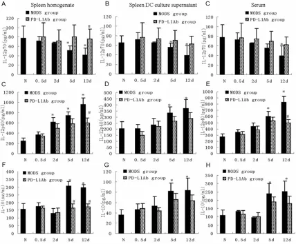
Changes of IL-12 and IL-10 in spleen and serum
IL-12(IL-12p70), a heterodimer composed of p35 and p40 subunits, is mainly derived from mononuclear macrophages and DCs having ability of antigen presenting. IL-12p70 binds to IL-12 receptor of T-cells promoting immune response while IL-12p40, in contrary, is the natural antagonist of IL-12 [27]. Under condi-tion of physiology or pathology, IL-12p70/p40 induces T-helper cell (Th1/Th2) shift and modu-lates immune response through modulating Th cells. Therefore, spleen change of IL-12p70/
p40 reflects the immune states of antigen pre -senting cells including DCs. After zymosan injection, the content of IL-12p70 in spleen gradually decreases while IL-12p40 gradually increases. At 5 d and 12 d, the IL-12p70
reached the bottom while IL-12p40 reached the peak (vs control group, P<0.05; Figure 3A-E). Spleen inhibitory cytokine IL-10 was increased at 5 d and 12 d after zymosan injury (vs control group, P<0.05; Figure 3G, 3H). PD-L1 antibody blocked the imbalance between
superficial DC co-stimulator and co-inhibitor
induced by zymosan and affected the secretion
of IL-12 and IL-10, resulting in significant differ -ence between PD-L1 antibody group and MODS group at 12 d (P<0.05, Figure 3).
The serum concentration of IL-12p70 was only 50% of control group at 12 d after injury (P<0.05) while IL-12p40 was greatly increased at late stage of MODS. The serum concentra-tion of IL-10 was largely increased at 5 d and 12 d (vs control group, P<0.05; Figure 3C, 3F, 3I). In PD-L1 antibody group, the serum content of IL-12p70 was kept at control level while IL-12p40 was lower than MODS group at 12 d. The serum IL-10 was mild lower than MODS group at 5 d and 12 d (P>0.05, Figure 3C, 3F,
3I).
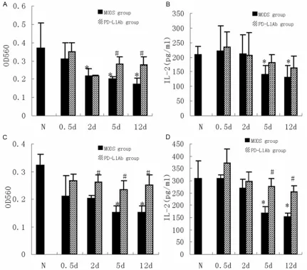
Protection of PD-L1 antibody on T-cell prolifera-tion induced by zymosan
At 2 d after zymosan injection, the proliferative activity of spleen T-cells was largely decreased from 2 d to 12 d (P<0.05). PD-L1 antibody recovered the proliferation of T-cells at 5 d and
12 d, significantly higher than MODS group
secretion of IL-2 by T-cells in vitro culture was
significantly decreased at 5 d and 12 d
(P<0.05), PD-L1 antibody increased the
secre-tion of IL-2 but without significant difference
(P>0.05) (Figure 4B). The serum concentration of IL-2 showed same trend with the secretion of IL-2 by T-cells in culture, suggesting that the
immunity of T-cells at 5 d and 12 d was signifi -cantly decreased while PD-L1 antibody had protective effect on T-cells activity.
Improvement of DC and T-cells by PD-L1 anti-body in vitro
In order to investigate if the inhibition T-cell in MODS was correlated with the formation of tol-erant DC through PD-L1/PD-1 pathway, we iso-lated spleen DCs at different stage of MODS mice and cultured the DCs with normal spleen T-cells. The result indicated that the spleen DCs
of MODS mice had strong inhibitory effect on the proliferation of T-cells of normal spleen at 5 d and 12 d, and inhibited the secretion of IL-2 (P<0.05); PD-L1 antibody could improve the proliferation of T-cells and increase the secre-tion of IL-2 (P<0.05) (Figure 4C, 4D).
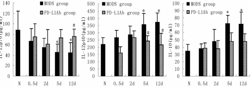
In addition, PD-L1 antibody increased the centration of IL-12p70 and decreased the con-centrations of IL1-2p40 and IL-10 by DC in the medium co-culture of spleen DC from MODS mice and normal T-cell (Figure 5).
Discussion
[image:6.612.93.521.72.426.2]decreased. The tolerant DC of MODS mice had inhibitory effects on T-cells. These changes in late stage of MODS mice were blocked by PD-L1 antibody.
As one pro-inflammatory agent, zymosan can induce systemic inflammatory reaction, typical
clinic signs of sepsis and MODS, demonstrated
as excessive inflammatory response at early
stage and immune inhibition at late stage, and corresponding 2 death peaks [23, 28]. The development stages of this animal model are
easily to be identified with clear different
immune features at different stages, and there-fore is ideal model for study of pathogenic mechanism and prevention strategy of MODS. The present study showed similar developmen-tal stages of MODS mice induced by zymosan
with previous studies [23, 28], suggesting suc-cessful establishment of MODS model.
[image:7.612.92.523.72.450.2]HMGB1, which is the important reason for immune inhibition at late stage of sepsis, MODS onset and death [31]. Splenectomy or apoptosis inhibitor can reduce the release of HMGB1 and decrease the mortality [32]. Consistently, in the present study, we found downregualtion of IL-12p70, IL-2 and T-cell func-tion, and upregulation of IL-10 and IL-12p40 at late stage of MODS mice induced by zymosan, accompanied with the second death peak.
Under the condition of insufficient positive sig -nals, the negative signal provided by PD-L1/ PD-1 pathway took dominant advantage, con-trolled the stimulation intensity of T-cells
through limiting the release of IL-2 and IFN-γ,
and inhibited activities of T-cell [14, 33]. Our study indicated that the positive immune mol-ecule CD86 on spleen DCs at late stage of MODS were down-regulated while negative co-stimulator PD-L1/PD-1 was up-regulated. These results suggested a correlation between the immune reduction of DCs and the imbal-ance of co-stimulator and co-inhibitor expres-sion. This is also consistent with our previous study that administration of immune enhancer Flt3 improved the mature of tolerant DC to recover immunity and reduce mortality [34]. Previous studies have proved that PD-L1 was the major inhibitory modulation receptor in PD-1 knockout mice suffering autoimmune dis-eases [19] and that blockade of the interaction between PD-L1 and PD-1 improved the immune condition [22, 35]. Consistently, the present result indicated that the functions of spleen DCs and T-cells were greatly improved during
[image:8.612.95.522.74.222.2]expression has important effect on the
forma-tion of tolerant DCs and is specific marker judg -ing formation of tolerant DCs [24, 41]. Consistently, we found that PD-L1 antibody had inhibitory effect on the expression of PIR-B on spleen DCs, resulting in upregulation of tran-scription and translation of CD86, IL-12p70 and MHC-II on DCs. Analysis of the relative ratio of PIR-B to MHC-II and CD86 showed that PD-L1 antibody decreased the ratio of PIR-B/MHC-II and PIR-B/CD86 at late stage after zymosan injury. These studies suggested that PD-L1 blockade could improve DC activities through downregulation of PIR-B, which may be one mechanism for PD-L1 antibody to recover the activation of tolerant DCs.
In summary, our study indicated that the imbal-ance between co-stimulator and co-inhibitor of spleen DCs at late stage of MODS mice induced by zymosan mediated the formation of tolerant DCs and decreased the activities of T-cells. Blockade of PD-L1/PD-1 pathway improved the immune disorder at late stage of MODS induced by sepsis. These results suggested that block-ade of PD-L1/PD-1 pathway may be valuable strategy in preventing and treating immune tol-erance at late stage of sepsis.
Acknowledgements
The study was supported by the Capital Medical Development Foundation, 2011-5002-02.
Disclosure of conflict of interest
None.
Address correspondence to: Dr. Jiangyang Lu, Department of Pathology, The First Affiliated Hospital of General Hospital of PLA, Beijing 100048, China. Tel: 68689159; Fax: +86-010-68689159; E-mail: [email protected]
References
[1] Deans KJ, Haley M, Natanson C, Eichacker PQ and Minneci PC. Novel therapies for sepsis: a review. J Trauma 2005; 58: 867-874.
[2] Riedemann NC, Guo RF and Ward PA. The enig-ma of sepsis. J Clin Invest 2003; 112: 460-467.
[3] Hotchkiss RS and Karl IE. The pathophysiology and treatment of sepsis. N Engl J Med 2003; 348: 138-150.
[4] Rittirsch D, Flierl MA and Ward PA. Harmful molecular mechanisms in sepsis. Nat Rev Im-munol 2008; 8: 776-787.
[5] Moraes TJ and Downey GP. Death of the septic monocyte: is more better? Crit Care 2006; 10: 146.
[6] Laudanski K and Wyczechowska D. Monocyte-related immunopathologies in trauma pa-tients. Arch Immunol Ther Exp (Warsz) 2005; 53: 321-328.
[7] Carlet J, Cohen J, Calandra T, Opal SM and Ma-sur H. Sepsis: time to reconsider the concept. Crit Care Med 2008; 36: 964-966.
[8] dib-Conquy M and Cavaillon JM. Compensatory anti-inflammatory response syndrome. Thromb Haemost 2009; 101: 36-47.
[9] Remick DG. Pathophysiology of sepsis. Am J Pathol 2007; 170: 1435-1444.
[10] Boomer JS, To K, Chang KC, Takasu O, Osborne DF, Walton AH, Bricker TL, Jarman SD, Kreisel D, Krupnick AS, Srivastava A, Swanson PE, Green JM and Hotchkiss RS. Immunosuppres-sion in patients who die of sepsis and multiple organ failure.JAMA 2011; 306: 2594-2605. [11] Mebius RE and Kraal G. Structure and function
of the spleen. Nat Rev Immunol 2005; 5: 606-616.
[12] Wu L and Dakic A. Development of dendritic cell system. Cell Mol Immunol 2004; 1: 112-118.
[13] Banchereau J, Briere F, Caux C, Davoust J, Leb-ecque S, Liu YJ, Pulendran B and Palucka K. Immunobiology of dendritic cells. Annu Rev Im-munol 2000; 18: 767-811.
[14] Sato K, Yamashita N, Baba M and Matsuyama T. Modified myeloid dendritic cells act as regu-latory dendritic cells to induce anergic and regulatory T cells. Blood 2003; 101: 3581-3589.
[15] Scumpia PO, McAuliffe PF, O’Malley KA, Unga-ro R, Uchida T, Matsumoto T, Remick DG, Clare-Salzler MJ, Moldawer LL and Efron PA. CD11c+ dendritic cells are required for survival in mu-rine polymicrobial sepsis. J Immunol 2005; 175: 3282-3286.
[16] Flohe SB, Agrawal H, Schmitz D, Gertz M, Flohe S and Schade FU. Dendritic cells during poly-microbial sepsis rapidly mature but fail to initi-ate a protective Th1-type immune response. J Leukoc Biol 2006; 79: 473-481.
[17] Dong H and Chen X. Immunoregulatory role of B7-H1 in chronicity of inflammatory responses. Cell Mol Immunol 2006; 3: 179-187.
[18] Poirier N, Blancho G and Vanhove B. A more selective costimulatory blockade of the CD28-B7 pathway. Transpl Int2011; 24: 2-11. [19] Okazaki T and Honjo T. PD-1 and PD-1 ligands:
from discovery to clinical application. Int Im-munol 2007; 19: 813-824.
[21] Keir ME, Butte MJ, Freeman GJ and Sharpe AH. PD-1 and its ligands in tolerance and immuni-ty. Annu Rev Immunol 2008; 26: 677-704. [22] Zhang Y, Zhou Y, Lou J, Li J, Bo L, Zhu K, Wan X,
Deng X and Cai Z. PD-L1 blockade improves survival in experimental sepsis by inhibiting lymphocyte apoptosis and reversing monocyte dysfunction. Crit Care 2010; 14: R220. [23] Jansen MJ, Hendriks T, Verhofstad AA, Lange
W, Geeraedts LM Jr and Goris RJ. Gradual de-velopment of organ damage in the murine zy-mosan-induced multiple organ dysfunction syndrome. Shock 1997; 8: 261-267.
[24] Liu Z, Li W, Zhang M, Zhou H, Han H and Zou P. Paired immunoglobin-like receptors A and B are new targets for inducing dendritic cells tol-erance in mice. J Huazhong Univ Sci Technolog Med Sci2007; 27: 252-256.
[25] Mitsuhashi Y, Nakamura A, Endo S, Takeda K, Yabe-Wada T, Nukiwa T and Takai T. Regulation of plasmacytoid dendritic cell responses by PIR-B. Blood 2012; 120: 3256-3259.
[26] Selenko-Gebauer N, Majdic O, Szekeres A, Ho-fler G, Guthann E, Korthauer U, Zlabinger G, Steinberger P, Pickl WF, Stockinger H, Knapp W and Stockl J. B7-H1 (programmed death-1 li-gand) on dendritic cells is involved in the in-duction and maintenance of T cell anergy. J Immunol2003; 170: 3637-3644.
[27] Gillessen S, Carvajal D, Ling P, Podlaski FJ, Stremlo DL, Familletti PC, Gubler U, Presky DH, Stern AS and Gately MK. Mouse interleukin-12 (IL-12) p40 homodimer: a potent IL-12 antago-nist. Eur J Immunol1995; 25: 200-206. [28] Volman TJ, Hendriks T and Goris RJ.
Zymosan-induced generalized inflammation: experimen-tal studies into mechanisms leading to multi-ple organ dysfunction syndrome. Shock 2005; 23: 291-297.
[29] de HM, Oldenhove G, Urbain J, Thielemans K, Maliszewski C, Leo O and Moser M. Depending on their maturation state, splenic dendritic cells induce the differentiation of CD4 (+) T lymphocytes into memory and/or effector cells in vivo. Eur J Immunol 2004; 34: 1861-1869. [30] Belz GT, Heath WR and Carbone FR. The role of
dendritic cell subsets in selection between tol-erance and immunity. Immunol Cell Biol 2002; 80: 463-468.
[31] Huston JM, Wang H, Ochani M, Ochani K, Ro-sas-Ballina M, Gallowitsch-Puerta M, Ashok M, Yang L, Tracey KJ and Yang H. Splenectomy protects against sepsis lethality and reduces serum HMGB1 levels. J Immunol 2008; 181: 3535-3539.
[32] Qin S, Wang H, Yuan R, Li H, Ochani M, Ochani K, Rosas-Ballina M, Czura CJ, Huston JM, Mill-er E, Lin X, ShMill-erry B, Kumar A, Larosa G, New-man W, Tracey KJ and Yang H. Role of HMGB1 in apoptosis-mediated sepsis lethality. J Exp Med 2006; 203: 1637-1642.
[33] Riley JL. PD-1 signaling in primary T cells. Im-munol Rev 2009; 229: 114-125.
[34] Tian G, Lu JY, Wang HW, Liu Q and Yang Y. Flt3 ligand in recovering the function of the splenic dendritic cells and the immune system in mice with multiple organ dysfunction syndrome. Zhongguo Wei Zhong Bing Ji Jiu Yi Xue2008; 20: 45-48.
[35] Karwacz K, Bricogne C, MacDonald D, Arce F, Bennett CL, Collins M and Escors D. PD-L1 co-stimulation contributes to ligand-induced T cell receptor down-modulation on CD8+ T cells. EMBO Mol Med 2011; 3: 581-592.
[36] Li ZH, Lu JY, Wang HW, Yang Y and Tong X. The expression of IL-2 in mice of multiple organ dysfunctional syndromes and its role in imbal-ance of immunity. Medical Journal of Chinese People’s Liberation Army 2003; 28: 887-889. [37] Chang KC, Burnham CA, Compton SM, Rasche
DP, Mazuski R, Smcdonough J, Unsinger J, Kor-man AJ, Green JM and Hotchkiss RS. Blockade ofthe negative co-stimulatory molecules PD-1 and CTLA-4 improves survival in primary and secondary fungal sepsis. Crit Care 2013; 17: R85.
[38] de Jong EC, Smits HH and Kapsenberg ML. Dendritic cell-mediated T cell polarization. Springer Semin Immunopathol 2005; 26: 289-307.
[39] Chang CC, Ciubotariu R, Manavalan JS, Yuan J, Colovai AI, Piazza F, Lederman S, Colonna M, Cortesini R, la-Favera R and Suciu-Foca N. To-lerization of dendritic cells by T(S) cells: the crucial role of inhibitory receptors ILT3 and ILT4. Nat Immunol 2002; 3: 237-243.
[40] Munitz A, Cole ET, Beichler A, Groschwitz K, Ahrens R, Steinbrecher K, Willson T, Han X, Denson L, Rothenberg ME and Hogan SP. Paired immunoglobulin-like receptor B (PIR-B) negatively regulates macrophage activation in experimental colitis. Gastroenterology 2010; 139: 530-541.