ASSOCIATIONS BETWEEN QUADRICEPS MUSCLE QUALITY, QUADRICEPS FUNCTION, AND FUNCTIONAL ABILITY
Ashley Dibbert
A thesis submitted to the faculty at the University of North Carolina at Chapel Hill in partial fulfillment of the requirements for the degree of Bachelor of Arts with honors in the Exercise and
Sport Science Department.
Chapel Hill 2019
ABSTRACT
Ashley Dibbert: Associations Between Quadriceps Muscle Quality, Quadriceps Function, and Functional Ability
(Under the direction of Troy Blackburn)
Anterior cruciate ligament (ACL) injury and surgical reconstruction (ACLR) increase the risk of post-traumatic knee osteoarthritis (PTOA). Quadriceps dysfunction is a common,
individuals with ACLR. We hypothesized that higher EI values (i.e. poorer QMQ) would be associated with lower quadriceps function (PT) and poorer functional outcomes (i.e. single-leg hop). EI, PT, and single-leg hop distance were assessed in 36 ACLR individuals. EI was assessed as the average grayscale value of the muscle cross-sectional area (CSA), excluding the muscle fascia and subcutaneous fat thickness. PT was assessed as the maximum torque generated by the quadriceps. Single-leg hop distance was assessed as the average of three hop trials for maximum distance on the ACLR limb. The primary findings of this study were that there was a moderate negative correlation between EI and PT and a strong negative correlation between EI and single-leg hop distance in the ACLR limb. These findings are consistent with the experimental
hypotheses. Our findings suggest that muscle quality is indicative of quadriceps function and functional ability in individuals with ACLR, and may be a potential RTS criterion following ACLR and the rehabilitation process. Therefore, strategies to improve QMQ may result in an increase in functional performance, improve RTS outcomes, and decrease the risk of knee PTOA in ACLR individuals. Future research may evaluate QMQ in the clinical setting for RTS
TABLE OF CONTENTS
LIST OF TABLES………..vi
CHAPTER 1: INTRODUCTION………..………..………1
CHAPTER 2: LITERATURE REVIEW……….6
CHAPTER 3: METHODS……….17
CHAPTER 4: RESULTS………...21
CHAPTER 5: DISCUSSION……….…25
LIST OF TABLES
CHAPTER 1: INTRODUCTION
In the United States, there are between 100,000 and 250,000 anterior cruciate ligament (ACL) injuries each year. The majority of these injuries occur in individuals 15 to 45 years of age, and 70% result from sports participation (1). ACL reconstruction (ACLR) is a less costly and more effective treatment for ACL tears compared to non-surgical rehabilitation (2). However, ACLR does not restore the knee to pre-injury function and increases the risk of post-traumatic knee osteoarthritis (PTOA), with those sustaining any knee injury being (3) over four times as likely to develop PTOA than those without a history of knee injury (4). Culvenor et al. (5) demonstrated that PTOA is evident as early as one year following ACLR, with 38 (34.2%) of 111 subjects diagnosed with knee PTOA via MRI.
Quadriceps dysfunction is a common, lingering complication following ACLR that may contribute to the heightened risk of knee PTOA (6,7). The quadriceps is responsible for
dissipating forces across the knee and stresses to surrounding cartilage, thus inadequate quadriceps function may lead to breakdown of cartilage and PTOA. High loading rates, which compress the cartilage and put stress on the knee joint, have been identified in the ACLR limb during walking and are attributable to quadriceps dysfunction (8,9). Additionally, individuals who develop PTOA within 5 years post-ACLR display smaller sagittal plane moments during walking than those without PTOA, and these biomechanical outcomes are influenced by
quadriceps dysfunction (10–12). These findings suggest that quadriceps dysfunction is associated with aberrant gait biomechanics and may be a contributor to the risk of PTOA following ACLR.
mechanism. The quadriceps is unable to fully contract even though the muscle and its nervous supply are not directly affected by the ACL injury. AMI may also limit the efficacy of
rehabilitation by contributing to muscle weakness, muscle atrophy, and immobilization (6,13). Another potential contributor to quadriceps dysfunction is an increase in intramuscular adipose tissue (IMAT), which has been associated with decreased strength, decreased muscle activation, and limited mobility (14). All of these may occur after ACLR, and can therefore lead to
increased levels of IMAT. Consequently, these complications can also lead to further inactivity and increasing levels of IMAT, initiating a continuing cycle. This relationship between
quadriceps dysfunction and IMAT has been demonstrated to subsequently result in difficulties with functional activities (14). However, this association has only been demonstrated in elderly individuals, and the influence of muscle composition on strength and activation following ACLR is unknown (14–18). Understanding the mechanisms of progression from ACLR to knee PTOA and underlying causes is critical for developing effective rehabilitation methods.
of which are correlated with QMQ (15–17,21,22). Ultrasound measurements of CSA and MT may provide corresponding information to EI regarding quadriceps musculature (23). However, CSA and MT differentiate size, which is less variable across individuals and indicative of muscle atrophy whereas QMQ represents muscle composition. QMQ may be a better measure for
assessing quadriceps function to evaluate above and beyond what thickness and CSA provide because it has greater variability and can differ between quadriceps with similar CSA and MT values (23).
Factors that influence QMQ include fat and connective tissue infiltration. Greater fat content may influence the intensity of the ultrasound echo because sound passes through organized muscle fibers while differing tissues, such as fat, reflect more sound and appear brighter. If a muscle is composed of greater fat content within its CSA, this may influence its ability to generate force because IMAT has been demonstrated to influence the effectiveness of muscle fiber shortening during contractions (14,24). Additionally, a larger percentage of fat within a muscle cross-sectional area necessarily infers a smaller percentage is composed of contractile tissue. Since muscle quality is inversely represented by EI, greater amounts of IMAT decrease muscle quality (24). IMAT in turn affects the rate of torque development (RTD) and the relationship between EI and knee extension peak torque (PT) (18).
quality is less, it may not function as appropriately and effectively compared with prior to the injury. If the muscle quality is poor, it may be unable to withstand force and strain and put the rehabilitated limb at risk. Muscle ultrasound imaging is an affordable tool to measure percent intramuscular fat and assess EI due to its accessibility and reproducibility (21,23,27).
Apart from RTD and knee extension PT, functional outcomes such as single-leg hop, triple hop, and crossover hop are important in making RTS decisions post-ACLR and are the most commonly used determinants due to their clinical feasibility (28). Even so, RTS criteria vary among healthcare professionals, and there are no consistently and universally implemented guidelines (10,29,30). Physical performance alone may not be the ideal postoperative outcome, but rather a combination of different variables that capture various aspects (31). EI may be an appropriate measure of quadriceps function and functional outcomes post-ACLR, but the association between EI and functional test performance is unclear (17). Since EI is correlated with strength (32), it may prove of use for making RTS decisions. Additionally, quadriceps strength predicts hop test performances, which may indicate a possible relationship between functional outcomes and QMQ (33).
Quadriceps dysfunction following ACLR is a likely contributor to the development of knee PTOA. Resulting alterations in QMQ due to neural changes are not well understood. However, muscle composition is also important because it influences both muscle strength and activation in elderly individuals, but the influence post-ACLR is unknown. By understanding quadriceps activation failure in post-ACLR individuals, interventions can aim at improving outcomes and potentially reducing the occurrence of knee PTOA in this population (6). Strategies to improve quadriceps muscle quality may result in an increase in functional
(18). Therefore, the purpose of this investigation is to evaluate the associations between QMQ (EI), quadriceps function (knee extension PT and RTD), and functional outcomes (single-leg hop) in individuals with ACLR.
Research Aims
● To evaluate associations between quadriceps muscle quality (ultrasound echo intensity) and quadriceps function (knee extension peak torque and rate of torque development) in individuals with ACLR.
○ H1: We expect QMQ (echo-intensity values) to be negatively associated with quadriceps function. As higher values of EI represent poorer QMQ, we expect participants with poorer QMQ to have lower quadriceps function.
● To evaluate the associations between functional outcomes (single-leg hop) and quadriceps muscle quality in individuals with ACLR.
CHAPTER 2: LITERATURE REVIEW
ACL Injury
Each year, there are between 100,000 and 250,000 ACL injuries in the United States alone. Most ACL injuries occur in individuals between 15 and 45 years of age (1), with half of all ACL injured individuals between the ages of 15 and 25 (34). Seventy percent of ACL injuries result from sports participation (1). Incidence rates for the general population are known for several countries. The United Kingdom (0.01%-0.02%) reports lower rates, while Australia (0.05%) reports higher incidence rates. The United States among other countries fall in the middle, with incidence rates between 0.03% and 0.04%. Among amateur athletes, the annual ACL injury incidence rate varies up to 1.62%, much higher than the general population rates. In military personnel and professional athletes, the rates are even greater, upwards of 3.67% (35). ACL tears are one of the most common injuries (36), and while they primarily affect young and active individuals, they are not limited to these populations and also impact middle-aged to older individuals (34).
There are two traditional treatment methods, surgical reconstruction and non-surgical rehabilitation (2). Treatment for ACL injuries is expensive to society, with ACLR and rehabilitation together costing $3 billion annually in America (37). Additionally, it costs approximately $38,121 and $88,538 for a typical patient undergoing either ACLR or
rehabilitation throughout their lifetime, respectfully (2). This financially differentiates the two treatments, with a $50,417 long-term savings in choosing the surgical intervention.
premature post-traumatic osteoarthritis (PTOA) compared with those who undergo conservative rehabilitation (36). Those sustaining any knee injury are over four times as likely to develop PTOA than those with no knee injury history (4). Reported incidence rates of PTOA following ACL injury vary from 10 to 90% (36). Culvenor et al. (5) found 38 (34.2%) of 111 subjects with PTOA via MRI only one year post-ACLR. Within 10 to 20 years, approximately 20 to 50% of ACLR individuals will have evidence of PTOA (38). These studies not only demonstrate the association between ACLR and PTOA, but also the financial impact of ACLR treatments to society.
PTOA
Osteoarthritis is one of the leading causes of disability and the most common joint disorder (39), affecting an estimated 27 million adults in the United States and continuing to increase in incidence (40). OA is defined by degeneration of the articular cartilage, resulting in joint pain, stiffness, and ultimately disability (41). Symptomatic knee OA is characterized the same as osteoarthritis, but is accompanied with radiographic OA resulting from degeneration of articular cartilage (41,42). It is estimated to develop in 40% and 47% of men and women during their lifetime, respectfully (43). This chronic OA results from aging as the greatest risk factor, and is also influenced by body weight, joint injury, repetitive joint use, bone density, muscle weakness, and laxity in weight-bearing joints.
make up two million of these cases, and six million between ages 45 and 65 (44). Luc et al. (3) found that PTOA developed in 44% of those who underwent ACLR compared with 37% of those who remained ACL deficient. Culvenor et al. (5) demonstrated that PTOA is evident as early as one year following ACLR, with 34.2% of the subjects developing radiographic PTOA.
Comorbidities, such as meniscal injuries, increased the risk of PTOA development, with 52% of individuals with ACLR developing PTOA (3). PTOA is a significant public health burden due to the high incidence rate of knee injuries and the strong correlation between joint injury and PTOA development (41). However, the direct underlying causes of PTOA are unknown.
Quadriceps Function Following ACLR
Quadriceps dysfunction is a common complication following ACLR that may contribute to the increased risk of knee PTOA (6,7). While the quadriceps are important for joint motion, they also are critical in joint protection by absorbing impact forces during tasks such as walking and running. The quadriceps dissipate forces across the knee and stresses to surrounding
cartilage. The body’s natural defenses following joint injury are to alter neural drive to the surrounding musculature, contributing to quadriceps dysfunction (11). If this neuromuscular protection mechanism is impaired following ACLR, then stress loading of the joint may result in PTOA (45). Becker et al. (45) demonstrated that deficits in quadriceps muscle activation and negatively affected voluntary contraction following ACLR may be contributors to early PTOA development.
moment during walking is lower in those who develop PTOA 5 years following ACLR
compared to those who do not (10,11), and Lewek et al. (12) demonstrated that the sagittal plane moment is influenced by quadriceps dysfunction. These data suggest that quadriceps dysfunction is linked to aberrant gait biomechanics associated with PTOA risk.
AMI of the quadriceps is an inhibitory reflex response that occurs after knee injury that is likely a joint protection mechanism by forcing the individual to rest as to not aggravate the injury (13). AMI is a process in which neural inhibition causes quadriceps activation failure (46). This results in an impaired ability of the quadriceps to fully contract even though the ACL injury does not directly affect the muscle or its nervous supply. AMI also contributes to muscle weakness, muscle atrophy, instability, poor function, chronic knee pain, immobilization, and early PTOA which may limit the effectiveness of rehabilitation (6,13,46). AMI results from pain, disuse, and a lack of knee extension and impaired contraction following ACLR (46). Attenuation of the afferent input from the quadriceps in the spinal cord of the afferent input from the quadriceps may be the strongest determinant associated with AMI (47,48), and ACL injuries have been shown to result in quadriceps inhibition (49,50). AMI is important when considering PTOA contributors because it may lead to quadriceps weakness which likely contributes to the development of PTOA after ACLR (51).
Intramuscular Adipose Tissue and Quadriceps Dysfunction
levels of IMAT. These complications can also lead to further inactivity and increased levels of IMAT, precipitating a cycle. The relationship between quadriceps dysfunction and IMAT has been demonstrated to subsequently result in difficulties with functional activities (14). Increases in IMAT have been linked as a possible contributor to lesser strength and poorer muscle quality, and may contribute to muscle inhibition and quadriceps dysfunction (52,53). Research regarding the association between quadriceps dysfunction and IMAT has only been evaluated in elderly individuals, thus the relationship between muscle composition, strength, and activation following ACLR is unknown (14–18). Understanding the mechanisms for the progression of ACLR to knee PTOA and underlying causes is crucial in forming effective rehabilitation methods. If a muscle has greater IMAT content, it may generate less force because IMAT has been demonstrated to affect the ability of muscle fibers to shorten during muscle contractions (12,23). A larger percentage of fat within a muscle means that a smaller percentage is composed of contractile tissue.
Echo Intensity and Quadriceps Muscle Quality Association
Echo intensity (EI) can assess quadriceps muscle quality (QMQ) and represent changes caused by IMAT (15), where higher EI values represent poorer QMQ. EI can also reveal the lean muscle tissue and connective tissue which contribute to QMQ in ultrasound images (21).
RTD is a reliable, safe measure of power production (17). Muscle power, the ability to generate a rapid increase in muscle force (54), has a positive association with functional capacity (19,20). RTD is affected by co-activation and a loss of skeletal muscle fibers (55), as seen in elderly individuals and post-ACLR. Rech et al. (17) found that in elderly individuals, RTD was significantly correlated with physical performance in various functional tests. IMAT affects the rate of RTD and the relationship between EI and PT (18).
Cross-sectional area (CSA) and muscle thickness (MT) are other ultrasound outcomes related to quadriceps function, which may lead to similar results to EI since they are all correlated with QMQ (15–17,21,22). CSA and MT ultrasound measurements may provide
complementary information to EI regarding quadriceps musculature (23). While QMQ represents muscle composition, CSA and MT represent size and muscle atrophy, which are less variable compared with QMQ. Due to the lower variability in CSA and MT, these measures are not sensitive enough to distinguish differences in quadriceps function, but QMQ may. QMQ may be a better measure for assessing quadriceps function to evaluate more than MT and CSA because of its greater variability, even distinguishing between quadriceps with similar CSA and MT values (23).
Return to Sport Criteria and Barriers
Return to sport (RTS) after ACLR is defined as participation in regular season
competition or physician clearance for an athlete to return to training. RTS criteria vary among healthcare professionals, as there are no consistent, universal guidelines (10,29,30). This
variance influences the safety of RTS, and may not minimize risk of reinjury or developing long-term complications such as PTOA. Functional outcome tests such as single-leg hop, triple hop, and crossover hop are the most commonly used predictors due to their ease and accessibility. However, decisions solely based on physical performance may not be the ideal basis for RTS as opposed to a combination of different variables (31). Performance on functional tests increases with time after surgery (28), and one of the most common RTS criteria used by healthcare professionals is time post-ACLR (56). However, there is no correlation between time from surgery and RTS measurements such as functional performance, strength, or limb symmetry (56). Time alone is an insufficient indicator for healthcare professionals and may contribute to not only ACL reinjury, but also contralateral injury (57). Limb symmetry, strength assessments, and hop tests do not guarantee that prior functional levels have been met and may not provide valid RTS criteria (10).
contralateral knees compensate for the ACLR knee, which contribute to altered joint loading and increased risk of PTOA development (59). Following ACLR, there is an increased role in the hip and ankle joints in controlling deceleration, or slowing down the body’s motion. Compensation at the ankle is associated with greater peak internal ankle plantar flexor moments and indicate less recovery (60). Individuals with knee pathology shift more of the burden to the hip and ankle extensors to avoid using the knee extensors. This may mask quadriceps function and make it appear that the ACLR individual has adequate overall function when evaluating functional tests, such as hop distance.
The quadriceps are important for performance on single-leg hop test, among other
functional tests, because the force generated by the muscle indicates properties of the quadriceps that may be influenced by neural alterations resulting from ACLR. Sueyoshi et al. (30)
demonstrated that knee flexion strength deficits were correlated with single-leg hop test
performance. Therefore, quadriceps dysfunction may influence the outcomes by reflecting lower muscle strength and power. Following ACLR, there may be a decrease in the correlation between the affected limb’s quadriceps strength and functional test performance. There may also be an increase in the correlation between the uninvolved limb’s strength and functional performance, corresponding with the former relationship.
healing, and impairment and functional improvement. Grade C suggests that quadriceps strength should be measured and symmetry restored to limit aberrant movements within sport.
RTD is defined as the maximum rate of increase in muscle force and is important functionally because it determines the force that can be generated in the beginning of muscle contraction (61). RTD is correlated with quadriceps dysfunction in that individuals with ACLR who demonstrate lesser RTD also demonstrate aberrant gait kinetics that are associated with cartilage degradation (26). It has been demonstrated that there are deficits in RTD in ACLR affected limbs (62). When considering RTD in RTS decisions, normal RTD and interlimb gait mechanics should be restored prior to RTS (62).
Peak torque (PT) during quadriceps activation is a valuable measurement, but does not reflect how the quadriceps function during dynamic activity. Therefore, other measures are needed that assess the quadriceps’ ability to generate force quickly, such as RTD (62). PT is negatively correlated with EI (15,17), so if PT is a valuable outcome for RTS decisions, then EI may be as well. PT is not associated with gait biomechanical variables (26). EI could be a potential factor in making RTS decisions since it is also correlated with strength (32), but the association between EI and functional test performance is unclear (17).
Also associated with RTD is peak vertical ground reaction force (vGRF) and instantaneous loading rates (26). Walking loading rates contribute to cartilage degeneration which may lead to knee PTOA. Altered mechanical knee loading may negatively affect joint tissues such as cartilage, menisci, and bone (25). It has been demonstrated that a higher vGRF in the ACLR limb is associated with cartilage breakdown (63).
return, putting them at a 30 to 40 times greater risk of ACL injury compared with uninjured adolescents (67). Grindem et al. (68) found that patients who returned to their sport less than nine months and more than nine months post-ACLR sustained 39.5% and 19.4% reinjuries,
respectfully. Additionally, each month delay in RTS reduced reinjury by 51%.
Functional hop tests, including single-leg hop, triple hop, and crossover hop, are cited as one of the most common criteria in RTS decisions (29). Since the single-leg hop functional test uses bilateral comparisons, it may not be the best since one limb may not have been equal to the other preinjury. Functional hop tests are recommended by the International Knee Documentation Committee (IKDC) in considering RTS decisions (29). Patient-reported outcomes, such as IKDC Scale, Knee Osteoarthritis Outcomes Scale (KOOS), Tegner activity scale, and others, may provide insightful and psychological information. Isokinetic strength tests (maximum force/PT, angle-specific torque, RTD), gait analysis, Lower Extremity Functional Test (LEFT), and sport-specific testing are other RTS testing considerations (29).
Summary
they are less variable measures and may provide limited information (23). When making RTS decisions for individuals following ACLR, there are no consistently implemented criteria
CHAPTER 3: METHODS
Subjects
Approximately 30 individuals with unilateral ACLR between ages 18-35 years were recruited. Additional inclusion criteria included being at least 6 months post-ACLR; having no neurological disorder history or injury to either leg within 6 months prior to participation (other than the initial ACLR); being cleared by a physician for return to physical activity; and
participating in physical activity for at least 20 minutes 3 times per week. Exclusion criteria included pregnancy, ACL injury in both legs, having reinjured the ACL and/or required an additional ACL surgery, history of or symptoms of knee osteoarthritis, and/or inflammatory arthritis or other diseases affecting joints.
Experimental Design
A cross-sectional experimental design was used to evaluate quadriceps muscle quality (QMQ), quadriceps function, and functional ability in individuals with ACL injury. Each participant underwent a series of assessments including ultrasound (US) imaging of the
quadriceps, quadriceps functional assessments via isometric dynamometry, and functional ability assessment via single-leg hop for distance. The data reported here are from a larger study that also collected data regarding body composition, blood samples, cartilage deformation, and gait biomechanics. Only the muscle ultrasound, quadriceps function, and functional outcomes procedures are described here.
Upon arrival to the laboratory, subjects were given an overview of the testing procedures. Subjects completed 3 surveys related to knee function and physical activity: International Knee Documentation Committee Subjective Knee Evaluation Form (IKDC), Knee Injury
Osteoarthritis Outcome Score (KOOS), and Tegner Activity Scale. These questionnaires were completed using Qualtrics software and stored on the UNC-Chapel Hill server. The first session evaluated quadriceps muscle quality and function, gait biomechanics, functional outcomes, and body composition. The three following sessions, part of the larger study, involved various treadmill walking conditions in a block-randomized order. Informed consent was obtained after the participants were informed of the procedures.
Functional Ability Assessment
Quadriceps Muscle Quality Assessment
Ultrasound images of the quadriceps (vastus lateralis, rectus femoris) and hamstrings (biceps femoris, semitendinosus, semimembranosus) muscles were obtained bilaterally. Images were taken at 50% of the femur length (greater trochanter to lateral epicondyle) with the knee bolstered to approximately 50° as described by Kleinberg et al (69). All US settings were held constant with a depth of 6 cm, frequency of 10 MHz, and gain 56 dB. The linear US probe was moved lateral to medial in the transverse plane, perpendicular to the thigh at a consistent speed. Only data from the quadriceps are reported here. Subcutaneous fat thickness (SFT), muscle cross-sectional area, and echo-intensity (muscle quality) were assessed using Image J processing software (National Institutes of Health). The image with the clearest borders of each muscle was analyzed. Muscle cross-sectional area was obtained by using the polygon function to outline the muscle, excluding the respective fascia. The EI of this same area was determined by calculating the average grayscale value. SFT was determined by measuring the distance from the innermost skin border to the outermost fascia surface in three different locations (lateral, middle, medial). Subcutaneous fat thickness is important to collect in order to correct for EI among groups with different levels of body fat. When obtaining the ultrasound images, the sounds beam must pass through the subcutaneous fat before producing an image of the muscle of interest. SFT can alter the brightness of the image, so using the regression equation (corrected EI = raw EI + [SFT x 40.5278]) provides a corrected value (21). Since EI is determined from ultrasound QMQ, it is related to both total body and limb-specific body composition (70). Corrected values suggest that QMQ decreases with greater total body and limb-specific fat while uncorrected EI values
correcting EI values, higher levels of IMAT would incorrectly indicate better QMQ since this relationship is actually the opposite with lower levels of IMAT indicating better QMQ (70).
Quadriceps Function Assessment
Muscle strength was assessed using an isokinetic dynamometer. The knee was in 90° of flexion and participants were asked to contract the quadriceps maximally and as quickly as possible by “kicking out”, following a warmup. Torque data were sampled at 2000 Hz and lowpass filtered at 150 Hz. From this, the rate of torque development (RTD) and peak torque (PT) were determined. Three trials were performed, with one minute of rest between each trial. RTD was calculated as the slope of the torque vs. time curve from 0-100 ms following the initial contraction and PT was identified as the maximum torque value. PT and RTD were normalized to body mass to account for differences due to weight, also making these variables more
standardized among participants. For PT, the torque value was divided by the mass (kg) of the subject and reported as body weight (BW). For RTD, BW was divided by seconds.
Statistical Analysis
CHAPTER 4: RESULTS
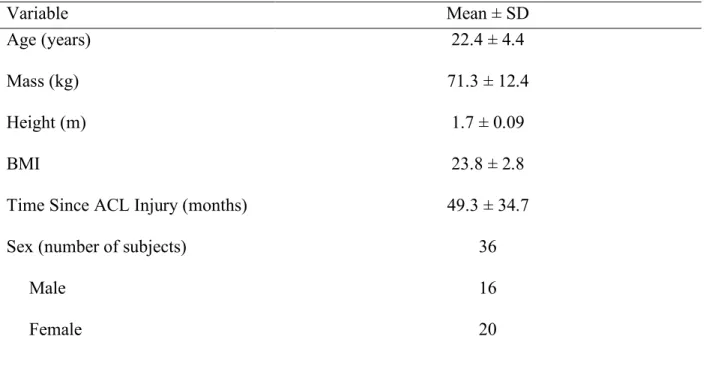
Thirty-six subjects were included for statistical analysis. Demographic data for these subjects is presented in Table 1.
Relationships Between PT, CSA, and EI of the Injured Limb
There were no significant relationships between CSA and normalized PT in either the vastus lateralis (r = 0.215, p = 0.215) or rectus femoris (r = 0.134, p = 0.44) of the injured limb. However, there was a significant moderate negative correlation between EI and PT in both the vastus lateralis (r = -0.514, p = 0.002) and rectus femoris (r = -0.554, p = 0.001) of the injured limb, indicating that poorer QMQ was associated with lower quadriceps strength. (See Table 2)
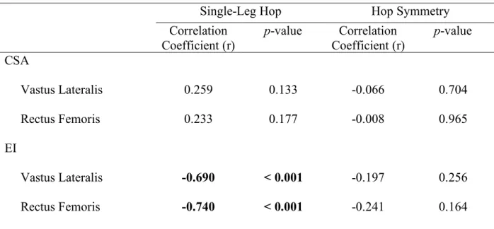
Relationship Between Single-Leg Hop Distance, CSA, and EI
There were no significant relationships between CSA and single-leg hop distance for either the vastus lateralis (r = 0.259, p = 0.133) or rectus femoris (r = 0.233, p = 0.177). Additionally, there were no significant correlations between CSA and single-leg hop limb
Table 1: Demographics of Subjects Included in Analysis (n = 36)
Variable Mean ± SD
Age (years) Mass (kg) Height (m) BMI
Time Since ACL Injury (months) Sex (number of subjects)
Male Female
22.4 ± 4.4 71.3 ± 12.4
1.7 ± 0.09 23.8 ± 2.8 49.3 ± 34.7
Table 2: Partial Correlations between EI, CSA values and PT in ACLR Limb (n = 36)
Correlation Coefficient (r) p-value CSA
Vastus Lateralis Rectus Femoris EI
Vastus Lateralis Rectus Femoris
0.215
0.134
-0.514
-0.554
0.215 0.444
0.002
0.001
Table 3: Partial Correlations between EI, CSA values and Single-Leg Hop (n = 36)
Single-Leg Hop Hop Symmetry
Correlation Coefficient (r)
CHAPTER 5: DISCUSSION
The primary findings of this study were that there was a moderate negative correlation between EI and PT and a strong negative correlation between EI and single-leg hop distance in the ACLR limb. These findings are consistent with the experimental hypotheses. It was
hypothesized that higher EI values (i.e. poorer QMQ) would be associated with lower quadriceps function (PT) and poorer functional outcomes (i.e. single-leg hop). Our findings suggest that muscle quality is indicative of quadriceps function and functional ability in individuals with ACLR, and may be a potential RTS criterion following ACLR and the rehabilitation process.
Our finding that EI was significantly correlated with PT for both the vastus lateralis and rectus femoris is supported by previous research. Wilhelm et al. (18) evaluated the correlation between EI of the four quadriceps muscles and muscular performance in elderly men, and reported significant negative correlations between EI and PT, consistent with our findings. Watanabe et al. (15) also demonstrated a negative correlation between EI and knee extension strength in elderly men, which was also found in our study. Furthermore, Rech et al. (17) found significant negative correlations between rectus femoris EI and PT in elderly women. These results coupled with our own indicate that poorer QMQ is associated with lower muscle strength (32). Ours is the first study, to our knowledge, to report this association in young individuals following ACLR. These findings are significant because fewer contractile fibers may
of an altered ratio of contractile fibers and IMAT, indicating that an underlying mechanism as to why muscle strength is compromised may be a shift towards a higher proportion of IMAT post-ACLR. Additionally, EI may indicate the extent of quadriceps dysfunction due to this
association. Previous research has suggested that contributors to quadriceps dysfunction may include AMI (6,13,14). Our study demonstrated that another potential origin of quadriceps dysfunction following ACLR may be IMAT since it can limit PT, which can be a rehabilitative focus coupled with AMI and IMAT. Other ways to measure IMAT include MRI and computed tomography (CT) (71). However, while these imaging techniques have high resolution, they are both costly and not always clinically feasible (71). Ultrasound is safe and widely available in the clinical setting, which is why ultrasound might be advantageous compared with other methods that assess IMAT.
Our study did not find a significant correlation between CSA and PT in either the vastus lateralis or rectus femoris. The main reason that this correlation was not significant is because we evaluated PT normalized to body mass to account for the notion that larger individuals have larger muscles and therefore produce more force. Body mass is typically used as a proxy for muscle CSA because it is easier to measure. However, had strength and CSA not been
the hop distance in the involved limb was correlated with EI but when the involved limb hop distance was expressed as a ratio including the uninvolved limb hop distance, there was no significant association. Hegedus et al. (72) demonstrated that when compared to a healthy
control group, the uninvolved leg in ACLR individuals is also significantly weaker in addition to the involved leg. Wellsandt et al. (10) evaluated the use of the uninvolved limb single leg hop distance in calculating LSI as a RTS criterion and demonstrated that considering the uninvolved limb in LSI may not be optimal. Additionally, Wellsandt et al. (10) demonstrated that meeting LSI criteria for RTS did not ensure that prior functional levels were met. Potential reasons contributing to weaker quadriceps in the contralateral limb are decreased physical activity aside from walking and activities of daily living during rehabilitation prior to RTS (10) as well as bilateral AMI (6). This implies that the uninvolved limb is also affected by the ACL injury which means that LSI may underestimate performance deficits (73,74). This may be a reason why EI was not significantly associated with LSI but was significantly associated with single-leg hop distance of the injured limb. However, if the contralateral limb was also impaired to a similar magnitude to the ACLR limb, resulting in a greater LSI closer to 1.00 for all subjects such that there was little between-subject variability in one of the variables, this mathematically would make the correlation smaller because there would be no variability, limiting significant findings. Decreased functional performance of the contralateral limb may overestimate LSI, therefore indicating a potentially inaccurate representation of the functional ability of the involved limb (10).
since CSA does not indicate the composition or quality of the muscle (23). The vagueness of this variable may contribute to why these results were not significant not just for LSI, but also single-leg hop distances of the involved single-leg.
These findings are clinically relevant because they may be useful in making RTS decisions. Werner et al. (31) demonstrated that RTS decisions primarily based on physical performance may not be ideal. Wellsandt et al. (10) performed a study in which 70 athletes completed a quadriceps strength test and 4 single-leg hop tests pre-ACLR and 6 months post-ACLR, from which LSIs were calculated. They demonstrated that LSIs often overestimated knee function and that limb symmetry, strength assessments, and hop tests may not be consistent and reliable RTS criteria because they do not guarantee prior function levels have been met.
Therefore, other measures may need to be considered such as QMQ which can be assessed with ultrasound EI. Although single-leg hop distance is cheaper and easier to implement in the clinic than ultrasound imaging, there are several limitations of using this for RTS decisions even though QMQ and single-leg hop distance are correlated. One of these is that other muscle groups may compensate for the quadriceps when doing a single-leg hop, therefore masking the
Our study demonstrated that EI and single-leg hop distances were significantly associated. While single-leg hop tests are one of the primary and most commonly used RTS criteria (29), some literature indicates concerns that single-leg hop distances may not be valid RTS criteria (10,31). Assessing QMQ may eliminate areas of concern in leg hop distances. On single-leg hop tests, the force generated by the quadriceps may be influenced by neural alterations from ACLR. Ultrasound EI does not consider this, as it is a more passive measurement. Furthermore, the ACLR patient may compensate via heightened ankle plantarflexor effort during single-leg hop tests (60). Button et al. (60) not only demonstrated that there was increased peak internal ankle plantarflexion moment during single-leg hop tests, but also that this is a consistently used compensation strategy post-ACLR. There may also be compensation by the hip extensors, therefore limiting the use of the knee extensors (60). By using these compensatory strategies during single-leg hop tests, the results may not accurately reflect quadriceps function.
Ultrasound EI eliminates the possibility of musculature crossing other joints compensating for the knee, as it solely considers the quadriceps rather than including potential measurements at the ankle and hip joints. Therefore, assessing QMQ may be a potential RTS criterion post-ACLR.
Age-related changes in QMQ may have occurred independent of ACLR, therefore being a potential confounding variable. Fellner et al. (75) evaluated age-related changes in both the QMQ and quadriceps muscle quantity via MRI in a rat model to relate these results to the field of geriatrics. They found that advancing age up to 21 months led to significantly decreased body weight (BW), CSA, CSA/BW, and lower QMQ due to an increase in IMAT. This may also be the case in post-ACLR individuals and be a confounding variable when relating QMQ to RTS decisions post-ACLR. Our study addressed this limitation to some level by limiting the age range from 18 to 35 years, controlling for potential age-related changes in QMQ. However, this is still a range of 17 years. Other variables affecting QMQ need to be considered in future studies. Another limitation in our study was the sample size, as this study only included 36 subjects for statistical analysis.
REFERENCES
1. Panzarella M. ACL Injury 101. American Orthopaedic Society for Sports Medicine [Internet]. 2015 [cited 2018 Aug 16];(Winter 2015). Available from:
https://www.sportsmed.org/AOSSMIMIS/members/downloads/InMotionArchives/2015Wi nter.pdf
2. Mather RC, Koenig L, Kocher MS, Dall TM, Gallo P, Scott DJ, et al. Societal and Economic Impact of Anterior Cruciate Ligament Tears. J Bone Joint Surg Am. 2013 Oct 2;95(19):1751–9.
3. Luc B, Gribble PA, Pietrosimone BG. Osteoarthritis Prevalence Following Anterior Cruciate Ligament Reconstruction: A Systematic Review and Numbers-Needed-to-Treat Analysis. J Athl Train. 2014;49(6):806–19.
4. Muthuri SG, McWilliams DF, Doherty M, Zhang W. History of knee injuries and knee osteoarthritis: a meta-analysis of observational studies. Osteoarthritis and Cartilage. 2011 Nov;19(11):1286–93.
5. Culvenor AG, Collins NJ, Guermazi A, Cook JL, Vicenzino B, Khan KM, et al. Early knee osteoarthritis is evident one year following anterior cruciate ligament reconstruction: a magnetic resonance imaging evaluation. Arthritis & Rheumatology (Hoboken, NJ). 2015 Apr;67(4):946–55.
6. Hart JM, Pietrosimone B, Hertel J, Ingersoll CD. Quadriceps Activation Following Knee Injuries: A Systematic Review. J Athl Train. 2010;45(1):87–97.
7. Tengman E, Brax Olofsson L, Stensdotter AK, Nilsson KG, Häger CK. Anterior cruciate ligament injury after more than 20 years. II. Concentric and eccentric knee muscle strength. Scandinavian Journal of Medicine & Science in Sports. 2014 Dec;24(6):e501-509.
8. Radin EL, Burr DB, Caterson B, Fyhrie D, Brown TD, Boyd RD. Mechanical determinants of osteoarthrosis. Semin Arthritis Rheum. 1991 Dec;21(3 Suppl 2):12–21.
9. Ewers BJ, Jayaraman VM, Banglmaier RF, Haut RC. Rate of blunt impact loading affects changes in retropatellar cartilage and underlying bone in the rabbit patella. J Biomech. 2002 Jun;35(6):747–55.
10. Wellsandt E, Failla M, Snyder-Mackler L. Limb Symmetry Indexes Can Overestimate Knee Function After ACL Injury. J Orthop Sports Phys Ther. 2017 May;47(5):334–8. 11. Khandha A, Manal K, Wellsandt E, Capin J, Snyder-Mackler L, Buchanan TS. Gait
mechanics in those with/without medial compartment knee osteoarthritis 5 years after anterior cruciate ligament reconstruction. J Orthop Res. 2017;35(3):625–33.
13. Hopkins JT, Ingersoll CD. Arthrogenic muscle inhibition: a limiting factor in joint rehabilitation. Journal of Sport Rehabilitation. 2000 May;9(2):135–59.
14. Addison O, Marcus RL, LaStayo PC, Ryan AS. Intermuscular Fat: A Review of the Consequences and Causes. Int J Endocrinol [Internet]. 2014 [cited 2018 Jun 11];2014. Available from: https://www.ncbi.nlm.nih.gov/pmc/articles/PMC3910392/
15. Watanabe Y, Yamada Y, Fukumoto Y, Ishihara T, Yokoyama K, Yoshida T, et al. Echo intensity obtained from ultrasonography images reflecting muscle strength in elderly men. Clin Interv Aging. 2013;8:993–8.
16. Fukumoto Y, Ikezoe T, Yamada Y, Tsukagoshi R, Nakamura M, Mori N, et al. Skeletal muscle quality assessed from echo intensity is associated with muscle strength of middle-aged and elderly persons. Eur J Appl Physiol. 2012 Apr;112(4):1519–25.
17. Rech A, Radaelli R, Goltz FR, da Rosa LHT, Schneider CD, Pinto RS. Echo intensity is negatively associated with functional capacity in older women. Age (Dordr) [Internet]. 2014 Oct [cited 2018 Jun 11];36(5). Available from:
https://www.ncbi.nlm.nih.gov/pmc/articles/PMC4453939/
18. Wilhelm EN, Rech A, Minozzo F, Radaelli R, Botton CE, Pinto RS. Relationship between quadriceps femoris echo intensity, muscle power, and functional capacity of older men. Age (Dordr) [Internet]. 2014 Jun [cited 2018 Jun 11];36(3). Available from:
https://www.ncbi.nlm.nih.gov/pmc/articles/PMC4082605/
19. Akima H, Yoshiko A, Tomita A, Ando R, Saito A, Ogawa M, et al. Relationship between quadriceps echo intensity and functional and morphological characteristics in older men and women. Archives of Gerontology and Geriatrics. 2017 May;70:105–11.
20. Pinto RS, Correa CS, Radaelli R, Cadore EL, Brown LE, Bottaro M. Short-term strength training improves muscle quality and functional capacity of elderly women. Age (Dordr). 2014 Feb;36(1):365–72.
21. Young H-J, Jenkins NT, Zhao Q, McCully KK. Measurement of Intramuscular Fat by Muscle Echo Intensity. Muscle Nerve. 2015 Dec;52(6):963–71.
22. Kuenze CM, Blemker SS, Hart JM. Quadriceps function relates to muscle size following ACL reconstruction. J Orthop Res. 2016;34(9):1656–62.
23. Palmer TB, Akehi K, Thiele RM, Smith DB, Thompson BJ. Reliability of Panoramic Ultrasound Imaging in Simultaneously Examining Muscle Size and Quality of the Hamstring Muscles in Young, Healthy Males and Females. Ultrasound in Medicine & Biology. 2015 Mar;41(3):675–84.
25. Andriacchi TP, Briant PL, Bevill SL, Koo S. Rotational Changes at the Knee after ACL Injury Cause Cartilage Thinning. Clinical Orthopaedics and Related Research®. 2006 Jan;442:39.
26. Blackburn JT, Pietrosimone B, Harkey MS, Luc BA, Pamukoff DN. Quadriceps Function and Gait Kinetics after Anterior Cruciate Ligament Reconstruction. Medicine & Science in Sports & Exercise. 2016 Sep;48(9):1664.
27. Tillquist M, Kutsogiannis DJ, Wischmeyer PE, Kummerlen C, Leung R, Stollery D, et al. Bedside Ultrasound Is a Practical and Reliable Measurement Tool for Assessing
Quadriceps Muscle Layer Thickness. JPEN J Parenter Enteral Nutr. 2014 Sep;38(7):886– 90.
28. Abrams GD, Harris JD, Gupta AK, McCormick FM, Bush-Joseph CA, Verma NN, et al. Functional Performance Testing After Anterior Cruciate Ligament Reconstruction. Orthop J Sports Med [Internet]. 2014 Jan 21 [cited 2018 May 31];2(1). Available from:
https://www.ncbi.nlm.nih.gov/pmc/articles/PMC4555525/
29. Davies GJ, McCarty E, Provencher M, Manske RC. ACL Return to Sport Guidelines and Criteria. Curr Rev Musculoskelet Med. 2017 Jul 12;10(3):307–14.
30. Sueyoshi T, Nakahata A, Emoto G, Yuasa T. Single-Leg Hop Test Performance and Isokinetic Knee Strength After Anterior Cruciate Ligament Reconstruction in Athletes. Orthop J Sports Med [Internet]. 2017 Nov 14 [cited 2018 May 31];5(11). Available from: https://www.ncbi.nlm.nih.gov/pmc/articles/PMC5692146/
31. Werner JL, Burland JP, Mattacola CG, Toonstra J, English RA, Howard JS. Decision to Return to Sport Participation After Anterior Cruciate Ligament Reconstruction, Part II: Self-Reported and Functional Performance Outcomes. Journal of Athletic Training [Internet]. 2018 May 18 [cited 2018 Jun 5]; Available from:
http://natajournals.org/doi/10.4085/1062-6050-328-16
32. Cadore EL, González-Izal M, Pallarés JG, Rodriguez-Falces J, Häkkinen K, Kraemer WJ, et al. Muscle conduction velocity, strength, neural activity, and morphological changes after eccentric and concentric training. Scand J Med Sci Sports. 2014 Oct;24(5):e343-352. 33. SCHMITT LC, PATERNO MV, HEWETT TE. The Impact of Quadriceps Femoris
Strength Asymmetry on Functional Performance at Return to Sport Following Anterior Cruciate Ligament Reconstruction. J Orthop Sports Phys Ther. 2012 Sep;42(9):750–9. 34. Griffin LY, Albohm MJ, Arendt EA, Bahr R, Beynnon BD, Demaio M, et al.
Understanding and preventing noncontact anterior cruciate ligament injuries: a review of the Hunt Valley II meeting, January 2005. Am J Sports Med. 2006 Sep;34(9):1512–32. 35. Moses B, Orchard J, Orchard J. Systematic Review: Annual Incidence of ACL Injury and
36. Dare D, Rodeo S. Mechanisms of Post-traumatic Osteoarthritis After ACL Injury. Current Rheumatology Reports [Internet]. 2014 Oct [cited 2018 Oct 6];16(10). Available from: http://link.springer.com/10.1007/s11926-014-0448-1
37. Brophy RH, Wright RW, Matava MJ. Cost Analysis of Converting from Single-Bundle to Double-Bundle Anterior Cruciate Ligament Reconstruction. Am J Sports Med. 2009 Apr 1;37(4):683–7.
38. Sepúlveda F, Sánchez L, Amy E, Micheo W. Anterior Cruciate Ligament Injury: Return to Play, Function and Long-Term Considerations. Curr Sports Med Rep. 2017 Jun;16(3):172– 8.
39. Felson DT, Lawrence RC, Dieppe PA, Hirsch R, Helmick CG, Jordan JM, et al.
Osteoarthritis: new insights. Part 1: the disease and its risk factors. Ann Intern Med. 2000 Oct 17;133(8):635–46.
40. Lawrence RC, Felson DT, Helmick CG, Arnold LM, Choi H, Deyo RA, et al. Estimates of the prevalence of arthritis and other rheumatic conditions in the United States. Part II. Arthritis Rheum. 2008 Jan;58(1):26–35.
41. Thomas AC, Hubbard-Turner T, Wikstrom EA, Palmieri-Smith RM. Epidemiology of Posttraumatic Osteoarthritis. Journal of Athletic Training. 2017 Jun 2;52(6):491–6. 42. Zhang Y, Jordan JM. Epidemiology of Osteoarthritis. Clin Geriatr Med. 2010
Aug;26(3):355–69.
43. MURPHY L, SCHWARTZ TA, HELMICK CG, RENNER JB, TUDOR G, KOCH G, et al. Lifetime Risk of Symptomatic Knee Osteoarthritis. Arthritis Rheum. 2008 Sep
15;59(9):1207–13.
44. Deshpande BR, Katz JN, Solomon DH, Yelin EH, Hunter DJ, Messier SP, et al. The number of persons with symptomatic knee osteoarthritis in the United States: Impact of race/ethnicity, age, sex, and obesity. Arthritis Care Res (Hoboken). 2016 Dec;68(12):1743– 50.
45. Becker R, Berth A, Nehring M, Awiszus F. Neuromuscular quadriceps dysfunction prior to osteoarthritis of the knee. J Orthop Res. 2004 Jul;22(4):768–73.
46. Sonnery-Cottet B, Saithna A, Quelard B, Daggett M, Borade A, Ouanezar H, et al.
Arthrogenic muscle inhibition after ACL reconstruction: a scoping review of the efficacy of interventions. British Journal of Sports Medicine. 2018 Sep 7;bjsports-2017-098401. 47. L Latash M. Neurophysiological Basis of Movement. 2008.
50. Newham D, Hurley M, Jones DW. Ligamentous knee injuries and muscle inhibition. J Orthop Rheumatol. 1989 Jan 1;2:163–73.
51. Palmieri-Smith RM, Thomas AC. A neuromuscular mechanism of posttraumatic
osteoarthritis associated with ACL injury. Exerc Sport Sci Rev. 2009 Jul;37(3):147–53. 52. Yoshida Y, Marcus RL, Lastayo PC. Intramuscular adipose tissue and central activation in
older adults. Muscle & Nerve. 46(5):813–6.
53. MARCUS RL, ADDISON O, KIDDE JP, DIBBLE LE, LASTAYO PC. SKELETAL MUSCLE FAT INFILTRATION: IMPACT OF AGE, INACTIVITY, AND EXERCISE. J Nutr Health Aging. 2010 May;14(5):362–6.
54. Suetta C, Aagaard P, Rosted A, Jakobsen AK, Duus B, Kjaer M, et al. Training-induced changes in muscle CSA, muscle strength, EMG, and rate of force development in elderly subjects after long-term unilateral disuse. Journal of Applied Physiology. 2004 Nov 1;97(5):1954–61.
55. Nilwik R, Snijders T, Leenders M, Groen BBL, van Kranenburg J, Verdijk LB, et al. The decline in skeletal muscle mass with aging is mainly attributed to a reduction in type II muscle fiber size. Experimental Gerontology. 2013 May 1;48(5):492–8.
56. Menzer H, Slater LV, Diduch D, Miller M, Norte G, Goetschius J, et al. The Utility of Objective Strength and Functional Performance to Predict Subjective Outcomes After Anterior Cruciate Ligament Reconstruction. Orthop J Sports Med [Internet]. 2017 Dec 18 [cited 2018 May 31];5(12). Available from:
https://www.ncbi.nlm.nih.gov/pmc/articles/PMC5753987/
57. Joreitz R, Lynch A, Rabuck S, Lynch B, Davin S, Irrgang J. PATIENT-SPECIFIC AND SURGERY-SPECIFIC FACTORS THAT AFFECT RETURN TO SPORT AFTER ACL RECONSTRUCTION. Int J Sports Phys Ther. 2016 Apr;11(2):264–78.
58. de David A, Stergiou P, Stefanyshyn D. JOINT MOMENTS DURING WALKING AND RUNNING AT DIFFERENT SPEEDS. :2.
59. Zabala ME, Favre J, Scanlan SF, Donahue J, Andriacchi TP. Three-dimensional knee moments of ACL reconstructed and control subjects during gait, stair ascent, and stair descent. Journal of Biomechanics. 2013 Feb;46(3):515–20.
60. Button K, Roos PE, van Deursen RWM. Activity progression for anterior cruciate ligament injured individuals. Clin Biomech (Bristol, Avon). 2014 Feb;29(2):206–12.
62. Kline PW, Morgan K, Johnson DL, Ireland ML, Noehren B. Impaired quadriceps rate of torque development and knee mechanics after anterior cruciate ligament reconstruction with patellar tendon autograft. Am J Sports Med. 2015 Oct;43(10):2553–8.
63. Pietrosimone B, Blackburn JT, Harkey MS, Luc BA, Hackney AC, Padua DA, et al. Greater Mechanical Loading During Walking Is Associated With Less Collagen Turnover in Individuals With Anterior Cruciate Ligament Reconstruction. The American Journal of Sports Medicine. 2016 Feb;44(2):425–32.
64. Sandberg R, Balkfors B. Reconstruction of the anterior cruciate ligament. A 5-year follow-up of 89 patients. Acta Orthop Scand. 1988 Jun;59(3):288–93.
65. Otto D, Pinczewski LA, Clingeleffer A, Odell R. Five-Year Results of Single-Incision Arthroscopic Anterior Cruciate Ligament Reconstruction with Patellar Tendon Autograft. The American Journal of Sports Medicine. 1998 Mar;26(2):181–8.
66. Bak K, Scavenius M, Hansen S, Nørring K, Jensen KH, Jørgensen U. Isolated partial rupture of the anterior cruciate ligamentLong-term follow-up of 56 cases. Knee Surgery. 1997 Jun 1;5(2):66–71.
67. Wiggins AJ, Grandhi RK, Schneider DK, Stanfield D, Webster KE, Myer GD. Risk of Secondary Injury in Younger Athletes After Anterior Cruciate Ligament Reconstruction. Am J Sports Med. 2016 Jul;44(7):1861–76.
68. Grindem H, Snyder-Mackler L, Moksnes H, Engebretsen L, Risberg MA. Simple decision rules can reduce reinjury risk by 84% after ACL reconstruction: the Delaware-Oslo ACL cohort study. Br J Sports Med. 2016 Jul;50(13):804–8.
69. Kleinberg CR, Ryan ED, Tweedell AJ, Barnette TJ, Wagoner CW. Influence of Lower Extremity Muscle Size and Quality on Stair-Climb Performance in Career Firefighters. J Strength Cond Res. 2016;30(6):1613–8.
70. Ryan ED, Shea NW, Gerstner GR, Barnette TJ, Tweedell AJ, Kleinberg CR. The influence of subcutaneous fat on the relationship between body composition and ultrasound-derived muscle quality. Appl Physiol Nutr Metab. 2016 Oct;41(10):1104–7.
71. Heymsfield SB, Gonzalez MC, Lu J, Jia G, Zheng J. Skeletal muscle mass and quality: evolution of modern measurement concepts in the context of sarcopenia. Proceedings of the Nutrition Society. 2015 Nov;74(04):355–66.
72. Hegedus EJ, McDonough S, Bleakley C, Cook CE, Baxter GD. Clinician-friendly lower extremity physical performance measures in athletes: a systematic review of measurement properties and correlation with injury, part 1. The tests for knee function including the hop tests. Br J Sports Med. 2015 May 1;49(10):642–8.
ligament reconstruction in recreational athletes. Human Movement Science. 2015 Feb 1;39:73–87.
74. Gokeler A, Welling W, Benjaminse A, Lemmink K, Seil R, Zaffagnini S. A critical analysis of limb symmetry indices of hop tests in athletes after anterior cruciate ligament
reconstruction: A case control study. Orthopaedics & Traumatology: Surgery & Research. 2017 Oct 1;103(6):947–51.