SHORT PALATE LUNG AND NASAL EPITHELIUM 1 AND AIRWAY DISEASE
Julianne Jahui Huang
A dissertation submitted to the faculty at the University of North Carolina at Chapel Hill in partial fulfillment of the requirements for the degree of Doctor of Philosophy in the
Department of Chemistry (Biological Division).
Chapel Hill 2016
Approved by:
Matthew R. Redinbo
Stephen L. Tilley
Robert Tarran
Eric M. Brustad
ii © 2016
iii
ABSTRACT
Julianne Jahui Huang: Short Palate Lung and Nasal Epithelium Clone 1 and Airway Disease
(Under the direction of Matthew R. Redinbo and Stephen L. Tilley) Airway disease such as asthma and infection is the cause substantial
morbidity and mortality in the world today. Although modern medicine has developed many drugs for these conditions, these diseases remain highly prevalent and are often difficult to treat.
Short palate, lung and nasal epithelium clone 1 (SPLUNC1) is an abundant multi-functional protein in the airway. It has been reported to have
immune-modulatory, surfactant and anti-microbial functions, and it regulates the airway surface liquid (ASL) height through the epithelial sodium channel (ENaC). This study focuses on utilizing SPLUNC1’s protective properties in combatting airway disease.
iv
Pseudomonas aeruginosa, a primary lung pathogen in nosocomial pneumonia and in lung diseases such as cystic fibrosis and chronic obstructive pulmonary disease (COPD), causes considerable morbidity and mortality. SPLUNC1 has been shown to neutralize and combat P. aeruginosain vivo and in vitro. Here, we sought to establish a model for evaluating delivery of exogenous SPLUNC1 in acute lung infection and provide evidence that preemptive administration of SPLUNC1 may decrease bacterial burden.
Lastly, we suggest that the administration of SPLUNC1 which we propose for asthma and lung infection results in SPLUNC1 mediated SPLUNC1 release in the lungs, effectively increasing the local protein concentration. This effect may utilize SPLUNC1’s natural protective properties to combat airway disease.
v
ACKNOWLEDGEMENTS
First and foremost, I must thank my PIs Matt and Steve for this incredible journey. Matt, even though you are a crystallographer, you allowed me (after solving my obligatory crystal structure) to wander off into uncharted territory on a new
project, and start a new collaboration based in cell and mouse work! The confidence you and Steve both had in me to get the work done played an enormous part in my success. You were both always there for support when I needed it but largely left me to my own devices, resulting in my independence and ownership of my project. You have left a strong impression with the scientific integrity both of you have shown throughout my time in graduate school that I will carry throughout the rest of my career. Steve, it has been amazing coming to your lab and having the opportunity to do this translational research. I have learned so much from your approach to
science. You are hilarious for frequently trying to have lab meeting at TOPO on Friday afternoons instead of running experiments. These attitudes set the tone for morale in the lab and for that I am extremely grateful.
Thanks to Rob Tarran for the financial support, scientific ideas and for
including me in social and scientific activities with your lab. Thanks to Andy Ghio for the human samples and Hong Dang for the help with stats.
vi
the first publication and for the friendships you helped pave when I first arrived. Thanks to Bill and Mike for all the proteins. I’m so thankful for not having to spend very much time in that cold cold lab, waiting for my OD600 and for the ÄKTA to spit out my proteins. Thanks to Herodes for hanging the drops leading to my crystal structure and to Ashley for help with cloning PilY1. Rebecca, you are amazing for always keeping things organized and for having answers to tons of miscellaneous scientific and administrative questions I had from across campus.
vii
contributions to my project. Thanks especially to Dan and Austin for your high quality work that has led to figures in my manuscript and dissertation.
To my family, for all your support. Thanks for picking up my calls and listening to my stories through all that life has thrown my way. Thanks, Mom, for always checking up on me and for listening to all my pipe leaking and appliance breaking stories and for coming to make sure my house is still intact each year. Thanks for your never-ending support in life no matter what I’ve chosen to get myself into. Chuck, thanks for all the car advice from across the country (I’ve needed a lot of advice!) and for making sure I’m okay in the event of natural disasters. Jaching, my favorite family story related to grad school was the time you mistook my “making protein” for cooking steak. Even when you didn’t understand what I was doing, you always had my back and I felt that.
To all the friends I’ve made in graduate school, you have been integral in helping me get through this! I would not have made it out with as much sanity as I have left without you. Jet, my cubby buddy in Kenan, thanks for your friendship and all the scientific and nonscientific conversations over the years. And thanks for helping to get my butt to the gym. Thanks, Mike, for being there from quals to the busiest part of my time here. Thanks for accompanying me to campus, for the rides home and for your patience. Erica, I’m so grateful I met you during interview
viii
ix
TABLE OF CONTENTS
LIST OF FIGURES ... xiii
LIST OF ABBREVIATIONS AND SYMBOLS ...xv
CHAPTER 1: INTRODUCTION ... 1
Asthma ... 1
SPLUNC1 ... 6
Summary ... 10
CHAPTER 2: SPLUNC1 AND ASTHMA/ALLERGIC INFLAMMATION ... 13
Introduction ... 13
Materials and Methods ... 13
Animals ... 13
HDM-induced Allergic Airway Inflammation ... 14
BAL Fluid from Humans and Mice ... 14
Human Bronchial Epithelial Cell Culture ... 14
Measurement of AHR ... 15
SPLUNC1 ELISA ... 15
Western Blot ... 15
Protein Preparation ... 15
x
Bone Marrow Mast Cell Culture ... 16
Bone Marrow Mast Cell IL-13 Assay ... 16
Bone Marrow Mast Cell Hexosaminidase Release ... 17
Statistics ... 17
Study Approval ... 17
Results ... 18
SPLUNC1 is an Epithelial-derived Factor that is Reduced in Asthmatic Human and Allergic Mouse BAL ... 18
SPLUNC1 Regulates AHR... 19
K138E Mutant Structure and Function ... 19
The Molecular Basis for SPLUNC1’s Effects on AHR ... 20
SPLUNC1’s Effect on AHR is Independent of Effects on ASL Height ... 20
SPLUNC1’s N-terminus is Critical for AHR Reduction ... 21
SPLUNC1’s Effect on AHR is Dependent on the Coordination of its N-terminus with an Electrostatic Patch on the Protein Body ... 22
SPLUNC1’s Mechanism of Action for AHR Reduction ... 23
Discussion ... 24
CHAPTER 3: SPLUNC1 AND LUNG INFECTION... 40
Introduction ... 40
Materials and Methods ... 41
Animals ... 41
Protein Preparation ... 41
Bacterial and SPLUNC1 Dosing ... 41
xi
Statistics ... 42
Study Approval ... 42
Results ... 42
Discussion ... 43
Future Directions ... 44
CHAPTER 4: ADMINISTRATION OF SPLUNC1 RESULTS IN SECRETION OF SPLUNC1 ... 48
Introduction ... 48
Materials and Methods ... 48
Animals ... 48
HDM-induced Allergic Airway Inflammation ... 49
SPLUNC1 Dosing and BAL Collection ... 49
Cell Culture ... 49
SPLUNC1 ELISA ... 50
Western Blot ... 50
Protein Preparation ... 50
Statistics ... 50
Study Approval ... 51
Results ... 51
Administration of SPLUNC1 Results in Secretion of SPLUNC1 ... 51
SPLUNC1 Secretion is Specific to SPLUNC1 Administration ... 52
SPLUNC1 Concentration Increase is Not Sustained in Allergic Inflammation ... 52
xii
Future Directions ... 53
APPENDIX: P. aeruginosa Infection and PilY1 ... 60
Introduction ... 60
Aims ... 60
Preliminary Progress ... 62
P. aeruginosa PilY1 PAK Strain Amino Acids 532-1163 and 191-1163 and PA14 Strain Amino Acids 200-1170 Cloning ... 62
PAK PilY1 532-1163 Cloning into Avidity Vector for Biotinylation ... 63
Administration of Cyclic RGD Peptides to Mice ... 63
xiii
LIST OF FIGURES
Figure 1.1 SPLUNC1 and BPI Structural Alignment ... 12
Figure 2.1. SPLUNC1 is Reduced in Allergic Airways ... 29
Figure 2.2 HBECs Secrete SPLUNC1 ... 30
Figure 2.3 SPLUNC1-/- Mice are Hyperresponsive to Methacholine ... 31
Figure 2.4 Administration of Wildtype SPLUNC1Δ19 Abolishes Allergen-induced AHR ... 32
Figure 2.5 SPLUNC1K138E Protein Restoration of ASL-height at Acidic pH is Not the Result of a Structural Change ... 33
Figure 2.6. SPLUNC1’s Effect on AHR is Independent of Effects on ASL Height .... 34
Figure 2.7 SPLUNC1’s N-terminus is Critical for AHR Reduction ... 35
Figure 2.8 Both the N-terminus and Electrostatic Patch are Necessary for AHR-reduction ... 37
Figure 2.9 SPLUNC1 Exhibits Dose-dependent Reduction of IL-13 Secretion in Mast Cells ... 38
Figure 2.10 SPLUNC1 Does Not Reduce Allergen-induced Mast Cell Degranulation ... 39
Figure 3.1. The Jackson Laboratory vs. In-house Mouse PAO1 Infection ... 46
Figure 3.2 SPLUNC1 Reduces CFU Load in PAO1 Lung Infection ... 47
Figure 4.1. SPLUNC1 Increases SPLUNC1 Secretion by HBECs ... 56
xiv
Figure 4.3 SPLUNC1 Secretion in Mice is Specific to SPLUNC1 Administration ... 58
Figure 4.4 SPLUNC1 Secretion Remains High at least 24 h after SPLUNC1
xv
LIST OF ABBREVIATIONS AND SYMBOLS
% percent
° degree
± plus or minus
< less than
> greater than
µg microgram
µL microliter
µmol micromol
Å angstrom
A alanine
AAs allergic asthmatics
AHR airway hyperresponsiveness APC antigen presenting cell ASL airway surface liquid ASM airway smooth muscle ATP adenosine triphosphate BAL bronchoalveolar lavage
B. cenocepacia Burkholderia cenocepacia
BMMC bone marrow mast cell
BPI bactericidal/permeability-increasing
BPIFA1 bactericidal/permeability-increasing fold containing family member A1
xvi
C cysteine
cAMP cyclic adenosine monophosphate CD4 cluster of differentiation 4
CF cystic fibrosis
CFTR cystic fibrosis transmembrane conductance regulator CFUs colony forming units
CO2 carbon dioxide
COPD chronic obstructive pulmonary disease C-terminus carboxyl terminus
d day(s)
D aspartic acid
DNA deoxyribonucleic acid DNase deoxyribonuclease DNP dinitrophenyl albumin
E glutamic acid
EDSMRF epithelial-derived smooth muscle relaxing factor ENaC epithelial sodium channel
ELISA enzyme-linked immunosorbent assay EP electrostatic patch
FCεRI high-affinity receptor for the Fc region of immunoglobulin E
G glycine
h hour(s)
xvii
HBEC human bronchial epithelial cell HDM house dust mite
H. influenzae Haemophilus influenzae
His6 6 x histidine affinity tag HRP horseradish peroxidase
IACUC Institutional Animal Care and Use Committee ICS inhaled corticosteroids
ICU intensive care unit IgE immunoglobulin E
IL interleukin
i.n. intranasal
IPTG isopropyl-1-thio-D-galactopyranoside IRB Institutional Review Board
i.t. intratracheal
K lysine
kDa kilodalton
kg kilogram
LABA long-acting beta agonist
LB lysogeny broth
LIC ligation independent cloning
LPS lipopolysaccharide
M molar
xviii
MANOVA multivariate analysis of variance MBP maltose binding protein
MCC mucociliary clearance Mch methacholine
MDR multi-drug resistant
mg milligram
mL milliliter
mm millimeter mmol millimole
N asparagine
N-terminus amino terminus
ng nanogram
nm nanometer
NVs normal volunteers
OD600 optical density at 600 nm wavelength
OVA ovalbumin
P p-value
PanK pantothenate kinase
P. aeruginosa Pseudomonas aeruginosa
PAGE polyacrylamide gel
PBS phosphate-buffered saline
pH negative log (base 10) of the molar concentration of hydronium ions
xix
RANTES regulated on activation, normal T cell expressed and secreted; chemokine ligand 5
S serine
S18 N-terminal 18 amino acids of SPLUNC1 protein G22-A39
S. aureus Staphylococcus aureus
SDS sodium dodecyl sulfate SEM standard error of mean
SPLUNC1 short palate, lung and nasal epithelium clone 1 TBSA total body surface area
Th2 type 2 T helper cell
TMB 3,3',5,5'-tetramethylbenzidine Tris tris(hydroxymethyl)aminomethane
UNC University of North Carolina at Chapel Hill VAP ventilator associated pneumonia
vs versus
1
CHAPTER 1: INTRODUCTION
Asthma
Asthma, the most common chronic inflammatory lung disease in developed countries, affects approximately 26 million people annually in the United States alone and 300 million worldwide. Asthma has been estimated to cost 56 billion dollars in the United States and treating 20-30% of asthmatics constitutes 80% of the cost of managing all asthmatics (1, 2). The disease is influenced by both genetic and environmental components, such as pathogens and pollution, and has higher frequency and severity in boys compared to girls 0 to 14 years of age; interestingly, this prevalence reverses in adulthood with women having a higher incidence of asthma than men (3-5). The gender differences are attributed to sex hormones influencing β2 adrenoreceptors. Approximately 70% of asthmatics also have allergies and half of all asthmatics experience asthma attacks annually.
Asthma is characterized by three cardinal features: airway inflammation, airflow obstruction, and airway hyperresponsiveness (AHR). Allergic inflammation involves many steps, affects patients from mild to severe asthmatics, and results in airway remodeling that can be correlated to the severity of symptoms (6). Airway edema, mucus hypersecretion, and smooth muscle remodeling all contribute to airflow obstruction and ultimately exacerbations (7, 8). AHR or bronchial
2
features translate clinically into a number of symptoms including cough, wheezing, chest tightness, and shortness of breath. Untreated symptoms can progress into status asthmaticus (also termed near-fatal asthma), which results in respiratory failure and sometimes death. Approximately 4,000 people per year die from status asthmaticus in the United States.
Fortunately, a number of highly effective therapies exist to treat the underlying inflammation in asthma. These drugs control asthma well when taken on a regular basis. Inhaled corticosteroids (ICS) are the mainstay of controller medicines for asthma. They work by a number of mechanisms, including the inhibition of
proinflammatory cytokine synthesis. When ineffective alone, ICSs are combined with long-acting beta agonists (LABAs), long-acting anti-muscarinics, and leukotriene receptor antagonists. In addition to acting as long-term controller medicines, beta agonists are also used acutely to reverse bronchospasm by activating beta adrenergic receptors on airway smooth muscle (ASM), leading to rises in
intracellular cAMP which promotes smooth muscle relaxation. This action translates clinically to bronchodilation and a reduction of airflow obstruction.
The primary cell types mediating allergic asthma pathogenesis are dendritic cells, which are antigen presenting cells (APCs), B cells, mast cells, eosinophils, and T lymphocytes (T cells), specifically T helper 2 (Th2) cells that play a large role in allergic inflammation. Dendritic cells are the first cells activated in the pathway to allergic inflammation. They are residents on mucosal surfaces and are located near the basement membrane of the respiratory epithelium and function as cellular
3
in asthmatics and in an ovalbumin-sensitized allergic lung inflammation rat model (11, 12). They have been shown to play roles in AHR, increasing IgE concentration and in eosinophilia in the allergic airways. Dendritic cells are professional APCs and are key in adaptive immunity and also sensitization to antigens which results in allergic asthma via Th2 cells.
Th2 CD4+ T helper cells are responsible for the release of many cytokines relevant to asthma. Although they secrete cytokines, including IL-2, IL-3, IL-4, IL-5, IL-7, IL-9, IL-13, IL-15, IL-16 and IL-17, they are most well-characterized for their secretion of IL-4, IL-5 and IL-13 in asthma. IL-4 has been reported to be responsible for skewing to a Th2 phenotype by promoting Th2 cell differentiation and inhibiting Th1 differentiation, eosinophil expansion, B-cell growth resulting in IgE production, and mucus production (5, 13, 14). IL-5 is well known for causing eosinophilia by promoting proliferation and survival of eosinophils in the airway. When IL-5 was reduced in eosinophilic asthma by anti-IL-5 antibodies, lung function was improved, exacerbations were reduced and the quality of life was improved (15). IL-13 effects have overlap with IL-4 in that they enhance IgE and mucus production, induce eosinophilic inflammation and also play a role in AHR and airway remodeling (16, 17).
The principal involvement of B cells in asthma is in the production of IgE antibodies, which is a key mediator of the allergic response and has been linked directly to disease severity (5). B cells are influenced by IL-4 to undergo
4
Circulating IgE, released by B cells, binds to mast cells via their high affinity FCεRI receptors.
Mast cells are mucosal sentinels which, in normal tissues, have a role in angiogenesis, homeostasis, elimination of pathogens and play a role in innate and adaptive immunity (18). Although typically residing in the mucosa and connective tissues, they additionally congregate in smooth muscles and submucosal glands in asthmatic but not normal patients. Mast cells are activated in the asthmatic lung by a number of mechanisms. The best characterized mechanism is IgE-dependent
activation. Mast cells have high affinity FCεRI receptors that bind IgE which, after encountering antigens, cross-link leading to mast cell activation (19).
IgE-independent activation also occurs in asthma when mast cells are stimulated by mediators such as ATP and adenosine, which activate purinergic receptors exposed on the cell surface. Mast cell activation results in degranulation, lipid mediator
release, and cytokine synthesis. Mast cells, following activation by both
IgE-dependent and IgE-inIgE-dependent mechanisms, release a number of proinflammatory mediators including histamine, serotonin, prostaglandins and leukotrienes which cause smooth muscle contraction and airflow obstruction. Additionally, mast cells secrete the asthma-related cytokines IL-4, IL-5 and IL-13 which have previously been described as playing roles in eosinophilic airway inflammation and IgE synthesis. They also secrete profibrogenic cytokines leading to airway remodeling (20).
5
and inform management strategy (13). The presence of eosinophils is associated with the Th2 cytokines IL-4, IL-5, which cause eosinophil expansion and survival and IL-13 expression. Eosinophil recruitment is due to eotaxin, RANTES and IL-5 and one of the main eosinophil products, major basic protein, damages the epithelium, increases AHR and causes mast cell degranulation. Eosinophils also cause goblet cell metaplasia, matrix deposition and smooth muscle hypertrophy (21). In addition to playing a critical role in the pathogenesis of Th2-high allergic asthma, eosinophils also mediate non-allergic eosinophilic asthma. In this asthma endotype, cytokines produced by the airway epithelium (thymic stromal lymphopoietin, IL-25, and IL-33) stimulate innate lymphoid cells to produce IL-5 and IL-13. In contrast to allergic eosinophilic asthma mediated by Th2 cells, IL-4 is not produced by the innate
lymphoid cells, therefore IgE levels are low. Elucidation of these biological pathways involving mast cells and eosinophils has led to the development of new therapies including anti-IgE antibodies and anti-IL-5 antibodies, typically reserved for patients with severe asthma (22-24).
6
While mechanisms that underlie the development of airway inflammation and airflow obstruction are well-characterized, the pathophysiology of AHR remains less well understood. AHR is defined as heightened contraction of ASM following
exposure to stimuli that fail to or minimally stimulate contraction of ASM in normal subjects. Out of all the immune cells described above, only mast cells have been shown to play a critical role in AHR development. Mast cells infiltrate the ASM of asthmatics, and mediators released from mast cells are believed to contribute to AHR development. Whether or not ASM is intrinsically abnormal in asthma and in itself a major contributor to AHR development remains controversial. ASM
hypertrophy and hyperplasia have been reported to contribute to AHR pathogenesis in asthma (25, 26). However, some investigations of airway smooth muscle (ASM) from asthmatic subjects suggest that it is intrinsically normal but becomes
dysfunctional in the milieu of the asthmatic airway (27, 28).
Over 30 years ago, a critical role for the epithelium in producing mediators that limit AHR was demonstrated when enhanced contraction was observed after the epithelia was denuded from the bronchi of dogs (29). This observation has been confirmed in multiple species, including mice and humans (30-64). Collectively, these studies strongly support the existence of an epithelial-derived smooth muscle relaxing factor (EDSMRF) that limits AHR. However, to date the identity of this EDSMRF has remained elusive.
SPLUNC1
7
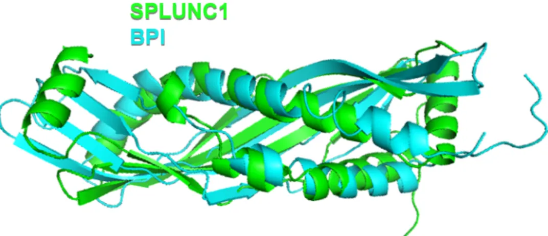
one of the most abundantly secreted proteins in mammalian airways, comprising up to 10% of total protein found in the airway surface liquid (ASL) (65). It was first discovered in mice in 1999 in a search for genes responsible for facial development and SPLUNC1 is primarily found in the airways and oral cavity; however, here we will mostly limit the discussion to airway involvement (66, 67). SPLUNC1 is encoded by genes on the long arm of chromosome 20 in the human genome and displays sequence and structural homology to the N-terminal portion of the antimicrobial bactericidal/permeability-increasing (BPI) protein (Figure 1.1) (68). It is a 256 amino acid long approximately 25 kDa protein which has a 19 amino acid signal sequence at its N-terminus that is cleaved off prior to the delivery of the mature protein to its final destination on mucosal surfaces.
SPLUNC1 is a multi-functional protein that regulates ASL height, possesses immunomodulatory and antimicrobial properties, and demonstrates surfactant actions (69). SPLUNC1 controls airway hydration by its inhibition of the sodium epithelial channel (ENaC) (70). This regulation results in homeostatic fluid
absorption in the airway and is believed to be important for maintaining normal ASL height, which is required for proper mucociliary clearance (MCC) essential for debris and pathogen removal in the lungs. SPLUNC1 accomplishes its regulation of ENaC through its N-terminal G22-A39 “S18” domain (71). Additionally, an electrostatic surface patch on the protein is responsible for presenting its N-terminal S18 region to ENaC in order to effect ASL height regulation (72).
8
allergic airway inflammation in mice (73, 74). SPLUNC1 levels are reduced in mouse lungs following OVA sensitization and knockout of the protein results in increased eosinophilic inflammation after OVA challenge (75). Additionally, Nogo-B, in the reticulon family of proteins, has been shown to regulate SPLUNC1 in the OVA model (76). Nogo-B-deficient mice demonstrate augmented eosinophilic lung inflammation in this model and SPLUNC1 was substantially reduced in the Nogo-B knockout mice.Interestingly, out of 40,000 genes examined in a genome-wide microarray comparing RNA expression from lungs of Nogo-deficient and wildtype mice, only SPLUNC1 was markedly reduced (278-fold or 95%). Restoration of SPLUNC1 by transgenic expression in the airway epithelium of Nogo-deficient mice rescued the phenotype, reverting inflammation back to wildtype levels. Collectively, these studies suggest that SPLUNC1 acts to limit the development of allergic airway inflammation, albeit by unknown mechanisms. Clinical data supporting these observations in mouse studies include studies showing that SPLUNC1 levels are low in the nasal lavage fluid and nasal tissues of patients with allergic rhinitis and chronic
rhinosinusitis particularly within nasal polyps (77, 78)
Due to SPLUNC1’s sequence similarity with the antimicrobial protein BPI, it was originally expected to have antimicrobial capabilities. Indeed, it has been shown to have activity against many bacteria including Pseudomonas aeruginosa,
Burkholderia cenocepacia, Haemophilus influenzae and others (79-83). Part of its antimicrobial effects stem from its capacity to affect MCC through ASL height
9
expected to disrupt biofilm formation potentially through its ability to function as a surfactant (84).
SPLUNC1 has been reported to spread the ASL at the air-liquid interface. While the purpose of liquid-spreading surfactants in the alveoli has been extensively studied, the function of surfactants, such as SPLUNC1, in the conducing airways is less well characterized. There are reports that surfactants in the conducting airways aid MCC by increasing ciliary beating. Surfactants regulate immune cells and bind allergens, and a lack of surfactant is correlated with increased airway resistance in the lungs (85). Therefore, the ability of SPLUNC1 to function as a surfactant is yet another method in which the protein is able to protect the lungs.
SPLUNC1 has been implicated in the pathogenesis of a number of disease states including cystic fibrosis (CF), lung cancer, allergy (described above) and chronic obstructive pulmonary disease (COPD). Both the cystic fibrosis
transmembrane conductance regulator (CFTR) and SPLUNC1 serve to negatively regulate ENaC. In CF, mutations in CFTR results in the decreased presence of this critical chloride channel on epithelial cells. The decreased pH in the CF lung reduces SPLUNC1’s ability to inhibit ENaC (72). The combination of the inability CFTR and SPLUNC1 to inhibit CFTR in CF results in the dehydration of the ASL due to excess sodium transport and leads to a dysfunction in MCC. Despite the increased
SPLUNC1 in the CF lung, there is an increased incidence of bacterial colonization, particularly with P. aeruginosa, B. cepacia and H. influenzae (86-89).
10
detected outside its locations in the normal lung. It has been found in pleural effusions and lymph nodes and has been suspected of protectively reducing an oncogene correlated with nasopharyngeal carcinoma (69). Therefore, SPLUNC1 regulation in terms of cancer depends on the particular disease.
Finally, SPLUNC1 has been investigated in COPD and the results are varied. In one study, SPLUNC1 levels have been shown to be increased in sputum from patients with COPD (90). However, another study was not able to confirm this finding, showing instead no difference between patients with COPD and normal subjects (91). Therefore, more studies are necessary to determine whether or not SPLUNC1 may play a role in this common airway disease.
Summary
Asthma is a common chronic inflammatory airway disease affecting
approximately 8% of adults and 10% of children in the United States. It is influenced by genetic and environmental components and causes wheezing, shortness of breath, coughing and chest tightness in patients with the disease. Its defining
characteristics include airway inflammation resulting in airway remodeling, reversible airflow obstruction, mucus hypersecretion and AHR. Although the pathobiology of airway inflammation and airflow obstruction is well characterized, AHR is less well defined. Studies in multiple species, including mouse and human, strongly suggest the existence of an EDSMRF; however, the identity of such a factor(s) has remained elusive.
11
tension reduction, antimicrobial function, and modulation of immune cell function. Its location in the airways facilitate these many protective abilities and its dysregulation has been indicated in several disease states including CF, lung cancer, and allergy. Although SPLUNC1 has been investigated in terms of allergic rhinitis and chronic rhinosinusitis, it has not been investigated in human allergic asthma.
Here, we report SPLUNC1 levels in allergic asthmatics and house dust mite (HDM)-allergic mice, and present data suggesting a non-redundant role for
12
Figure 1.1 SPLUNC1 and BPI Structural Alignment
13
CHAPTER 2: SPLUNC1 AND ASTHMA/ALLERGIC INFLAMMATION
Introduction
SPLUNC1 is a multifunctional protective protein in the human airways that has been implicated in allergic airway inflammation such as in allergic rhinitis and chronic rhinosinusitis. It has also been investigated in mouse models of allergic inflammation. However, its involvement in human allergic asthma and one of the defining features of asthma, AHR, has not been investigated. Many studies involving mice and humans have pointed to the existence of an EDSMRF but have not
conclusively found such a factor. In this study, we report SPLUNC1 levels in allergic asthmatics and HDM-allergic mice and suggest that SPLUNC1 is the elusive
EDSMRF that has been long sought after in the literature.
Materials and Methods
Animals
Female C57BL/6 mice were purchased from The Jackson Laboratory.
SPLUNC1-/- mice were a kind gift from Dr. Y. Peter Di and Dr. Paul B. McCray Jr. at the University of Pittsburgh. SPLUNC1-/- mice were backcrossed 3 generations to the C57BL/6 background. SPLUNC1+/- N3 heterozygote breeders were used to generate SPLUNC1-/- mice and SPLUNC1+/+ littermate controls. All mice were
14
HDM-induced Allergic Airway Inflammation
Allergic airway inflammation was induced in 7-13 week old female C57BL/6 mice by intranasal (i.n.) administration of 25 µg of HDM (Greer Laboratories) for 12 days. HDM was administered once a day for 5 consecutive days followed by 2 non-treatment days, and this cycle repeated until 12 total doses of HDM were
administered. Isoflurane was used for anesthesia for all i.n. instillations.
BAL Fluid from Humans and Mice
Bonchoalveolar lavage (BAL) fluid was collected from healthy and mild allergic asthmatic volunteers undergoing bronchoscopy at the U.S. Environmental Protection Agency in Chapel Hill, NC as previously described (92). BAL was performed on HDM-allergic and PBS-control mice with 0.8 mL of Hank’s Balanced Salt Solution via tracheal cannula.
Human Bronchial Epithelial Cell Culture
15
Measurement of AHR
AHR was measured in tracheotomized, mechanically ventilated mice previously described (94). Briefly, mice were anesthetized with pentobarbital (70 mg/kg), tracheotomy performed and an 18 gauge tracheostomy cannula inserted. Mice were then ventilated using a small animal ventilator (FlexiVent, Scireq) and paralyzed with atracurium. Airway mechanics were measured at baseline and following increasing doses of aerosolized methacholine (Mch) using a single sinusoidal frequency applied to the airway. Lung resistance was calculated using equation of motion-single compartment model.
SPLUNC1 ELISA
A sandwich ELISA was developed and used to detect SPLUNC1 protein in BAL supernatants. Primary monoclonal and secondary biotinylated polyclonal anti-SPLUNC1 antibodies (R&D Systems) were used along with Avidin-HRP and TMB substrate (eBioscience).
Western Blot
SDS-PAGE was run on BAL supernatants and proteins transferred to a nitrocellulose membrane. Blots were incubated in primary anti-SPLUNC1 antibody and secondary HRP-conjugated antibodies (R&D Systems). Enhanced
chemiluminescent substrate was used for detection. Densitometry analysis was performed using Image J software and normalized to a PonceauS 70 kDa band.
Protein Preparation
16
transformed with the expression plasmid of interest and cultured in LB with ampicillin (100 μg/mL), chloramphenicol (34 μg/mL) and antifoam (50 μL) with shaking at 37°C until the OD600 reached 0.6. The cells were induced with 0.1 mM isopropyl-1-thio-D-galactopyranoside (IPTG) and the temperature was reduced to 18°C for overnight growth. Cell pellets were lysed using sonication in the presence of lysozyme, DNase 1, and protease inhibitor cocktail. Nickel and size exclusion chromatography were used for purification, and tobacco etch virus protease removed the histidine tag from the protein.
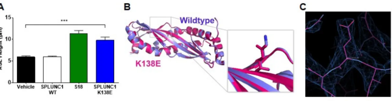
Crystallization of SPLUNC1K138E
Crystals of SPLUNC1K138E were grown in 6M ammonium nitrate, and 0.1M Tris (pH 8.5) and cryoprotected in 15% glycerol. Crystals diffracted to 2.7 Å with space group C2221. Molecular replacement with wildtype SPLUNC1Δ19 was used as a search model.
Bone Marrow Mast Cell Culture
Bone barrow cells were isolated from the femurs of 8-12 week old C57BL/6 mice and grown in tissue culture for 6 weeks. Media was changed twice weekly and non-adherent cells were enriched for mast cells with IL-3. By 4 weeks, pure
populations of mast cells were obtained.
Bone Marrow Mast Cell IL-13 Assay
Bone marrow mast cells (BMMCs) were treated with LPS from P. aeruginosa
17
Bone Marrow Mast Cell Hexosaminidase Release
BMMCs were coated with a monoclonal IgE specific for human dinitrophenyl albumin (DNP, 100 ng/mL) and incubated overnight at 37°C. The next day, IgE-loaded mast cells were stimulated with DNP antigen (5, 50 ng/mL) and incubated for 20 m. Mast cell degranulation was assessed through detection of hexosaminidase in the media and comparing the hexosaminidase in the media to the total
hexosaminidase present.
Statistics
All analyses were performed by Mann-Whitney test, Wilcoxon matched pairs-signed rank test (GraphPad Prism) or MANOVA as indicated in figure legends. For MANOVA, total airway resistance data were log2 transformed and modeled as repeated measures in a multivariate model with genotype, treatment (where applicable), and Mch dose as fixed effect, and individual mouse as random effect factors. Between-subject effects fitted by the sum of the repeated measures were tested for significant differences between genotype, treatment, and Mch dose using F-statistics (JMP v.12.0.1, SAS). All data represent mean ± SEM.
Study Approval
The IACUC and IRB of the University of North Carolina at Chapel Hill
18
Results
SPLUNC1 is an Epithelial-derived Factor that is Reduced in Asthmatic Human and Allergic Mouse BAL
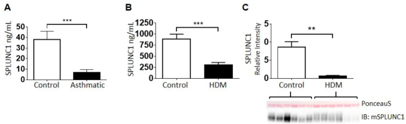
Mild allergic asthmatics (AAs) and normal volunteers (NVs) underwent bronchoscopy, and SPLUNC1 protein levels were measured in BAL fluid by ELISA. SPLUNC1 levels were markedly reduced or undetectable in samples from AAs (9.3+/-3.4 ng/mL). In contrast, most NVs had detectable SPLUNC1 at levels that were significantly higher than AAs (38.0+/-8.0 ng/mL) (Figure 2.1A). To determine if mice with asthma-like airway disease would also develop a relative SPLUNC1-deficiency in their airways, BAL fluid was collected following 12 days of mucosal sensitization with HDM. As shown in Figure 2.1B and similar to our findings in human BAL, SPLUNC1 levels in the airways of HDM-allergic mice were markedly lower than levels in PBS-treated controls (301.8+/-61.8 ng/mL vs. 885.2+/-113.8 ng/mL). Western blots confirmed the results obtained by ELISA, showing markedly reduced SPLUNC1 protein in the HDM-treated animals (Figure 2.1C).
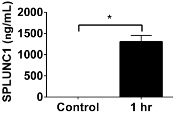
Next, to demonstrate that airway epithelia secrete SPLUNC1, we cultured HBECs at air-liquid interface and measured SPLUNC1 levels in the apical wash by ELISA. HBECs secrete abundant SPLUNC1 (Figure 2.2). Our findings of reduced SPLUNC1 levels in HDM-allergic mice and asthmatic humans are consistent with in vitro
19
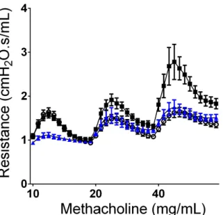
SPLUNC1 Regulates AHR
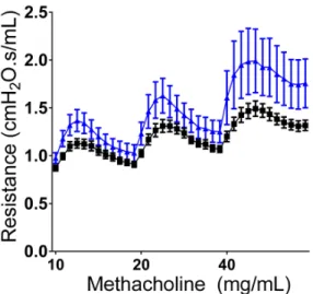
To investigate whether SPLUNC1-deficiency might contribute to AHR, airway mechanics during a graded Mch challenge were examined in naïve SPLUNC1 -/-mice and their wildtype littermate controls. SPLUNC1-/- mice showed greater responsiveness to Mch, suggesting a link between SPLUNC1-deficiency and AHR (Figure 2.3). We next tested whether intratracheal (i.t.) instillation of SPLUNC1 to HDM-allergic mice would reduce AHR. Addition of SPLUNC1 1h prior to Mch challenge reduced AHR to levels similar to controls (Figure 2.4). These results indicate that SPLUNC1 plays a critical role in controlling AHR, and lead us to posit that restoration of SPLUNC1 may be therapeutic.
K138E Mutant Structure and Function
It has been reported that exercise in asthmatics leads to dehydrated or low ASL height, and that this effect on the airway produces AHR (9, 97). A
well-characterized function of SPLUNC1 is its capacity to regulate and restore low ASL height through its inhibition of ENaC (70, 71). We therefore hypothesized that SPLUNC1 reduces AHR through the regulation of ASL height and sought to
20
2.5A). We were interested in determining the structure of this salient point mutant to obtain insight into a structure-function relationship.
We solved the crystal structure of SPLUNC1K138E to 2.67 Å which revealed no major structural differences between the wildtype protein and the SPLUNC1K138E mutant (Figure 2.5B). This suggests that a structural change is not likely to cause the change in function. However, a limitation of this method is that the crystallization condition was basic (pH 8.5) rather than acidic. Since the functional differences between the wildtype and mutant protein exists in acidic rather than basic conditions, the next step would be to determine whether any structural differences exist at a more acidic condition.
The Molecular Basis for SPLUNC1’s Effects on AHR
SPLUNC1’s Effect on AHR is Independent of Effects on ASL Height
21
(102), and SPLUNC1S190A and SPLUNC15XHis which both lack the ability to regulate ASL height (Figures 2.6C, 2.6D, 2.6E). All three mutants were able to reduce AHR, providing further evidence that SPLUNC1’s effect on AHR is independent of ENaC-mediated changes in ASL-height.
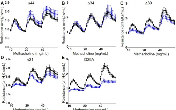
SPLUNC1’s N-terminus is Critical for AHR Reduction
22
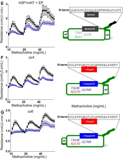
SPLUNC1’s Effect on AHR is Dependent on the Coordination of its N-terminus with an Electrostatic Patch on the Protein Body
23
Lastly, as reported previously, deletions of helix 4 and helix 6 from SPLUNC1 do not alter its overall structure, and thus are expected to leave both the S18 and electrostatic regions intact (82). These two proteins reduced AHR in HDM-allergic mice to levels similar to that observed with wildtype SPLUNC1Δ19 (Figures 2.8F, 2.8G). Collectively, these data indicate that the N-terminal region of SPLUNC1 and the electrostatic patch work in concert to control AHR.
SPLUNC1’s Mechanism of Action for AHR Reduction
Mast cells are the most critical leukocyte in AHR development and we and others have shown that the absence of mast cells results in an inability to exhibit allergen-induced AHR (103, 104). Due to the co-localization of mast cells and
SPLUNC1 in the mucosa, the capacity of mast cells to cause AHR, and the ability of SPLUNC1 to abolish AHR in allergic mice, we hypothesized that SPLUNC1
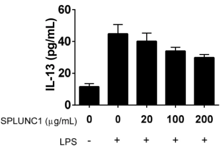
functions to reduce AHR by inhibiting the activation of mast cells. To test this hypothesis, we isolated bone marrow cells from the femurs of C57BL/6 wildtype mice and grew them in culture, enriching for mast cells with IL-3. To test whether SPLUNC1 could inhibit IL-13 production by mast cells, a Th2 cytokine important for AHR, BMMCs were treated with LPS to induce IL-13 secretion. Cells were then administered SPLUNC1 and secreted IL-13 was measured in the media 24 h later. We observed a SPLUNC1 dose-dependent trend toward reduced IL-13 secretion (Figure 2.9).
24
stimulated with DNP antigen and incubated for 20 m. Mast cell activation was assessed by measuring hexosaminidase in the media as a percentage of the total hexosaminidase present (105). SPLUNC1 did not alter the antigen-mediated mast cell degranulation (Figure 2.10).
These studies suggest that the presence of SPLUNC1 may reduce the secretion of IL-13 from mast cells. If the trends we observed are confirmed with more mast cell cultures, then it is possible that inhibition of mast cell IL-13 synthesis may in part be responsible for SPLUNC1’s ability to attenuate AHR.
Discussion
Using samples from allergic asthmatics, we have shown that levels of the normally abundant airway protein SPLUNC1 are significantly reduced. Similarly, the induction of allergic airway inflammation in mice results in reduced SPLUNC1 levels. SPLUNC1-deficient mice are hyper-responsive to Mch, and restoring SPLUNC1 to the airway of HDM-allergic mice reverses the AHR that develops in this model. Collectively, these data suggest that SPLUNC1 is an epithelial-derived relaxing factor, something that has been sought for 30 years. Further, they suggest that SPLUNC1 deficiency may contribute to AHR development in asthma.
SPLUNC1 is a multi-functional protein in the airway with antimicrobial, surfactant, ASL-height regulating, and immunomodulatory activities. Mice lacking SPLUNC1 show enhanced eosinophilic airway inflammation and goblet cell
hyperplasia following OVA sensitization (75). And allergic inflammation significantly reduces SPLUNC1 levels in the lungs (76). These studies on inflammation,
25
importance in AHR, suggest that correction of SPLUNC1-deficiency in asthmatics may be therapeutic.
One of SPLUNC1’s well-characterized functions is its ability to regulate ASL height through ENaC (70, 71). Here we report that the ENaC-regulating SPLUNC1 peptide S18 is unable to reduce AHR, giving an indication that ENaC-regulated changes in ASL height is not likely to be the route of AHR reduction. In support of this finding, the ENaC inhibitor amiloride also failed to reduce AHR. It is known that asthma exacerbations are correlated with acidification of the airways (98). Our group has previously shown that the S18 peptide, and point mutant SPLUNC1D193N are able to regulate ASL height at both normal and acidic pH (72). Despite the fact that both are able to regulate ASL height independent of pH,
SPLUNC1D193N reduced AHR while S18 did not. We additionally tested two mutants which do not regulate ASL height (SPLUNC1S190A, SPLUNC15XHis) and showed that these proteins significantly reduced AHR. Therefore, we conclude that SPLUNC1’s ability to alter ASL height via ENaC inhibition is not the mechanism for its ability to affect AHR.
Since SPLUNC1Δ44, which lacks the first 18 amino acids of the protein, did not reduce AHR, we felt confident that the N-terminus of the protein was critical for the observed effects on AHR. Interestingly, the first two N-terminal residues of
SPLUNC1 exhibit structural similarity to the muscarinic receptor antagonist,
26
inducing AHR when given before Mch challenge. However, when we removed the two N-terminal residues from the protein, its ability to abolish AHR was not
eliminated. These data suggest that muscarinic receptor antagonism is not responsible for the effects of SPLUNC1 on AHR.
We previously reported that an “electrostatic patch” on SPLUNC1 is involved in presenting the protein’s N-terminus (72). We hypothesized that the electrostatic patch may be involved in N-terminus presentation for AHR-reduction as well. Indeed, we were able to support this hypothesis by preparing a few mutant proteins. The first was a mouse-human chimeric protein that naturally does not have an electrostatic patch. This protein, containing the mouse body and human N-terminus was not able to reduce AHR. However, when the hybrid protein was conferred the human
27
Thus, these data provide further evidence that both the N-terminus and electrostatic patch are necessary for SPLUNC1 to effect AHR.
Defining the portions of the SPLUNC1 protein that mediate its effect on AHR reduction is critical for the development of SPLUNC1-derived proteins as
therapeutics for asthma. Equally important is understanding the cellular and
molecular mechanisms by which SPLUNC1 influences AHR. To begin to define this mechanism, we chose to examine SPLUNC1’s effect on mast cell activation. Mast cells are critical effector cells in AHR development, in part through their synthesis of IL-13 (106, 107). Our data suggests that SPLUNC1 may inhibit IL-13 synthesis by mast cells. Since we only observed a trend toward IL-13 reduction, more
experiments with additional BMMC cultures are warranted. Human mast cells can also be cultured in vitro, and similar experiments with human mast cells could further support this mechanism of action in IL-13 inhibition is seen in human cells. IL-13 has long been recognized as a key mediator in AHR development. It is tempting to speculate that the down-regulation of SPLUNC1 in the allergic airway results in increased IL-13 synthesis by mast cells, resulting in AHR development.
In summary, we have demonstrated that one of the most abundant proteins in the normal airway, SPLUNC1, is reduced in allergic asthmatics and in mice
28
29
Figure 2.1. SPLUNC1 is Reduced in Allergic Airways
A. Human BAL was obtained from normal volunteers (Control) and allergic asthmatics (Asthmatic) undergoing bronchoscopy and SPLUNC1 levels were measured in BAL supernatants by ELISA. n=20 for Control group and n=21 for Asthmatic group.
B. C57BL/6 mice were treated with HDM (25 μg i.n.) or PBS (Control) for 12d, then SPLUNC1 levels were measured in BAL supernatants by ELISA. n=17 for both groups.
30
Control
1 hr
0
500
1000
1500
2000
*
Figure 2.2 HBECs Secrete SPLUNC1
31
Figure 2.3 SPLUNC1-/- Mice are Hyperresponsive to Methacholine
32
Figure 2.4 Administration of Wildtype SPLUNC1Δ19 Abolishes Allergen-induced
AHR
33
Figure 2.5 SPLUNC1K138E Protein Restoration of ASL-height at Acidic pH is Not
the Result of a Structural Change
A. Mean ASL height in HBEC cultures at pH 6.0 8 h after addition of wildtype (WT) SPLUNC1, S18 peptide or SPLUNC1K138E (n ≥ 7 for each group). Mann-Whitney test. ***P<0.0005
B. Crystal structure of SPLUNC1K138E mutant (pink) at 2.7 Å resolution
superimposed on wildtype protein (purple), with a close-up of the normal K138 and mutant E138
34
Figure 2.6. SPLUNC1’s Effect on AHR is Independent of Effects on ASL Height
C57BL/6 mice were treated with HDM (25 μg i.n.) for 12d. One day after the last HDM challenge, mice were dosed with
A. S18 (6 mmol i.t.)
B. Amiloride (0.1 µmol i.t.)
C. Amiloride (0.5 µmol i.t.)
D. Amiloride (3.75 µmol i.t.)
E. SPLUNC1D193N (150 μg i.t.)
F. SPLUNC1S190A (150 μg i.t.)
G. SPLUNC15XHis (150 μg i.t)
35
Figure 2.7 SPLUNC1’s N-terminus is Critical for AHR Reduction
C57BL/6 mice were treated with HDM (25 μg i.n.) for 12d. One day after the last HDM challenge, mice were dosed with
A. SPLUNC1Δ44 (150 μg i.t.)
B. SPLUNC1Δ34 (150 μg i.t.)
C. SPLUNC1Δ30 (150 μg i.t.)
D. SPLUNC1Δ21 (150 μg i.t.)
E. SPLUNC1D29A (150 μg i.t.)
37
Figure 2.8 Both the N-terminus and Electrostatic Patch are Necessary for AHR-reduction
C57BL/6 mice were treated with HDM (25 μg i.n.) for 12d. One day after the last HDM challenge, mice were dosed with
A. SPLUNC1mSP1hNT (150 μg i.t.)
B. SPLUNC1mSP1hNT+EP (150 μg i.t.)
C. SPLUNC1hSP1mNT (150 μg i.t.)
D. SPLUNC1mSP1Δ45 (150 μg i.t.)
E. SPLUNC1hSP1mNT+EP (150 μg i.t.)
F. SPLUNC1Δα4 (150 μg i.t.)
G. SPLUNC1Δα4 (150 μg i.t.)
38
IL
-1
3
(p
g
/m
L
)
Figure 2.9 SPLUNC1 Exhibits Dose-dependent Reduction of IL-13 Secretion in Mast Cells
39
Figure 2.10 SPLUNC1 Does Not Reduce Allergen-induced Mast Cell Degranulation
Mast cells coated in anti-dinitrophenyl albumin (DNP) IgE were treated with SPLUNC1 at 0, 1, 4, 16, 250, 1000 and 2250 µg/mL and stimulated with DNP
40
CHAPTER 3: SPLUNC1 AND LUNG INFECTION
Introduction
Pneumonia kills approximately 35% of patients with healthcare-associated infections in the US, and the common opportunistic pathogen Pseudomonas aeruginosa is the primary Gram-negative infectious bacterium cultured from the lungs of patients with hospital-acquired pneumonia (108, 109). Additionally, P. aeruginosa colonizes the airway of 80-85% of patients with cystic fibrosis by age 20, and infects patients with moderate, severe, and exacerbated COPD (110-112). Although antibiotics such as antipseudomonal beta-lactams, aminoglycosides and quinolones are available, the prevalence of antimicrobial resistance is extensive and growing (113-116). Thus, new methods to help eradicate P. aeruginosa infections are urgently needed.
41
Materials and Methods
Animals
Female C57BL/6 mice bred in-house or purchased from The Jackson
Laboratory were used for these studies. Mice were housed in specific pathogen-free animal facilities with 12 h day and night cycles and provided food and water ad lib.
Protein Preparation
Protein was expressed in BL21-CodonPlus cells and purified as previously described (72). BL21-CodonPlus competent cells were transformed with the expression plasmid of interest and cultured in LB with ampicillin (100 μg/mL), chloramphenicol (34 μg/mL) and antifoam (50 μL) with shaking at 37°C until the OD600 reached 0.6. The cells were induced with 0.1 mM IPTG and the temperature was reduced to 18°C for overnight growth. Cell pellets were lysed using sonication in the presence of lysozyme, DNase 1, and protease inhibitor cocktail. Nickel and size exclusion chromatography were used for purification, and tobacco etch virus
protease removed the histidine tag from the protein.
Bacterial and SPLUNC1 Dosing
42
peptone 1 h later. Mice were kept on heating pads overnight and left lungs were harvested 1 d later.
Lung Harvest
Isoflurane euthanasia was performed and the left lung removed from each mouse. The lung was weighed, minced, and stored in PBS with 7% fetal bovine serum and 2% Triton X-100. Lungs were homogenized with metal beads in a Next Advance Bullet Blender. Lung homogenates were plated on LB agar plates overnight and colonies counted 1 d later.
Statistics
Analyses were performed by Mann-Whitney test. All data represent mean ± SEM.
Study Approval
The IACUC of the University of North Carolina at Chapel Hill approved all experiments.
Results
In order to establish an acute lung infection, we tested a 5 x 10⁶ CFU P.
aeruginosa PAO1 strain dose on C57BL/6 mice bred in-house or purchased from The Jackson Laboratory. Lungs were harvested one day later and CFU/mg of lung determined. We successfully established infection and discovered that The Jackson Laboratory animals had higher CFU/mg lung than in-house bred animals (Figure 3.1).
43
had a higher CFU/mg lung count. We administered SPLUNC1 1 h prior to infection with PAO1 and discovered a trend toward reduction of CFU/mg of lung with
SPLUNC1 administration (Figure 3.2). In summary, we show that we were able to establish a model for the delivery of exogenous SPLUNC1 to an acute lung infection and provide evidence that SPLUNC1 may reduce bacterial load in this model.
Discussion
SPLUNC1 has previously been shown to possess antimicrobial properties, which includes activity against the Gram-negative bacterium, P. aeruginosa (79, 80, 117-119). The published studies examined SPLUNC1’s activity against P.
aeruginosa either in vitro, or in vivo in knockout and transgenic mice. No study to date has reported administering exogenous SPLUNC1 to mice. In order to determine whether SPLUNC1 has the potential to be used therapeutically against P.
aeruginosa infection, and to determine the molecular basis for SPLUNC1’s ability to combat infection in vivo, we needed to be able to deliver exogenous SPLUNC1 in acute lung infection. In a model of P. aeruginosa pneumonia, we show that the delivery of SPLUNC1 trends towards reduction of bacterial burden in mice.
Interestingly, we observed a trend toward increased bacterial burden in purchased mice compared to mice bred in our animal facility when given P. aeruginosa alone. One potential explanation for this observation is stress induced by travel and acclimation to a new housing environment. Stress is known to be
44
Using purchased mice, we were able to show a trend towards lower CFUs in mice treated with rSPLUNC1 (Figure 3.2). However, the variability in the severity of infection was high, thus contributing to the lack of statistical significance. More
experiments with larger numbers of mice will be necessary to determine whether this trend that we observed is a real effect of SPLUNC1, and if so, would suggest that administration of rSPLUNC1 be evaluated further for use in treating lung infections such as P. aeruginosa.
Future Directions
In this study, we begin to evaluate the ability of exogenous SPLUNC1 administration to reduce bacterial burden in vivo. One challenge mentioned above was that the error was higher than desired in the infection group which affected our ability to detect statistical significance from the results obtained. Therefore, future directions include more practice with infectious dosing to normalize the dose delivered for a more consistent result. Once more consistent infections are
established, rSPLUNC1 would be tested again and statistical significance evaluated. Our group has published in vitro studies which detail the structural elements critical for SPLUNC1’s antimicrobial properties (82). If anti-infective actions of rSPLUNC1 are established in vivo, then future directions include testing mutants with reduced surfactant functions as well as bacteriostatic and LPS-binding activity against P. aeruginosa in vivo. As we also have mutants with reduced activity against
45
46
Figure 3.1. The Jackson Laboratory vs. In-house Mouse PAO1 Infection
47
Figure 3.2 SPLUNC1 Reduces CFU Load in PAO1 Lung Infection
48
CHAPTER 4: ADMINISTRATION OF SPLUNC1 RESULTS IN SECRETION OF SPLUNC1
Introduction
Studies from our group and others address SPLUNC1’s protective properties in the context of allergic inflammation and infection (75, 76, 79-83, 96, 118-122). Chapters 2 and 3 suggest that administration of SPLUNC1 or methods to increase the endogenous SPLUNC1 be investigated for treatment of asthma and lung
infection. Overall, we have shown that the local increase in SPLUNC1 concentration in the lung acts protectively in these diseases. While the literature has shown that delivery of drugs such as α1-antitrypsin, beta-2-agonists and glucocorticoids results in an increase in SPLUNC1 concentration, we advocate that exogenous SPLUNC1 delivery should also be considered (78, 95, 119, 123). Here, we show that the delivery of exogenous SPLUNC1 results in feedback releasing endogenous SPLUNC1. We explore the possibility of utilizing this effect to assist in combatting AHR and lung infection as described in chapters 2 and 3.
Materials and Methods
Animals
Female C57BL/6 mice were purchased from The Jackson Laboratory.
49
generate SPLUNC1-/- mice and SPLUNC1+/+ littermate controls. All mice were
housed in specific pathogen-free animal facilities with 12 h day and night cycles and provided food and water ad lib.
HDM-induced Allergic Airway Inflammation
Allergic airway inflammation was induced in 7-13 week old female C57BL/6 mice by i.n. administration of 25 µg of HDM (Greer Laboratories) for 12 days. HDM was administered once a day for 5 consecutive days followed by 2 non-treatment days, and this cycle repeated until 12 total doses of HDM were administered. Isoflurane was used for anesthesia for all i.n. instillations.
SPLUNC1 Dosing and BAL Collection
SPLUNC1 was administered to mice (150 µg i.t.) and BAL performed with 0.8 mL of Hank’s Balanced Salt Solution via tracheal cannula 1, 12, 24, 48 and 72 h after administration.
Cell Culture
50
SPLUNC1 ELISA
A sandwich ELISA was used to detect SPLUNC1 protein in BAL supernatants and HBEC samples. Primary monoclonal and secondary biotinylated polyclonal anti-SPLUNC1 antibodies (R&D Systems) were used along with Avidin-HRP and TMB substrate (eBioscience).
Western Blot
SDS-PAGE was run on BAL supernatants and proteins transferred to a nitrocellulose membrane. Blots were incubated in primary anti-SPLUNC1 antibody and secondary HRP-conjugated antibodies (R&D Systems). Enhanced
chemiluminescent substrate was used for detection.
Protein Preparation
Proteins were expressed in BL21-CodonPlus cells and purified as previously described (72). BL21-CodonPlus competent cells were transformed with the
expression plasmid of interest and cultured in LB with ampicillin (100 μg/mL), chloramphenicol (34 μg/mL) and antifoam (50 μL) with shaking at 37°C until the OD600 reached 0.6. The cells were induced with 0.1 mM IPTG and the temperature was reduced to 18°C for overnight growth. Cell pellets were lysed using sonication in the presence of lysozyme, DNase 1, and protease inhibitor cocktail. Nickel and size exclusion chromatography were used for purification, and tobacco etch virus
protease removed the histidine tag from the protein.
Statistics
51
Study Approval
The IACUC of the University of North Carolina at Chapel Hill approved all experiments.
Results
Administration of SPLUNC1 Results in Secretion of SPLUNC1
HBECs cultured at air-liquid interface were exposed to wildtype SPLUNC1Δ19 applied to their apical surface and an apical wash was collected 0.5, 1, 1.5, 2, 2.5 and 3 h after SPLUNC1 delivery. SPLUNC1 levels measured in apical wash samples by ELISA were significantly higher in SPLUNC1-treated as compared to PBS-treated cells (Figure 4.1). To determine whether this would also be the case in vivo, we dosed mice with SPLUNC1Δ19 i.t. and collected BAL at various time points post-administration. We show here that SPLUNC1 levels were more than 90X higher 1 h after SPLUNC1 treatment as compared to PBS treatment taking into account the dose administered (Figure 4.2A). The SPLUNC1 levels remained above baseline for at least 24 h after SPLUNC1 administration. We supplement these data with
Western blot results to illustrate that the increase in SPLUNC1 is not simply due to measuring the administered protein in the ELISA. First, we provide evidence that the exogenous recombinant SPLUNC1 administered runs at a smaller size than the endogenous protein (Figure 4.2B). We show this by comparing naïve wildtype mouse BAL which only contains endogenous SPLUNC1 with SPLUNC1-/- mouse BAL and recombinant protein. The endogenous wildtype SPLUNC1 runs at 35 kDa while the recombinant protein runs at 25 kDa. No protein was detected in the
52
kDa SPLUNC1 protein band intensity increased (Figure 4.2C). These results show both in vitro in HBECs and in vivo in mice that the administration of SPLUNC1 results in the release of SPLUNC1.
SPLUNC1 Secretion is Specific to SPLUNC1 Administration
To show SPLUNC1 secretion is specific to SPLUNC1 administration, we administered a 25 kDa protein pantothenate kinase (PanK), which was prepared in the laboratory using a similar method as SPLUNC1, and LPS. We show in Figure 4.3 that only the administration of SPLUNC1 resulted in the abundant secretion of SPLUNC1.
SPLUNC1 Concentration Increase is Not Sustained in Allergic Inflammation
Thus far, we have shown that SPLUNC1 administration results in the secretion of SPLUNC1 in normal HBECs and naïve animals and that this effect is specific to the administration of SPLUNC1. We suggest in chapter 2 that SPLUNC1 administration be evaluated for reducing AHR in allergic inflammation. We
hypothesized that the release of endogenous SPLUNC1 contributes to the protective effect observed in Mch challenge and sought to evaluate the ability of mice to
53
concentration of SPLUNC1 in the BAL, the duration for the increase is much longer in the PBS-treated than the HDM-allergic mice. Since SPLUNC1 has been shown to be reduced in the lungs during allergic inflammation, we speculate that the allergic inflammatory milieu may diminish the ability to sustain a SPLUNC1 concentration increase through suppression of transcription or reduction of intracellular SPLUNC1 stores.
Discussion
We suggest in chapters 1 and 2 that SPLUNC1 administration or methods to increase endogenous SPLUNC1 be investigated for treatment of asthma and lung infection. We show here that administration of SPLUNC1 itself results in the release of endogenous SPLUNC1 and that this effect is specific to the administration of SPLUNC1. We also note that the increase in SPLUNC1, while sustained in naïve and PBS-treated mice, had a shorter duration of increase in our preliminary studies with allergic inflammation. Thus far, it is unclear whether this effect has therapeutic significance in terms of AHR as SPLUNC1Δ19 and SPLUNC1Δ44 had similar
preliminary results for the release of endogenous SPLUNC1 while the two proteins had substantially differing results with regard to AHR (Figure 2.4 and 2.7A). More investigation is needed to confirm the preliminary findings and to determine what the results may mean with regard to AHR and infection.
Future Directions
Although we begin in this chapter to unravel the idea that SPLUNC1
54
mice should be given SPLUNC1 i.t. and BAL collected at the 1, 12, 24, 48 and 72 h time points (Figure 4.2A). The goals are to increase the n numbers and to see if error may be reduced with more animals especially at the 1 h time point.
Additionally, more mouse BAL should be tested by Western blot and the results normalized to obtain a quantitative evaluation of the increase in SPLUNC1 through Western blot. This method could exclude the exogenously administered 25 kDa SPLUNC1 which the ELISA technique is not able to separate out due to the antibody recognition of both endogenous and recombinant proteins.
More mice also need to be given PanK and LPS in order to increase the n numbers to show that only SPLUNC1 administration results in the release of SPLUNC1 (Figure 4.3). Additionally, the drugs which have been published to increase the SPLUNC1 concentration in the lungs should be tested and BAL collected for evaluation of SPLUNC1 content. The goal is to compare the
administration of SPLUNC1 with α1-antitrypsin, beta-2-agonists and glucocorticoids. We would like to investigate whether the amount of SPLUNC1 released differs between the various treatments currently shown to increase SPLUNC1. Future directions also include testing these drugs in HDM-allergic mice and the evaluation of AHR.
55
56
Figure 4.1. SPLUNC1 Increases SPLUNC1 Secretion by HBECs
HBECs were cultured at air-liquid interface for 4-6 weeks and SPLUNC1 levels were measured in apical wash samples 1 h after the addition of apical PBS (white bars) or SPLUNC1 (black bars, 50 µg) by ELISA. n=5. P<0.05 between PBS and SPLUNC1 groups by Wilcoxon matched-pairs signed rank test.
S
P
L
U
N
C
1
(
g
/m
L
57
Figure 4.2. SPLUNC1 Administration Increases SPLUNC1 Secretion in Mouse BAL up to 48 Hours Post-administration
A. C57BL/6 mice were dosed with SPLUNC1 (80 µg i.t.) or PBS ((-) Control). BAL was collected at the indicated time points and SPLUNC1 levels measured in the BAL supernatants by ELISA. Inset shows combined (-) Control PBS result from all time points. n=3-4 for all SPLUNC1 groups and n=11 for (-) Control. Mann-Whitney test. *P<0.05 **P<0.005
B. BAL was collected from C57BL/6 (WT) and SPLUNC1-/- mice and SPLUNC1 was evaluated in BAL supernatants by Western blot. Rightmost lane shows recombinant wildtype SPLUNC1Δ19 alone.
C. C57BL/6 and SPLUNC1-/- mice were dosed with SPLUNC1 (80 µg i.t.). BAL was collected 1 h later and SPLUNC1 was evaluated in BAL supernatants by Western blot. Rightmost lane shows recombinant wildtype SPLUNC1Δ19 alone.
58
Figure 4.3 SPLUNC1 Secretion in Mice is Specific to SPLUNC1 Administration
59
Figure 4.4 SPLUNC1 Secretion Remains High at least 24 h after SPLUNC1 Administration in Controls but Decreases Rapidly in HDM-Allergic Mice.
C57BL/6 mice were treated with HDM (25 μg i.n.) or PBS for 12 d. One day after the last HDM challenge, mice were dosed with
A. Wildtype SPLUNC1Δ19 (150 μg i.t.) or
B. SPLUNC1Δ44 (150 μg i.t.)
60
APPENDIX: P. aeruginosa Infection and PilY1 Introduction
As introduced in chapter 2, P. aeruginosa in a common opportunistic
pathogen. This bacterium routinely affects patients with burns, cystic fibrosis, COPD, those who are immunocompromised, and is a primary Gram-negative infectious bacterium in patients with hospital-acquired pneumonia.Critical care infection rates have been reported as greater than 70% in ICU patients remaining at the hospital for longer than 7 days and P. aeruginosa is the leading multi-drug resistant (MDR) Gram-negative organism causing ventilator associated pneumonia (VAP) in ICU and burn patients (124-126). With increasing infection rates and incidence of MDR P. aeruginosa, we sought to investigate new potential prophylactic agents for this pathogen.
As previously published by our group, P. aeruginosa binds to integrins via an RGD (arginine-glycine-aspartic acid) domain on the type IV pilus-associated PilY1 protein (127). This binding is involved in P. aeruginosa attachment to human cells, and consequently PilY1 has a role in mediating infection. We sought to elucidate the mechanistic details of Pseudomonas infection via the type IV pilus and also
investigate potential prophylactic therapies for bacterial pneumonia.
Aims
61
to amino acids 532-1163 and 191-1163 which both include the critical pilus RGD motif (128). An alternative is to use PA14 strain PilY1 (amino acids 200-1170). Obtaining an extended crystal structure would facilitate a detailed understanding of how the RGD is presented for integrin binding to human cells. These data are expected to lead to the development of specific, directed prophylactic measures for preventing binding and subsequent infection by P. aeruginosa. 2) Administer PilY1 to healthy and damaged lung epithelial cells in vitro in collaboration with the