Structure-Based Design of Inhibitors Selective for Human
Proteasome
β
2c or
β
2i Subunits
Bo-Tao Xin,
†,#Eva M. Huber,
‡,#Gerjan de Bruin,
†Wolfgang Heinemeyer,
‡Elmer Maurits,
†Christofer Espinal,
†Yimeng Du,
†Marissa Janssens,
†Emily S. Weyburne,
∥Alexei F. Kisselev,
∥,⊥Bogdan I. Florea,
†Christoph Driessen,
§Gijsbert A. van der Marel,
†Michael Groll,
*
,‡and Herman S. Overkleeft
*
,††
Gorlaeus Laboratories, Leiden Institute of Chemistry and Netherlands Proteomics Centre, Einsteinweg 55, 2333 CC Leiden,
Netherlands
‡
Center for Integrated Protein Science at the Department Chemie, Lehrstuhl fu
̈
r Biochemie, Technische Universität Mu
̈
nchen,
85748 Garching, Germany
§
Department of Hematology and Oncology, Kantonsspital St. Gallen, 9007 St. Gallen, Switzerland
∥
Department of Molecular and Systems Biology and Norris Cotton Cancer Center, Geisel School of Medicine at Dartmouth, 1
Medical Centre Drive HB7936, Lebanon, New Hampshire 03756, United States
*
S Supporting InformationABSTRACT:
Subunit-selective proteasome inhibitors are valuable tools to assess the
biological and medicinal relevance of individual proteasome active sites. Whereas the
inhibitors for the
β
1c,
β
1i,
β
5c, and
β
5i subunits exploit the di
ff
erences in the
substrate-binding channels identi
fi
ed by X-ray crystallography, compounds selectively targeting
β
2c
or
β
2i could not yet be rationally designed because of the high structural similarity of
these two subunits. Here, we report the development, chemical synthesis, and biological
screening of a compound library that led to the identi
fi
cation of the
β
2c- and
β
2i-selective
compounds LU-002c (4; IC
50β
2c: 8 nM, IC
50β
2i/
β
2c: 40-fold) and LU-002i (5; IC
50β
2i: 220 nM, IC
50β
2c/
β
2i: 45-fold), respectively. Co-crystal structures with
β
2
humanized yeast proteasomes visualize protein
−
ligand interactions crucial for subunit
speci
fi
city. Altogether, organic syntheses, activity-based protein pro
fi
ling, yeast
muta-genesis, and structural biology allowed us to decipher signi
fi
cant di
ff
erences of
β
2
substrate-binding channels and to complete the set of subunit-selective proteasome
inhibitors.
■
INTRODUCTION
Proteasomes are proteolytic machines responsible for the
degradation of misfolded proteins localized in the cytosol and
nucleus of eukaryotic cells.
1Their 20S core particles (CPs) are
C2-symmetrical barrel-shaped complexes assembled of 28
subunits that are arranged in four stacked seven-membered
rings.
2The two outer rings are made of seven
α
subunits (
α
1
−
7) and the two inner rings consist of seven homologous yet
distinct
β
subunits (
β
1
−
7). In ubiquitously expressed
constitutive proteasomes, the proteolytic activities reside
within the subunits
β
1c (caspase-like activity),
β
2c
(trypsin-like activity), and
β
5c (chymotrypsin-like activity).
3In
lymphoid tissues, these subunits are replaced by their
interferon-
γ
-inducible counterparts,
β
1i (LMP2),
β
2i
(MECL-1), and
β
5i (LMP7),
4yielding the so-called
immunoproteasome particles (iCPs) that preferentially
gen-erate antigenic peptides with high a
ffi
nity for major
histocompatibility complex (MHC) class I receptors.
5Proteasomes are validated drug targets in oncology, and
numerous structurally diverse inhibitors of natural and
nonnatural origin have been reported so far.
6Most synthetic
compounds are N-terminally capped peptides of two to four
residues with a C-terminal electrophilic warhead that forms a
covalent linkage with the nucleophilic hydroxyl group and
possibly the free N terminus of threonine-1 (Thr1) of the
catalytically active proteasomal
β
subunits.
7Subunit speci
fi
city
of peptidic ligands is largely determined by the sequence of the
peptide fragment, although the nature of the warhead can
confer selectivity as well.
8The
fi
rst-generation boronic acid
bortezomib and the second-generation epoxyketone car
fi
lzo-mib target more than one subunit at a time and therefore are
considered broad-spectrum proteasome inhibitors.
6aBortezo-mib and car
fi
lzomib are now approved drugs for the treatment
of multiple myeloma.
9,10Current industrial and academic drug
design e
ff
orts focus on the development of subunit-selective
proteasome inhibitors and their potential therapeutic use in
chronic in
fl
ammatory diseases. For instance, the
fi
rst
immunoproteasome-selective compound KZR-616,
11an
ana-log of ONX 0914,
12has recently entered phase 1b/2 clinical
Received: November 30, 2018
Published: January 18, 2019
Article
pubs.acs.org/jmc Cite This:J. Med. Chem.XXXX, XXX, XXX−XXX
© XXXX American Chemical Society A DOI:10.1021/acs.jmedchem.8b01884
J. Med. Chem.XXXX, XXX, XXX−XXX
Derivative Works (CC-BY-NC-ND) Attribution License, which permits copying and redistribution of the article, and creation of adaptations, all for non-commercial purposes.
Downloaded via LEIDEN UNIV on February 12, 2019 at 15:57:34 (UTC).
trials for the treatment of lupus erythematosus. Besides medical
issues, selective inhibition of individual proteasome subunits
may aid investigations on the involvement of these sites in
di
ff
erent cellular pathways including MHC class I antigen
presentation and control of cytokine levels. Although there is
an overlap in the substrate preferences of the cCP and iCP
subunits, distinct structural features and amino acid linings of
the substrate-binding channels
β
1c and
β
1i as well as
β
5c and
β
5i could be identi
fi
ed and subsequently allowed for the
development of speci
fi
c inhibitors.
12,13The design of inhibitors
targeting exclusively
β
2c or
β
2i however remained a challenge
because of the high structural similarity between the
trypsin-like active sites
13dIn 2018, Liskamp and co-workers reported a
set of
β
2-selective inhibitors. However, these compounds,
which are characterized by a sulfonyl
fl
uoride as the C-terminal
electrophile, a basic P1 residue, and a free N terminus, display
limited preference for either
β
2c or
β
2i.
14In addition, Kezar
Life Sciences developed an epoxyketone inhibitor with
moderate selectivity for human
β
2i.
11Recently, we published a set of activity-based
protein-pro
fi
ling (ABPP) probes and inhibitors selective for each of the
six catalytic activities of human cCP and iCP, including
compounds LU-002c (
β
2c) and LU-002i (
β
2i;
Figure 1
).
15Here, we describe the design, synthesis, and screening of
focused compound libraries that allowed us to identify these
β
2c and
β
2i inhibitors, respectively. Crystallographic data on
humanized yeast proteasomes in complex with selective ligands
provide insights into their mode of binding and reveal so far
unnoticed di
ff
erences in substrate and inhibitor speci
fi
city for
the trypsin-like active sites of cCP and iCP.
■
RESULTS
Development of Selective Inhibitors for Subunit
β
2c.
The previously identi
fi
ed vinyl sulfone inhibitor LU-102
(
Figure 1
), which inhibits
β
2c and
β
2i with similar potency,
16was used as a starting point for creating selective
β
2c ligands.
We generated a compound library based on the vinyl sulfone
warhead and the 4-aminomethylphenyl side chain on P1 of
LU-102, as these moieties proved to be crucial for
β
2
selectivity in general.
16In a
fi
rst step, we replaced the N cap
of LU-102 by a set of groups often found in peptide-based
proteasome inhibitors (6
−
12). Next, we synthesized
com-pounds with relatively small amino acid side chains in the P2
position (4,
13
−
20) and
fi
nally incorporated bulky aliphatic
side chains at P2 and P3 (21
−
36). In total, 32 compounds
were prepared using established protocols for the chemical
synthesis of the 4-aminomethylphenylalanine vinyl sulfone
warhead and solution-phase coupling of the peptide vinyl
sulfones to the corresponding alpha-amino acids (see
Supporting Information
).
27All compounds were evaluated for
β
2c/
β
2i inhibition by our
competitive ABPP assay at the
fi
nal concentrations of 0.01, 0.1,
1.0, and 10.0
μ
M, and the apparent IC
50values were
determined (
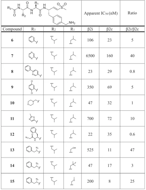
Table 1
). Among the N-cap series
6
−
12,
compound
7
(pyrazine N cap) showed the highest selectivity
for
β
2c over
β
2i (40-fold), but also decreased potency for
β
2c
compared to LU-102 (23-fold). Screening of small P2 residues
(compounds
4,
13
−
20) identi
fi
ed several ligands with both
good selectivity and potency for
β
2c:
4
(P2 alanine; 10 nM,
32-fold selectivity over
β
2i),
13
(P2 serine; 11 nM, 47-fold),
15
(P2 methoxyserine; 8 nM, 25-fold),
16
(P2 threonine; 8
nM, 41-fold), and especially
18
(P2 glycine; 26 nM, 224-fold).
Combining 2-methylthiazole N caps (20) with bulky P2 or P3
residues (21
−
36) revealed several potent and selective
β
2c
compounds as well: see for instance, compounds
20
(P2
methoxyserine, P3 leucine; 72 nM, 14-fold),
22
(P2 leucine,
P3 cyclohexyl; 18 nM, 30-fold),
30
(P2
cyclohexyl-homoalanine, P3 leucine; 11 nM, 25-fold), and
36
(P2 and
P3 cyclohexyl; 40 nM, 10.5-fold). Altogether, based on the
data shown in
Table 1
, we conclude that (1) subunit
β
2c
accepts small as well as bulky P2 residues but disfavors
oversized P3 side chains and that (2)
β
2i disfavors small P2
side chains and large P3 groups.
To establish the apparent IC
50values more accurately and to
obtain insights into the coinhibition of
β
1c,
β
1i,
β
5c, and
β
5i
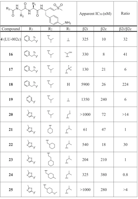
activities, we selected the compounds
4,
7,
13,
16,
18,
20,
22,
and
25
for further analysis. In our competitive ABPP assay
using Raji cell extracts (containing both cCPs and iCPs), a
wider range of
fi
nal concentrations were tested. All compounds
inhibited
β
2c at low nanomolar concentrations (
Table 2
). The
inhibitors
4,
13,
18, and
20, featuring small side chains on P2,
displayed considerably enhanced selectivity for
β
2c over
β
2i
(
≥
27-fold) compared to LU-102 (1.6-fold;
Table 2
), with
18
being the most selective (54-fold).
Next, we assessed the inhibitory e
ff
ects in living RPMI-8226
cells (
Table 3
). Initial screenings identi
fi
ed compound
4
as the
most active, and we included this compound as LU-002c in our
suite of subunit-selective proteasome inhibitors.
15In
subse-Figure 1.Chemical structures and IC50values for the lead structures
LU-102 (1),16LU-112 (2),16and ONX 0914 (3)12that guided the development of theβ2c- andβ2i-selective compounds LU-002c (4) and LU-002i (5), respectively. IC50 values were measured by
competitive ABPP.
Table 1. Chemical Structures of Compounds 4, 6
−
36 and Their Inhibitory Activity (Apparent IC
50Values) against
β
2c and
β
2i
(Determined by Competitive ABPP)
aTable 1. continued
Table 1. continued
a
A highβ2i/β2c ratio indicates selectivity forβ2c. Raw data used for the calculations of IC50values are in theSupporting Information.
Table 2. Apparent IC
50Values of Compounds 1 (LU-102), 4, 7, 13, 16, 18, 20, 22, and 25 for the Six Catalytic Sites from
Human cCPs and iCPs in Raji Cell Lysates, as Established by Competitive ABPP
apparent IC50(μM) ratio
compound β2c β2i β5c β5i β1c β1i β2i/β2c β1i/β2c β1c/β2c β5i/β2c β5c/β2c
1(LU-102) 0.013 0.020 1.33 1.17 >100 >100 2 >7700 >7700 90 102
4(LU-002c) 0.0050 0.14 1.3 2.8 >100 >100 27 >19 000 >19 000 540 250
7 0.17 2.9 >100 >100 >100 >100 17 >600 >600 >600 >600
13 0.0060 0.23 1.4 2.2 >100 >100 40 >17 000 >17 000 380 241
16 0.0070 0.11 0.75 2.1 >100 >100 16 >14 000 >14 000 300 107
18 0.046 2.5 8.6 12.7 >100 >100 54 >2200 >2200 187 276
20 0.077 4.0 45.3 57.1 >100 >100 52 >1300 >1300 740 590
22 0.065 0.42 >100 >100 >100 >100 6 >1500 >1500 >1500 >1500
25 0.44 3.1 >100 >100 >100 >100 7 >220 >220 >220 >220
quent studies, we identi
fi
ed compound
16
to be even more
potent and selective, and we dubbed this compound LU-012c.
Development of
β
2i-Selective Inhibitors.
For the
development of
β
2i-selective compounds, we used ONX
0914 (3)
12as the starting point (
Figure 1
). Though ONX
0914 is a
β
5i-selective inhibitor, it also targets other
proteasome subunits
12,13b(
Figure 1
) and shows slight
selectivity for
β
2i over
β
2c (IC
50(
β
2i) 0.59
μ
M; IC
50(
β
2c)
1.1
μ
M, 1.9-fold).
13bDuring our e
ff
orts to create
β
5i-selective
compounds, we noted that the substitution of P1
phenyl-alanine in ONX 0914 for cyclohexylphenyl-alanine enhances the
selectivity for both
β
5i and
β
2i over the respective constitutive
subunits (ratio
β
2c/
β
2i = 6) and that any additional
modi
fi
cations of the P2 and P3 positions as well as the N
cap led to the loss of activities for the trypsin-like sites.
13bOn
the basis of these observations, we reasoned that large aliphatic
amino acid residues at P1 might lead to
β
2i-selective inhibitors.
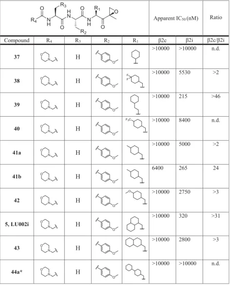
To probe this hypothesis, a set of epoxyketone inhibitors with
large hydrophobic P1 residues (compounds
5,
37
−
53,
Table
4
) was synthesized (for details, see
Supporting Information
).
The compounds were tested at the
fi
nal concentrations of
0.01, 0.1, 1.0, and 10.0
μ
M by our competitive ABPP assay,
and the apparent IC
50values for the inhibition of
β
2c and
β
2i
were determined (
Table 4
). In this
fi
rst evaluation step,
compounds
5
(P1 1-decalanine; 320 nM, >31-fold),
39
(P1
cyclohexyl-homoalanine; 215 nM, >46-fold),
41b
(methyl-cyclohexylalanine; 265 nM, 24-fold), and
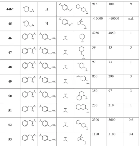
44b
(bicyclohex-ylalanine; 100 nM, 9-fold) showed the highest selectivity for
β
2i over
β
2c.
Next, the inhibition of all six sites by compounds
5
and
39
were tested at a wider range of
fi
nal concentrations (
Table 5
).
In this setup, compound
5
proved to be the most selective
β
2i
ligand (ratio
β
2c/
β
2i: 67) as it did not inhibit any of the
β
1
and
β
5 proteasome subunits. By contrast, epoxyketone
39
proved to be a dual inhibitor of both
β
2i and
β
5i with high
selectivity over the corresponding constitutive subunits (ratio
β
2c/
β
2i: 44; ratio
β
5c/
β
5i: 109).
Epoxyketone
5, the most selective
β
2i inhibitor of the series,
was termed LU-002i and published as part of a set of
compounds and ABPP probes to visualize all the six catalytic
activities of human constitutive and immunoproteasomes.
15However, the decalin moiety of
5
was synthesized as a mixture
of stereoisomers that could not be separated. To address the
question whether one or both of the possible stereomers are
active, the following attempts were undertaken to synthesize a
stereomerically pure analogue of
5
(LU-002i). First,
com-pounds with partially reduced naphthyl rings containing only
one chiral carbon center within the bicyclic system were
synthesized:
68
(R) and
71
(S) (
Scheme 1
;
Supporting
Information
). In the competitive ABPP assay in Raji cell lysates
(
Table 6
),
68
was inactive, whereas
71
selectively targeted
β
2i,
though with a dramatic loss of potency (IC
502.5
μ
M)
compared to
5
(IC
500.18
μ
M).
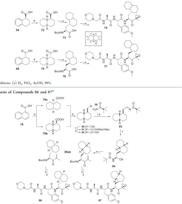
In a second approach to unravel the active stereomer of
5,
fully reduced decalin systems were produced, yielding the
peptide epoxyketones
74
and
77, respectively (
Scheme 2
;
Supporting Information
). Competitive ABPP revealed that
74
inhibits
β
2i with an IC
50of 12.0
μ
M without touching the
other
fi
ve active sites of cCP and iCP particles (
Table 6
).
Compound
77
in turn proved to be a potent
β
2i inhibitor
(IC
500.38
μ
M) with some cross-reactivity against
β
2c (IC
5028
μ
M). Notably, the absolute stereochemistry of the P1 side
chain in
77
matches that of the corresponding carbon center in
ligand
71, but it appears that decalin at P1 (77) is more
e
ff
ective for
β
2i inhibition than the corresponding partially
oxidized bicyclic system (71).
With this information in hand, an enantiomerically pure
diastereomeric set of peptide epoxyketones
86
and
87
was
synthesized (
Scheme 3
;
Supporting Information
). Compound
86
appeared to be a weak (IC
5034
μ
M) but selective
β
2i
inhibitor, whereas epoxyketone
87
strongly inhibits
β
2i (IC
500.19
μ
M) with
β
2c,
β
1c, and
β
5i as o
ff
-targets at high
micromolar concentrations (
Table 6
). On the basis of the
assumption that carbon 1 in the decalin system of compound
87
has the (S) con
fi
guration as in
71
and
77, and assuming
that the catalytic hydrogenation proceeded to deliver decalin
with cis stereochemistry, the observed results strongly suggest
that the stereochemistry of the most active and selective
β
2i
inhibitor is as shown in structure
87
(
Scheme 3
).
To test whether compound
87
is the major active
component of the stereomeric mixture that makes up
compound
5
(the previously described
β
2i-selective inhibitor,
LU-002i
15), both were assessed in a competitive ABPP assay in
Raji cell extracts at
fi
nal inhibitor concentrations ranging from
0 to 3
μ
M (
Figure 2
). As both preparations are about equally
active and selective, diastereomer
87
appears to be indeed the
main active component in the stereomeric mixture that has
previously been reported as LU-002i.
15Next, compound
87
was tested in intact RPMI-8226 cell
lines, in comparison with the dual
β
2i/
β
5i inhibitor
39. The
cells were
fi
rst treated with the inhibitor at various
concentrations, then lysed, incubated with the ABPP mixture,
denatured, and resolved by sodium dodecyl
sulfate-polyacry-lamide gel electrophoresis (SDS-PAGE), as described before.
Like in Raji cell lysates, compound
87
selectively targeted only
β
2i (IC
500.159
μ
M) without a
ff
ecting the remaining
proteolytically active proteasome subunits, whereas
epoxyke-tone
39
inhibited both
β
2i (IC
500.124
μ
M) and
β
5i (IC
500.183
μ
M) (
Figure 3
). Thus, inhibitor
39
represents a
co-inhibitor of
β
2i and
β
5i with potential medicinal relevance,
especially because targeting of
β
2 has previously been shown to
sensitize cells to
β
5 inhibitors,
17and dual subunit inhibition is
required for suppressing autoin
fl
ammatory reactions.
11As the next research objective, we decided to investigate
whether a
β
2i-selective activity-based probe (ABP) could be
derived from LU-002i (5). As the attachment of a
fl
uorescent
tag at the N terminus of subunit-selective inhibitors may be
detrimental to selectivity, we decided to graft the reporter
group onto the tyrosine residue at P2 by substituting the
Table 3. Inhibition of Proteasome Activities by Compounds
1 (LU-102), 4 (LU-002c), 7, 13, 16 (LU-012c), 18, 20, 22,
and 25 in Intact RPMI-8226 Cells
apparent IC50(μM) ratio
compound β2c β2i β5c β5i β1c β1i β2i/β2c
1(LU-102)a 0.29 0.41 >10 >10 >10 >10 1.4 4(LU-002c)a 1.80 >10 >10 >10 >10 >10 >5.6
7 >10 >10 >10 >10 >10 >10 n.d.
13 2.00 >10 >10 >10 >10 >10 >5
16(LU-012c) 1.250 >10 >10 >10 >10 >10 >8
18 >10 >10 >10 >10 >10 >10 n.d.
20 >10 >10 >10 >10 >10 >10 n.d.
22 >10 >10 >10 >10 >10 >10 n.d.
25 >10 >10 >10 >10 >10 >10 n.d.
a
Data cited from the literature; n.d., not determined.
Table 4. Structures of Compounds 5, 37
−
53 and Their Inhibitory Activity (Apparent IC
50Values) against
β
2c and
β
2i
(Determined by the Competitive ABPP Assay)
amethyl group for an appropriately functionalized alkyl group
(
Scheme 4
). The resulting ABP
97
was tested in Raji cell
lysates to pro
fi
le the proteasome activities. At a
fi
nal
concentration of 3
μ
M,
β
2i labeling was selective and could
be easily distinguished (
Figure 4
A). In a competitive ABPP
assay with probe
97, labeling of
β
2i could be completely
abolished by preincubation with LU-002i (5,
β
2i) at 3
μ
M.
The
β
2i signal was partially reduced after treatment with
LU-002c (4,
β
2c) at high concentrations and completely abolished
after preincubation with LU-102 (1,
β
2c/
β
2i) (
Figure 4
B).
Finally, a competitive ABPP assay with probe
97
side-by-side
with the three-probe mixture used previously in competitive
ABPP experiments was carried out. This time, treatment with
LU-002i (5,
β
2i) selectively blocked
β
2i labeling by the three
probes at 3
μ
M, whereas LU-002c (4,
β
2c) completely
prevented
β
2c identi
fi
cation (
fi
nal concentration of 0.3
μ
M)
and partially inhibited
β
2i labeling. Furthermore, LU-102 (1,
β
2c/
β
2i) blocked both
β
2c and
β
2i labeling at 1
μ
M (
Figure
4
B). These results match those published earlier on these
compounds against the same set of probes.
15Altogether, these
data demonstrate that ABP
97
is a potent and highly selective
ABP for visualizing
β
2i activities of human
immunoprotea-somes.
X-ray Structures of Selected Inhibitors in Complex
with Yeast and Humanized CPs.
To obtain more insights
into the structural features that drive either
β
2c or
β
2i
selectivity of ligands, we aimed at determining the X-ray
structures of selected compounds in complex with CPs. As
structural data on human apo iCP are not available, we recently
developed chimeric yeast proteasomes, which feature the key
elements of human
β
5 subunits, as structural tools.
18On the
basis of this work, we created here
β
2 humanized yeast
proteasomes.
Although the yeast proteasome (yCP)
α
subunits can be
easily exchanged by human counterparts, the replacement of
most
β
entities, that is,
β
1,
β
2,
β
5,
β
6, and
β
7 is lethal to
yeast.
13d,18,19Strikingly, however, the single-point mutation
S171G su
ffi
ces to rescue the lethal phenotype that is caused by
the substitution of the endogenous yeast (y)
β
2 subunit with
the human (h)
β
2c counterpart.
19We created the respective
Table 4. continued
a
A high β2c/β2i ratio indicates selectivity for β2i. bn.d., not determined.*Compounds 44a and 44b are diastereomers; for details on stereochemistry, seeSupporting Information.
Table 5. Apparent IC
50(
μ
M) Values of Compounds 5 and 39 against the Six Catalytic Active Sites from Human cCPs and
iCPs, as Determined in Raji Cell Lysates by Competitive ABPP
compound β2i β2c β5i β5c β1i β1c ratioβ2c/β2i ratioβ5c/β5i
5(LU-002i) 0.18 12.1 >100 >100 >100 >100 67 ∼1
39 0.057 2.5 0.046 5.0 >100 >100 44 109
β
2c chimeric yeast strain (
Figures 5
A,
S8
), puri
fi
ed, and
crystallized its mutant proteasome. The X-ray structure (
Table
S13
) revealed that the
β
2 propeptide was released from the
active site Thr1 and that the overall fold of the subunit was
intact (
Figure 6
A). Although the S171G mutation had no
obvious impact on the structure of the matured mutant
proteasome, it likely supports subunit folding and proteasome
assembly. Any pronounced e
ff
ects of Gly171 on
β
2 activity are
excluded, as yeast viability does not depend on peptide bond
hydrolysis by
β
2.
20As no rescuing mutation for the h
β
2i subunit is known to
date, we created various chimeric h
β
2i-y
β
2 constructs and
tested whether they can substitute wild-type (WT) y
β
2.
Surprisingly, only a construct featuring the
β
2i amino acids 1
−
53 was viable (
Figure 5
B). As this sequence covers the entire
β
2 substrate-binding channel, we used this construct for
structural analyses (
Table S13
).
The superposition of ligand-free
β
2c/i chimeric structures
with the natural mouse counterpart
13dproved their structural
similarity (
Figure 6
A,B). The subsequent crystal soakings with
ONX 0914 as a reference compound con
fi
rmed that the
β
2
proteolytic centers were reactive (
Figure S9
) and visualized a
similar binding mode for the inhibitor as in the respective
mouse crystal structures
13d(
Figure 6
C,D). The
β
2 subunits
can accommodate bulky P1 residues without any pronounced
conformational changes of the protein backbone (
Figure
S10A,B
). The corresponding spacious P1 binding site is
created by Gly45 at the bottom of the S1 pocket.
13dAlthough
the chemical nature and the orientation of amino acid 45 di
ff
er
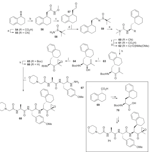
Scheme 1. Synthesis of Compounds 68 and 71
aa
Reagents and conditions: (a) (i) LiAlH4/Et2O, 99%; (ii) TsCl/triethylamine (TEA)/dichloromethane (DCM), 97%; (iii) NaCN/
dimethylformamide (DMF), 95%; (b) (i) KOH/ethylene glycol; (ii) N,O-dimethylhydroxylamine hydrochloride, 2-(6-chloro-1H -benzotriazol-1-yl)-1,1,3,3-tetramethyluronium hexafluorophosphate (HCTU)/N,N-diisopropylethylamine (DiPEA)/DCM, 49% over two steps; (c) LiAlH4/
Et2O; (d)58/CuSO4/DCM, 84% two-step yield; (e) Et2AlCN/i-PrOH/tetrahydrofuran (THF), 58%; (f) (i) 6 M HCl, reflux; (ii) Boc2O/TEA/
THF/H2O, 58% over two steps; (g)N,O-dimethylhydroxylamine hydrochloride, HCTU/DiPEA/DCM, 77%; (h)tBuLi/2-bromopropene/Et2O,
−78°C, 78%; (i) NaBH4/CeCl3·7H2O/MeOH, 59%; (j) (1) VO(acac)2/tBuOOH/DCM; (2) Dess−Martin periodinane/DCM, 33% over two
steps; (k) trifluoroacetic acid (TFA), quantitative yield; (l) (1)67, tBuONO/HCl (4N in dioxane) DCM/DMF,−30°C; (2)66, DiPEA, DMF, 40% over two steps.
Table 6. Apparent IC
50(
μ
M) Values of Compounds 68, 71,
74, 77, 86, and 87 against the Six Catalytic Active Sites from
Human cCPs and iCPs, Determined in Raji Cell Lysates by
Competitive ABPP
compound β2i β2c β5i β5c β1i β1c
68 >100 >100 >100 >100 >100 >100
71 2.5 >100 >100 >100 >100 >100
74 12.0 >100 >100 >100 >100 >100
77 0.38 28 >100 >100 >100 >100
86 34.0 >100 >100 >100 >100 >100
87 0.19 19 28.40 >100 >100 53
among most proteasome subunits, Gly45 has been preserved in
β
2 subunits throughout evolution.
13dThough the mutation of
Gly45 to Ala does neither impair yeast growth nor a
ff
ect
subunit folding and ligand binding, any additional increase of
residue 45 is predicted to sterically interfere with the
surrounding protein side chains (
Figures S8, S11, and S12
,
Table S13
).
On the basis of the structural similarity of human
−
yeast
chimeric and mouse
β
2 active sites, a set of 29 ligand complex
structures was determined with WT and
β
2 chimeric yeast
proteasomes (
Table S13
).
The
β
2c-selective compound
4
(LU-002c) was found to be
well-stabilized in the
β
2c and
β
2i active sites. The interactions
of the 4-aminomethylphenyl group at P1 with the carboxylic
Scheme 2. Synthesis of Compounds 74 and 77
aaReagents and conditions: (a) H
2, PtO2, AcOH, 99%.
Scheme 3. Synthesis of Compounds 86 and 87
aa
Reagents and conditions: (a) H2, PtO2, AcOH, quantitative yield; (b) (i) LiAlH4/Et2O, 92%; (ii) TsCl/TEA/DCM, 95%; (3) NaCN/DMF, 83%;
(c) (i) KOH/ethylene glycol; (ii)N,O-dimethylhydroxylamine hydrochloride, HCTU/DiPEA/DCM, 88% over two steps; (d) LiAlH4/Et2O; (e) 58/CuSO4/DCM, 85% over two steps; (f) Et2AlCN/i-PrOH/THF, 75%.
Figure 2.Comparative ABPP assay of compounds5(LU-002i) and
87, determined in Raji cell lysates.
Figure 3.Inhibition profiles of compounds39and87, determined in intact RPMI-8226 cell lines.
Scheme 4. Synthesis of Probe 97
aa
Reagents and conditions: (a)89, K2CO3/DMF, 80%; (b) (i) TFA, 99%; (ii) Boc-Ala-OH, HCTU/DiPEA/DCM, 93%; (c) (i) TFA, 99%; (ii)
2-morpholino acetic acid, HCTU/DiPEA/DCM, 32%; (d) N2H4·H2O, MeOH, 99%; (e)tBuONO/HCl (4N in dioxane), DCM/DMF (1/1, v/v),
−30°C, 56%; (f) CuSO4, sodium ascorbate, DMF, 18%.
Figure 4.(A) Activity-based proteasome profiling using probe97at different concentrations. Cocktail ABPs were added as control. (B) Left: competitive ABPP assay using ABP97and the inhibitors1(LU-102, 0.1μM),4(LU-002c, 0.3μM), and5(LU-002i, 3μM). Right: competitive ABPP assay with probe97side-by-side with the three-probe mixture used previously in competitive ABPP experiments and the inhibitors1 (LU-102, 0.1μM),4(LU-002c, 0.03μM), and5(LU-002i, 3μM).
Figure 5.Schematic representation of yeast (y) and human (h)β2 subunits and their propeptides. Secondary structure elements, helices (H), and sheets (S) are numbered. (A) The full-length hβ2c (green) and hβ2i (pink) subunits cannot substitute the endogenous yβ2 subunit (gray), neither with their natural propeptides (pp; colored) nor with the yβ2 one (gray) (for details, see the experimental procedures). Strikingly, the humanβ2c subunit can replace the yeast counterpart when featuring the single-point mutation S171G.19(B) Schematic illustration of human−yeast chimeric
β2i constructs according to panel (A). Sequences highlighted in pink were taken from humanβ2i, whereas the gray ones originate from the yeastβ2 entity. All tested variants, except for the construct encoding the residues 1−53 from humanβ2i, caused lethality when expressed in apup1Δyeast strain.
amino acid side chains in position 53 are supposed to be the
driving forces for the general
β
2 selectivity of
4
(LU-002c) as
well as the related compounds LU-102 (1) and LU-112 (2)
(
Figure 1
).
16The selectivity for subunit
β
2c might be gained
by dual anchoring of the 4-aminomethylphenyl group to Asp53
in
β
2c versus a single interaction with Glu53 in
β
2i (
Figure
7
A,C). In addition, the shorter P2 Ala side chain of
4
(LU-002c) compared to Leu in LU-102 increases
β
2c selectivity by
reducing the potency for
β
2i (
Figure 1
). Most likely, small P2
residues like Ala fail to undergo favorable van der Waals
interactions with Val48 in
β
2i (
Figure 7
C) and thereby lead to
the observed
β
2c selectivity of
4
(LU-002c).
For the most selective
β
2i inhibitor, compound
5
(LU-002i),
crystallographic data could only be obtained with WT yCP
(
Figure S13
,
Table S14
). We assume that the ligand could not
be trapped at the mutant
β
2 active site, as the reactivity of
chimeric subunits is impaired
18and as compound
5
is poorly
soluble in aqueous solutions because of its apolar decalin
moiety. Chimeric proteasome structures in complex with
39
however could be achieved. Compounds
5
(LU-002i) and
39
are derived from the epoxyketone inhibitor ONX 0914.
Epoxyketones have recently been shown to form
seven-membered,
21instead of six-membered,
22ring structures with
the nucleophilic Thr1 residue of the proteasomal
β
subunits.
Although the 1,4-oxazepane (seven-membered) ring structure
fi
ts our experimental electron densities in most cases, we also
have structural data which match better the six-membered
1,4-morpholine system (e.g., see
Figure S13A,B
). However, the
kind of irreversible covalent structure inhibitors formed with
Thr1 has no further implications for drug development, as
subunit selectivity of epoxyketone inhibitors is mostly gained
by the interactions of the ligands
’
side chains with the protein
surroundings.
ONX 0914 slightly favors
β
2i over
β
2c,
12which may be
supported by an advantageous hydrophobic interaction of its
P2-methoxy group with Val48 of
β
2i, a contact that is not
provided in subunit
β
2c (
Figure 6
C,D). Furthermore, Asn22
forms hydrogen bonds with the amide oxygen atom of the
morpholine cap of ONX 0914, whereas Glu22 in subunit
β
2c
fails to provide this additional stabilization (
Figure 6
C,D). The
interaction with Asn22 in
β
2i is also observed with other
tripeptide ligands like
39
(
Figure 7
G,H), implying that peptide
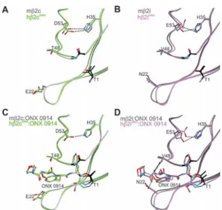
Figure 6.Structural superpositions of the natural mouseβ2c (A,C) and β2i (B,D) subunits with their human−yeast chimeric counter-parts in the ligand-free (A,B) and ONX 0914-bound (C,D) states. Amino acids are labeled by the one-letter code. Hydrogen bonds are depicted by black dashed lines. Hydrophobic interactions are highlighted by double arrows. Color coding is according toFigure 5. Note that ONX 0914 has been previously modeled into the mouse
β2 subunits as a morpholine adduct with Thr1,13d whereas in the chimeric subunits it was built as a seven-membered ring structure according to the revised reaction mechanism of epoxyketones with Thr1.21PDB IDs: 3UNE (mouse cCP), 3UNH (mouse iCP), 3UNB (mouse cCP:ONX 0914), 3UNF (mouse iCP:ONX 0914), 6HTB (hβ2c chimera), 6HV3 (hβ2i chimera), 6HTC (hβ2c chimera:ONX 0914), 6HV4 (hβ2i chimera:ONX 0914).
Figure 7. Human−yeast chimeric proteasomes in complex withβ2c (4; green)- andβ2i (39; purple)-selective inhibitors. (A,C,E,G) 2FO−FC
electron density maps for the compounds bound to theβ2c (green) andβ2i (purple) chimeric subunits, respectively, are shown as blue meshes contoured to 1σ. (B,D,F,H) Structural superposition of ligand-free and ligand-bound chimericβ2c andβ2i subunits. Polar and hydrophobic interactions are depicted according toFigure 6. PDB IDs: 6HTB (hβ2c chimera), 6HTD (hβ2c chimera:4), 6HUV (hβ2c chimera:39), 6HV3 (hβ2i chimera), 6HV5 (hβ2c chimera:4), 6HVV (hβ2i chimera:39).
substrates in general might be better stabilized in the
β
2i
substrate-binding channel than in the
β
2c one. Notably, a
similar observation has previously been reported for Thr22 in
subunit y
β
1/
β
1c.
13aThe co-crystal structure of the
β
2i chimera with compound
39
shows a well-de
fi
ned 2F
O−
F
Celectron density map for the
ligand (
Figure 7
G). A comparison of the ligand-free and
ligand-bound states of the
β
2i chimera indicates a movement
of His35 upon inhibitor binding (
Figure 7
H). Despite this
structural
fl
exibility and plasticity of the S1 pocket, the
hydrogen bond between His35 and Glu53 remains intact.
Compared to
β
2i, the
β
2c active site appears to be more rigid,
as binding of
39
does not trigger any structural changes of
His35 (
Figure 7
F). Presumably, the P1 side chain of
39
is less
well-de
fi
ned in the
β
2c active site because of the tight
anchoring of and the resulting steric hindrance with His35
(
Figure 7
E,F). Thus, although the
β
2 subunits in general
accept large P1 side chains, it appears that the plasticity of the
β
2i active site tolerates bulky residues even more readily than
β
2c.
■
DISCUSSION AND CONCLUSIONS
Here, we describe the development and evaluation of a set of
potent and selective inhibitors of human
β
2c and
β
2i
proteasome activities. Because of the structural similarities of
the mammalian
β
2c and
β
2i subunits, no key guidelines for
compound design strategies could be derived from the crystal
structures so far.
13dThus, we used the previously described
inhibitors LU-102 (1),
16LU-112 (2),
16and ONX 0914 (3)
12as the starting points, which have no or only moderate
preference for one of the two human
β
2 subunits over the
other. By changing the P sites of the ligands, we disfavored the
most closely related subunit, either
β
2i or
β
2c, and gained
selectivity.
Substantial organic synthesis e
ff
orts and thorough empiric
screening of compound libraries derived from these lead
structures
fi
nally led to the identi
fi
cation of selective
compounds and to the development of suitable probes for
ABPP assays. Furthermore, previously unaddressed
stereo-chemistry issues on LU-002i (5) have now been resolved and
the exact con
fi
guration of the bioactive compound has been
determined.
Selected
β
2c and
β
2i inhibitors were analyzed by X-ray
crystallography in complex with the WT yeast CP and with
chimeric human
−
yeast proteasomes, incorporating key
ele-ments of the human
β
2c and
β
2i substrate-binding channels,
respectively. Despite the arti
fi
cial character of chimeras, they
were previously shown to serve as excellent structural tools
18and now again prove valuable for explaining the selectivity
patterns observed for the
β
2 compound libraries described
here. Both
β
2c and
β
2i can incorporate large P1 residues in
their spacious S1 pocket. Because of the favorable hydrogen
bond interactions with Asp/Glu53, LU-102 derivatives with
their 4-aminomethylphenyl side chain at P1 are in general
more potent
β
2 inhibitors than ONX 0914-based compounds,
featuring apolar P1 residues.
16Selectivity for
β
2c was gained by
installing small P2 residues on LU-102. Epoxyketones with
bulky hydrophobic P1 residues and small P3 side chains were
found to show
β
2i selectivity. Because of the plasticity of the
S1 pocket and the
fl
exibility of His35 in subunit
β
2i, large
apolar P1 side chains can be better accommodated in
β
2i than
in
β
2c.
Taken together, we here present the most selective
β
2c and
β
2i ligands reported so far. As part of a set of inhibitors and
ABPs that is capable of disabling and visualizing the individual
activities of human constitutive and immunoproteasomes,
15these compounds might become valuable tools for
fundamen-tal as well as applied biochemical and biomedical research on
proteasomes and hopefully elucidate more details on the
biological role and impact of the trypsin-like active sites of
human proteasomes.
■
EXPERIMENTAL SECTION
General Procedures.All reagents were of commercial grade and used as received unless indicated otherwise. The purity of all tested compounds is >95% on the basis of liquid chromatography−mass spectrometry (LC-MS) and nuclear magnetic resonance (NMR).1
H-and 13C NMR spectra were recorded on a Bruker AV-400 (400
MHz), AV-600 (600 MHz), or AV-850 (850 MHz) spectrometer. Chemical shifts are given in ppm (δ) relative to CD3OD or CDCl3as
an internal standard. Coupling constants are given in Hz, and peak assignments are based on 2D1H correlation spectroscopy and 13C
heteronuclear single quantum coherence NMR experiments. All13C
attached proton test spectra are proton-decoupled. LC-MS analysis was performed on a Finnigan Surveyor high-performance liquid chromatography (HPLC) system with a Gemini C18 50×4.60 mm column (detection at 200−600 nm) coupled to a Finnigan LCQ Advantage Max mass spectrometer with electrospray ionization (ESI). Methods used are: 15 min (0−0.5 min: 10% MeCN; 0.5−10.5 min: 10−90% MeCN; 10.5−12.5 min: 90% MeCN; 12.5−15 min: 90− 10% MeCN) or 12.5 min (0−0.5 min: 10% MeCN; 0.5−8.5 min: 10−90% MeCN; 8.5−10.5 min: 90% MeCN; 10.5−12.5 min: 90−− 10% MeCN). HRMS was recorded on an LTQ Orbitrap (ThermoFinnigan). For reverse-phase HPLC purification, an automated Gilson HPLC system equipped with a C18 semiprep column (Phenomenex Gemini C18, 5 μm 250 × 10 mm) and a GX281 fraction collector was used.
General Procedure for Boc Removal. The appropriate Boc-protected C-terminally modified leucine derivative was dissolved in TFA and stirred for 20 min. Co-evaporation with toluene (3×) afforded the TFA salt, which was used without further purification.
General Procedure for Azide Couplings.Compounds6−53,
68,71,74,77,86,87,and97were prepared via azide coupling of the appropriate protected tripeptide hydrazide and either an epoxyketone amine or a vinyl sulfone amine. Peptide hydrazides were prepared by hydrazinolysis of peptide methyl esters synthesized as described in the
Supporting Information. The hydrazide was dissolved in 1:1 DMF/ DCM (v/v) and cooled to−30°C.tBuONO (1.1 equiv) and HCl (4 N solution in 1,4-dioxane, 2.8 equiv) were added, and the mixture was stirred for 3 h at −30 °C, after which thin-layer chromatography analysis (10% MeOH/DCM, v/v) showed the complete consumption of the starting material. The epoxyketone or vinyl sulfone amine was added as a free amine to the reaction mixture as a solution in DMF with 5.0 equiv of DiPEA. The mixture was allowed to warm to room temperature overnight. The mixture was diluted with ethyl acetate (EtOAc) and extracted with H2O (3×) and brine. The organic layer
was dried over MgSO4 and purified by reverse-phase HPLC. For
compounds featuring Boc-protecting groups, TFA was added, and the reaction mixture was stirred for 30 min. The crude was purified by reverse-phase HPLC.
N3Phe-Leu-Ser-Phe(4-CH2NH2)VS TFA salt (13). The synthesis
of tripeptide hydrazide N3Phe-Leu-Ser(tBu)-NHNH2is described in
the Supporting Information. The title compound was prepared according to the general procedure for azide coupling on a 50μmol scale and purified by HPLC (30−40% MeCN−H2O) to yield 2.8 mg
172.23, 171.81, 146.65, 139.69, 137.84, 133.00, 131.84, 131.26, 130.42, 130.24, 129.65, 128.10, 65.38, 62.75, 56.70, 53.80, 52.56, 44.09, 42.77, 41.38, 40.25, 38.72, 25.82, 23.46, 21.84. LC−MS (linear gradient 10 →90% MeCN/H2O, 0.1% TFA, 15.0 min):Rt(min):
6.27 (ESI−MS (m/z): 628.20 (M + H)+). HRMS calcd for
C30H41N7O6S, 628.29118 [M + H]+; found, 628.29123.
Morp-Ala-Tyr(Me)-HomoCha-EK TFA salt (39). The synthesis of Boc-HomoCha-EK is described in the Supporting Information, and the Boc-protecting group was removed according to the general procedure. The title compound was prepared according to the general procedure for azide coupling on a 50 μmol scale and purified by HPLC (30−45% MeCN−H2O) to yield 12.3 mg (17.2μmol, 34%). 1
H NMR (600 MHz, MeOD):δ7.25−7.01 (m, 2H), 6.91−6.67 (m, 2H), 4.60−4.57 (m, 1H), 4.48−4.28 (m, 2H), 3.77 (s, 3H), 3.71− 3.70 (m, 4H), 3.21 (d,J= 4.9 Hz, 1H), 3.09−2.88 (m, 4H), 2.84− 2.79 (m, 1H), 2.56−2.37 (m, 4H), 1.83−1.63 (m, 6H), 1.52−1.39 (m, 4H), 1.38−1.16 (m, 9H), 0.97−0.83 (m, 2H).13C NMR (150
MHz, MeOD): δ 209.19, 174.20, 173.30, 171.99, 159.94, 131.41, 130.02, 114.75, 67.85, 62.40, 60.01, 55.75, 55.60, 54.71, 53.06, 52.92, 49.65, 38.48, 38.15, 34.61, 34.02, 29.11, 27.72, 27.44, 27.38, 18.65, 16.84. LC−MS (linear gradient 10→90% MeCN/H2O, 0.1% TFA,
12.5 min): Rt (min): 6.23 (ESI−MS (m/z): 601.33 (M + H)+).
HRMS calcd for C32H48N4O7, 601.35958 [M + H]+; found,
601.35945.
Morp-Ala-Tyr(Me)-1-(R)-TetraNal-EK TFA salt (68). The syn-thesis of Boc-1-TatraNal-EK is described in the Supporting Information, and the Boc protecting group was removed according to the general procedure. The title compound was prepared according to the general procedure for azide coupling on a 56μmol scale and purified by HPLC (30−45% MeCN−H2O) to yield 14.2 mg (22.4
μmol, 40%).1H NMR (400 MHz, MeOD):δ7.22−7.13 (m, 2H),
7.12−7.01 (m, 4H), 6.88−6.77 (m, 2H), 4.63−4.59 (m, 2H), 4.41− 4.36 (m, 1H), 4.06−3.84 (m, 6H), 3.78 (s, 3H), 3.17−2.98 (m, 2H), 2.93−2.67 (m, 5H), 2.14−1.54 (m, 6H), 1.45 (s, 3H), 1.35 (d,J= 7.1 Hz, 3H). 13C NMR (100 MHz, MeOD):δ209.40, 174.19, 173.10,
165.04, 160.01, 140.66, 137.91, 131.40, 130.18, 129.98, 129.94, 127.07, 126.54, 114.84, 64.84, 59.89, 58.37, 55.83, 55.67, 53.93, 52.88, 51.64, 50.57, 39.22, 37.99, 36.17, 30.02, 29.64, 20.16, 18.11, 16.76. LC−MS (linear gradient 10→90% MeCN/H2O, 0.1% TFA, 12.5
min):Rt(min): 6.10 (ESI−MS (m/z): 635.00 (M + H)+). HRMS
calcd for C35H46N4O7, 635.34393 [M + H]+; found, 635.34371.
Morp-Ala-Tyr(Me)-1-(S)-TetraNal-EK TFA salt (71). The syn-thesis of Boc-1-TatraNal-EK is described in the Supporting Information, and the Boc-protecting group was removed according to the general procedure. The title compound was prepared according to the general procedure for azide coupling on a 50μmol scale and purified by HPLC (30−45% MeCN−H2O) to yield 16.5 mg (26.0
μmol, 52%).1H NMR (400 MHz, MeOD):δ7.24−7.13 (m, 3H), 7.13−6.98 (m, 3H), 6.86−6.78 (m, 2H), 4.77−4.59 (m, 2H), 4.41− 4.36 (m, 1H), 4.00−3.86 (m, 6H), 3.21 (d,J= 5.0 Hz, 1H), 3.10 (dd, J= 14.0, 6.0 Hz, 1H), 3.03−2.64 (m, 5H), 2.02−1.60 (m, 6H), 1.45 (s, 3H), 1.33 (d,J= 7.2 Hz, 3H).13C NMR (100 MHz, MeOD):δ
208.97, 174.18, 173.57, 164.98, 160.01, 141.21, 138.13, 131.41, 130.09, 130.01, 129.75, 126.83, 126.76, 114.84, 114.75, 64.82, 60.04, 58.35, 55.89, 55.66, 53.91, 53.05, 51.05, 50.54, 38.67, 38.02, 35.13, 30.57, 27.13, 20.23, 18.10, 16.86. LC−MS (linear gradient 10→90% MeCN/H2O, 0.1% TFA, 12.5 min):Rt(min): 6.18 (ESI−MS (m/z):
635.07 (M + H)+). HRMS calcd for C
35H46N4O7, 635.34393 [M +
H]+; found, 635.34370.
Morp-Ala-Tyr(Me)-1-DecAla-EK TFA salt (74). The synthesis of Boc-1-DecAla-EK is described in theSupporting Information, and the Boc-protecting group was removed according to the general procedure. The title compound was prepared according to the general procedure for azide coupling on a 50μmol scale and purified by HPLC (40−50% MeCN−H2O) to yield 14.6 mg (22.8 μmol,
46%).1H NMR (400 MHz, MeOD):δ7.20−7.11 (m, 2H), 6.86− 6.78 (m, 2H), 4.62−4.51 (m, 2H), 4.38−4.33 (m, 1H), 4.07−3.83 (m, 6H), 3.77 (d,J= 3.7 Hz, 3H), 3.22 (d,J= 12 Hz, 1H), 3.06−3.01 (m, 1H), 2.95 (d,J= 12 Hz, 1H), 2.85−2.79 (m, 1H), 1.84−1.13 (m, 25H). 13C NMR (100 MHz, MeOD): δ 209.81, 174.11, 173.35,
164.81, 159.95, 131.42, 130.00, 114.77, 64.78, 60.08, 58.26, 55.64, 53.89, 52.81, 50.50, 50.29, 39.17, 38.74, 38.20, 33.84, 29.05, 27.91, 26.60, 22.38, 20.22, 18.11, 16.88. LC−MS (linear gradient 10→90% MeCN/H2O, 0.1% TFA, 12.5 min):Rt(min): 6.75 (ESI−MS (m/z):
641.13 (M + H)+). HRMS calcd for C
35H52N4O7, 641.39088 [M +
H]+; found, 641.39081.
Morp-Ala-Tyr(Me)-1-DecAla-EK TFA salt (77). The synthesis of Boc-1-DecAla-EK is described in theSupporting Information, and the Boc- protecting group was removed according to the general procedure. The title compound was prepared according to the general procedure for azide coupling on a 23μmol scale and purified by HPLC (40−50% MeCN−H2O) to yield 6.8 mg (10.6μmol, 46%
s).1H NMR (400 MHz, MeOD):δ7.19−7.13 (m, 2H), 6.84−6.80
(m, 2H), 4.63−4.60 (m, 1H), 4.53−4.50 (m, 1H), 4.40−4.34 (m, 1H), 4.01−3.90 (m, 6H), 3.78 (d,J= 1.9 Hz, 3H), 3.17 (d,J= 5.1 Hz, 1H), 3.07−3.02 (m, 1H), 2.94 (d,J= 5.1 Hz, 1H), 2.88−2.80 (m, 1H), 1.85−1.07 (m, 25H).13C NMR (100 MHz, MeOD):δ209.46,
174.13, 173.41, 164.82, 159.97, 131.43, 129.96, 114.77, 64.78, 59.91, 58.27, 55.62, 53.90, 52.92, 50.97, 50.52, 43.08, 39.49, 39.11, 38.25, 36.48, 33.73, 27.99, 27.69, 26.76, 26.54, 22.32, 21.21, 18.10, 16.87. LC−MS (linear gradient 10→90% MeCN/H2O, 0.1% TFA, 12.5
min):Rt(min): 6.79 (ESI−MS (m/z): 641.07 (M + H)+). HRMS
calcd for C35H52N4O7, 641.39088 [M + H]+; found, 641.39070.
Morp-Ala-Tyr(Me)-1-DecAla-EK TFA salt (86). The synthesis of Boc-1-DecAla-EK is described in theSupporting Information, and the Boc-protecting group was removed according to the general procedure. The title compound was prepared according to the general procedure for azide coupling on a 25μmol scale and purified by HPLC (30−45% MeCN−H2O) to yield 8.6 mg (11.4μmol, 46%). 1H NMR (500 MHz, MeOD):δ7.15 (d,J= 8.7 Hz, 2H), 6.85−6.80
(m, 2H), 4.62−4.59 (m, 1H), 4.55−4.52 (m, 1H), 4.40−4.35 (m, 1H), 4.01−3.91 (m, 6H), 3.78 (s, 3H), 3.21 (d,J= 5.1 Hz, 1H), 3.07−3.03 (m, 1H), 2.95 (d,J= 5.1 Hz, 1H), 2.85−2.81 (m, 1H), 1.83−1.52 (m, 10H), 1.47−1.41 (m, 5H), 1.37−1.18 (m, 10H).13C
NMR (125 MHz, MeOD):δ209.83, 174.13, 173.36, 164.80, 159.96, 131.42, 129.99, 114.77, 64.78, 60.09, 58.26, 55.64, 53.90, 52.81, 50.49, 50.29, 39.21, 39.17, 38.75, 38.20, 34.85, 33.84, 29.05, 27.92, 27.91, 26.60, 22.38, 20.22, 18.12, 16.87. LC−MS (linear gradient 10→90% MeCN/H2O, 0.1% TFA, 12.5 min):Rt(min): 6.92 (ESI−MS (m/z):
641.13 (M + H)+). HRMS calcd for C
35H52N4O7, 641.39088 [M +
H]+; found, 641.39065.
Morp-Ala-Tyr(Me)-1-DecAla-EK TFA salt (87). The synthesis of Boc-1-DecAla-EK is described in theSupporting Information, and the Boc-protecting group was removed according to the general procedure. The title compound was prepared according to the general procedure for azide coupling on a 44μmol scale and purified by HPLC (30−45% MeCN−H2O) to yield 12.7 mg (16.8 μmol,
38%).1H NMR (500 MHz, MeOD): δ7.15 (d, J= 8.6 Hz, 2H), 6.84−6.79 (m, 2H), 4.66−4.58 (m, 1H), 4.53−4.50 (m, 1H), 4.39− 4.35 (m, 1H), 4.02−3.92 (m, 6H), 3.17 (d,J= 5.1 Hz, 1H), 3.07− 3.03 (m, 1H), 2.94 (d,J= 5.1 Hz, 1H), 2.86−2.82 (m, 1H), 1.84− 1.18 (m, 25H). 13C NMR (125 MHz, MeOD): δ209.47, 174.13,
173.41, 164.81, 159.96, 131.43, 129.96, 114.77, 64.76, 59.91, 58.24, 55.64, 53.89, 52.93, 50.96, 50.55, 43.06, 39.48, 39.10, 38.25, 36.48, 33.72, 27.98, 27.68, 26.75, 26.53, 22.31, 21.21, 18.08, 16.87. LC−MS (linear gradient 10→90% MeCN/H2O, 0.1% TFA, 12.5 min): Rt
(min): 6.88 (ESI−MS (m/z): 641.13 (M + H)+). HRMS calcd for
C35H52N4O7, 641.39088 [M + H]+; found, 641.39077.
Morp-Ala-Tyr(O−C2H4-BODIPY(FL))-1-DecAla-EK (97). The synthesis of compound95is described in theSupporting Information. Compound95(23 mg, 33μmol) and BODIPY-FL-alkyne9623(13 mg, 40μmol, 1.2 equiv) were dissolved in DMF. An aqueous solution of sodium ascorbate (100μL, 25μmol, 0.75 equiv) and an aqueous solution of CuSO4(100μL, 17μmol, 0.5 equiv) were added. The
reaction mixture was stirred at room temperature overnight. The reaction mixture was concentrated in vacuo, and purification by HPLC (50−70% MeCN−H2O) yielded the title compound (6.7 mg,
5.9μmol, 18%).1H NMR (600 MHz, MeOD):δ7.91 (s, 1H), 7.17
(d,J= 8.2 Hz, 2H), 6.84 (dd,J= 12.1, 8.3 Hz, 2H), 6.14 (s, 2H), 4.82 (t,J= 5.0 Hz, 2H), 4.64 (dt,J= 8.3, 5.5 Hz, 1H), 4.57 (ddd,J= 13.6,
10.7, 3.3 Hz, 1H), 4.46−4.34 (m, 4H), 3.99 (dd,J= 24.6, 16.0 Hz, 8H), 3.34−3.16 (m, 3H), 3.16−3.03 (m, 4H), 2.98 (dt,J= 11.4, 5.6 Hz, 1H), 2.87 (t,J= 7.2 Hz, 4H), 2.51 (s, 8H), 2.44 (s, 8H), 1.99 (q, J= 7.4 Hz, 3H), 1.93−1.80 (m, 3H), 1.75 (ddd,J= 16.6, 8.4, 3.4 Hz, 6H), 1.69−1.54 (m, 6H), 1.52 (d,J= 2.8 Hz, 1H), 1.49 (d,J= 2.9 Hz, 5H), 1.45−1.37 (m, 5H), 1.37−1.18 (m, 7H).13C NMR (150
MHz, MeOD): δ 209.76, 209.41, 174.09, 173.28, 173.22, 172.91, 164.76, 161.88, 161.64, 158.47, 154.93, 148.55, 147.84, 142.18, 132.57, 131.57, 130.91, 130.89, 130.77, 124.23, 122.62, 115.63, 115.58, 67.69, 64.80, 64.75, 60.04, 59.87, 58.23, 55.64, 53.89, 52.90, 52.77, 50.96, 50.52, 50.20, 49.43, 49.28, 49.14, 49.00, 48.86, 48.72, 48.57, 43.06, 39.48, 39.22, 39.11, 38.74, 38.28, 38.23, 36.50, 35.66, 34.99, 34.89, 33.84, 33.73, 32.20, 32.15, 30.72, 30.63, 29.08, 29.04, 27.99, 27.91, 27.69, 27.58, 27.00, 26.76, 26.59, 26.53, 25.88, 22.38, 22.31, 21.21, 18.14, 16.88, 16.49, 14.48. LC−MS (linear gradient 10
→90% MeCN/H2O, 0.1% TFA, 12.5 min):Rt(min): 8.07 (ESI−MS
(m/z): 1024.33 (M + H)+). HRMS calcd for C
55H78BF2N9O7,
1024.60108 [M + H]+; found, 1024.60174.
Biological and Structural Analysis.Competition Assays in Cell Lysates. Lysates of Raji cells were prepared by sonication in three volumes of lysis buffer containing 50 mM Tris pH 7.5, 1 mM DTT, 5 mM MgCl2, 250 mM sucrose, 2 mM ATP, and 0.05% (w/v)
digitonin. The protein concentration was determined by the Bradford assay. Cell lysates (diluted to 5μg of total protein in buffer containing 50 mM Tris pH 7.5, 2 mM DTT, 5 mM MgCl2, 10% (v/v) glycerol,
and 2 mM ATP) were exposed to the inhibitors for 1 h at 37°C prior to incubation with cocktail ABPs for another 1 h, followed by 3 min boiling with a reducing gel-loading buffer and fractionation on 12.5% SDS-PAGE. In-gel detection of residual proteasome activity was performed in the wet gel slabs directly on a ChemiDoc MP system using Cy2 settings to detect BODIPY(FL)-LU-112, Cy3 settings to detect BODIPY(TMR)−NC−005-VS, and Cy5 settings to detect Cy5-NC-001. The intensities of bands were measured byfluorescent densitometry and normalized to the intensity of bands in the mock-treated extracts. The average values of three independent experiments were plotted against the inhibitor concentrations (in the initial screening, experiments were only carried out one time). The IC50
(ligand concentrations giving 50% inhibition) values were calculated using GraphPad Prism software.
Competition Assays in Living RPMI-8226 Cells.RPMI-8226 cells were cultured in RPMI-1640 media supplemented with 10% (v/v) fetal calf serum, GlutaMAX, and penicillin/streptomycin in a 5% CO2
-humidified incubator. An amount of (5−8)×105cells/mL cells was
exposed to inhibitors for 1 h at 37°C. The cells were harvested and washed twice with phosphate-buffered saline. The cell pellets were treated with lysis buffer (50 mM Tris pH 7.5, 2 mM DTT, 5 mM MgCl2, 10% (v/v) glycerol, 2 mM ATP, 0.05% (w/v) digitonin) on
ice for 1 h, followed by centrifugation at 14 000 rpm for 15 min. Proteasome inhibition in the obtained cell lysates was determined using the method described above. The intensities of bands were measured byfluorescent densitometry and divided by the intensity of bands in the mock-treated extracts. Gels were stained by Coomassie Brilliant Blue, which was used to correct for gel-loading differences. The average values of three independent experiments were plotted against the inhibitor concentrations. The IC50 (compound
concen-trations causing 50% inhibition) values were calculated using GraphPad Prism software.
ABPP Assays in Raji Cell Lysates.Raji cell lysates (diluted to 5μg of total protein in buffer containing 50 mM Tris pH 7.5, 2 mM DTT, 5 mM MgCl2, 10% (v/v) glycerol, and 2 mM ATP) were exposed to
the probe for 1 h at 37°C, followed by 3 min boiling with a reducing gel-loading buffer and fractionation by 12.5% SDS-PAGE. Separation was obtained by electrophoresis for 15 min on 80 V, followed by 120 min on 130 V. In-gel detection of residual proteasome activity was performed in the wet gel slabs directly on a ChemiDoc MP system using Cy2 settings.
Yeast Mutagenesis.hPSMB7 and hPSMB10 encoding the human
β2c and β2i proteasome subunits, respectively, were purchased as yeast codon-optimized, synthetic gene fragments, each with a 30 bp 5′ overhang corresponding to the yeastPUP1(yβ2) promoter sequence
preceding the start ATG and a 40 bp 3′overhang corresponding to thePUP1terminator sequence following the stop codon. An AgeI site at the codons for Gly-1/Thr1 was incorporated into both genes.
The human PSMB7/10 ORFs were fused to thePUP1promoter and terminator by recombinant polymerase chain reaction (PCR): both genes were amplified with the primers PSMB-for and PSMB-rev (Table S15). ThePUP1 promoter was amplified from the template plasmid p15-PUP1-new with the primers pBS-rev and PUP1-prom-rev and the terminator with the primers PUP1-ter-for and pBS-uni (Table S15). The promoter fragment and the ORF fragments were fused by recombinant PCR in the presence of pBS-rev and PSMB-rev. The resulting fragment was then fused by recombinant PCR with the terminator fragment in the presence of pBS-rev and pBS-uni.
The recombinant gene fragments were cut with SacI and HindIII and ligated with SacI/HindIII cut vector pUC19 and afterward transferred into the shuttle vector pRS315, yielding p15-fl-PSMB7 and p15-fl-PSMB10. The S171G mutant version of PSMB7 was created by recombinant PCR with the pUC19 construct as the template and mutagenic primers S171G-for and PSMB7-S171G-rev (Table S15) and cloning of the resulting SacI/HindIII cut product into pRS315, yielding p15-fl-PSMB7*.
For replacement of the genuine human propeptide-encoding sequences by thePUP1 propeptide sequence, the PUP1 promoter, together with the propeptide encoding region, was amplified from p15-PUP120b with the primers pBS-rev and PUP1-Age-rev (Table
S15), which introduces an AgeI site at the corresponding Gly-1/Thr1-encoding position ofPUP1. The PCR product was cut with HindIII and AgeI and ligated with the respective AgeI/SacI fragments from p15-fl-PSMB7, p15-fl-PSMB7*, and p15-fl-PSMB10 into HindIII/ SacI cut pRS315 to obtain the plasmids PSMB7, p15-P1pp-PSMB7*, and p15-P1pp-PSMB10.
Genes encoding the hybrids of yβ2 and hβ2i were constructed by recombinant PCR. For the hβ2i1−129 construct, an N-terminal
fragment resulting from a PCR with primers pBS-rev and beta2i-129-rev on template p15-P1pp-PSMB10 was fused with a C-terminal fragment made by PCR with primers beta2i-129-for and pBS-uni on template p15-PUP1-new (Table S15). Accordingly, the hybrids hβ2i1−93and hβ2i1−52were constructed employing primers
beta2i-1-93-for/beta2i-1-93-rev and beta2i-1-52-for/beta2i-1-52-rev, respec-tively. For the hβ2i1−52/93−129 hybrid, the N-terminal fragment was obtained by PCR on the hβ2i1−52template with primers pBS-rev and
y93-rev, and for the C-terminal fragment, the hβ2i1−129template was
used with primers 2i93-for and pBS-uni (Table S15).
All pRS315-based constructs were introduced into the yeast strain YWH10,20bwhich has the chromosomal PUP1 gene deleted and a WTPUP1 copy in aURA3-marked plasmid. After selection against URA3 on 5′-fluorouracil, clones that were viable without the WT PUP1gene were recovered.
Crystallographic Analysis. WT and mutant yCP crystals were grown by hanging drop vapor diffusion as previously described.24 Inhibitor complex structures were obtained by incubating crystals in 5
μL cryobuffer (20 mM magnesium acetate, 100 mM 2-(N -morpholino)ethanesulfonic acid, pH 6.8, and 30% (v/v) 2-methyl-2,4-pentanediol) supplemented with 0.5μL of inhibitor (50 mM in dimethyl sulfoxide) for up to 48 h. Diffraction data were collected at the Paul Scherrer Institute, SLS, Villigen, Switzerland and the ESRF, Grenoble, France (λ= 1.0 Å). The evaluation of reflection intensities and data reduction was performed with the program package XDS.25 Molecular replacement was carried out with the coordinates of the yeast 20S proteasome (PDB entry code: 5CZ426) by rigid body refinements (REFMAC527). COOT28 was used to build models. Translation/libration/screw refinements finally yielded excellent R factors as well as root-mean-square deviation bond and angle values. The coordinates, proven to have good stereochemistry from the Ramachandran plots, were deposited in the RCSB Protein Data Bank. For accession codes, seeTable S13.