THE ROLE OF BP180 IN GRANULOPOIESIS AND NLRP3 INFLAMMASOME ACTIVATION IN BULLOUS PEMPHIGOID
Lin Lin
A dissertation submitted to the faculty of the University of North Carolina at Chapel Hill in partial fulfillment of the requirements for the degree of Doctor of Philosophy in the
Curriculum of Oral Biology, School of Dentistry
Chapel Hill 2016
Approved by: Liu, Zhi
ii ©2016 Lin Lin
iii
ABSTRACT
Lin Lin: The role of BP180 in granulopoiesis and NLRP3 inflammasome activation in bullous pemphigoid
(Under the direction of Zhi Liu)
BP180 (also called collagen XVII) is a hemidesmosomal protein. Autoantibodies directly against it cause bullous pemphigoid (BP). Gene mutations of BP180 result in generalized atrophic benign epidermolysis bullosa (GABEB). BP patients’ autoantibodies primarily recognize NC16A domain of BP180; however, there is no cross reactivity between human NC16A and mouse homologue termed NC14A. To investigate the pathogenesis of BP and identify new functions of BP180, we established humanized BP180NC16A (NC16A) mice, and subsequently NC16A domain truncated (ΔNC16A) mice.
iv
induces NF-kB signaling pathway activation that leads to elevated G-CSF secretion in bone marrow and blood. G-CSF regulates granulopoiesis in bone marrow. These findings provide a mechanism for clinical relevant pathophysiological observation in BP and Junctional Epidermolysis Bullosa (JEB).
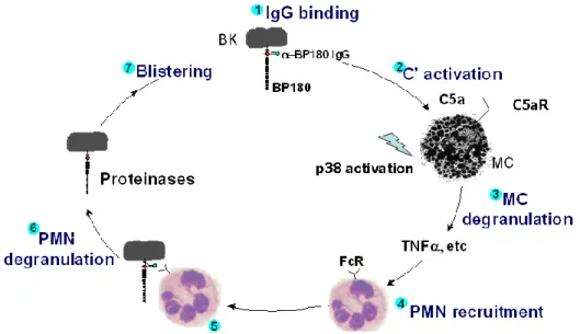
NC16A mice injected with anti-NC16A autoantibodies develop skin disease that closely mimics the clinical, immunological and histological features of human BP. Previous works have shown that pathological autoantibodies against NC16A, when binding to the target on basal keratinocytes, trigger complement activation to generate C5a. C5a binding to the C5a receptor on mast cells leads to TNFα release. Mast cell recruitment of polymorphonuclear neutrophils is required for disease. However, the mechanisms by which mast cells recruit neutrophils to the antibody-binding sites are not completely known. We used mice lacking key components of NLRP’s inflammasome to test whether mast cell-released TNF-α would act on keratinocyte as first signal and pathogenic IgG binding to BP180 as a second signal to activate NLRP3 inflammasome in basal keratinocyte. We found that pathogenic antibody-induced BP depends on NLRP3 activation, leading to IL-1β release and subsequent neutrophil recruitment. Furthermore, we showed that IL-1R antagonist, Anakinra, could be an effective drug to treat experimental BP. In this study, we suggest a molecular mechanism for mast cells recruitment of neutrophils in experimental BP. More importantly, our findings identify potential new therapeutic targets for treatment of BP.
v
vi
ACKNOWLEDGMENTS
I would like to express my gratitude to my mentor Dr. Zhi Liu, who always supports and encourages me throughout my graduate school career. I have learnt a lot from him as a critical thinker, an enthusiasm scientist, and a thoughtful story teller.
To the current and past members of the laboratory of Liu, I would like to thank all of you who have accompanied with me ups and downs. Without your help and friendship, I could not accomplish this journey. I would like to thank Bin-Jin Hwang for discussing our projects, troubleshooting for experiments and covering each other for mice colonies. I also thank Jaime Brozowski, Lisa Heimbach, Kayla Shumate, Susan Burette, Peng Geng, Zhen Liu, Jianxun Yang, Jinbo Chen, Qiaoling Zhang, Bin Peng and Yang Zhang. I also would like thank my committee members, Dr. Ed Miao, Dr. Barbara Vilen, Dr. Jenny Ting, Dr. Roland Arnold, and Dr. Steve Clarke. Their value suggestions and advice made my research progress. I also want to thank members in Oral Biology program, Dr. Patrick Flood, Dr. Ceib Phillips, Dr. Eric Everett, Cindy Blake, Nathan Kotecki, Meagan Elizabeth Solloway and Department of Dematology, Dr. Ning Li, Dr. Ye Qian, Dr. Donna Culton and Dr. Luis Diaz. To all my classmates and friends in Oral Biology Program, I would like thank all of you for your support and friendship.
vii
TABLE OF CONTENTS
LIST OF FIGURES ...ix
LIST OF ABBREVATIONS ...xi
CHAPTER ONE BACKGROUND AND SIGNIFICANCE ... 1
1.1 BP180 ... 1
1.1.1 General background ... 1
1.1.2 Gene and cDNA of BP180 ... 1
1.1.3 Biochemical graph of BP180 ... 2
1.1.4 Other functions of BP180 ... 4
1.1.5 Bullous pemphigoid ... 5
1.1.6 Junctional epidermolysis bullosa (EB) ... 8
1.2 Inflammasome ... 11
1.2.1 General background ... 11
1.2.2 Mechanisms of NLRP3 Inflammasome activation ... 12
1.2.3 Inflammasome related diseases and therapeutics... 13
1.3 Granulopoiesis... 14
1.3.1 Granulocyte development ... 14
1.3.2 Extrinsic factor for granulopoiesis ... 15
1.3.3 Granulocyte circulation ... 15
viii
1.3.5. Granulocytes and B cell development ... 18
1.4 References ... 20
CHAPTER TWO DEFICIENCY OF THE HEMIDESMOSOMAL PROTEIN BP180 FUNCTION LEADS TO ALTERED GRANULOPOIESIS IN MICE ... 28
2.1 Introduction ... 28
2.2 Materials and methods ... 31
2.3 Results ... 35
2.4 Discussion ... 42
2.5 References ... 46
CHAPTER THREE NLRP3 INFLAMMASOME ACTIVATION IS REQUIRED FOR THE CUTANEOUS AUTOIMMUNE DISEASE BULLOUS PEMPHIGOID ... 70
3.1 Introduction ... 70
3.2 Materials and methods ... 73
3.3 Results ... 77
3.4 Discussion ... 83
3.5 References ... 87
CHAPTER FOUR DISCUSSION AND FUTURE DIRECTIONS ... 105
4.1 Summary of findings ... 105
4.2 New function of BP180 ... 106
4.3 Granulocytes in B cell development and a possible mechanism in autoimmune diseases ... 109
4.4 Possible inflammasome mechanism in experimental BP ... 110
ix
LIST OF FIGURES
Figure 1.1 The molecular structure of BP180……… 3
Figure 1.2 The pathogenesis of experimental bullous pemphigoid……… 8
Figure 1.3 The categories of epidermolysis bullosa (EB) ……… 9
Figure 2.1 Granulocyte hyperplasia in ΔNC16A mice……… 50
Figure 2.2 Increased granulocyte-monocyte progenitor in ΔNC16A mice……… 52
Figure 2.3 Granulocyte hyperplasia in ΔNC16A mice were caused by extrinsic factors……… 54
Figure 2.4 ΔNC16A mice showed elevated G-CSF level and increased activation of NF-kB signaling pathway in bone marrow……… 56
Figure 2.5 Bone marrow-derived mesenchymal stem cells (MSC) from ΔNC16A mice showed increased activation of NF-kB pathway and secreted higher level of G-CSF……… 58
Figure 2.6 Blocking G-CSF restored normal granulopoiesis in ΔNC16A mice… 59 Figure 2.7 K14Cre/ΔNC16A mice had normal granulopoiesis……… 61
Figure 2.8 Characterization of ΔNC16A mice……… 63
Figure 2.9 Four week old ΔNC16A mice showed no skin lesion and altered granulopoiesis……… 65
Figure 2.10 Increased autoantibody and affected B cell development in ΔNC16A mice……… 66
Figure 2.11 Decreased B cell populations in ΔNC16A mice……… 68
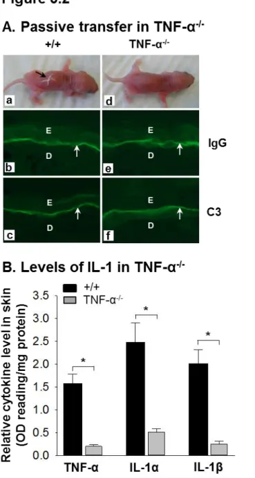
Figure 3.1 Increased levels of TNF-α, IL-1α and IL-1β in blister fluid of mouse BP……… 91
x
Figure 3.3 Rabbit anti-mBP180 IgG-injected mice lacking NLRP 3 inflammation activation are resistant to experimental BP……… 93 Figure 3.4 Anti-NC16A IgG autoantibody-injected NC16A mice lacking NLRP 3 inflammation activation are resistant to experimental BP……… 95 Figure 3.5 Activation of NLRP3 inflammasome in experimental BP………… 97
Figure 3.6 Blockade of caspase-1 abolishes anti-NC16A IgG-induced BP in
NC16A mice……… 99
Figure 3.7 IL-1β-IL-1R interaction is required for experimental BP……… 101
Figure 3.8 IL-1R antagonist Anakinra blocks anti-NC16A IgG-induced BP in
xi
LIST OF ABBREVATIONS
ATP Adenosine triphosphate
BM Bone marrow
BP Bullous Pemphigoid
CAPS Cryopyin-Associated Periodic
Syndromes
CINCA Chronic infantile neurologic cutaneous and articular syndrome
CLP Common lymphoid progenitor
CMP Common Myeloid progenitors
CMP Common myeloid progenitor
CNS Central Nervous System
COL Collagen
COPD Chronic obstructive pulmonary disease
CXCR4 C-X-C chemokine receptor type 4
DAMP Damage-associated molecular pattern
EB Epidermolysis Bullosa
ECM Extracellular Matrix
EDTA Ethylenediaminetetraacetic acid
FCAS Familial cold autoinflammatory
syndrome
GABEB Generalized Atrophic Benign
Epidermolysis Bullosa
xii
G-CSF Granulocyte-colony stimulating factor
GM-CSF Granulocyte macrophage colony-
stimulating factor
GMP Granulocyte/macrophage progenitor
HSC Hematopoietic Stem Cells
i.p. Intraperitoneal injection
i.v. Intravenous injection
JEB Junctional Epidermolysis Bullosa
MAPK Mitogen-activated protein kinases
MC Mast cells
MEP Megakaryocyte-erythrocyte progenitor
MMP2 Matrix metallopeptidase 2
MMP9 Matrix metallopeptidase 9
MPO Myeloperoxidase
MSC Mesenchymal stem cells
mtDNA Mitochondrial DNA
MWS Muckle-Wells syndrome
NC Non-Collagen
NE Neutrophil Elastase
NLR Nucleotide binding domain, Leucine
rich repeats containing proteins
ORF Open Reading Frame
PAMP Pathogen- associated molecular
xiii
PRR Pattern Recognition Receptor
RA Rheumatoid arthritis
ROS Reactive Oxygen Species
SDF-1 Stromal cell-derived factor 1
SEM Standard error of measurement
SLE Systemic lupus erythematosus
TNF-α Tumor necrosis factor α
1
CHAPTER 1: BACKGROUND AND SIGNIFICANCE
1.1 BP180
1.1.1 General background
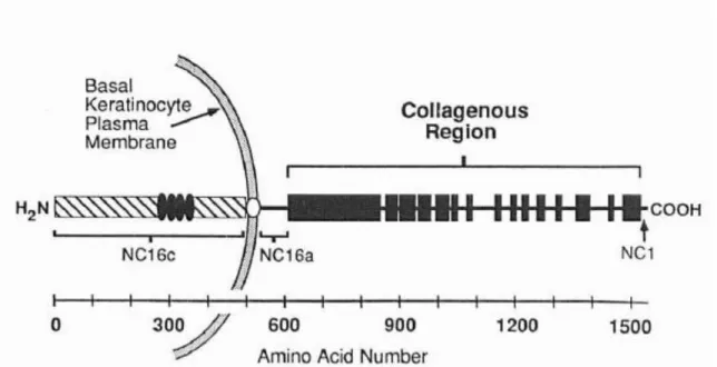
Bullous pemphigoid is an autoimmune skin blister disease characterized by the presence of the separation of epidermis and dermis, deposition of autoantibodies against BP180 and inflammatory infiltration in the dermis(1). The gene of BP180 was first isolated from human keratinocytes in 1990 and identified as a hemidesmosomal protein by the group of Dr. Luis A. Diaz, using sera from patients with bullous pemphigoid(2). The cDNA clones encompass 4,699 bases of the BP180 transcript. Structural analysis reveals that BP180 is a type II transmembrane glycoprotein: the carboxyl-terminal half of BP180 consists of 15 collagen domains and 16 non-collagen domains in the extracellular part. The main function of BP180 is that it serves as a cell-matrix adhesion molecule through its collagenous region, which interacts with basement membrane components(1)(3)(4).
BP180 is a highly conserved collagenous protein of the stratified squamous epithelium evidenced by the similarity of human, mouse and chicken BP180 sequences(5). However, when compared, mouse and human sequences have 86% homology(6). Nonetheless, There is poor cross reactivity between human and mouse autoantigens, especially in the NC16A domain(7).
1.1.2 Gene and cDNA of BP180
2
keratinocytes, while mouse BP180 cDNA was cloned in the same year(5). Sequential nucleotide screening of two independently derived human cDNA libraries leads to the isolation of a set of overlapping cDNA clones spanning 4,669 bases of the BP180 transcript and one long open reading frame (ORF), while mouse has a single ~6kb mRNA transcript(5). The C-terminal half of human BP180 contains 916 amino acids. Primary structure demonstrated tandem repeats of the Glycine-X-Y triple peptides, indicating a collagen triple helix component. On the other hand, one clone of mouse BP180 was 598 amino acids, including ten Glycine-X-Y triplets. In human, within the long collagenous region are 15 stretches of non-collagen domains, ranging from 15 to 242 amino acids. Seventy-six amino acids upstream of the collagenous region is a putative membrane-spanning domain (position 502-524), which is flanked by an upstream lysine and two downstream glutamate residues. In mouse, another feature of BP180 is the collagenous domains separated by non-collagenous segments. However, a computer software prediction of BP180 showed it contained one and possibly two transmembrane segments. Also, Dr. Li and colleagues identified 7 amino acid segments (amino acids 178-184) as potential antigenic sites for development of autoantibodies(8). By prokaryotic expression constructs, Dr. Giudice et al. found an autoantibody-reactive site within the protein sequence, RSILPYGDSMDRIE (BP180AA#542-555), corresponding to a portion of the NC16A noncollagenous domain(9).
1.1.3 Biochemical graph of BP180
3
adhesion. The extracellular domain consists of a C-terminal 1007 amino acid stretch that is predicted to extend into the basal lamina region adjacent to the basal keratinocytes, which has a series of 15 collagen domains (COL1 through COL15; solid box) and stretches of non-collagen sequence (NC1 through NC16a; thin lines). Numbering of the collagen and non-collagen domain starts at the C-terminus. The non-collagen domain, NC16, encompasses the membrane spanning domain, NC16b, as well as flanking regions to the C-terminal and N-terminal sides (NC16a and NC16c, respectively). Domain NC16c, the N-terminal segment predicted to be an intracellular domain, includes the four tandem repeats(5).
Noncollagenous domain (NC16A) is reported to interact with integrin α6, and the N-terminal of BP180 interacts with BP230 N-terminal amino acid residues 1-171 and
4
700(10), while the C-terminal of BP180 is an extracellular domain that interacts with the lamina densa.
BP180, along with BP230, plectin, keratin 5, 14, integrin α6β4, Laminin and type VII collagen compose the hemidesmosome. Super resolution microscopy shows the hemidesmosome structure in cultured keratinocytes and human skin. Nascent hemidesmosomes are associated with individual keratin filaments and β4 integrin. BP180 surrounds a central core of BP230 molecules(11).
1.1.4 Other functions of BP180
After cloning BP180, the homology at both the nucleotide and amino acid levels was found with chick cornea collagen. Striking conservation in the sizes and positions of their collagen domains was observed. In addition to the skin, collagen XVII has been reported to be expressed in the buccal mucosa, upper esophagus, ocular cornea, conjunctiva, bladder, umbilical cord, placenta, retina and neurons of the central nervous system.
5
muscle and spleen. Therefore, BP180 expresses in a variety of normal tissues with an epithelial component.
1.1.5 Bullous pemphigoid
Bullous pemphigoid (BP) was first described by Lever in 1953, the hallmark of which includes tense, sub-epidermal bullae associated with inflammatory infiltration (15)(16).
6
7
are activating FcR, while FcγRII is inhibiting FcR. To elucidate the role of different FcγR on neutrophils in bullous pemphigoid, FcγRI-/-, FcγRII-/-, FcγRIII -/- genes deficient mice were used. FcγRIII-/- mice were resistant to BP, which indicates that FcRn is a potential therapeutic target for IgG mediated autoimmune diseases(27)(28).
Neutrophil recruitment is an important event in experimental bullous pemphigoid, since using different methods to deplete neutrophils or neutropenia mice are resistant to bullous pemphigoid(20). Neutrophils, along with other inflammatory cells infiltrate the skin blister, and release granules(23). Neutrophil granules contain a variety of proteolytic enzymes, including elastase(NE), cathepsin, collagenase, and gelatinase B(GB) (29)(30)(31). Taking advantage of gene deficient mice, Dr. Liu and colleagues have demonstrated that both NE-/- and GB-/- mice are resistant to disease by passive
transferred pathogenic BP IgG autoantibodies(23)(32). Both NE-/- and GB-/- mice did not
show affected of neutrophil migration to skin at 4 hour time course(32). Furthermore, the function of GB and NE in experimental BP has been elucidated. GB acts upstream to
regulate NE activity by inactivating of α1-proteinase inhibitor (α1-PI)(33). Two types of bullous pemphigoid mouse model have been established: one is passive
8
1.1.6 Junctional epidermolysis bullosa (EB)
Another BP180 related disease is called epidermolysis bullosa (EB). EB refers to a group of heterogenous heritable disorders characterized by formation of blisters at sites of minor friction or trauma(36). 14 gene mutations have been identified within the cutaneous basement membrane zone(37).
There are four types of epidermolysis bullosa based on the level of tissue separation within the cutaneous basement membrane zone: epidermolysis bullosa simplex, junctional epidermolysis bullosa, dystrophic epidermolysis bullosa and Kindler syndrome (38)(39).
Figure 1.2 The pathogenesis of experimental bullous pemphigoid. Pathogenic autoantibody binds to BP180 on basal keratinocytes and triggers complement
9 .
Figure 1.3 The categories of epidermolysis bullosa (EB). Based on the level of tissue separation, there are four types of EB: epidermolysis bullosa simplex; junctional epidermolysis bullosa; dystrophic epidermolysis bullosa and Kindler syndrome.
(Adapted from Daisuke Sawamura, J. Dermatol. 2010;37(3):214–219.)
10
staining for BP180, which might imply GABEB patient could have anti-BP180 autoantibodies. Furthermore, GABEB caused by deficiency of BP180 is characterized as the autosome recessive form(42).
The clinical features of GABEB are continuous blistering since birth, cigarette paper-like atrophic depigmented skin at sites of recurrent blistering (no scarring or milia), normal growth and lack of anemia, moderate improvement during aging, dystrophic nails and dentition, mild mucous membrane involvement, and a typical pattern baldness with significant scalp atrophy, partial absence of eyelashes and eyebrows, and absence of pubic and axillary hair. GABEB could also be caused by lamini-5 deficiency. Although there is no specific or effective treatment for epidermolysis bullosa yet, gene therapy, protein replacement therapy, and cell-based therapies have been developed(44)(45)(46)(47)(48).
Generalized JEB is distinct from Herlitz disease by its absence of anemia and growth retardation, however in the BP180 gene deficient mouse model, the phenotype of runt was observed.
11
1.2 Inflammasome
1.2.1 General background
12
molecular pattern (DAMP), rather that it senses cellular homeostasis changes(63). In this introduction, we focused on the NLRP3 inflammasome.
1.2.2 Mechanisms of NLRP3 Inflammasome activation
After the NLRP3 inflammasome senses the disturbances in cellular homeostasis, there are some common cellular events that serve as the activation signals for the NLRP3 inflammasome, including potassium efflux, generation of reactive oxygen species, and cathepsin B release(64). The NLRP3 inflammasome is not activated when K+ efflux is
prevented by a drug or a high concentration in the media(65). The mechanism is that extracellular ATP engages the ATP-gated cation channel P2X7R whereas bacterial toxins cause membrane pore formation to trigger K+ efflux(66). The generation of reactive
oxygen species (ROS) is considered a cellular event that causes NLRP3 inflammasome activation, evidenced by ROS blockade via chemical inhibitors or gene knockdown suppressed inflammasome activation(67)(68). The third molecular mechanism which activates the NLRP3 inflammasome is cathepsin B release. Uptake the crystalline and particulate activators causes destabilization of the acidic lysosomal compartment and releases cathepsin B trigger inflammasome activation(69).
13
some cues for NLRP3 activation, such as mitochondrial reactive oxygen species (mROS), mitochondrial DNA (mtDNA), and phospholipid cardiolipin(63). ASC relocalization from the nucleus to the cytosol, and forming a large perinuclear aggregate is also an indicator for inflammasome activation(72).
1.2.3 Inflammasome related diseases and therapeutics
14
channels(79). MCCP950 is a highly selective inhibitor of the NLRP3 inflammasome and a diarylsulfonylurea-containing compound, by molecular targeting of NLRP3 itself or is closely linked to the activation of NLRP3, instead of blocking K+ efflux or Ca2+ influx by
mechanisms of which structurally related drug Glyburide, so MCCP950 blocked both canonical and noncanonical NLRP3-dependent inflammasome activation(80)(81).
1.3 Granulopoiesis
1.3.1 Granulocyte development
Neutrophils are the most abundant leukocytes in blood, comprising 60-70% of all circulating white blood cells. Neutrophil differentiation from bone marrow hematopoietic stem cells is regulated by the coordinated expression of three myeloid transcription factors, GFI-1, PU.1 and members of the CCAAT enhancer binding protein family (C/EBPs). The balance between PU.1 and C/EBPα determines whether myeloblasts differentiate into granulocytes (high C/EBPα) or monocytes (high PU.1)(82).
Pluripotent hematopoietic stem cells (HSC) could give rise to a lin-IL7R-KIT+
Sca-1-CD34+FcrRlo common myeloid progenitor (CMP), CMPs can give rise to all classes of
15
1.3.2 Extrinsic factor for granulopoiesis
NF-kB, a transcription factor, is involved in cellular processes of proliferation and differentiation of hematopoietic lineages. IkBα is an inhibitor of NF-kB in its canon signaling pathway. IkBα was phosphorylation by kinase, followed by degradation of IkBα and subsequent translocation of NF-kB to the nucleus. Mice deficient in IkBα develop a hypergranulopoiesis. However, specific deletion of IkBα in neutrophils and macrophages or hematopoietic stem cells did not result in granulopoiesis, despite constitutive NF-kB activation in these cells. This establishes the relevance that NF-kB signaling pathway of nonhematopoietic cells control and regulate granulopoiesis. The signaling pathway in stromal cells could induce granulopoiesis. Extensive investigation reports ubiquitous deletion of IkBα in non-hematopoietic cells(86)(87).
The granulocytes of IkBα gene deficient mice express high levels of the transmembrane receptor Notch1, a key regulator of cell-fate decisions during differentiation.
1.3.3 Granulocyte circulation
16
infection or the stimulation of inflammation, granulocytes would emigrate from bone marrow by gradient change of cytokines and chemokines(88).
Mature granulocyte egress from bone marrow to the circulation is the process for the body to respond to infection or inflammation. Mobilization of granulocytes from the bone marrow to sites of inflammation is regulated by cytokines or chemokines, such as chemokine macrophage inflammatory protein-2(cxcl2)(89); KC(cxcl1); leukotriene B4, C5a and IL-8 et al(90)(91)(92). CXCR4 is important for granulocyte retention in bone marrow by interacting with bone marrow stromal cell-derived factor (SDF-1a)(93). Using CXCR4 antagonist demonstrated that rapid release of granulocytes indicates that SDF-1a is a critical factor for detaining granulocytes in bone marrow, whereas mutation of CXCR4 in patients with hypogammaglobulinaemia, infection and myelokathexis (WHIM) syndrome showed normal bone marrow granulocytes but neutropenia in the circulation(94).
In gene deficient G-CSF or G-CSFR mice, there are very few neutrophils, both in the bone marrow and the blood, which demonstrates that G-CSF regulates both granulopoiesis and mobilization of granulocytes from bone marrow. G-CSF’s control of granulocyte mobilization may result from reduced production of SDF-1a or down-regulated CXCR4 expression on granulocytes(95).
Since the half- life of neutrophils in circulation is about 6.5hr, and 1011 cells are
17
home back to the bone marrow under homeostatic conditions, which is cleaned by resident macrophages(97).
1.3.4 Granulocytes’ function and disease
18
which neutrophils clear high local concentration of microbes are neutrophil extracellular traps. Upon cues from outside, neutrophils undergo NETosis, an active form of cell death that leads to release of decondensed chromatin into the extracellular space(110). The failure to remove apoptotic neutrophils leads to the accumulation of cytotoxic substances and is associated with cystic fibrosis, chronic obstructive pulmonary disease (COPD) and rheumatoid arthritis (RA)(111)(112)(113). Inflammation is also involved in cancer. Neutrophil derived ROS can initiate tumor formation by genotoxic stress and induction of genomic instability(114)(115). Neutrophils produce angiogenic factors, enhance metastasis, and suppress the antitumor immune response functions to promote tumor growth(116)(117). Neutrophils also are associated with autoimmune diseases due to deregulated cell death and clearance. NETosis may related to autoimmune disease systemic lupus erythematosus (SLE)(118). Recruitment of inflammatory cells to joints is the hallmark of rheumatoid arthritis (RA). Neutrophils are the critical immune cells necessary for the development of joint inflammation and damage. NETs are observed in the synovial tissue of RA and serum levels of MPO increase in patients with RA(119).
1.3.5. Granulocytes and B cell development
19
20
REFERENCES
1. Giudice GJ et al. Identification of two collagen domains within the bullous pemphigoid autoantigen, BP180. [Internet]. J. Clin. Invest. 1991;87(2):734–8.
2. Diaz LA et al. Isolation of a human epidermal cDNA corresponding to the 180-kD autoantigen recognized by bullous pemphigoid and herpes gestationis sera.
Immunolocalization of this protein to the hemidesmosome [Internet]. J Clin Invest
1990;86(4):1088–1094.
3. Borradori L et al. Role of the bullous pemphigoid antigen 180 (BP180) in the assembly of hemidesmosomes and cell adhesion--reexpression of BP180 in
generalized atrophic benign epidermolysis bullosa keratinocytes [Internet]. Exp Cell Res
1998;239(2):463–476.
4. Van den Bergh F et al. Type XVII collagen (BP180) can function as a cell-matrix adhesion molecule via binding to laminin 332 [Internet]. Matrix Biol 2011;30(2):100–108.
5. Giudice GJ et al. Cloning and primary structural analysis of the bullous pemphigoid autoantigen BP180 [Internet]. J Invest Dermatol 1992;99(3):243–250.
6. Li K et al. Cloning of Partial cDNA for Mouse 180-kDa Bullous Pemphigoid Antigen (BPAG2), a Highly Conserved Collagenous Protein of the Cutaneous Basement Membrane Zone [Internet]. J. Invest. Dermatol. 1992;99(3):258–263.
7. Giudice GJ et al. Bullous pemphigoid and herpes gestationis autoantibodies recognize a common non-collagenous site on the BP180 ectodomain.. J. Immunol.
1993;151(10):5742–5750.
8. Li K et al. Cloning of type XVII collagen. Complementary and genomic DNA sequences of mouse 180-kilodalton bullous pemphigoid antigen (BPAG2) predict an interrupted collagenous domain, a transmembrane segment, and unusual features in the 5’-end of the gene and the 3' [Internet]. J Biol Chem 1993;268(12):8825–8834.
9. Giudice GJ et al. Identification of two collagen domains within the bullous pemphigoid autoantigen, BP180 [Internet]. J Clin Invest 1991;87(2):734–738.
10. Hopkinson SB et al. The N terminus of the transmembrane protein BP180 interacts with the N-terminal domain of BP230, thereby mediating keratin cytoskeleton anchorage to the cell surface at the site of the hemidesmosome [Internet]. Mol Biol Cell
2000;11(1):277–286.
11. Nahidiazar L et al. The molecular architecture of hemidesmosomes as revealed by super-resolution microscopy [Internet]. J. Cell Sci. 2015;128(20):3714–9.
12. Hurskainen T et al. Transmembrane collagen XVII is a novel component of the glomerular filtration barrier. Cell Tissue Res. 2012;348(3):579–588.
21
for an anchoring complex in the central nervous system. J. Comp. Neurol.
2005;487(2):190–203.
14. Aho S et al. 180-kD bullous pemphigoid antigen/type XVII collagen: Tissue-specific expression and molecular interactions with keratin 18. J. Cell. Biochem.
1999;72(3):356–367.
15. Vieira JP et al. Some Recent Studies in Brazilian Pemphigus. Am J Trop Med Hyg
1954;3(5):868–877.
16. Nousari HC et al. Pemphigus and bullous pemphigoid. Lancet 1999;354(9179):667–
672.
17. Liu Z, et al. Bullous pemphigoid: end of the century overview. [Internet]. J. Dermatol.
2001;28(11):647–650.
18. Liu Z et al. A passive transfer model of the organ-specific autoimmune disease, bullous pemphigoid, using antibodies generated against the hemidesmosomal antigen, BP180. J. Clin. Invest. 1993;92(5):2480–2488.
19. Liu Z et al. The role of complement in experimental bullous pemphigoid. J. Clin. Invest. 1995;95(4):1539–1544.
20. Liu Z et al. A major role for neutrophils in experimental bullous pemphigoid. [Internet]. J. Clin. Invest. 1997;100(5):1256–63.
21. Liu Z et al. Synergy between a plasminogen cascade and MMP-9 in autoimmune disease [Internet]. J Clin Invest 2005;115(4):879–887.
22. Liu Z et al. A critical role for neutrophil elastase in experimental bullous pemphigoid. [Internet]. J. Clin. Invest. 2000;105(1):113–23.
23. Liu Z et al. Gelatinase B-deficient mice are resistant to experimental bullous pemphigoid. [Internet]. J. Exp. Med. 1998;188(3):475–82.
24. Nelson KC et al. Role of different pathways of the complement cascade in experimental bullous pemphigoid. J. Clin. Invest. 2006;116(11):2892–2900.
25. Chen R et al. Mast cells play a key role in neutrophil recruitment in experimental bullous pemphigoid2001;108(8):1151–1158.
26. Heimbach L et al. The C5a receptor on mast cells is critical for the autoimmune skin-blistering disease bullous pemphigoid. J. Biol. Chem. 2011;286(17):15003–15009.
27. Zhao M et al. Role of FcRs in animal model of autoimmune bullous pemphigoid.. J. Immunol. 2006;177(5):3398–405.
28. Li N et al. Complete FcRn dependence for intravenous Ig therapy in autoimmune skin blistering diseases. J. Clin. Invest. 2005;115(12):3440–3450.
22
30. Buck MR et al. Degradation of extracellular-matrix proteins by human cathepsin B from normal and tumour tissues. Biochem. J. 1992;282 ( Pt 1:273–8.
31. Janoff A. Elastase in tissue injury. Annu. Rev. Med. 1985;36:207–216.
32. Liu Z et al. A critical role for neutrophil elastase in experimental bullous pemphigoid.
J. Clin. Invest. 2000;105(1):113–123.
33. Liu Z et al. The Serpin α 1-Proteinase Inhibitor Is a Critical Substrate for Gelatinase B / MMP-9 In Vivo2000;102:647–655.
34. Hirose M et al. Repetitive immunization breaks tolerance to type XVII collagen and leads to bullous pemphigoid in mice. J. Immunol. 2011;187(3):1176–83.
35. Hurskainen T et al. Deletion of the Major Bullous Pemphigoid Epitope Region of Collagen XVII Induces Blistering, Autoimmunization, and Itching in Mice [Internet]. J Invest Dermatol [published online ahead of print: 2014]; doi:10.1038/jid.2014.443 36. Lin AN et al. Epidermolysis bullosa. Annu. Rev. Med. 1993;44:189–99.
37. Uitto J et al. Progress in epidermolysis bullosa research: toward treatment and cure. [Internet]. J. Invest. Dermatol. 2010;130(7):1778–84.
39. Sawamura D et al. Overview of epidermolysis bullosa. J. Dermatol. 2010;37(3):214–
219.
40. Pasmooij AMG et al. Features of epidermolysis bullosa simplex due to mutations in the ectodomain of type XVII collagen. Br. J. Dermatol. 2004;151(3):669–674.
41. Ruzzi L et al. A homozygous nonsense mutation in type XVII collagen gene (COL17A1) uncovers an alternatively spliced mRNA accounting for an unusually mild form of non-Herlitz junctional epidermolysis bullosa. J. Invest. Dermatol.
2001;116(1):182–187.
42. La Placa M et al. Generalized atrophic benign epidermolysis bullosa. J. Eur. Acad. Dermatology Venereol. 1996;7(2):167–170.
43. Jonkman MF et al. 180-kD bullous pemphigoid antigen (BP180) is deficient in generalized atrophic benign epidermolysis bullosa [Internet]. J. Clin. Invest.
1995;95(3):1345–1352.
44. Pasmooij AM et al. Natural gene therapy may occur in all patients with generalized non-Herlitz junctional epidermolysis bullosa with COL17A1 mutations [Internet]. J Invest Dermatol 2012;132(5):1374–1383.
45. Osborn MJ et al. TALEN-based gene correction for epidermolysis bullosa. [Internet].
Mol. Ther. 2013;21(6):1151–9.
46. Hovnanian A. Systemic protein therapy for recessive dystrophic epidermolysis bullosa: how far are we from clinical translation?. J. Invest. Dermatol.
23
47. Kiuru M et al. Bone marrow stem cell therapy for recessive dystrophic epidermolysis bullosa. Dermatol. Clin. 2010;28(2):371–382.
48. Fine J-D. Inherited epidermolysis bullosa: recent basic and clinical advances.. Curr. Opin. Pediatr. 2010;22(4):453–8.
49. Mazzanti C et al. 180-kDa bullous pemphigoid antigen defective generalized atrophic benign epidermolysis bullosa: Report of four cases with an unusually mild phenotype. Br. J. Dermatol. 1998;138:859–866.
50. Roth RR et al. Eosinophilic infiltrates in epidermolysis bullosa [Internet]. Arch Dermatol 1990;126(9):1191–1194.
51. Fu CL et al. Protein structural analysis of BP180 mutant isoforms linked to non-Herlitz junctional epidermolysis bullosa [Internet]. J Invest Dermatol 2006;126(1):232–
234.
52. Esposito S et al. Epidermolysis bullosa and the partnership with autoimmunity: what should we assimilate? [Internet]. Immunol Res [published online ahead of print: 2014]; doi:10.1007/s12026-014-8583-3
53. Martinon F et al. The Inflammasome: A molecular platform triggering activation of inflammatory caspases and processing of proIL-β. Mol. Cell 2002;10(2):417–426.
54. Li P et al. Mice deficient in IL-1β-converting enzyme are defective in production of mature IL-1β and resistant to endotoxic shock. Cell 1995;80(3):401–411.
55. Kuida K et al. Altered cytokine export and apoptosis in mice deficient in interleukin-1 beta converting enzyme. Science 1995;267(5206):2000–2003.
56. Miao EA et al. Caspase-1-induced pyroptotic cell death. Immunol. Rev.
2011;243(1):206–214.
57. Yamamoto M et al. ASC is essential for LPS-induced activation of procaspase-1 independently of TLR-associated signal adaptor molecules. Genes to Cells
2004;9(11):1055–1067.
58. Poyet JL et al. Identification of Ipaf, a Human Caspase-1-activating Protein Related to Apaf-1. J. Biol. Chem. 2001;276(30):28309–28313.
59. Levinsohn JL et al. Anthrax lethal factor cleavage of Nlrp1 is required for activation of the inflammasome. PLoS Pathog. 2012;8(3). doi:10.1371/journal.ppat.1002638 60. Zhao Y et al. The NLRC4 inflammasome receptors for bacterial flagellin and type III secretion apparatus. Nature 2011;477(7366):596–600.
61. Miao EA et al. Cytoplasmic flagellin activates caspase-1 and secretion of interleukin 1beta via Ipaf. Nat Immunol 2006;7(6):569–575.
24
63. Wen H et al. Mechanisms of NOD-like receptor-associated inflammasome activation. Immunity 2013;39(3):432–441.
64. Jin C et al. Molecular mechanism of NLRP3 inflammasome activation. J. Clin. Immunol. 2010;30(5):628–631.
65. Dostert C et al. Innate immune activation through Nalp3 inflammasome sensing of asbestos and silica. Science 2008;320(5876):674–7.
66. Mariathasan S et al. Cryopyrin activates the inflammasome in response to toxins and ATP. Nature 2006;440(7081):228–232.
67. Cruz CM et al. ATP activates an ros-dependent oxidative stress response and secretion of pro-inflammatory cytokines in macrophages. J Biol Chem 2006;
68. Naik E et al. Mitochondrial reactive oxygen species drive proinflammatory cytokine production. J. Exp. Med. 2011;208(3):417–20.
69. Hornung V et al. Silica crystals and aluminum salts activate the NALP3
inflammasome through phagosomal destabilization. Nat. Immunol. 2008;9(8):847–856.
70. Subramanian N et al. The adaptor MAVS promotes NLRP3 mitochondrial localization and inflammasome activation. Cell 2013;153(2):348–361.
71. Zhou R et al. A role for mitochondria in NLRP3 inflammasome activation. Nature
2011;469(7329):221–225.
72. Bryan NB et al. Activation of inflammasomes requires intracellular redistribution of the apoptotic speck-like protein containing a caspase recruitment domain. J. Immunol.
2009;182(5):3173–3182.
73. Jin Y et al. NALP1 in vitiligo-associated multiple autoimmune disease. N Engl J Med
2007;356(12):1216–1225.
74. Magitta NF et al. A coding polymorphism in NALP1 confers risk for autoimmune Addison’s disease and type 1 diabetes. Genes Immun. 2009;10(2):120–124.
75. Agostini L et al. NALP3 forms an IL-1β-processing inflammasome with increased activity in Muckle-Wells autoinflammatory disorder. Immunity 2004;20(3):319–325.
76. Hoffman HM et al. Mutation of a new gene encoding a putative pyrin-like protein causes familial cold autoinflammatory syndrome and Muckle-Wells syndrome. Nat. Genet. 2001;29(3):301–5.
77. Goldbach-mansky R. Current Status of Understanding the Pathogenesis and Management of Patients With NOMID/CINCA. Curr. Opin. Microbiol. 2011;13(2):123–
131.
78. Zhang H et al. Anti-IL-1β therapies. Recent Pat. DNA Gene Seq. 2011;5(2):126–
25
79. Lamkanfi M et al. Glyburide inhibits the Cryopyrin/Nalp3 inflammasome. J. Cell Biol.
2009;187(1):61–70.
80. Coll RC et al. A small-molecule inhibitor of the NLRP3 inflammasome for the treatment of inflammatory diseases. Nat. Med. 2015;21(3):248–255.
81. Guo H et al. Inflammasomes: mechanism of action, role in disease, and therapeutics. Nat. Med. 2015;21(7):677–687.
82. Friedman a D. Transcriptional control of granulocyte and monocyte development.
Oncogene 2007;26(47):6816–6828.
83. Akashi K et al. A clonogenic common myeloid progenitor that gives rise to all myeloid lineages [Internet]. Nature 2000;404(6774):193–197.
84. Manz MG et al. Prospective isolation of human clonogenic common myeloid progenitors. Proc. Natl. Acad. Sci. U. S. A. 2002;99(18):11872–11877.
85. Amulic B et al. Neutrophil Function: From Mechanisms to Disease. Annu. Rev. Immunol. 2012;30(1):459–489.
86. Beg AA et al. Constitutive NF-kappa B activation, enhanced granulopoiesis, and neonatal lethality in I kappa B alpha-deficient mice. [Internet]. Genes Dev.
1995;9(22):2736–46.
87. Rupec RA et al. Stroma-mediated dysregulation of myelopoiesis in mice lacking IκBα. Immunity 2005;22(4):479–491.
88. Furze RC et al.Neutrophil mobilization and clearance in the bone marrow.
Immunology 2008;125(3):281–288.
89. Martin C et al. Chemokines acting via CXCR2 and CXCR4 control the release of neutrophils from the bone marrow and their return following senescence. Immunity
2003;19(4):583–593.
90. Jagels MA et al. Neutrophil chemotactic factors promote leukocytosis. A common mechanism for cellular recruitment from bone marrow. J. Immunol. 1992;148(4):1119–
1128.
91. van Eeden SF et al. Interleukin 8 (IL-8) and the release of leukocytes from the bone marrow. Leuk. Lymphoma 2000;37(3-4):259–71.
92. Jagels MA et al. C5a- and tumor necrosis factor-alpha-induced leukocytosis occurs independently of beta 2 integrins and L-selectin: differential effects on neutrophil
adhesion molecule expression in vivo. Blood 1995;85(10):2900–2909.
93. Bleul CC et al. A highly efficacious lymphocyte chemoattractant, stromal cell-derived factor 1 (SDF-1). J. Exp. Med. 1996;184(3):1101–9.
26 2003;34(1):70–74.
95. Petit I et al. G-CSF induces stem cell mobilization by decreasing bone marrow SDF-1 and up-regulating CXCR4. Nat. Immunol. 2002;3(7):687–694.
96. Rankin SM. The bone marrow: a site of neutrophil clearance. J. Leukoc. Biol.
2010;88(2):241–251.
97. Furze RC et al. The role of the bone marrow in neutrophil clearance under homeostatic conditions in the mouse. FASEB J. 2008;22(9):3111–9.
98. Nusse O et al. The dynamics of exocytosis in human neutrophils. J. Cell Biol.
1988;107(6 I):2117–2123.
99. Faurschou M et al. Neutrophil granules and secretory vesicles in inflammation.
Microbes Infect. 2003;5(14):1317–1327.
100. Borregaard N et al. Neutrophil granules: a library of innate immunity proteins.
Trends Immunol. 2007;28(8):340–345.
101. Soehnlein O et al.Neutrophils launch monocyte extravasation by release of granule proteins. In: Thrombosis and Haemostasis. 2009:198–205
102. Schroeder BO et al. Reduction of disulphide bonds unmasks potent antimicrobial activity of human β-defensin 1.. Nature 2011;469(7330):419–423.
103. Canny G et al. Bactericidal/permeability-increasing protein (BPI) and BPI homologs at mucosal sites. Trends Immunol. 2008;29(11):541–547.
104. Markart P et al. Mouse lysozyme M is important in pulmonary host defense against Klebsiella pneumoniae infection. Am. J. Respir. Crit. Care Med. 2004;169(4):454–458.
105. Shimada J et al. Lysozyme M deficiency leads to an increased susceptibility to Streptococcus pneumoniae-induced otitis media. BMC Infect. Dis. 2008;8:134. 106. Corbin BD et al. Metal chelation and inhibition of bacterial growth in tissue abscesses. Science 2008;319(5865):962–5.
107. Bullen JJ. Iron and Infection. Eur. J. Clin. Microbiol. 1986;5(1):132.
108. Nauseef WM. How human neutrophils kill and degrade microbes: An integrated view. Immunol. Rev. 2007;219(1):88–102.
109. Shao B et al. Methionine sulfoxide and proteolytic cleavage contribute to the
inactivation of cathepsin G by hypochlorous acid: An oxidative mechanism for regulation of serine proteinases by myeloperoxidase. J. Biol. Chem. 2005;280(32):29311–29321.
110. Brinkmann V et al. Neutrophil extracellular traps kill bacteria. Science .
2004;303(5663):1532–1535.
27
neutrophilic inflammation. Science 2010;330(6000):90–4.
112. Tanaka D et al. Essential role of neutrophils in anti-type II collagen antibody and lipopolysaccharide-induced arthritis. Immunology 2006;119(2):195–202.
113. Chou RC et al. Lipid-Cytokine-Chemokine Cascade Drives Neutrophil Recruitment in a Murine Model of Inflammatory Arthritis. Immunity 2010;33(2):266–278.
114. Shacter E et al. Corrigendum: Activated neutrophils induce prolonged DNA damage in neighboring cells. Carcinogenesis 1989;10(3):627.
115. Dizdaroglu M et al. Modification of DNA bases in chromatin of intact target human cells by activated human polymorphonuclear leukocytes. Cancer Res 1993;53(6):1269–
1272.
116. Shojaei F et al. Role of Bv8 in neutrophil-dependent angiogenesis in a transgenic model of cancer progression. Proc. Natl. Acad. Sci. U. S. A. 2008;105(7):2640–5.
117. Pekarek LA et al. Inhibition of tumor growth by elimination of granulocytes. J. Exp. Med. 1995;181(1):435–440.
118. Garcia-Romo GS et al. Netting neutrophils are major inducers of type I IFN production in pediatric systemic lupus erythematosus. Sci. Transl. Med.
2011;3(73):73ra20.
119. Wright HL et al. The multifactorial role of neutrophils in rheumatoid arthritis. Nat. Publ. Gr. 2014;10(10):593–601.
120. Defrance T et al. T cell-independent B cell memory. Curr. Opin. Immunol.
2011;23(3):330–336.
121. Parker DC. T cell-dependent B cell activation. Annu. Rev. Immunol. 1993;11:331–
28
CHAPTER 2: DEFICIENCY OF THE HEMIDESMOSOMAL PROTEIN BP180 FUNCTION LEADS TO ALTERED GRANULOPOIESIS IN MICE
2.1 Introduction
BP180 (also termed type XVII collagen) is a transmembrane hemidesmosomal protein and plays a critical role in cell-cell matrix adhesion in the skin. This structural property of BP180 was identified based on two human subepidermal blistering diseases: the anti-BP180 autoantibody-mediated autoimmune blistering disease bullous pemphigoid (BP)(1)(2)(3), and BP180 gene mutation-caused generalized atrophic benign epidermolysis bullosa (GABEB)(4)(5)(6). However, other functions of BP180 are largely unknown.
29
between these two species (3). Animal models of BP demonstrated that anti-NC14A and anti-NC16A antibodies induce subepidermal blistering in mice associated with neutrophil infiltration (2) (3).
Junctional epidermolysis bullosa (JEB) is a group of rare genetic diseases characterized by marked skin fragility and blister formation following minor trauma, sometimes along with eosinophilia, eosinophils and/or neutrophils infiltrated in the dermis and autoantibodies deposited at the basement membrane zone(14). GABEB is a subset of EB and caused by BP180 gene mutations(15). Patients with GABEB develop subepidermal blistering(15)(16). Previous reports demonstrated that deletion of NC14A domain of mouse BP180 led to loss of cell-cell matrix adhesion and granulocyte infiltration in the skin(17). However, the mechanisms underlying the increased granulocytes in JEB patients and mice lacking BP180 function are unknown.
Pluripotent hematopoietic stem cells (HSC) could give rise to a lin-IL7R-KIT+
Sca-1-CD34+FcrRlo common myeloid progenitor (CMP), and CMPs could differentiate into
30
Deficiency of G-CSF or G-CSF receptor exhibits neutropenia in mice, demonstrating that G-CSF is an important extrinsic regulator for neutrophils(22).
Homeostasis of hematopoietic stem cells (HSCs) in bone marrow and peripheral are dependent on multiple signaling pathways. Notch and NF-kB are two of the most investigated signaling pathways(23)(24). Previous study showed that bone marrow niche regulates hematopoiesis in a NF-kB dependent manner(25)(26). NF-kB transcription factor activation leads to proinflammatory cytokines and G-CSF production(27)(28). Loss of transcriptional repression of NF-kB activation in IκBα-deficient mice leads to an uncontrolled expansion of myeloid cells via G-CSF(29)(30). BP180 was reported to be involved in NF-kB signaling pathway in cultured keratinocyte (31).
31
2.2Materials and methods
Generation of ΔNC16A mice
Humanized NC16A mice were described previously (2) Briefly, mouse BP180 Exons 17 and 18 encoding NC14A domain were replaced by human BP180 exons 18 and 19 encoding NC16A domain flanked by loxp sites (also see Figure 1A). NC16A mice (on C57BL/6J background) were then bred with germline Cre mice (on C57BL/6J background, The Jackson Laboratory, Maine, USA); leading to removal of human exons 17 and 18 cassettes and expression of NC16A truncated BP180 in ΔNC16A mice. Mice with skin-specific deletion of NC16A were generated by crossing NC16A mice with K14Cre mice (on C57BL/6J background)(32)(33). All the mice were bred and hosted under specific pathogen-free conditions in the animal facilities of the University of North Carolina, Chapel Hill. Animal care and animal experiments were conducted in accordance with the Animal Care Committee at the University of North Carolina, Chapel Hill.
Transplantation
C57BL/6 (CD45.1and CD45.2) mice were purchased from National Cancer Institute and housed under specific pathogen-free conditions in the animal facilities of the University of North Carolina, Chapel Hill. To make mixed bone marrow chimera, bone marrow from ΔNC16A CD45.2 and wild type CD45.1 mice were mixed at 1:1 ratio, and then 5X10⁶ cells were transplanted to sub-lethal irradiated (700 cGy) WT CD45.1.2
recipient mice by I.V. administration(34).
32
Bone marrow cell isolation and culture were as described(35). Briefly, bone marrow cell suspensions were isolated from 8-12 weeks ΔNC16A and WT control mice by flushing the marrow from femurs a few times with 10 ml PBS/2%FBS in syringe with a 22-G needle. The cells were centrifuged for 5 min at 1000 rpm, 4°c after red blood cells were lysed and removed. Cells were counted and seeded with MSC culture medium (MesenCultTM MSC culture medium, stem cell technologies, Vancouver, Canada)
according to the protocol provided by the manufacture.
CFU assay
The colony forming capacity of bone marrow cells was estimated in mouse methylcelluose complete media, according to the manufacturer’s instructions (R&D System Inc. MN). Briefly, 3×10⁴ bone marrow mononuclear cells were mixed well in 1 ml
media, plated in 3 cm diameter culture dishes, and then incubated at 37°C in a 5% CO2
incubator. After 9 day culture, the total number of colonies in each dish was counted under a microscopy(36) .
Flow cytometry
Apc-33
cy7, anti-CD11b, anti-CD3, anti-CD19, anti-Gr.1 PerCp, anti-CD11b PerCp, Anti-CD3 PerCp, anti-CD19PerCp, anti-CD127 PE, anti-CD34 Pacific Blue, anti-CD16, anti-CD19 PO, anti-Sca-1 FITC, anti-c-kit Pe-cy7. Anti-Gr.1, anti-CD11b antibodies were used to detect granulocytes. Anti- CD3 antibody was used to detect T cells. Anti-CD19 antibody was used to detect B cells. Anti-CD127, CD34, CD16, Sca-1 and c-Kit antibodies were used to identify different population of granulocyte progenitors (Figure 3A). These antibodies were purchased from Biolegend (San Diego, CA).
Immunoblotting and immunofluorescence
Immunoblotting and immunofluorescence for BP180 were performed by using standard techniques as previously described (2). The anti-NC16A antibody was from BP patient serum, while rabbit anti-NC1-3 polyclonal antibody was made in house. Femurs were fixed in 4% paraformaldehyde overnight and then decalcification by 14% EDTA overnight and then tissue was rinsed by PBS 3 times. The femurs were fixed in OCT (Tissue-TEK®, CA, USA). Cryosections of the OCT-embedded femurs were stained and were imaged by a Zeiss LSM 700 Confocal Laser Scanning Microscope and ZEN imaging software. The pictures were analyzed by Image-J software. Bone marrow cells and bone marrow derived mesenchymal stem cell (1x10⁶) were used for NF-kB signaling pathway
34
Quantification of G-CSF, GM-CSF by ELISA
Levels of G-CSF and GM-CSF in bone marrow supernatant and serum samples from ΔNC16A, K14Cre ΔNC16A and WT control mice were quantified by ELISA kits according to the procedures provided by the manufacture (Abcam, MA, USA). ELISA Max™ Mouse GM-CSF ELISA kits were purchased from Biolegend. To collect bone marrow supernatants, marrows from femurs were flushed out with PBS and centrifuged. Collected supernatants were stored in -80°C before use.
Statistical analysis
35
2.3 Results
Generation and characterization of conditional ΔNC16A mice
36
ΔNC16A mice develop myeloid hyperplasia
To explain these gross alterations in spleen and bone marrow, we analyzed the hematopoiesis system of ΔNC16A mice by hematological examination and flow cytometry. Histological examination showed increased granulocytes in blood and dermis, increased cellularity in bone marrow, and increased red pulp in spleen of ΔNC16A mice as compared to WT control (Figure 2.1D). Hematological test showed significantly increased total cell number (Figure 2.1E) and percentage (Figure 2.1F) of granulocytes in circulation in ΔNC16A compared to WT mice. Normal granulocytes number range of WT mice is less than 20 percentage in white blood cells(37), while ΔNC16A mice manifested an abnormally high percentage of granulocytes in white blood cells and granulocytes number in the peripheral blood. Flow cytometry analysis also showed significantly increased granulocytes in bone marrow, spleen and peripheral blood in ΔNC16A mice as compared to WT controls using forward scatter (cell size) and side scatter (granularity) (Figure 2.1G) and granulocyte number was calculated based on surface markers CD11b and Gr.1 (Figure 2.1H). These results demonstrated that lacking of BP180 function caused myeloid hyperplasia.
ΔNC16A mice have more granulocyte-macrophage progenitors
37
ΔNC16A mice were changed by flow cytometry using Lin- IL-7R-SCA-1-
C-kit+CD34+CD16+ surface markers combination panel (Figure 2.2A)(39)(40). We found that
there was more than two folds increase in GMP population in ΔNC16A mice compared to WT control (Figure 2.2B), which was statistically significant (Figure 2.2C). Interestingly, megakaryocyte-erythrocyte progenitor (MEP) populations in ΔNC16A mice were significantly decreased compared to WT control (Figure 2.2B, 2.2C), which could cause abnormal bone marrow color change in ΔNC16A mice. By cell culture, significantly higher colony-forming units of granulocytes/monocytes from ΔNC16A bone marrow than WT control mice (Figure 2.2D). These results demonstrate that lack of BP180 leads to myeloid progenitor lineage skewing toward GMPs, resulting in increased granulocytes production.
Granulopoiesis defects in ΔNC16A mice arise from non-cell-autonomous
mechanism
Knowing that ΔNC16A mice develop granulocyte hyperplasia, we then determined if the significantly elevated granulocytes came from cell-autonomous or non-cell-autonomous mechanisms. We performed bone marrow chimera and bone marrow transplantation experiments. Taking advantage of congenic marker, bone marrows of CD45.1 WT mice and CD45.2 ΔNC16A mice were mixed at 1:1 ratio and transferred to sub-lethally irradiated WT CD45.1.2 recipients (Figure 2.3A). After 8 weeks reconstitution, there was no difference between granulocyte populations derived from WT and ΔNC16A mice in bone marrow and spleen of host animals (Figure 2.3B). Gr.1+ cell populations in
38
populations in ΔNC16A mice transplanted with WT bone marrow were similar to ΔNC16A mice, while WT mice transplanted with ΔNC16A bone marrow showed normal granulopoiesis (Figure 2.3D). These data demonstrated that altered granulocyte development in ΔNC16A mice was from a non-cell autonomous source.
ΔNC16A mice exhibit elevated G-CSF levels and increased activation of NF-kB signaling pathway in bone marrow mesenchymal stem cells
Since altered granulopoiesis occurs in the bone marrow stromal cell compartment, we determined whether BP180 is expressed in bone marrow and/or mesenchymal stem cells (MSC). By immunoblotting, anti-NC16A antibody detected the full-length BP180 in bone marrow protein extract of WT and not ΔNC16A mice, whereas anti-NC1-3 antibody recognized both full-length and NC16A truncated BP180 in bone marrow of both WT and
ΔNC16A mice (Figure 2.4A). Indirect immunofluorescence using anti-NC16A antibody stained bone marrow cryosection from WT and not ΔNC16A mice (Figure 2.4B). Since G-CSF is a key regulator for granulopoiesis of non-cell autonomous mechanism and myeloid cell proliferation (41)(42), we used ELISA and quantified G-CSF expression levels both in bone marrow supernatant and serum from ΔNC16A and WT mice. ΔNC16A mice had significantly higher G-CSF levels in bone marrow and serum compared to WT mice (Figure 2.4C, 2.4E). As a control, levels of GM-CSF in bone marrow and serum in
39
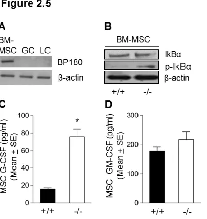
signaling pathway activation in bone marrow of ΔNC16A mice by Western blot. ΔNC16A mice showed increased activation of JNK and IκBα since levels of phosphorylated JNK and IκBα in total bone marrow cell protein extracts of ΔNC16A mice were increased as compared to WT control (Figure 2.4G). Densitometry analysis also demonstrated significantly elevated phosphorylation of these signaling molecules (Figure 2.4H). To further confirm these findings, cultured bone marrow MSCs were assayed for activation of NF-kB signaling pathway and release of G-CSF. BP180 expression was detected in cultured bone marrow derived MSC but not granulocyte or lymphocyte (Figure 2.5A). Cultured bone marrow MSC from ΔNC16A mice showed significant increase of NF-κB signaling pathway (Figure 2.5B) and released significantly higher levels of G-CSF than WT mice (Figure 2.5C). In contrast, there is no significant change of GM-CSF release in ΔNC16A mice as compared to WT mice (Figure 2.5D). These results suggest that increased activation of NF-κB signaling pathway and release of G-CSF in bone marrow MSC contribute to the altered granulopoiesis and subsequently elevated number of granulocytes in bone marrow, spleen, and peripheral blood.
Blocking G-CSF restores normal granulopoiesis in ΔNC16A mice
40
test (Figure 2.6A). Restored granulocyte population frequency was also observed in ΔNC16A mice treated with G-CSF neutralizing antibody and not matched control antibody (Figure 2.6B). To further investigate if the granulocyte-macrophage progenitors (GMPs) were restored to normal, Lin- IL-7R-SCA-1-C-kit+CD34+CD16+ surface marker
combination panel were used by flow cytometry. As expected, the granulocyte-macrophage progenitors (GMPs) in anti-G-CSF neutralizing antibody-treated ΔNC16A mice were also significantly reduced and close to levels found in WT mice (Figure 2.6C). These results suggest that G-CSF elevation in bone marrow is a key in myeloid hyperplasia in ΔNC16A mice.
Granulopoiesis in K14Cre-driven skin-specific ΔNC16A mice is normal
41
ΔNC16A mice were comparable to WT mice (Figure 2.7D). More importantly, the frequency of populations in bone marrow, spleen and blood were also comparable between K14Cre/ΔNC16A mice and WT mice (Figure 2.7D). Furthermore, levels of G-CSF and GM-G-CSF in the serum of K14Cre/ΔNC16A mice were similar to WT mice (Figure 2.7E, 2.7F). These results demonstrated that altered granulopoiesis in ΔNC16A mice is caused by loss of BP180 function in bone marrow MSC and not in the skin.
ΔNC16A mice at 4 weeks old show no skin lesion and exhibit abnormal granulopoiesis
42
2.4 Discussion
BP180 is well documented as a key cell-cell matrix adhesion molecule in the skin; however, other biological functions remain largely unknown (43). In this study, we generated a new BP180 loss-of-function mouse model (termed ΔNC16A mice) by deletion of NC16A domain of humanized NC16A mice (2). Using this model, we demonstrated that loss of BP180 function leads to altered granulopoiesis, myeloid progenitor lineage being skewed toward GMPs, resulting in significantly increased Gr.1 positive granulocytes in immune organs including bone marrow, spleen and blood and the skin. We further showed that the altered granulopoiesis in ΔNC16A mice is caused by NC16A domain deletion in bone marrow. These findings suggest that BP180 as a cell-cell matrix adhesion molecule also plays a role in granulopoiesis.
Abnormal granulopoiesis could arise from cell-autonomous or non-cell-autonomous (44). By bone marrow chimera and bone marrow transplantation experiments, we demonstrated that altered granulocyte development in ΔNC16A mice are from non-cell autonomous source. By western blotting we found that bone marrow stromal cells and not granulocytes in WT and ΔNC16A mice express BP180. G-CSF is the most important regulator that drives hematopoiesis stem cells to differentiate into common myeloid progenitor and granulocyte-macrophage progenitor (21). Granulocytes express G-CSF receptor (G-CSFR). Mice lacking G-CSF or G-CSFR show granulopenia (45)(46)(47)(48)-(49). We found that increased granulocytes in ΔNC16A mice were accompanied with increased level of G-CSF in bone marrow. Bone marrow MSCs from
43
normal granulocyte numbers in circulation in ΔNC16A mice. Taken together, these findings suggest that lack of BP180 function in MSCs leads to upregulation of G-CSF and subsequently altered granulopoiesis.
Transcription factor NF-kB is critical in upregulation of G-CSF and other pro-inflammatory cytokine (27)(28). Mice lacking IκBα have increased granulocyte numbers(31). Cultured human keratinocytes lacking BP180 were reported to secrete inflammatory cytokines associated with activated NF-kB signaling pathway (50). To determine whether activation of NF-kB pathway is the underlying mechanism in BP180 functional deficiency-caused increased G-CSF and granulocytes, we analyzed the status of NF-kB signaling pathway activation in bone marrow and bone marrow MSCs of
ΔNC16A mice by western blot. We found an increased activation of NF-kB signaling pathway in ΔNC16A mice as compared to WT control mice. These results suggest that increased activation of NF-kB signaling pathway and release of G-CSF in bone marrow MSC contribute to the altered granulopoiesis and subsequently elevated number of granulocytes in bone marrow, spleen, and peripheral blood.
44
organs. We found that K14Cre/ΔNC16A mice phenocopied skin lesion of ΔNC16A mice but had normal granulopoiesis and compatible granulocyte numbers in bone marrow, spleen and blood as WT control mice. To further sustain that skin lesion/skin inflammation is not the main cause for the altered granulopoiesis in ΔNC16A mice, we quantified granulocytes in 4 weeks old ΔNC16A mice when no skin abnormality presents clinically and histologically. Like 8 weeks old ΔNC16A mice, 4 weeks old ΔNC16A mice also exhibited significantly increased granulocytes as compared to WT control. These results suggest that BP180 in bone marrow regulates normal granulopoiesis.
Increased granulocytes in the skin and circulation are present in some of GABEB patients(56)(57). Mice with deletion of NC14A domain of BP180 (termed ΔNC14A mice) also show granulocyte infiltration in the skin (17). But, the mechanism(s) how lack of BP180 function in GABEB and NC14A deletion in ΔNC14A mice leads to granulocyte infiltration in the skin are unknown. Our current findings provide a molecular and cellular mechanism that explains the increased granulocytes in these pathological conditions due to loss of BP180 function. What the exact physiological/pathological consequence of the elevated infiltrating granulocytes in the skin remains to be determined.
45
46
REFERENCES
1. Diaz LA et al. Isolation of a human epidermal cDNA corresponding to the 180-kD autoantigen recognized by bullous pemphigoid and herpes gestationis sera:
Immunolocalization of this protein to the hemidesmosome. J. Clin. Invest.
1990;86(4):1088–1094.
2. Liu Z et al. Subepidermal blistering induced by human autoantibodies to BP180 requires innate immune players in a humanized bullous pemphigoid mouse model. [Internet]. J. Autoimmun. 2008;31(4):331–8.
3. Liu Z et al. A passive transfer model of the organ-specific autoimmune disease, bullous pemphigoid, using antibodies generated against the hemidesmosomal antigen, BP180. J. Clin. Invest. 1993;92(5):2480–2488.
4. Foldes C et al.. Generalized atrophic benign form of junctional epidermolysis bullosa [Internet]. Dermatologica 1988;176:83–90.
5. Mazzanti C et al. 180-kDa bullous pemphigoid antigen defective generalized atrophic benign epidermolysis bullosa: Report of four cases with an unusually mild phenotype.
Br. J. Dermatol. 1998;138:859–866.
6. T.N. D et al. Generalized atrophic benign epidermolysis bullosa. Adv. Dermatol.
1997;13:87–119; discussion 120.
7. Nousari HC et al.. Pemphigus and bullous pemphigoid. Lancet 1999;354(9179):667–
672.
8. Balding SD et al.. A recombinant form of the human BP180 ectodomain forms a collagen-like homotrimeric complex [Internet]. Biochemistry 1997;36(29):8821–8830.
9. Hirako Y et al. Demonstration of the molecular shape of BP180, a 180-kDa bullous pemphigoid antigen and its potential for trimer formation. J. Biol. Chem.
1996;271(23):13739–13745.
10. Fitzpatrick TB et al. Fitzpatrick’s dermatology in general medicine [electronic
resource]. 2008:
11. Giudice GJ et al. Cloning and primary structural analysis of the bullous pemphigoid autoantigen BP180 [Internet]. J Invest Dermatol 1992;99(3):243–250.
12. Giudice GJ et al. Bullous pemphigoid and herpes gestationis autoantibodies recognize a common non-collagenous site on the BP180 ectodomain. J. Immunol.
1993;151(10):5742–5750.
13. Zillikens D et al. Tight clustering of extracellular BP180 epitopes recognized by bullous pemphigoid autoantibodies. J. Invest. Dermatol. 1997;109(4):573–9.
47 2013;133(9):2121–2126.
15. Jonkman MF et al. 180-kD bullous pemphigoid antigen (BP180) is deficient in generalized atrophic benign epidermolysis bullosa [Internet]. J. Clin. Invest.
1995;95(3):1345–1352.
16. Yuen WY et al. Junctional epidermolysis bullosa of late onset explained by mutations in COL17A1 [Internet]. Br J Dermatol 2011;164(6):1280–1284.
17. Hurskainen T et al. Deletion of the Major Bullous Pemphigoid Epitope Region of Collagen XVII Induces Blistering, Autoimmunization, and Itching in Mice. J. Invest. Dermatol. 2014;1–8.
18. Amulic B et al. A. Neutrophil Function: From Mechanisms to Disease. Annu. Rev. Immunol. 2012;30(1):459–489.
19. Paukovits WR. The regulation system of granulopoiesis. Osterr. Z. Onkol. 1977;4(2-3):52–54.
20. Yoda M et al. Dual functions of cell-autonomous and non-cell-autonomous ADAM10 activity in granulopoiesis. [Internet]. Blood 2011;118(26):6939–42.
21. Basu S et al. G-CSF: function and modes of action (Review). Int. J. Mol. Med.
2002;10(1):3–10.
22. Boettcher S et al. Endothelial cells translate pathogen signals into G-CSF-driven emergency granulopoiesis [Internet]. Blood 2014;124(9):1393–1403.
24. Yao D et al. Protein O -fucosyltransferase 1 ( Pofut1 ) regulates lymphoid and myeloid homeostasis through modulation of Notch receptor ligand interactions. Blood
2011;117(21):5652–5662.
25. Khalaj M et al. Notch one up to stroma: endothelial notch prevents inflammation and myeloproliferation [Internet]. Cell Stem Cell 2014;15(1):1–2.
26. Wang L et al. Notch-dependent repression of miR-155 in the bone marrow niche regulates hematopoiesis in an NF-kappaB-dependent manner [Internet]. Cell Stem Cell
2014;15(1):51–65.
27. Himes SR et al. HTLV-1 tax activation of the GM-CSF and G-CSF promoters requires the interaction of NF-kB with other transcription factor families [Internet].
Oncogene 1993;8(12):3189–3197.
28. Grigoriadis G et al. The Rel subunit of NF-kappaB-like transcription factors is a positive and negative regulator of macrophage gene expression: distinct roles for Rel in different macrophage populations. [Internet]. EMBO J. 1996;15(24):7099–107.
29. Beg AA et al. Constitutive NF-kappa B activation, enhanced granulopoiesis, and neonatal lethality in I kappa B alpha-deficient mice. [Internet]. Genes Dev.