PROBING THE MECHANISM OF BINDING AND RECOGNITION OF METHYLATED LYSINE
Amber Lynn Koenig
A dissertation submitted to the faculty at the University of North Carolina at Chapel Hill in partial fulfillment of the requirements for the degree of Doctor of Philosophy in the
Department of Chemistry (Organic) in the College of Arts and Sciences
Chapel Hill 2016
ABSTRACT
Amber Lynn Koenig: Probing the Mechanism of Binding and Recognition of Methylated lysine
(Under the direction of Marcey Waters)
Lysine methylation is an important posttranslational modification that is responsible for the proper regulation of gene expression. The misregulation of these methylation marks has been linked to various diseases. Proteins that are involved in the regulation and
recognition of these marks are emerging therapeutic targets. Detailed understanding of the mechanism employed by these proteins to recognize their natural substrates would provide valuable information for the development of probes with the necessary affinity and
specificity required to provide activity and avoid off target effects.
Cation-π interactions are thought to be one of the major noncovalent interactions contributing to methylated lysine recognition. Here we have demonstrate that two tyrosine residues present in the binding pocket of the reader protein heterochromatin protein 1 (HP1) show differential contributions to trimethyllysine recognition. By incorporating unnatural amino acids containing substitutions on the aromatic rings, we tune the ability of these residues to participate in cation-π interactions, which influences overall binding affinity. We demonstrate a clear linear free energy relationship (LFER) at both tyrosine positions of different magnitudes.
amino acid mutagenesis. We demonstrate the synthesis of fmoc-protected fluorinated tryptophan for use in solid phase peptide synthesis, as well as an improved method for synthetically accessing long peptide or short protein sequences. By acetyl capping after coupling steps of solid phase peptide synthesis we have eliminated the possibility of deletion products arising from incomplete coupling reactions. By adding a polyhistidine tag at the N-terminus, we greatly simplified purification by incorporating an affinity tag that allows the isolation of only the fully synthesized protein. Furthermore, the function of the synthetic protein was confirmed by performing a binding assay with its native substrate.
Lastly, we discuss ongoing efforts to expand studies to other reader proteins, as well as other substrates, including dimethyllysine, inhibitors, and a neutral analog. By studying other proteins that contain different binding pockets, such as lower methylation state readers that incorporate salt bridges, we can develop a broader and more complete understanding of the mechanism of recognition of these post-translational modifications. This information not only provides information for therapeutic design, but a fully characterized system for
ACKNOWLEDGEMENTS
First, I would like to thank Marcey Waters for her mentorship and guidance over the past five years. She has truly created a supportive and encouraging lab environment. Every meeting with her left me excited about chemistry and often resulted in new, fun, and exciting ideas she allowed me to pursue. I am truly grateful for the opportunity to work on so many interesting and diverse projects during my time here.
I would also like to thank my committee members, Jeff Johnson, Simon Meek, Eric Brustad, and Mike Gagné, as well as Dave Nicewicz for serving on my orals committee. I appreciate all the support in guidance you all have provided me over the years: in the classroom, by providing me with opportunities for teaching experience, acting as job
references, and providing friendly and encouraging smiles on a daily basis. I am grateful for the fantastic faculty I have gotten to work with at Carolina.
Special thanks to my wonderful collaborators, Stef Baril and Eric Brustad, for being awesome to work with. All of your patience and willingness to work with us even when we requested crazy amounts of protein is truly appreciated. Through all the struggles and roadblocks we encountered, I always knew I could count on words of encouragement, someone to complain with, or a baked good when we needed a break.
I am extremely thankful for all of the members of the Waters lab, both past and present, for a truly supportive and welcoming work environment. You have all provided guidance and friendship over my time here. I’m thankful for all the conversations about science, late nights in lab together, volunteer partners, drinking buddies, sports teammates, travel mates, partners in crime, and even short-term roommates. I could not have asked for a better group of people to work with.
TABLE OF CONTENTS
LIST OF TABLES...xiv
LIST OF FIGURES...xv
LIST OF SCHEMES...xix
LIST OF ABBREVIATIONS...xx
CHAPTER 1: RECOGNITION OF METHYLATED LYSINE AND CATION-Π INTERACTIONS...1
1.1 Purpose of this Work...1
1.2 Epigenetics...1
1.2.1 Chromatin Structure...2
1.2.2 Posttranslational Modifications (PTMs)... 4
1.3 Lysine Methylation...5
1.3.1 Writers: Lysine Methyltransferases...6
1.3.2 Erasers: Lysine Demethylases...7
1.4 Cation-π Interactions...9
1.4.1 Description of Cation-π Interactions...9
1.4.2 Substituent Effects on Cation-π Interactions...11
1.4.3 Studying Cation-π Interactions in Biological Systems...12
1.4.4 Using Neutral Analogs to Probe Cation-π Interactions...13
1.5 Reader Proteins and Cation-π Interactions...16
1.5.1 Heterochromatin Protein 1...16
REFERENCES...19
CHAPTER 2: PROBING THE CONTRIBUTION OF TYROSINE RESIDUES TO CATION-Π INTERACTIONS OF HP1...24
2.1 Background...24
2.1.1 Tuning Cation-π Interactions of Ligand Gated Ion Channels...24
2.1.2 Tuning Cation-π Interactions of Enzymes...27
2.2 System Design...28
2.2.1 Unnatural Amino Acid Mutagenesis...28
2.2.2 HP1 Mutants...29
2.3.1 Circular Dichroism...30
2.3.2 X-Ray Structures...31
2.4 Binding Affinities of HP1 Mutants...32
2.4.1 Fluorescence Anisotropy...32
2.4.1.1 Linear Free Energy Relationships...33
2.4.2 Isothermal Titration Calorimetry...35
2.4.2.1 Linear Free Energy Relationships...37
2.4.2.2 Polarizability...40
2.3.2.3 Hydrophobicity...41
2.4.3 Surface Plasmon Resonance...43
2.4.3.2 Linear Free Energy Relationships...44
2.5 Discussion...45
2.6 Experimental...48
2.6.1 Peptide Synthesis...48
2.6.1.1 H3 1-15 for ITC...48
2.6.3 Fluorescence Polarization Binding Measurements...49
2.6.4 Isothermal Titration Calorimetry Binding Measurements...53
2.6.5 Surface Plasmon Resonance...66
REFERENCES...70
CHAPTER 3: SYNTHETIC METHODS FOR PROBING CATION-Π INTERACTIONS AT TRYPTOPHAN OF HP1...73
3.1 Cation-π Interactions at Tryptophan...73
3.1.1 Tuning Tryptophan Cation-π Interactions...73
3.2 Synthesis of Fluorinated Tryptophan...75
3.2.1 Synthesis of Alkyl Bromide Side Chain...75
3.2.2 Asymmetric Alkylation...76
3.3 Native Chemical Ligation...78
3.3.1 Background...78
3.3.2 NCL of HP1...80
3.3.2.1 Thioester Fragment...82
3.3.2.2 Aspartamide Formation...83
3.5 Binding Measurements of HP1...88
3.6 Discussion...90
3.7 Experimental...92
3.7.1 Procedure for Synthesis of Fmoc Fluorotryptophan...92
3.7.1.1 Synthesis of Compound 2...92
3.7.1.2 Synthesis of Compound 3...93
3.7.1.3 Synthesis of Compound 4...93
3.7.1.4 Synthesis of Compound 5...94
3.7.1.5 Synthesis of Compound 6...95
3.7.1.6 Synthesis of Compound 7...95
3.7.1.7 Synthesis of Compound 8...96
3.7.1.8 Synthesis of Compound 9...97
3.7.2 Peptide Synthesis...97
3.7.2.1 Synthesis of C-terminal Fragment...97
3.7.2.2 Synthesis of Thioester Fragment...98
3.7.2.5 Synthesis of FAM-capped H3 3-15...101
3.7.3 Native Chemical Ligation Reactions...101
3.7.4 Fluorescence Anisotropy Binding Measurements...102
REFERENCES...103
CHAPTER 4: PROBING CATION-Π INTERACTIONS WITH DIFFERENT HISTONE SEQUENCES AND ADDITIONAL NONCOVALENT INTERACTIONS...105
4.1 Significance...105
4.2 Reader Protein Inhibitors...106
4.2.1 Studying Inhibitor Interactions...107
4.3 Salt Bridges in Reader Proteins...108
4.3.1 E52Q Mutation of HP1...109
4.4 Binding Neutral Analogs...111
4.4.1 Neutral Analog and E52Q Mutant...111
4.4.2 Neutral Analog and HP1 Mutants...114
4.4.3 Neutral Analog and Other Reader Proteins...115
4.5 Experimental...116
4.2.1.2 Synthesis of FAM-capped Peptides...117
4.2.1.3 Synthesis of KMe3 and microwave Methylation...117
4.5.2 Fluorescence Anisotropy Binding Experiments...118
LIST OF TABLES
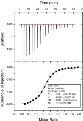
Table 2.1 Mutations of HP1 chromodomain and the calculated cation-π
binding energies of the substituent on benzene...30!
Table 2.2 Fluorescence polarization binding data for wild type HP1 and mutants...33
Table 2.3 Binding constants for HP1 mutants as measured by ITC...37
Table 2.4 Binding constants of HP1 mutants measured by SPR...44
Table 4.1 Dissociation constants of E25Q mutant with di- and trimethyllysine...110
LIST OF FIGURES
Figure 1.1 Crystal structure of DNA wrapped around histone protein octamer...3
Figure 1.2 Active euchromain and inactive heterochromatin...3
Figure 1.3 Various methylation states of lysine and arginine...4
Figure 1.4 Mechanism of SET7/9 methylation of lysine...7
Figure 1.5 Depiction of cavity insertion recognition, surface groove recognition, and aromatic box motif...8
Figure 1.6 Location of partial charges on a quaternary ammonium...10
Figure 1.7 Electrostatic potential maps of cation-π interaction and substituted benzenes...11
Figure 1.8 Neutral isosteres used to study cation-π interactions...14
Figure 1.9 Indole linked to cationic ammonium in folded conformation...15
Figure 1.10 Methylated base and neutral analog...16
Figure 1.11 Crystal structure of HP1 bound to H3K9Me3...17
Figure 1.12 Three stranded β-sheet formed between H3 tail and HP1...17
Figure 2.1 Aromatic residues of a binding region of an ACh binding protein...25
Figure 2.2 Electrostatic potential maps of fluorinated tryptophan and tyrosine...26
Figure 2.3 Linear free energy relationship of mutated aristocholene synthase...28
Figure 2.4 Electrostatic potential maps of unnatural amino acids...29
Figure 2.5 Circular dichroism of HP1 and unnatural mutants...31
Figure 2.6 Crystal structure overlays of WT with mutants...32
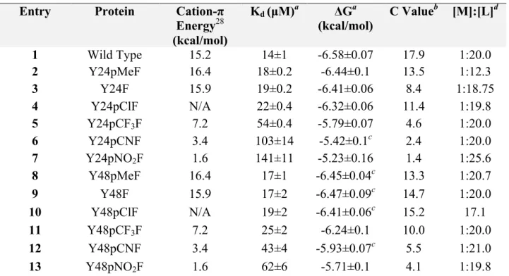
Figure 2.7 Linear free energy relationship of position 24 HP1 mutants...34
Figure 2.8 Three anisotropy runs on same batch of protein...35
Figure 2.9 Two ITC runs of WT HP1 with H3K9Me3...36
Figure 2.12 Plot of log(Ka) vs cation-π gas phase binding energies between
Na+ and C6H5X calculated by Wheeler and Houk...40
Figure 2.13 Relationship between log(Ka) and polarizability parameter (MR)...41
Figure 2.14 Relationship between log(Ka) and hydrophobicity parameter (π)...42
Figure 2.15. SPR data of Y48 HP1mutants plotted against binding energies calculated from electrostatic potential maps...44
Figure 2.16. Top view of Y24 (left) centered over 2 methyl groups and one methylene compared to top view of Y48 (right) centered over one methyl group...46
Figure 2.17. Distances between tyrosine residues and methyl groups and methylene of trimethyllysine...46
Figure 2.18 Binding curve of Wild Type HP1 titrated into FAM labeled H3K9Me3 peptide...50
Figure 2.19 Binding curve of HP1 Y24F titrated into FAM labeled H3K9Me3 peptide...51
Figure 2.20. Binding curve of HP1 Y24pClF titrated into FAM labeled H3K9Me3 peptide...52
Figure 2.21. Binding curve of HP1 Y24pCNF titrated into FAM labeled H3K9Me3 peptide...53
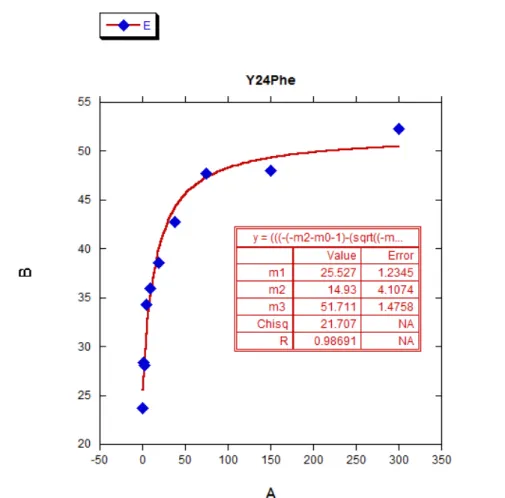
Figure 2.22 Wild Type ITC...54
Figure 2.23 Y24pMeF ITC...55
Figure 2.24 Y24F ITC...56
Figure 2.25 Y24pClF ITC...57
Figure 2.26 Y24pCF3F ITC...58
Figure 2.27 Y24pCNF ITC...59
Figure 2.28 Y24pNO2F ITC...60
Figure 2.29 Y468pMeF ITC...61
Figure 2.30 Y48F ITC...62
Figure 2.32 Y48pCF3F ITC...64
Figure 2.33 Y48pCNF ITC...65
Figure 2.34 Y48pNO2F ITC...66
Figure 2.35 Wild Type SPR...67
Figure 2.36 Y48F SPR...67
Figure 2.37 Y48pClF SPR...68
Figure 2.38 Y48pCF3F SPR...68
Figure 2.39 Y48pCNF SPR...69
Figure 2.40 Y48pNO2F SPR...69
Figure 3.1 Electrostatic potential maps of tryptophan (left) difluorotryptophan (middle) and tetrafluorotryptophan...73
Figure 3.2. Crystal structure of HP1 (top) with proposed sites of mutation to cysteine for ligation in purple...80
Figure 3.3. Binding curve of M38C HP1 titrated into FAM labeled H3K9Me3 peptide...81
Figure 3.4. Analytical HPLC trace of peptide fragments in ligation buffer...84
Figure 3.5. MALDI mass spec of crude ligation reaction...85
Figure 3.6. LC-MS of peak collected from ligation reactions showing the M+5, M+6, and M+7 peaks...85
Figure 3.7. HPLC trace of large scale (5 mL reaction volume) ligation reaction...86
Figure 3.8. Cartoon of example of types of products in crude peptide mixture (left) that can easily be purified out (right)...87
Figure 3.9. UPLC-MS trace of synthetic HP1 from CEM Corporation...88
Figure 3.10. Binding curve of synthetic HP1 titrated into FAM labeled H3K9Me3 peptide...89
Figure 3.11. Binding curve of synthetic HP1 containing Y48F5F mutation titrated into FAM labeled H3K9Me3 peptide...90 !
Figure 4.2 Inhibitors for KMe2 reader proteins...107
Figure 4.3 Partial charges on side chain of KMe2 compared to inhibitors...107
Figure 4.4. Approach for rapidly synthesizing peptides with a variety of side chain mimics...107
Figure 4.5. HP1 bound with KMe2 peptide containing a water-mediated salt bridge...109
Figure 4.6. Glutamic acid (left) and glutamine (right)...110
Figure 4.7. Neutral analog of trimethyllyinse used to probe cation-π interactions...111
Figure 4.8. Binding curve of Wild Type HP1 titrated into FAM labeled H3K9Me3 peptide...113
Figure 4.8. Binding curve of E52Q HP1 titrated into FAM labeled H3K9Me3 peptide...114
LIST OF SCHEMES
Scheme 3.1 Synthesis of alkyl bromide for tryptophan synthesis starting with the
corresponding indole...75
Scheme 3.2 Synthesis of tryptophan derivatives using the Schöllkopf auxiliary...76 Scheme 3.4 Synthesis of proline derived nickel complex...77 Scheme 3.5. Asymmetric alkylation of proline derived nickel complex
followed by release of the amino acid...78 Scheme 3.6. Native chemical ligation between peptide with C-terminal
LIST OF ABBREVIATIONS AND SYMBOLS
Ac acetyl
ACh acetylcholine ACN acetonitrile Ala, A alanine Arg, R arginine
aRMe2 asymmetric dimethyl arginine Asn, N asparagine
Asp, D aspartic acid
Bn benzyl
Boc tert-butyloxycarbonyl
CD circular dichroism
Cys, C cysteine
d doublet
DCM dichloromethane
dd doublet of doublet
DFT density functional theory DIPEA N,N’-diisopropylethylamine
DMF dimethylformamide
DNA deoxyribonucleic acid
DODT 2,2′-(Ethylenedioxy)diethanethiol DTT dithiothreitol
EDT 1,2-ethanedithiol ESI electrospray ionization FAM 5(6)-carboxyfluorescein Fmoc 9-fluorenylmethoxycarbonyl FPLC fast protein liquid chromatography Gln, Q glutamine
Glu, E glutamic acid Gly, G glycine
HBTU O-(benzotriazol-1-yl)-N,N,N’N’-tetramethyluronium hexafluorophosphate His, H histidine
HKMT histone lysine methyltransferase HOBt 1-hydroxybenzotriazole
HP1 heterochromatin protein 1
HPLC high performance liquid chromatography
HX histone protein X
Ile, I isoleucine
ITC isothermal titration calorimetry
K(Me) monomethyllysine
K(Me)2 dimethyllysine K(Me)3 trimethyllysine
Kd dissociation constant
LFER linear free energy relationship Log P octanol-water partition coefficient Lys, K lysine
m multiplet
MALDI matrix-assisted laser desorption ionization
MeOH methanol
Met, M methionine
MHz megahertz
mol mole
MR molar refractivity
MRE mean residue ellipticity
MS mass spectrometry
MTDB 7-methyl-1,5,7-Triazabicyclo[4.4.0]dec-1-ene n-BuLi n-butyllithium
nAChR nicotinic acetylcholine receptor
NBS N-Bromosuccinimide
NCL native chemical ligation NMP N-Methyl-2-pyrrolidone
NMR nuclear magnetic spectroscopy NTA nitrilotriacetic acid
PTM posttranslational modification
PyBOP (benzotriazol-1-yl-oxytripyrrolidinophosphonium hexafluorophosphate)
RMe monomethyl arginine
RNA ribonucleic acid
s singlet
SAH S-adenoxyl-L-homocysteine SAM S-adenosyl-L-methionine SAR structure activity relationship Ser, S serine
SPPS solid phase peptide synthesis SPR surface plasmon resonance sRMe2 symmetric dimethyl arginine
t triplet
tBu tert-butyl
TCEP (tris(2-carboxyethyl)phosphine) TFA trifluoroacetic acid
THF tetrahydrofuran Thr, T threonine
TIPS triisopropylsilane tRNA transfer ribonucleic acid Trp, W tryptophan
Trt trityl
Tyr, Y tyrosine
UV ultra violet
Val, V valine
CHAPTER 1: RECOGNITION OF METHYLATED LYSINE AND CATION-Π
INTERACTIONS 1.1 Purpose of this work
With the emergence of trimethyllysine reader proteins becoming important
therapeutic targets, an in depth understanding of their mechanism of recognition of their
substrates will be advantageous to drug design. Information on the contribution of each
residue to the binding interaction, as well as a broad comparison of reader proteins with
different binding pockets can guide the design of inhibitors or probes for broad classes of
reader proteins, as well as specific readers. While there have been some identified
inhibitors of these proteins, it remains unknown whether they bind by the same cation-π
mechanism as the natural substrate, or by an alternative mechanism.
The purpose of this work is to further probe reader protein binding mechanisms to
provide a detailed understanding of the nature of these cation-π interactions. A
systematic protein structure activity relationship (protein SAR) study, by systematically
altering the binding pocket of reader proteins is used to provide a detailed description on
the balance of forces in the binding of trimethyllysine.
1.2. Epigenetics
The human body consists of trillions of cells, all of which are genetically
identical.1 Despite containing identical DNA sequences, our cells are highly
differentiated and specialized for diverse and specific functions. Cells with the same
control and regulate gene expression in order to produce the diverse sets of phenotypes possible using the same genomic sequence. As a result, the field of epigenetics, the study of these differences in gene expression without differences in the nucleic acid sequence, has become increasingly popular.3 Moreover, the mechanism for regulating gene expression and silencing has risen in importance from a therapeutic standpoint, as the misregulation of expression is implicated in many diseases.4
1.2.1 Chromatin Structure
The extent of DNA packaging is responsible for whether or not a gene is
!
! 3
fiber for which several models have been created based on experimental data using cryogenic-electron microscopy (cryo-EM) and X-ray crystallography.8
This review focuses on recent advances in our understanding of the structure and function of the nucleosome and is divided into four parts. First, we present a primer covering the fundamentals of the nucleosome core particle structure determined at atomic scale by X-ray crystallography in 1997.9 Next we discuss recent insights into the role of DNA sequence in the structure of nucleosomal DNA based on structure− function studies of nucleosome core particles containing derivatives of the Widom 601 nucleosome positioning sequence.10 We then introduce patterns of nucleosome recognition by chromatin factors using recent crystal structures and NMR and cryo-EM models of peptide and protein macromolecular chromatin factors bound to the nucleosome core particle. Finally, we will compare a recent cryo-EM model for the 30 nm chromatin fiber11 to two previous models based on crystallographic and cryo-EM data.12,13
2. FUNDAMENTALS OF THE NUCLEOSOME CORE
PARTICLE STRUCTURE
While the composition of the nucleosome had long since been realized, the 1997 2.8 Å crystal structure of the nucleosome core particle (NCP) solved by Luger et al. afforded the first atomic depiction of this fundamental genomic unit.9 This structure showed 146 bp of the human alpha-satellite sequence wrapped 1.65 times around an octameric scaffold of Xenopus laevishistone proteins in a left-handed superhelix (Figure 1a). A single base pair is centered on the nucleosome dyad,14 which defines the pseudo-2-fold symmetry axis of the NCP. DNA locations are designated by superhelical locations (SHL)
representing superhelical turns from the dyad (SHL 0) and ranging from SHL −7 to SHL 7. The central histone octamer contains two copies of each of the core histone proteins, H2A, H2B, H3, and H4 as established by Arents and Moudrianakis in the 1991 3.1 Å crystal structure of the histone octamer.15 The core histones are assembled into four histone-fold heterodimers (two each of H2A/H2B and H3/H4). Ten flexible tails protrude from the NCP at defined locations, one N-terminal tail from each of the eight core histone proteins and two additional C-terminal tails contributed by H2A.
2.1. Histone-Fold Heterodimers
Each of the core histones contains a central α-helical region that forms a histone-fold motif, flanked by N- and C-terminal extensions. The histone-fold is constructed from threeαhelices connected by two intervening loops specified as α1-L1-α2-L2-α3 (Figure 1b,c).9,15,16 The two shorterα1 and α3 helices loop back to pack against the longer central α2 helix. Each histone-fold pairs with a complementary histone-histone-fold, H3 pairs with H4 and H2A pairs with H2B, to form a histone-fold heterodimer handshake motif. The antiparallel arrangement of this heterodimer approximates the L1 loop from one histone-fold and the L2 loop of the complementary histone-fold, placing one L1L2 pair at each end of the heterodimer. The result is a crescent-shaped heterodimer with the convex surface including the L1L2 loops and the α1 helices and the concave surface including the α3 and central α2 helices. The convex surface of the H2A/H2B and H3/H4 heterodimers carries a strong positive charge and constitutes the primary DNA binding element of each histone-fold heterodimer.
Figure 1.Nucleosome core particle structure and the histone-fold heterodimers. (a) Nucleosome core particle structure (PDB ID 1KX5). Histones and DNA are depicted in cartoon and sticks representations, respectively, and colored as indicated. (b) H3/H4 histone-fold heterodimer. (c) H2A/ H2B histone-fold heterodimer. Structures (top) and schemes (bottom) with secondary structure elements indicated. All molecular graphics in this review were prepared using PyMOL software (The PyMOL Molecular Graphics System, version 1.6, Schrodinger, LLC). All structures of NCP using high-resolution structure17 (PDB ID 1KX5) unless indicated otherwise.
Chemical Reviews Review
dx.doi.org/10.1021/cr500373h|Chem. Rev.2015, 115, 2255−2273 2256
Figure 1.1. Crystal structure of DNA wrapped around histone protein octamer forming a nucleosome with tails extending outward.6 Reprinted with permission from Chem. Rev. 2015, 115, 2255- 2273. Copyright 2015 American Chemical Society.
Figure 1.2. Active euchromatin (left) and inactive heterochromatin (right), with representative modifications often seen on each.5 From Science, 2001, 293, 1074-1080. Reprinted with permission from AAAS.
EPIGENETICS histone tails would induce interaction
ties for chromatin-associated proteins, and (ii) modifications on the same or different
histone tails may be interdependent and
erate various combinations on any one
cleosome.
Here, we wish to extend this concept for overall chromosome structure by proposing that (iii) distinct qualities of higher order chromatin, such as euchromatic or heterochromatic mains (7), are largely dependent on the local concentration and combination of differentially
modified nucleosomes (Fig. 1A). We envision
that this "nucleosome code" then permits the
assembly of different epigenetic states (7),
leading to distinct "readouts" of the genetic
information, such as gene activation versus
gene silencing or, more globally, cell ation versus cell differentiation. Any such el must account for how these epigenetic states are established, maintained, and stably inherited through mitosis and meiosis. Although there is
clear evidence for a "cellular memory" (8),
sudden switches in cell fate do occur, leading to variegating phenotypes. If the histone code pothesis is correct, at least in part, deciphering how that code is translated into biological
sponse remains an important and nontrivial
challenge. On the basis of current knowledge, other possibilities are likely to exist, including less stringent "charge patches" in histone tails
(9).
Clear evidence is beginning to link
ations in chromatin structure to cell cycle progression, DNA replication, DNA damage and its repair, recombination, and overall
chromosome stability (10). It also seems ly that the fundamental nature of
based epigenetics will have an impact on X inactivation, imprinting, developmental programming of cell lineages, and the
ticity of stem cells. The implications for
man biology and disease, including cancer
and aging, are far-reaching.
Su(var)s, Histone Methylation, and Heterochromatin
It is now widely recognized that heritable, but
reversible, changes in gene expression can occur without alterations in DNA sequence.
Pioneering studies on radiation-induced
mosomal translocations (11) provided some
of the earliest findings that epigenetic off" transcriptional states are largely dent on the position of a gene within an accessible (euchromatic) or an inaccessible
(heterochromatic) chromatin environment. This phenomenon, known as position-effect variegation (PEV), allowed the development of genetic screens in Drosophila (12) and S. pombe (13, 14) that have identified -30 to
40 loci involved in modifying PEV. Similar to PEV, mating-type switching in budding
(15) and fission (16) yeast represents another paradigm for a variegating mechanism where
the location of a gene within a distinct matin environment, the mat region, dictates the establishment of an active or a silent transcriptional state. In particular for S.
pombe, which appears to contain a higher
order chromatin structure more closely
sembling that of multicellular eukaryotes, heritance of silenced chromatin domains has
been shown to be remarkably stable during both mitosis and meiosis (16).
A euchromatin heterochromatin
B
E1 E(var) HT - Su(var)
ON/ OFF/
active IJ* J iL silenced
FEU - Su(var) HET] ----' E(var)
v , proteolysis ?C active/accessible inactive/condensed
[H3] ARTKQTARKSTGGKAPRKQL [H3] ARTKQTA RKSTGGKAPRKQL 910 14 10
[CENP-A] MGPRRRSRKPEAPRRRSPSP
7
[H3] ARTKQTARKSTGGKAPRKQL
4 14 4
t e> [H3] ARTKQTARKSTI IGKAPRKQL
9[H4] SGRGKGGKGLGKGGAKRHRK [H4] SGRGKGGKGLGKGGAKRHRK
3 5 12
D "0 Fig. 1. Models for euchromatic or hetero-
A chromatic histone tail modifications. (A)';| y^\^\~,~~ >Schematic representation of euchromatin
and heterochromatin as accessible orcondensed nucleosome fibers containing I + \ 'acetylated (Ac), phosphorylated (P), and ARTKQT ARKSTGGKAPRKQL...histone H3 methylated (Me) histone NH2-termini.
~~~~~~4 910 14 ~(B) Generic model for antagonistic E(var)
^"Yf7~~~~~~~ ^\ and Su(var) gene function in adding
~9 '^ a~ t . 9 chromatic (EU) or heterochromatic (HET)
* ~ 24 VSC~)0B *' ~ modification marks onto a nucleosomal
"^'pa/ c Y -template. In addition, Su(var)s also sGRGKGGKGLGKGGAKRHRK...histone H4 tion in removing euchromatic signals and 3 5 16 18 20 E(var)s can destabilize the
1.2.2 Posttranslational Modifications (PTMs)
The formation of heterochromatin and euchromatin is controlled by a complex series of posttranslational modifications (PTMs) on the histone proteins. PTMs are covalent modifications on the side chains of amino acids that are installed after the protein has been translated from RNA (Figure 1.3). Common examples of these modifications include methylation of lysine (mono, di, and tri), methylation of arginine (mono and di – either symmetrically or asymmetrically), acetylation of lysine, and phosphorylation of serine, threonine, and tyrosine.
These modifications are found on histone proteins – most abundantly on the unstructured tail regions on the histone N-terminus. The nucleosome consists of an octomer of histone proteins, containing two each of histone proteins H2A, H2B, H3 and H4, while H1 links adjacent nucleosomes.6 Posttranslational modifications marks are regulated and recognized by proteins known as “writers,” “readers,” and “erasers.”
Figure 1.3. Various methylation states of lysine (top) and arginine (bottom) Arginine can be methylated symmetrically (sRMe2) or asymmetrically (aRMe2). N H NH3 H N O N H
H2N
H N O N H H N H N O N H N H N O N H NH H N O NH2 H2N
N H NH H N O NH2 HN N H NH H N O H N HN N H NH H N O N H2N
Lys KMe KMe2 KMe3
Arg RMe sRMe2 aRMe2
N H H N O tButNle N H NH3 H N O N H
H2N
H N O N H H N H N O N H N H N O N H NH H N O NH2 H2N
N H NH H N O NH2 HN N H NH H N O H N HN N H NH H N O N H2N
Lys KMe KMe2 KMe3
Arg RMe sRMe2 aRMe2
termed the “histone code.”5 The modifications regulate chromatin formation both by altering interactions with DNA and between nucleosomes, as well as recruiting other proteins and enzymes involved in chromatin remodeling.7 They can also work together in the regulation of other modifications, known as histone “cross talk.”8
1.3 Lysine methylation
Lysine methylation, while one of the most common posttranslational
modifications, has not been extensively studied until recent years. Unlike acetylation and phosphorylation, which change the charge of the side chain, lysine methylation maintains the same overall side chain charge. Methylation is site-specific and the different levels (mono, di, and trimethylation) can effect gene expression differently, leading to added complexity with methylation compared to other posttranslational modifications. There have been various assays developed to map out the methylation pattern and determine the proteins that recognize these specific marks. The most characterized histone tail in terms of PTM’s has been the H3 tail. There are at least 4 lysine positions that can be
methylated on the H3 tail (K4, K9, K27, and K36).9 The regulation of these marks are important for cell cycle regulation, DNA damage response, and development and differentiation.10
1.3.1 Writers: Lysine Methyltransferases
The first histone lysine methyltransferase (HKMT) to be discovered was
SUV39H1, which methylates H3K9.11 The vast majority of HKMTs contain a domain
known as the SET domain (derived from the proteins it was identified in: Drosophila
Su(var)3-9 and ‘Enhancer of zeste’ proteins).12 These enzymes catalyze the transfer of a
methyl group from S-adenosylmethionine (SAM) to the amino group of the lysine side
chain (Figure 1.4). Most of these enzymes are very specific for a single site on a histone
tail, and can modify the ammonium to specific degree of methylation (mono-, di-, or tri-).
Often times this selectivity for methylation states is caused by a specific amino acid in
the active site of the enzyme.7 For example, one enzyme that can form trimethyllysine
(DIM5), contains a phenylalanine in the active site.13,14 Another enzyme, SET7/9,
contains the slightly larger tyrosine residue, and can only accommodate the monomethyl
product.15 This dependence on phenylalanine or tyrosine to control the degree of
methylation is thought to be general to these SET domain enzymes.16,17
O O O HN NH O N H H O H H O O O NH3 S O HO OH N N N N NH2 H H SAM Y305 295 290 H3K4Me3 Y245 O O O HN NH O N H H O H H O O O NH3 S O HO OH N N N N NH2 H H SAH Y305 295 290 H3K4Me3 Y245
1.3.2 Erasers: Lysine Demethylases
It was initially believed that lysine methylation was a permanent mark, and there
were no mechanisms for removal.7 In 2004, the first lysine demethylase (LSD1) was
discovered, which led to the conclusion that lysine methylation is a dynamic process.18 This enzyme, however, requires a protonated nitrogen in order to function, and thus,
cannot demethylate trimethyllysine. Another class of enzymes that contain a catalytic
jumonji domain was discovered in 2006, which was capable of demethylating
trimethyllysine.19 These enzymes utilize a radical mechanism for demethylation of
trimethyllysine. Histone methylation, while reversible, has a lower turnover rate than
some other modifications. Furthermore, some sites need to be maintained, and different
sites have different rates of turnover.10 1.3.3 Readers Proteins
Reader proteins are proteins that specifically recognize certain epigenetic marks
leading to gene expression, gene silencing, or regulation of other modifications.10 Some methyl binding domains commonly involved in the recognition of these marks include
PHD, WD40, CW, and PWWP domains, as well as the “royal family” which includes
tudor, chromo, MBT domains.20 As the level of methylation increases, the number of hydrogen bond donors on the sidechain decreases (with trimethyllysine unable to form a
hydrogen bond), and the size and hydrophobicity increase. This allows for the specific
recognition of different methylation states.20
There are two primary modes that reader proteins typically employ in order to
bind to methylated lysine (Figure 1.5). In a cavity insertion recognition mode, such as
!
! 8
protein cavity. This mode is often present with readers of lower methylation states, and
may also provide a way of discrimination of different states based on size.20 Proteins that
act via a surface groove recognition mode have a binding pocket that is more accessible,
providing slightly less specificity for methylation states. Domains that recognize
methylated lysine contain an “aromatic box” consisting of 2-4 aromatic residues that bind
to the cation, presumably via cation-π interactions, as described below.22
!
!
!
1.4 Cation-π Interactions
Cation-π interactions are prevalent across various biological systems are
particularly involved in protein-protein interactions and protein folding.23 While other
intramolecular interactions such as salt bridges, hydrogen bonding, and the hydrophobic
effect have been known and well studied in biological systems, advances demonstrating
the prevalence of cation-π interactions has been more recent.23,24
Figure 1.5. Depiction cavity insertion recognition (left), surface groove recognition (center) and aromatic box motif (right). Reprinted by permission from Macmillan Publishers Ltd: Nat. Struct. Mol. Biol., 2007, 14, 11, 1025-1040., Copyright 2007.
-" --""" %0 " -"+/"&*)+"'& ""
,) !)''- ++"('#+
"-%0 ."$+/( %0 +,') %0 %0 -"" %0 """
"/8,ESSONSLEARNEDFROMSTATESPECIFICREADOUTOFMETHYLLYSINEMARKS
3TRUCTURALSTUDIESOFMETHYLLYSINEEFFECTORSINCOGNATEPEPTIDEn BOUNDORAPOFORMSSUGGESTGENERALMECHANISMSTHATARE USEDBYBINDINGPOCKETSTOACHIEVESPECIFICITYTOWARDDISTINCT METHYLATIONSTATES(EREWEILLUSTRATETHESECOMMONFEATURES ANDTHEIRROLESINENGAGEMENT 3URFACEGROOVEVERSUSCAVITYINSERTIONRECOGNITIONMODES 4HEKNOWNSTRUCTURESOFMETHYLLYSINE EFFECTORCOMPLEXESCANBESUBDIVIDED INTOTHOSETHATUSEACAVITYINSERTION RECOGNITIONMODEI ASOBSERVEDIN COMPLEXESOFTHELOWERMETHYLATION STATEnBINDING"0TANDEMTUDOR DOMAINSAND,-"4,REF ANDTHOSETHATUSEASURFACE GROOVERECOGNITIONMODEII AS OBSERVEDINCOMPLEXESOFTHEHIGHER METHYLATIONSTATEnBINDING(0CHROMODOMAIN#($DOUBLECHROMODOMAINS*-*$!DOUBLETUDORDOMAINS AND0($FINGER)NTHECAVITYINSERTIONRECOGNITIONMODETHEMETHYLAMMONIUM GROUPOFMETHYLLYSINEISINSERTEDINTOANDBURIEDWITHINADEEPPROTEINCLEFTI POTENTIALLY ENDOWINGTHEPOCKETWITHSIZESENSITIVESELECTIONFILTERCAPABILITIES)NTHESURFACEGROOVE RECOGNITIONMODETHEBINDINGPOCKETSAREBOTHWIDERANDMOREACCESSIBLEII SUCHTHATTHE METHYLLYSINESIDECHAINLIESALONGAPROTEINSURFACEGROOVEANDTHEEFFECTORCOMPLEXESHAVE APPRECIABLEBUTLESSSTRINGENTPREFERENCESFORCERTAINLYSINEMETHYLATIONSTATES 3TATICRECOGNITIONPOCKETS 7HEREASTHEPEPTIDESCANUNDERGOBSTRANDBACKBONECONFORMATIONALCHANGESINDUCEDDURINGBINDINGTHEEFFECTORPROTEINSOFTENSHOW LITTLEORNOAPPRECIABLESTRUCTURALPERTURBATION4HECOMPACTNATUREANDAPPARENTMONOLITHICRIGIDITYOFEFFECTORFOLDSPRECLUDESSUBSTANTIAL MOVEMENTASACONSEQUENCEOFLIGANDENGAGEMENT&URTHERMOREFORMOSTSTRUCTURALLYCHARACTERIZEDEFFECTORSTHERESEEMSTOBELITTLEOR NOMOVEMENTOFSIDECHAINSINVOLVEDINRECOGNITION)NPARTICULARAROMATICCAGESINTHEBOUNDFORMAREVERYSTATICWITHRESPECTTOTHEIR UNLIGANDEDCOUNTERPARTSIII 0REORGANIZEDCAGESHAVEACLEARENERGETICBENEFITOVERFLEXIBLEBINDINGSITESINTHATTHECONFORMATIONAL ENTROPYLOSSUPONBINDINGISMINIMIZEDONTHEPROTEINSIDE.OTABLEEXCEPTIONSTOTHISRULEINCLUDESIDECHAINMOVEMENTSOBSERVEDUPON
COMPLEXATIONIN7$2AND"0REF
!ROMATICCAGEPOCKETS
!COMMONANDSTRIKINGFEATUREOFMETHYLLYSINEREADERDOMAINSISTHEPOSITIONINGOFTHEMETHYLAMMONIUMMOIETYWITHINANAROMATIC CAGECONSISTINGOFTWOTOFOURAROMATICRESIDUESOFTENSUPPLEMENTEDBYONEORMOREACIDICSIDECHAINS)NCONTRASTTOTHE0($FINGERS THATRECOGNIZETHEUNMODIFIEDLYSINEWHICHLACKANYSEMBLANCEOFAROMATICCAGINGRESIDUESIV THE"04&0($FINGERHASFOURAROMATIC RESIDUESVIII ANDTHE).'FINGERHASTWOAROMATICRESIDUESANDAHYDROPHOBICMETHIONINESIDECHAINVII 4HEMETHYLAMMONIUM GROUPWHICHCARRIESADIFFUSEPOSITIVECHARGESTACKSOVERTHEPARTIALNEGATIVECHARGEPERMEATINGTHEFACEOFTHEAROMATICRINGSWITHTHE ELECTROSTATICSTABILIZATIONMEDIATEDBYCATIONPINTERACTIONSVnVII ANDTOALESSEREXTENTBYHYDROPHOBICCONTACTS
3TRUCTURALBASISFORLOWERLYSINEMETHYLATIONSTATEnSPECIFICREADOUT
4HETANDEMTUDORDOMAINSOF"0REF ANDTHESECOND-"4POCKETOF,-"4,REF BOTHBINDMONOANDDIMETHYLLYSINE WITHINANAROMATICCAGEUSINGTHECAVITYINSERTIONRECOGNITIONMODEI TODISCRIMINATEAGAINSTTHETRIMETHYLLYSINECOUNTERPARTCOMPARE TOSURFACEGROOVERECOGNITIONII 4HESPECIFICITYFORLOWERLYSINEMETHYLATIONSTATEREADOUTORIGINATESINADIRECTHYDROGENBONDAND ELECTROSTATICINTERACTIONBETWEENTHEMETHYLAMMONIUMPROTONANDTHECARBOXYLATEOFANASPARTATERESIDUELININGTHEWALLSOFTHEAROMATIC CAGEVANDVI &URTHERMORETRIMETHYLLYSINEBINDINGWOULDBEDISFAVOREDBYANANTICIPATEDSTERICREPULSIONBETWEENTHEADDITIONALMETHYL GROUPANDTHEASPARTATESIDECHAIN)NADDITIONTHELIMITEDDIMENSIONSOFTHEBINDINGPOCKETSIN"0AND,-"4,COMPLEXESBOTH ATTHEENTRANCEANDWITHINTHECHANNELSEEMTORESTRICTTHEACCESSOFTHELARGERTRIMETHYLAMMONIUMGROUP %NGINEERINGMETHYLLYSINERECOGNITIONSPECIFICITY 4HEWILDTYPE"04&0($FINGERBINDSMETHYLATEDLYSINESINAN(+SEQUENCECONTEXTWITHAFFINITIESINTHEORDER+ME+ME +ME+MEASMEASUREDBY.-2TITRATIONSURFACEPLASMONRESONANCEANDFLUORESCENCEPOLARIZATIONSTUDIES)TWASANTICIPATED THATTHEAVAILABILITYOFAPROPERLYPOSITIONEDCARBOXYLATESIDECHAINCOULDALLOWFINETUNINGOFTHESTATESPECIFICREADOUTOFMETHYLATED LYSINES)NDEEDASINGLE4YRTO'LUSUBSTITUTIONINTHEAROMATICCAGEMOTIFOFTHE"04&0($FINGERREVERSESTHEBINDINGPREFERENCEFROM (+METO(+MEREF 4HECRYSTALSTRUCTUREOFTHE9%MUTANT"04&0($FINGERVIII ESTABLISHESTHATTHESPECIFICITYCHANGE ISASSOCIATEDWITHHYDROGENBONDINGBETWEENTHEMETHYLAMMONIUMPROTONANDTHECARBOXYLATEGROUPOFTHEGLUTAMATESIDECHAININTHE MUTANT)NTHISINSTANCETHESTRINGENCYOFDISCRIMINATIONBYSURFACEGROOVERECOGNITIONDOESNOTMATCHTHATDESCRIBEDABOVEFORCAVITY
2 % 6 ) % 7
-" --""" %0 " -"+/"&*)+"'& ""
,) !)''- ++"('#+
"-%0 ."$+/( %0 +,') %0 %0 -"" %0 """
"/8,ESSONSLEARNEDFROMSTATESPECIFICREADOUTOFMETHYLLYSINEMARKS
3TRUCTURALSTUDIESOFMETHYLLYSINEEFFECTORSINCOGNATEPEPTIDEn BOUNDORAPOFORMSSUGGESTGENERALMECHANISMSTHATARE USEDBYBINDINGPOCKETSTOACHIEVESPECIFICITYTOWARDDISTINCT METHYLATIONSTATES(EREWEILLUSTRATETHESECOMMONFEATURES ANDTHEIRROLESINENGAGEMENT 3URFACEGROOVEVERSUSCAVITYINSERTIONRECOGNITIONMODES 4HEKNOWNSTRUCTURESOFMETHYLLYSINE EFFECTORCOMPLEXESCANBESUBDIVIDED INTOTHOSETHATUSEACAVITYINSERTION RECOGNITIONMODEI ASOBSERVEDIN COMPLEXESOFTHELOWERMETHYLATION STATEnBINDING"0TANDEMTUDORDOMAINSAND,-"4,REF
ANDTHOSETHATUSEASURFACE GROOVERECOGNITIONMODEII AS OBSERVEDINCOMPLEXESOFTHEHIGHER METHYLATIONSTATEnBINDING(0
CHROMODOMAIN#($DOUBLECHROMODOMAINS*-*$!DOUBLETUDORDOMAINS
AND0($FINGER)NTHECAVITYINSERTIONRECOGNITIONMODETHEMETHYLAMMONIUM
GROUPOFMETHYLLYSINEISINSERTEDINTOANDBURIEDWITHINADEEPPROTEINCLEFTI POTENTIALLY ENDOWINGTHEPOCKETWITHSIZESENSITIVESELECTIONFILTERCAPABILITIES)NTHESURFACEGROOVE RECOGNITIONMODETHEBINDINGPOCKETSAREBOTHWIDERANDMOREACCESSIBLEII SUCHTHATTHE METHYLLYSINESIDECHAINLIESALONGAPROTEINSURFACEGROOVEANDTHEEFFECTORCOMPLEXESHAVE APPRECIABLEBUTLESSSTRINGENTPREFERENCESFORCERTAINLYSINEMETHYLATIONSTATES 3TATICRECOGNITIONPOCKETS
7HEREASTHEPEPTIDESCANUNDERGOBSTRANDBACKBONECONFORMATIONALCHANGESINDUCEDDURINGBINDINGTHEEFFECTORPROTEINSOFTENSHOW
LITTLEORNOAPPRECIABLESTRUCTURALPERTURBATION4HECOMPACTNATUREANDAPPARENTMONOLITHICRIGIDITYOFEFFECTORFOLDSPRECLUDESSUBSTANTIAL MOVEMENTASACONSEQUENCEOFLIGANDENGAGEMENT&URTHERMOREFORMOSTSTRUCTURALLYCHARACTERIZEDEFFECTORSTHERESEEMSTOBELITTLEOR NOMOVEMENTOFSIDECHAINSINVOLVEDINRECOGNITION)NPARTICULARAROMATICCAGESINTHEBOUNDFORMAREVERYSTATICWITHRESPECTTOTHEIR UNLIGANDEDCOUNTERPARTSIII 0REORGANIZEDCAGESHAVEACLEARENERGETICBENEFITOVERFLEXIBLEBINDINGSITESINTHATTHECONFORMATIONAL ENTROPYLOSSUPONBINDINGISMINIMIZEDONTHEPROTEINSIDE.OTABLEEXCEPTIONSTOTHISRULEINCLUDESIDECHAINMOVEMENTSOBSERVEDUPON
COMPLEXATIONIN7$2AND"0REF
!ROMATICCAGEPOCKETS
!COMMONANDSTRIKINGFEATUREOFMETHYLLYSINEREADERDOMAINSISTHEPOSITIONINGOFTHEMETHYLAMMONIUMMOIETYWITHINANAROMATIC CAGECONSISTINGOFTWOTOFOURAROMATICRESIDUESOFTENSUPPLEMENTEDBYONEORMOREACIDICSIDECHAINS)NCONTRASTTOTHE0($FINGERS THATRECOGNIZETHEUNMODIFIEDLYSINEWHICHLACKANYSEMBLANCEOFAROMATICCAGINGRESIDUESIV THE"04&0($FINGERHASFOURAROMATIC RESIDUESVIII ANDTHE).'FINGERHASTWOAROMATICRESIDUESANDAHYDROPHOBICMETHIONINESIDECHAINVII 4HEMETHYLAMMONIUM GROUPWHICHCARRIESADIFFUSEPOSITIVECHARGESTACKSOVERTHEPARTIALNEGATIVECHARGEPERMEATINGTHEFACEOFTHEAROMATICRINGSWITHTHE
ELECTROSTATICSTABILIZATIONMEDIATEDBYCATIONPINTERACTIONSVnVII ANDTOALESSEREXTENTBYHYDROPHOBICCONTACTS
3TRUCTURALBASISFORLOWERLYSINEMETHYLATIONSTATEnSPECIFICREADOUT
4HETANDEMTUDORDOMAINSOF"0REF ANDTHESECOND-"4POCKETOF,-"4,REF BOTHBINDMONOANDDIMETHYLLYSINE WITHINANAROMATICCAGEUSINGTHECAVITYINSERTIONRECOGNITIONMODEI TODISCRIMINATEAGAINSTTHETRIMETHYLLYSINECOUNTERPARTCOMPARE TOSURFACEGROOVERECOGNITIONII 4HESPECIFICITYFORLOWERLYSINEMETHYLATIONSTATEREADOUTORIGINATESINADIRECTHYDROGENBONDAND ELECTROSTATICINTERACTIONBETWEENTHEMETHYLAMMONIUMPROTONANDTHECARBOXYLATEOFANASPARTATERESIDUELININGTHEWALLSOFTHEAROMATIC CAGEVANDVI &URTHERMORETRIMETHYLLYSINEBINDINGWOULDBEDISFAVOREDBYANANTICIPATEDSTERICREPULSIONBETWEENTHEADDITIONALMETHYL GROUPANDTHEASPARTATESIDECHAIN)NADDITIONTHELIMITEDDIMENSIONSOFTHEBINDINGPOCKETSIN"0AND,-"4,COMPLEXESBOTH ATTHEENTRANCEANDWITHINTHECHANNELSEEMTORESTRICTTHEACCESSOFTHELARGERTRIMETHYLAMMONIUMGROUP %NGINEERINGMETHYLLYSINERECOGNITIONSPECIFICITY 4HEWILDTYPE"04&0($FINGERBINDSMETHYLATEDLYSINESINAN(+SEQUENCECONTEXTWITHAFFINITIESINTHEORDER+ME+ME
+ME+MEASMEASUREDBY.-2TITRATIONSURFACEPLASMONRESONANCEANDFLUORESCENCEPOLARIZATIONSTUDIES)TWASANTICIPATED
THATTHEAVAILABILITYOFAPROPERLYPOSITIONEDCARBOXYLATESIDECHAINCOULDALLOWFINETUNINGOFTHESTATESPECIFICREADOUTOFMETHYLATED LYSINES)NDEEDASINGLE4YRTO'LUSUBSTITUTIONINTHEAROMATICCAGEMOTIFOFTHE"04&0($FINGERREVERSESTHEBINDINGPREFERENCEFROM (+METO(+MEREF 4HECRYSTALSTRUCTUREOFTHE9%MUTANT"04&0($FINGERVIII ESTABLISHESTHATTHESPECIFICITYCHANGE ISASSOCIATEDWITHHYDROGENBONDINGBETWEENTHEMETHYLAMMONIUMPROTONANDTHECARBOXYLATEGROUPOFTHEGLUTAMATESIDECHAININTHE MUTANT)NTHISINSTANCETHESTRINGENCYOFDISCRIMINATIONBYSURFACEGROOVERECOGNITIONDOESNOTMATCHTHATDESCRIBEDABOVEFORCAVITY
2 % 6 ) % 7
.!452%3425#452!,-/,%#5,!2")/,/'96/,5-%.5-"%2./6%-"%2
-" --""" %0 " -"+/"&*)+"'& ""
,) !)''- ++"('#+
"-%0 ."$+/( %0 +,') %0 %0 -"" %0 """
"/8,ESSONSLEARNEDFROMSTATESPECIFICREADOUTOFMETHYLLYSINEMARKS
3TRUCTURALSTUDIESOFMETHYLLYSINEEFFECTORSINCOGNATEPEPTIDEn
BOUNDORAPOFORMSSUGGESTGENERALMECHANISMSTHATARE
USEDBYBINDINGPOCKETSTOACHIEVESPECIFICITYTOWARDDISTINCT
METHYLATIONSTATES(EREWEILLUSTRATETHESECOMMONFEATURES
ANDTHEIRROLESINENGAGEMENT
3URFACEGROOVEVERSUSCAVITYINSERTIONRECOGNITIONMODES
4HEKNOWNSTRUCTURESOFMETHYLLYSINE
EFFECTORCOMPLEXESCANBESUBDIVIDED
INTOTHOSETHATUSEACAVITYINSERTION
RECOGNITIONMODEI ASOBSERVEDIN
COMPLEXESOFTHELOWERMETHYLATION
STATEnBINDING"0TANDEMTUDOR
DOMAINS
AND,-"4,REF
ANDTHOSETHATUSEASURFACE
GROOVERECOGNITIONMODEII AS
OBSERVEDINCOMPLEXESOFTHEHIGHER
METHYLATIONSTATEnBINDING(0
CHROMODOMAIN
#($DOUBLECHROMODOMAINS
*-*$!DOUBLETUDORDOMAINS
AND0($FINGER
)NTHECAVITYINSERTIONRECOGNITIONMODETHEMETHYLAMMONIUM
GROUPOFMETHYLLYSINEISINSERTEDINTOANDBURIEDWITHINADEEPPROTEINCLEFTI POTENTIALLY
ENDOWINGTHEPOCKETWITHSIZESENSITIVESELECTIONFILTERCAPABILITIES)NTHESURFACEGROOVE
RECOGNITIONMODETHEBINDINGPOCKETSAREBOTHWIDERANDMOREACCESSIBLEII SUCHTHATTHE
METHYLLYSINESIDECHAINLIESALONGAPROTEINSURFACEGROOVEANDTHEEFFECTORCOMPLEXESHAVE
APPRECIABLEBUTLESSSTRINGENTPREFERENCESFORCERTAINLYSINEMETHYLATIONSTATES
3TATICRECOGNITIONPOCKETS
7HEREASTHEPEPTIDESCANUNDERGO
BSTRANDBACKBONECONFORMATIONALCHANGESINDUCEDDURINGBINDINGTHEEFFECTORPROTEINSOFTENSHOW
LITTLEORNOAPPRECIABLESTRUCTURALPERTURBATION4HECOMPACTNATUREANDAPPARENTMONOLITHICRIGIDITYOFEFFECTORFOLDSPRECLUDESSUBSTANTIAL
MOVEMENTASACONSEQUENCEOFLIGANDENGAGEMENT&URTHERMOREFORMOSTSTRUCTURALLYCHARACTERIZEDEFFECTORSTHERESEEMSTOBELITTLEOR
NOMOVEMENTOFSIDECHAINSINVOLVEDINRECOGNITION)NPARTICULARAROMATICCAGESINTHEBOUNDFORMAREVERYSTATICWITHRESPECTTOTHEIR
UNLIGANDEDCOUNTERPARTSIII 0REORGANIZEDCAGESHAVEACLEARENERGETICBENEFITOVERFLEXIBLEBINDINGSITESINTHATTHECONFORMATIONAL
ENTROPYLOSSUPONBINDINGISMINIMIZEDONTHEPROTEINSIDE.OTABLEEXCEPTIONSTOTHISRULEINCLUDESIDECHAINMOVEMENTSOBSERVEDUPON
COMPLEXATIONIN7$2
AND"0REF
!ROMATICCAGEPOCKETS
!COMMONANDSTRIKINGFEATUREOFMETHYLLYSINEREADERDOMAINSISTHEPOSITIONINGOFTHEMETHYLAMMONIUMMOIETYWITHINANAROMATIC
CAGECONSISTINGOFTWOTOFOURAROMATICRESIDUESOFTENSUPPLEMENTEDBYONEORMOREACIDICSIDECHAINS)NCONTRASTTOTHE0($FINGERS
THATRECOGNIZETHEUNMODIFIEDLYSINEWHICHLACKANYSEMBLANCEOFAROMATICCAGINGRESIDUESIV THE"04&0($FINGERHASFOURAROMATIC
RESIDUESVIII ANDTHE).'FINGERHASTWOAROMATICRESIDUESANDAHYDROPHOBICMETHIONINESIDECHAINVII 4HEMETHYLAMMONIUM
GROUPWHICHCARRIESADIFFUSEPOSITIVECHARGESTACKSOVERTHEPARTIALNEGATIVECHARGEPERMEATINGTHEFACEOFTHEAROMATICRINGSWITHTHE
ELECTROSTATICSTABILIZATIONMEDIATEDBYCATIONPINTERACTIONS
VnVII ANDTOALESSEREXTENTBYHYDROPHOBICCONTACTS
3TRUCTURALBASISFORLOWERLYSINEMETHYLATIONSTATEnSPECIFICREADOUT
4HETANDEMTUDORDOMAINSOF"0REF ANDTHESECOND-"4POCKETOF,-"4,REF BOTHBINDMONOANDDIMETHYLLYSINE
WITHINANAROMATICCAGEUSINGTHECAVITYINSERTIONRECOGNITIONMODEI TODISCRIMINATEAGAINSTTHETRIMETHYLLYSINECOUNTERPARTCOMPARE
TOSURFACEGROOVERECOGNITIONII 4HESPECIFICITYFORLOWERLYSINEMETHYLATIONSTATEREADOUTORIGINATESINADIRECTHYDROGENBONDAND
ELECTROSTATICINTERACTIONBETWEENTHEMETHYLAMMONIUMPROTONANDTHECARBOXYLATEOFANASPARTATERESIDUELININGTHEWALLSOFTHEAROMATIC
CAGEVANDVI &URTHERMORETRIMETHYLLYSINEBINDINGWOULDBEDISFAVOREDBYANANTICIPATEDSTERICREPULSIONBETWEENTHEADDITIONALMETHYL
GROUPANDTHEASPARTATESIDECHAIN)NADDITIONTHELIMITEDDIMENSIONSOFTHEBINDINGPOCKETSIN"0AND,-"4,COMPLEXESBOTH
ATTHEENTRANCEANDWITHINTHECHANNELSEEMTORESTRICTTHEACCESSOFTHELARGERTRIMETHYLAMMONIUMGROUP
%NGINEERINGMETHYLLYSINERECOGNITIONSPECIFICITY
4HEWILDTYPE"04&0($FINGERBINDSMETHYLATEDLYSINESINAN(+SEQUENCECONTEXTWITHAFFINITIESINTHEORDER+ME+ME
+ME+MEASMEASUREDBY.-2TITRATIONSURFACEPLASMONRESONANCEANDFLUORESCENCEPOLARIZATIONSTUDIES
)TWASANTICIPATED
THATTHEAVAILABILITYOFAPROPERLYPOSITIONEDCARBOXYLATESIDECHAINCOULDALLOWFINETUNINGOFTHESTATESPECIFICREADOUTOFMETHYLATED
LYSINES)NDEEDASINGLE4YRTO'LUSUBSTITUTIONINTHEAROMATICCAGEMOTIFOFTHE"04&0($FINGERREVERSESTHEBINDINGPREFERENCEFROM
(+METO(+MEREF 4HECRYSTALSTRUCTUREOFTHE9%MUTANT"04&0($FINGERVIII ESTABLISHESTHATTHESPECIFICITYCHANGE
ISASSOCIATEDWITHHYDROGENBONDINGBETWEENTHEMETHYLAMMONIUMPROTONANDTHECARBOXYLATEGROUPOFTHEGLUTAMATESIDECHAININTHE
MUTANT)NTHISINSTANCETHESTRINGENCYOFDISCRIMINATIONBYSURFACEGROOVERECOGNITIONDOESNOTMATCHTHATDESCRIBEDABOVEFORCAVITY
INSERTIONRECOGNITIONBECAUSEOFTHESTERICDEMANDSASSOCIATEDWITHTHELATTERRECOGNITIONMODE
1.4.1 Description of Cation-π Interactions
Cation-π interactions are an electrostatic interaction between a positively charged
cation and the partial negative face of an aromatic ring, arising from the quadrupole
moment.23 The dipole between the hydrogen atoms and the sp2 hybridized carbon atoms
in the ring create the quadrupole moment of aromatic compounds. While these
interactions are commonly seen in aromatic rings, the aromaticity is not necessary for the
electrostatic interaction. 25
These interactions were first seen in gas phase experiments where it was observed
that the interaction between metal cations with benzene was stronger than their
interaction with water. The ΔH of the interaction between potassium ions and benzene,
for example, was -19.2 kcal/mol, while the interaction between potassium and water was
-17.9 kcal/mol.26 Trimethylamonium cations also exhibited a difference with a delta H of
9.4 kcal/mol with benzene and 9.0 kcal/mol with water.27 The cation-π interaction
between these metal cations and benzene follow the traditional electrostatic model with
Li+>Na+>K+>Rb+.26,28–30
While these initial observations were all in the gas phase, the cation-π interaction
is also favorable in aqueous systems. Unlike amino acids that form salt-bridges and
hydrogen bonds, which are often well solvated and prefer to interact with water than each
other, the additional driving force for this interaction, which also includes van der Waals
interactions and the hydrophobic effect, make cation-π interactions favorable even in
aqueous systems, with countless examples in biological systems.24,31 Solvation of cations
desolvation cost to participate in cation-π interactions. This can be observed in aqueous
systems where the interaction of benzene with K+ is greater than with Na+.30
Within proteins, positively charged lysine and arginine residues interact with
aromatic phenylalanine, tyrosine, and tryptophan. These cations are often seen with the
alkyl chains packed against the aromatic face, for example the methyl groups of of
methylated lysine or the ε-methylene of unmethylated lysine, as this is where the partial
positive charge actually lies (Figure 1.6). Analysis of the PDB estimates that there is one
cation-π interaction for every 77 amino acid residues, and about 26% of all tryptophan
residues are involved in cation-π interactions.23,32 In intermolecular protein-protein interactions, cation-π interactions were present in about half of all complexes and one
third of all homodimers.32
Figure 1.6. Location of partial charges on a quaternary ammonium
H3C
N CH3
CH3
H3C
1.4.2 Substituent Effects on Cation-π Interactions
While the net interaction energy is dependent on several forces, including van der
Waals and the hydrophobic effect, the variance in the strength of cation-π interactions
result from differences in electrostatic interactions.33 With aromatic rings, this can be
estimated quite well using electrostatic potential maps (Figure 1.7).34 Electron
withdrawing substituents reduce the negative electrostatic potential of the ring. This
correlates nicely with the reduced binding energy measured for Na+ in the gas phase.
These energies also roughly correlate with σmeta values, as resonance effects do not play a
role in cation-π binding energies.31,33
Recent computation studies have provided an alternative model for substituent
effects on cation-π interactions.25,35–37 The results suggest that differences in energies are Figure 1.7. Electrostatic potential maps of a cation-π interaction between
primarily the result of through space interactions with the dipole between the carbon and
the substituent, rather than a change in the electron density of the π-cloud over the center of the ring.35,36
1.4.3 Studying Cation-π Interactions in Biological Systems
There have been many model systems developed to study and quantify cation-π
interactions, estimating them to be worth about 1-2 kcal/mol/ring.23 Among the first
small molecule model systems that pointed to the potential of cation-π interactions were cyclophane receptors.38,39 These receptors were known to bind small aromatic
compounds, but were also found to bind quaternary ammonium ions in aqueous
systems.40 Since then, a variety of synthetic receptors have been developed to bind
quaternary ammonium ions, and more specifically, cationic methylated amino acid
residues and peptides.41–54
Peptide models have incorporated cation-π interactions to increase structural
stability as well as to quantify the strength of the interaction.55 For example,
incorporation of an Arg/Lys and Trp pair at the i, i+4 positions of an α-helix stabilized
the structure by 0.4 kcal/mole, while the interaction of Phe with either Lys or Arg was
weaker, but still stabilizing by about 0.1 to 0.2 kcal/mol.56–58
β-hairpin peptides containing cross-strand cation-π interactions have provided
further insight into magnitude of these interactions.59–61 The cross-strand interaction
between cationic lysine and arginine and aromatic amino acids tryptophan and
phenylalanine stabilized the hairpins by about 0.2 to 0.5 kcal/mol.61
Studies with these hairpins were also done with methylated lysine and arginine
of this interaction in aqueous solutions.62–64 Unmethylated lysine is protonated and
therefore positively charged at neutral pH, and so it is still capable of participating in
cation-π interactions. However, increased amounts of methylation increases the
interaction, presumably due to decreasing the desolvation cost of the cation and increased
surface area for van der Waals interactions.
1.4.4 Using Neutral Analogs to Probe Cation-π Interactions
One approach that has been frequently used to study cation-π between aromatic
amino acid residues and trimethyllysine has been the replacement of the quaternary
ammonium of the trimethyllysine with a neutral isostere, a tert-butyl group. The analog
has similar size (and van der Waals interactions) and increased hydrophobicity but lacks
the positive charge. By replacing trimethyllysine with these neutral analogs in systems,
the role of the cation can be further investigated. This substitution has been used in
various systems including host-guest, peptide, inhibitor, and
protein-nucleotide to evaluate the contribution of electrostatics to the interaction.22,40,65,66
Interestingly, the results of these studies frequently measure very similar values, usually
One of the simplest examples using a neutral analog to demonstrate importance of
the cation is a small molecule that can fold and unfold.67 An indole ring connected with
a linker to a quaternary ammonium, can be compared to the molecule with a tert-butyl
group in place of the ammonium (Figure 1.9). X-ray analysis demonstrated that the
compound containing the ammonium was folded in a way that placed the ammonium
near the indole ring. The neutral analog, however, was unfolded. These differences
demonstrate the preference for the cation to interact with the indole ring.
Figure 1.8. Neutral isosteres used to determine electrostatic contributions of cation-p interactions. Interactions between reader proteins and trimethylysine (left),
Factor XA is a serine protease involved in blood coagulation that has been
similarly probed. It has three aromatic residues, Trp, Tyr, and Phe, that are proposed to
be involved in a cation-π interaction with an ammonium group of an inhibitor. 68 The
inhibitor was synthesized with a neutral tert-butyl group in place of the ammonium group
and the binding affinities were compared. Using this method, the electrostatics
contributed 2.8 kcal/mol to the binding.69
One of the most well studied examples of cation-π interactions between proteins
and nucleic acids has been with 7-methyl guanine. This methylation is an important part
of RNA processing, and forms the 5’ cap. Various proteins with different functions
recognize the 5’ cap.70 One such protein, eukaryotic initiation factor 4E, binds to the 5’
cap and triggers translation. Crystal structure analysis shows the methylated guanine
base sandwiched between two tryptophan residues.70–72 The positive charge of the
7-methyl G is thought to provide increased stability over π-π stacking interactions.
Computation studies were done with the neutral analog 7-methyl D, which is capable of
the stacking interaction but not the cation-π interaction (Figure 1.10). The binding N
H O O N
N H
O O
affinity was calculated to be reduced by 2.8 kcal/mol.73
1.5 Reader Proteins and Cation-π Interactions
As many reader proteins are thought to participate in cation-π interactions in the
recognition of post-translational modifications, these methods can also be used to begin
to probe and understand the mechanism of binding. Computation and experimental
studies were performed with reader proteins binding to histone peptides containing either
trimethyllysine or the neutral, tert-butyl containing analog.74 Most of the reader proteins
showed significant reduction in binding affinities with the neutral analog, often on the
order of 2 kcal/mol, similar in magnitude to that observed in other systems.
1.5.1 Heterochromatin Protein 1
The first reader protein to be crystalized was the chromodomain of
heterochromatin protein 1 (HP1) from Drosophilamelanogaster. 75 This protein binds to
di- and trimethyllyine 9 on histone 3 (H3K9Me3). This binding is associated with
heterochromatin formation and epigenetic silencing.
Figure 1.10. Methylated bases for 7-methyl G (m7G) and neutral analog 7-methyl D (m7D).
HN
N N
N
H2N
R O
HN
N N
H2N
R
O
Y48
Y24
W45
Figure 1.11. Crystal structure of HP1 (green) bound to H3K9Me3 (cyan) with binding pocket highlighted in purple (left), and zoom in of binding pocket with aromatic residues labeled (right). PDB: 1KNE75
It has been shown that the chomodomain alone of HP1 is sufficient for the
binding interaction with H3K9Me3. The chromodomain was crystalized with H3
peptides (residues 1-15: ARTKQTARK(MeX)STGGKA) containing both di- (KMe2) and
trimethyllysine (KMe3) at position 9. The crystal structure revealed a binding pocket
consisting of three aromatic residues. Two tyrosine residues and a tryptophan residue
surround the cationic ammonium (Figure 1.11), while a glutamic acid forms a water
mediated salt bridge with KMe2. Residues 5-8 (QTAR) of the H3 peptide form a three
standed β-sheet with the chromodomain (Figure 1.12), and serine 10 forms a hydrogen
bond. Binding affinities were measured by isothermal titration calorimetry (ITC). The
dissociation constant (Kd) for HP1 with H3 peptide with KMe3 was 2.5 µM, while the Kd
with KMe2 was around 7 µM. Mutation of any of the aromatic residues in the binding
pocket to alanine drastically reduced binding.75
Further evidence for cation-π interactions comes from previous work done in the
Waters’ lab (Figure 1.8, left panel).22 By using the neutral analog approach, the neutral
residue was both incorporated into a β-hairpin, as well as an H3 peptide. The neutral
analog destabilized the hairpin folding compared to trimethyllysine, and, when
incorporrated in the H3 peptide, exhibited reduced binding affinity with HP1 on the order
of 2 kcal/mol. This was the initial evidence for the critical role of the cation in this
REFERENCES
(1) Bianconi, E.; Piovesan, A.; Facchin, F.; Beraudi, A.; Casadei, R.; Frabetti, F.; Vitale, L.; Pelleri, M. C.; Tassani, S.; Piva, F.; Perez-Amodio, S.; Strippoli, P.; Canaider, S. Ann Hum Biol2013, 40 (6), 301–4460.
(2) Esteller, M. N. Engl. J. Med.2008, 358, 1148–1159.
(3) Egger, G.; Liang, G.; Aparicio, A.; Jones, P. a. Nature2004, 429 (6990), 457–463.
(4) Liu, Y.; Liu, K.; Qin, S.; Xu, C.; Min, J. Pharmacol. Ther.2014, 143 (3), 275– 294.
(5) Jenuwein, T.; Allis, C. D. Science (80-. ).2001, 293 (5532), 1074–1080.
(6) McGinty, R. K.; Tan, S. Chem. Rev.2015, 115 (6), 2255–2273.
(7) Bannister, A. J.; Kouzarides, T. Cell Res.2011, 21 (3), 381–395.
(8) Fischle, W.; Wang, Y.; Allis, C. D. Curr. Opin. Cell Biol.2003, 15 (2), 172–183.
(9) Lachner, M.; O’Sullivan, R. J.; Jenuwein, T. J. Cell Sci.2003, 116 (Pt 11), 2117– 2124.
(10) Greer, E. L.; Shi, Y. Nat. Rev. Genet.2012, 13 (5), 343–357.
(11) Rea, S.; Eisenhaber, F.; O’Carroll, D.; Strahl, B. D.; Sun, Z. W.; Schmid, M.; Opravil, S.; Mechtler, K.; Ponting, C. P.; Allis, C. D.; Jenuwein, T. Nature2000, 406 (6796), 593–599.
(12) Dillon, S. C.; Zhang, X.; Trievel, R. C.; Cheng, X. Genome Biol.2005, 6 (8), 227.
(13) Tamaru, H.; Zhang, X.; McMillen, D.; Singh, P. B.; Nakayama, J.; Grewal, S. I.; Allis, C. D.; Cheng, X.; Selker, E. U. Nat. Genet.2003, 34 (may), 75–79.
(14) Zhang, X.; Yang, Z.; Khan, S. I.; Horton, J. R.; Tamaru, H.; Selker, E. U.; Cheng, X. Mol. Cell2003, 12 (1), 177–185.
(15) Xiao, B.; Jing, C.; Wilson, J. R.; Walker, P. A.; Vasisht, N.; Kelly, G.; Howell, S.; Taylor, I. A.; Blackburn, G. M.; Gamblin, S. J. Nature2003, 421, 652–656.
(16) Cheng, X.; Collins, R. E.; Zhang, X. Annu. Rev. Biophys. Biomol. Struct.2005, 34, 267–294.
(18) Shi, Y.; Lan, F.; Matson, C.; Mulligan, P.; Whetstine, J. R.; Cole, P. A.; Casero, R. A.; Shi, Y. Cell 2004, 119 (7), 941–953.
(19) Tsukada, Y.; Fang, J.; Erdjument-Bromage, H.; Warren, M. E.; Borchers, C. H.; Tempst, P.; Zhang, Y. Nature 2006, 439 (February), 811–816.
(20) Taverna, S. D.; Li, H.; Ruthenburg, A. J.; Allis, C. D.; Patel, D. J. Nat. Struct. Mol. Biol. 2007, 14 (11), 1025–1040.
(21) Botuyan, M. V.; Lee, J.; Ward, I. M.; Kim, J. E.; Thompson, J. R.; Chen, J.; Mer, G. Cell 2006, 127 (7), 1361–1373.
(22) Hughes, R. M.; Wiggins, K. R.; Khorasanizadeh, S.; Waters, M. L. Proc. Natl. Acad. Sci. U. S. A. 2007, 104 (27), 11184–11188.
(23) Dougherty, D. A. Acc. Chem. Res. 2013, 46 (4), 885–893.
(24) Mahadevi, A. S.; Sastry, G. N. Chem. Rev. 2012, 113 (3), 2100–2138.
(25) Wheeler, S. E.; Bloom, J. W. G. J. Phys. Chem. A 2014, 118 (32), 6133–6147.
(26) Sunner, J.; Nishizawa, K.; Kebarle, P. J. Phys. Chem. 1981, 85 (13), 1814–1820.
(27) Meot-Ner, M.; Deakyne, C. a. J. Am. Chem. Soc. 1985, 107 (2), 474–479.
(28) Taft, R. W.; Anvia, F.; Gal, J.-F.; Walsh, S.; Capon, M.; Holmes, M. C.; Hosn, K.; Oloumi, G.; Vasanwala, R.; Yazdani, S. Pure Appl. Chem. 1990, 62 (1), 17–23.
(29) Guo, B. C.; Purnell, J. W.; Castleman, A. W. Chem. Phys. Lett. 1990, 168 (2), 155–160.
(30) Kumpf, R. A.; Dougherty, D. A. Science (80-. ). 1993, 261 (5129), 1708–1710.
(31) Ma, J. C.; Dougherty, D. A. Chem. Rev. 1997, 97 (5), 1303–1324.
(32) Gallivan, J. P.; Dougherty, D. a. Proc. Natl. Acad. Sci. U. S. A. 1999, 96 (17), 9459–9464.
(33) Mecozzi, S.; West, A. P.; Dougherty, D. A. J. Am. Chem. Soc. 1996, 118 (9), 2307–2308.
(34) Mecozzi, S.; West, A. P.; Dougherty, D. A. Proc. Natl. Acad. Sci. U. S. A. 1996, 93 (20), 10566–10571.
(36) Wheeler, S. E.; Houk, K. N. J. Am. Chem. Soc. 2009, 131 (9), 3126–3127.
(37) Wheeler, S. E. Acc. Chem. Res. 2012, 46 (4), 1029–1038.
(38) Shepodd, T. J.; Petti, M. A.; Dougherty, D. A. J. Am. Chem. Soc. 1986, 108 (19), 6085–6087.
(39) Diederich, F.; Dick, K. J. Am. Chem. Soc. 1984, 106 (26), 8024–8036.
(40) Petti, M. A.; Shepodd, T. J.; Barrans, R. E. J.; Dougherty, D. A. J. Am. Chem. Soc. 1988, 110 (1), 6825–6840.
(41) Murayama, K.; Aoki, K. Chem. Commun. 1997, No. 1, 119–120.
(42) Murayama, K.; Aoki, K. Chem. Commun. 1998, No. 12, 607–608.
(43) Shivanyuk, A.; Rebek, J. Proc. Natl. Acad. Sci. 2001, 98 (14), 7662–7665.
(44) Koh, K. N.; Araki, K.; Ikeda, a; Otsuka, H.; Shinkai, S. J. Am. Chem. Soc. 1996, 118 (4), 755–758.
(45) Lhoták, P.; Shinkai, S. J. Phys. Org. Chem. 1997, 10 (5), 273–285.
(46) Daze, K. D.; Pinter, T.; Beshara, C. S.; Ibraheem, A.; Minaker, S. A.; Ma, M. C. F.; Courtemanche, R. J. M.; Campbell, R. E.; Hof, F. Chem. Sci. 2012, 3 (9), 2695.
(47) Hof, F.; Trembleau, L.; Ullrich, E. C.; Rebek, J. Angew. Chemie - Int. Ed. 2003, 42 (27), 3150–3153.
(48) Gamal-Eldin, M. a; Macartney, D. H. Org. Biomol. Chem. 2013, 11 (3), 488–495.
(49) Corbett, P. T.; Leclaire, J.; Vial, L.; West, K. R.; Wietor, J.-L.; Sanders, J. K. M.; Otto, S. Chem. Rev. 2006, 106 (9), 3652–3711.
(50) Gagné, M. R.; Ghosh, S.; Ingerman, L. a.; Frye, A. G.; Lee, S. J.; Waters, M. L. Org. Lett. 2010, 12 (5), 1860–1863.
(51) Pinkin, N. K.; Waters, M. L. Org. Biomol. Chem. 2014, 7059–7067.
(52) E. Beaver, J.; C. Peacor, B.; V. Bain, J.; James, L. I.; L. Waters, M. Org. Biomol. Chem. 2015, 13 (11), 3220–3226.