THE EFFECT OF PRI PELVIC REPOSITIONING TECHNIQUE ON INNOMINA TE ROTA TION A ND HIP RA NGE OF MOTION
Carl A . W hite
A thesis submitted to the faculty of the University of North Carolina at Chapel Hill in partial fulfillment of the requirements for the degree of Masters of A rts in the Department
of Exercis e & Sport Science in the College of Arts and Sciences (A thletic Training).
Chapel Hill 2018
iii ABSTRA CT
Carl A. W hite: Effect of PRI Repositioning Exercis es on Innominate Rotation and Hip Range of Motion
(Under the direction of Meredith Petschauer)
Pos itional malalignment of the sacroiliac joint is a proposed mechanism of non-s pecific low back pain and a rinon-s k factor for lower extremity injury due to theoretical effects on hip range of motion. The Postural Res toration Institute™ (PRI) has introduced novel techniques for assessment and correction of these p ositional malalignments.
Twenty-four subjects displaying a left anterior innominate rotation, as defined by the PRI, participated in two data collection sessions: a control session and an intervention s ession, where each subject completed a three-exercise pelvic repositioning series. Sagittal and transverse innominate rotation, measured by palpation-digitization, and hip range of motion were collected at three time -points in each s ession: before intervention, after intervention, and after walking one half-mile. Two-way repeated measures
iv
TABLE OF CONTENTS
LIST OF FIGURES ... vii
LIST OF TABLES ... viii
LIST OF A BBREVIA TIONS ... ix
CHAPTER 1: INTRODUCTION ... 1
Clinical Significance ... 3
Research Questions and Hypotheses: ... 3
CHAPTER 2: REVIEW OF LITERA TURE ... 8
Functional Anatomy of the SIJ ... 8
Femoro-acetabular Motion... 11
Pathology and Epidemiology ... 12
Evaluation Techniques ... 15
Clinical Interventions ... 16
Theoretical Bas is of Postural Restoration ... 16
Postural Restoration Evaluation and Intervention ... 17
CHAPTER 3: METHODOLOGY ... 19
v
Participants... 19
Instrumentation ... 19
Procedures ... 20
Screening Protocol ... 20
Experimental Protocol... 21
Innominate Rotation Angles ... 21
Pass ive Range of Motion and Adduction Drop Test ... 22
Experimental Protocol: Control Sess ion ... 22
Experimental Protocol: Intervention Session... 23
Statistical Analys is ... 24
CHAPTER 4: RESULTS ... 28
Reliability (RQ1)... 28
Range of Motion (RQ2 & RQ4) ... 29
Innominate Rotation Angles (RQ 3 & 5) ... 30
Adduction Drop Test (RQ 6 & 7) ... 30
CHAPTER 5: DISCUSSION... 35
Reliability (RQ1)... 35
Range of Motion and Adduction Drop Test (RQ2, RQ4, RQ6, & RQ7) ... 36
Innominate Position (RQ3 & RQ5) ... 37
vi
Limitations ... 39
Clinical Implications ... 41
vii
LIST OF FIGURES
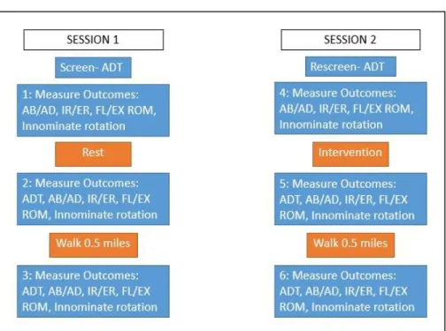
Figure 1. Outline of Data Collection……….………..………26
Figure 2. Model of Sagittal Innominate Rotation A ngle ………..………...27
Figure 3. Model of Transverse Innominate Rotation Angle …………..………..27
Figure 4. Left Adduction Range of Motion (p < 0.001)………...………34
viii
LIST OF TABLES
Table 1. Passive Range of Motion Measurement Procedures……….26
Table 2. Patient Demographic Information………....31
Table 3. Reliability of Range o f Motion and Innominate Position Measures ………..31
Table 4. Changes in Range of Motion in the Sagittal Plane……….32
Table 5. Changes in Range of Motion in the Frontal Plane………...………..32
Table 6. Changes in Range of Motion in the Transvers e Plane………...…33
ix
LIST OF ABBREVIATIONS
3D Three-dimensional
A CL A nterior cruciate ligament A DT A dduction drop test A IC A nterior inferior chain A SIS A nterior superior iliac s pine BMI Body-mass index
CA T Computed axial tomography ICC Inter-class coefficient
LBP Low back pain
LPHC Lumbo-pelvic-hip complex MDC Minimal detectable change MRI Magnetic resonance imagery PEC Pos terior exterior chain PRI Pos tural Restoration Institute PSIS Pos terior s uperior iliac s pine ROM Range of motion
RSA Roentgen s terophotogrammetric analysis SEM Standard error of measurement
1
CHAPTER 1: INTRODUCTION
Sacroiliac joint dysfunction is defined as degenerative change, positional malalignment, or kinematic fault of the sacroiliac joint (SIJ). SIJ dysfunction has been correlated with low back pain and limitation in hip range of motion, which has been identified as a ris k factor for lower extremity injury. Low back pain and lower extremity injury are two common and costly medical conditions; low back pain is estimated to have a lifetime prevalence of 80% in the United States, and more than 50% of all injuries s ustained in collegiate sports are lower extremity injuries.16,26 Sacroiliac joint dysfunction has been hypothesized to be involved in as many as 55%-61.5% of cases of non-specific low back pain.34 Deficit in hip range of motion has been associated with anterior cruciate ligament, hip, groin, and hamstring injuries, and is hypothesized to be involved in injury at the knee and ankle.7,33,48
2
electromagnetic tracking device has been introduced as a reliable and accurate assessment of innominate position and motion.1-5,11-13
Des pite the lack of s upport for palpation-based assessment, these techniques are widely us ed clinically alongside a variety of techniques designed to correct observed malalignments. These techniques include muscle energy techniques and mobilization or manipulation of the SIJ and lumbar s pine. Recently, the Postural Restoration Institute™ (PRI) has popularized a novel approach for assessing positional malalignment of the SIJ. PRI hypothesizes that anterior rotation of the left innominate is a natural resting position of the body, due to asymmetry of the diaphragm and viscera in the abdominal cavity. The PRI approach involves assessing innominate positioning through examination of concomitant changes in hip adduction range of motion. PRI als o proposes a novel s eries of is ometric exercises to correct for this common malalignment, focused on recruitment of abdominal, hamstring, gluteal, and adductor muscles to correct innominate position, which allows for greater hip range of motion. However, research is needed to s upport this theoretical basis of this approach. Namely, the ability of these repositioning exercis es to correct innominate positioning, and the ability of these repositioning exercises to alter hip range of motion must be s upported to validate the widespread clinical use of the technique.27-28
Through the use of the palpation-digitization technique, this study aims to accurately meas ure changes in innominate rotation at the s acroiliac joint after completion of PRI
3 Clinical Significance
This s tudy will build the base of evidence for a popular technique with varied and important clinical uses. If s upported, this technique provides a method to assess and correct positional malalignments that can be a causative fa ctor of low back pain as well as a number of other hip and pelvic pathologies. This technique is clinician-friendly, as it does not require s pecialized equipment and can be adapted to a varied patient population. If s upported, as sessment of pelvic alignment through its effect on range of motion could be a welcome alternative to the inaccurate and unreliable palpation assessment. This technique could also provide a method of correcting asymmetries in hip range of motion, which may decrease the ris k of lower extremity injury.
Research Questions and Hypotheses:
RQ1: Is palpation-digitization using an electromagnetic tracking system a reliable method for as sessing transverse and sagittal innominate rotation?
RH1: W e hypothesize that palpation-digitization using an electromagnetic tracking s ystem will be a reliable method for assessing transverse and sagitt al innominate rotation. RQ2: Does completion of PRI pelvic repositioning technique affect hip range of motion in individuals displaying a left anterior innominate rotation more than rest?
RQ2a: Does completion of PRI pelvic repositioning technique change hip frontal plane range of motion in individuals displaying a left anterior innominate ro tation more than rest?
RH2a: W e hypothesize that completion of PRI pelvic repositioning technique will
4
RQ2b: Does completion PRI pelvic repositioning change hip transverse plane range of motion in individuals displaying a left anterior innominate ro tation more than rest?
RH2b: W e hypothesize that completion of PRI pelvic repositioning technique will increase the angle of left hip internal rotation range of motion and d ecrease the angle of left hip external rotation range of motion, but will not alter total arc transverse plane range of motion in individuals displaying a left anterior innominate ro tation more than res t.
RQ2c: Does PRI pelvic repositioning change hip sagittal plane range of motion in individuals displaying a left anterior inn ominate rotation more than rest?
RH2c: W e hypothesize that completion of PRI pelvic repositioning technique will increase the angle of left hip extension range of motion and decrease t he angle of left hip flexion range of motion, but will not alter total arc sagittal plane range of motion in individuals displaying a left anterior innominate ro tation more than rest.
RQ3: Does completion of PRI pelvic repositioning technique change the a ngles of s agittal and transverse innominate rotation in individuals displaying a left anterior innominate rotation more than rest?
RQ3a: Does completion of PRI pelvic repositioning technique change the angles of sagittal and transverse left innominate rotation in individuals displaying a left anterior innominate rotation more than rest?
5
RQ3b: Does completion of PRI pelvic repositioning technique change the angles of s agittal and transverse right innominate rotation in individuals displaying a left anterior innominate rotation more than rest?
RH3b: W e hypothesize that completion of PRI pelvic repositioning technique will not change the angles of sagittal and transverse right innominate rotation in individuals dis playing a left anterior innominate rotation more than rest.
RQ3c: Does completion of PRI pelvic repositioning technique change the difference between left and right sagittal and transverse innominate rotation angles in individuals displaying a left anterior innominate rotation more than rest?
RH3c: W e hypothesize that completion of PRI pelvic repositioning technique will
decrease the difference between left and right sagittal and transverse innominate rotation angles in individuals displaying a left anterior innominate rotation more than rest. RQ4: Does walking one half mile immediately change left hip range of motion in individuals dis playing a left anterior innominate rotation after completion of PRI repositioning technique?
RQ4a: Does walking one half mile change hip frontal plane range of motion in individuals dis playing a left anterior innominate rotation after completion of PRI repositioning
technique?
RH4a: W e hypothesize that walking one half mile will not change right or left hip
6
RQ4b: Does walking one half mile change hip transverse plane range of motion in individuals displaying a left anterior innominate rotation after completion of PRI repositioning technique?
RH4b: W e hypothesize that walking one half mile will not change right or left hip internal rotation, external rotation, or total arc internal and external rotation range of motion in individuals displaying a left anterior innominate rotation after completion of PRI repositioning technique.
RQ4c: Does walking one half mile change hip frontal plane range of motion in individuals dis playing a left anterior innominate rotation after completion of PRI repositioning
technique?
RH4c: W e hypothesize that walking one half mile will not change right or left hip flexion, extens ion, or total arc flexion and extension range of motion in individuals dis playing a left anterior innominate rotation after completion of PRI repositioning technique.
RQ5: Does walking one half mile immediately change the angle of left or right sagittal and transverse innominate rotation in individuals displaying a left anterior innominate rotation after completion of PRI repositioning technique?
RH5: W e hypothesize that walking one half mile will not change the angles of left or right s agittal and transverse innominate rotation in individuals displaying a left anterior innominate rotation after completion of PRI repositioning technique.
7
RH6: W e hypothesize that completion of PRI repositioning technique will res ult in an increase in the adduction range of motion of the left hip, as examined by a change in
outcome of the left A dduction Drop Test.
RQ7: Does walking one half-mile res ult in a change in left hip adduction range of motion, as examined by a change in outcome of the left Adduction Drop Test, after completion of PRI
repositioning technique?
RH7: W e hypothesize that walking one half-mile will not res ult in a change in left hip adduction range of motion, as examined by a change in outcome of the left A dduction
8
CHAPTER 2: REVIEW OF LITERATURE
The s acroiliac joint (SIJ) is a complex joint with great impact on the kinematics of the lumbo-pelvic-hip complex due to its influence on the positioning of the innominates. Proper mobility at the SIJ is integral for both femoro -acetabular and lumbo-pelvic motion. Low back pain and lower extremity injury are two common and costly medical conditions associated with positional faults of the SIJ. Positional malalignments of the SIJ have historically been diagnosed clinically through palpation techniques, but these tests have been shown to be unreliable,
ins ensitive, and unspecific due to the difficulty in assessing the small magnitude of motion pres ent at the SIJ. There are also many proposed interventions to correct for positional faults, b ut there is a paucity of research supporting the ability of these techniques to alter the bony
positioning of the innominates. Recently, the Postural Restoration Institute™ has popularized a technique to assess and correct positional malalignments at the SIJ. In order to fully understand the mechanics of SIJ movement, this review will explore the functional anatomy of the pelvis, s acrum, and SIJ. This review will then discuss the theoretical and empirical links between the SIJ, low back pain and lower extremity injury, as well as the epidemiology and s ocietal costs of both of these conditions. This review will als o discuss the effectiveness of various techniques currently used to diagnose positional malalignment of the SIJ. Finally, the theoretical basis of the PRI techniques will be explored to make a case for examination of these techniques.
Functional Anatomy of the SIJ
9
However, more recent magnetic resonance imagery (MRI) and microscopic examination has prompted a reevaluation of this classification. Puhakka, et al., describe the SIJ to be a
“symphysis with some characteristics of a synovial joint;” the proximal portion two-thirds of the joint are defined by the thick, intertwined fibrocartilage and ligaments of the ilium and the s acrum, while the distal one-third possesses an “inner capsule with synovial cells,” on MR imagining and microscopy.40 However, a wide variation in SIJ anatomy has been observed. In the s ame s tudy, Puhakka, et al., noted that the transition between the proximal and distal portions of the sacroiliac joint was “microscopically rich in anatomical variants,” including “osseus clefts, cartilage, and subchondral defects.”40 Differences in SIJ anatomy between sexes has also been observed. Articular s urfaces in the female SIJ have been determined to be smoother, s horter, and more angled than in males. These adaptations decrease friction and allow for more motion at the SIJ, which is thought to allow for adaptions in bony geometry in pregnancy and childbirth .13 Thus, significant differences in SIJ range of motion have been observed between males and females .13 Similarly, s ignificant differences in motion have been observed between self-reported “dominant” and contralateral leg, suggesting that some variability in SIJ motion may be d ue to patterns of use.11-13
10
degrees along the Y-axis , and -0.5 to 8.0 degrees along the Z-axis . Translation ranged from -0.3 to 8.0mm along the X-axis , -0.2 to 7.0mm along the Y-axis , and -0.3 to 6.0mm along the Z-axis .22 Due to the complex s hape, articular structure, and anatomy of the SIJ, and despite the s mall magnitude of movement in each plane, these planar motions work in coordination to produce difficult-to-measure three dimensional (3D) motions of the innominate on the sacrum. The s mall magnitude of these motions also suggests that any deviation of the sacrum beyond its narrow range of motion could be a painful and important event.
Four positional malalignments of the innominate associated with SIJ dysfunction have been described: 1.) unilateral anterior tilt of one innominate bone, 2.) unilateral posterior tilt of one innominate bone, 3.) bilateral, antagonistic tilt of both innominates (one posteriorly tilted, one anteriorly tilted), and 4.) bilateral anterior tilt of both innominates, which is referred to as pelvic torsion.14-15 However, the biomechanical causes and effects of these positional faults have been underexamined. There is evidence that pelvic torsion is a common compensation for
individuals with an anatomical leg length discrepancy.15,29 In individuals with positional malalignment but without evidence of a leg length discrepancy, the prevailing theory of
malalignment causation is that a muscular imbalance, coupled with hypermobility of the SIJ, can cause lasting abnormal rotation of one or both innominates.14,47
It is theorized that rotation of the innominate could have effects on femoro-acetabular kinematics by 1.) altering the length-tension relationship of muscles crossing the SIJ and femoro-acetabular joint and 2.) altering the positioning of the acetabulum. The piriformis , which
11
s ome evidence connecting piriformis cross-sectional area, a measure associated with muscular s trength and endurance, and low back pain, s upporting the theory that the piriformis could play an undiscovered role in pelvic bony kinematics.32 The gluteus maximus als o crosses both the SIJ and the femoro-acetabular joint, and thus is hypothesized to be an additional influence on the positioning of the SIJ. The transverse abdominus, among other muscles involved in “local” s tability of the lumbo-pelvic-hip complex, has also been theorized to influence and stabilize the s acroiliac joint, but Gnat, et al., were unable to find a change in innominate rotation with
transverse abdominus contraction.21 Likewise, change to the innominate position could influence the line of force and function of muscles crossing the femoro-acetabular joint. Specifically, alteration to the line of pull of the obdurator internus, an important muscle for s tabilization of the femoro-acetabular joint, could significantly decrease its ability to provid e dynamic stabilization of the femoral head.25
Femoro-acetabular Motion
The femoro-acetabular joint, or hip joint, is the articulation between the innominate and the femur. This joint is a ball-and-socket joint which allows for motion in the transverse, frontal, and sagittal planes. The innominate is comprised of three bones, the ilium, the is chium, and the pubis, which fuse together to form the acetabulum, the s ocket of the hip joint. Motion at the femoro-acetabular joint occurs in three planes: abduction and adduction in the frontal plane, internal and external rotation in the transverse plane, and flexion and extension in the sagittal plane. Measurement of the range of motion in these planes using a goniometer has shown high intra- and inter-rater reliability, but low validity as compared to measurement via
12
Recently, the “total motion concept” has been popularized with regard to shoulder joint motion. This concept involves summing each motion to find the total arc range of motion for each plane. In the shoulder, differences in internal rotation range of motion between the
dominant and non-dominant limb are common. However, the total arc internal and external range of motion remains equal between limbs .50 This principle has been applied to rotational motion at the hip, as hip rotational motion has shown similar adaptations to limb dominance in baseball and tennis players.33 Comparis on of total arc range of motion offers another useful comparison between potentially asymmetric limbs .
The point of limitation in each range of motion may be due to soft tissue approximation, tension in the capsulo-ligamentous or muscular s tructures, or bony approximation. Recently, acetabular position has been found to be an important factor in range of motion limitations. Changes in acetabular position, due to unilateral or bilateral innominate tilt, may influence tension on capsulo-ligamentous structures or approximation of the femoral head-neck junction and the acetabulum. Ross, et al., concluded that dynamic changes in pelvic tilt “significantly influence the functional orientation of the acetabulum,” causing decreases in internal rotation range of motion in s ubjects displaying an anterior pelvic tilt.44 Similarly, Bagwell, et al., found a 1.2-1.6 degree increase of closed-kinetic-chain femoro-acetabular internal rotation for every 5-degree increase in anterior pelvic tilt as well as a converse relationship between posterior pelvic tilt and external rotation.6 This suggests that altered pelvic control or positioning has the
potential to influence transverse plane range of motion at the femoro-acetabular joint.6 Pathology and Epidemiology
non-13
s pecific low back pain. Non-specific low back pain is defined as back pain with no known underlying pathology, which makes up 90%-95% of all diagnosed low back pain.30 Low back pain is one of the most common and most costly musculoskeletal conditions, with point and lifetime prevalence estimated at 15% and 80%, res pectively, and causes an estimated $12.2-$90.6 billion in direct costs and $7.4-$28.2 billion in indirect costs per year in the United States.16 Through confirmation via intra-articular injections, SIJ dysfunction has shown to be involved in a minimum of 15%-30% of low back pain44, but s ome estimates based on clinical provocation tests have found SIJ dysfunction to be involved to as high as 55%-61.5% of low back pain34. In a s tudy of patients suffering from low back pain resulting from SIJ dysfunction, A dhia et al., found significant differences in innominate movement patterns between groups.3-4 W hile it is difficult to separate cause and effect of low back pain of s acroiliac origin and
innominate positional or kinematic abnormalities, there is a clear and strong correlation between the two conditions.
Likewis e, there is evidence of a correlation between limited hip range of motion (ROM), es pecially hip rotation, and low back pain. It is theorized that this correlation could be due to dysfunction of the SIJ, which is the direct connection between the femoro-acetabular joint and the lumbar spine, or that low back pain could be a result of compensation for limitations in rotation at the hip.23 Some have also postulated that muscular imbalances in the piriformis , which crosses both the SIJ and the femoro-acetabular joint, could cause movement deficiencies at both joints .14,47 LaBan, et al., found a more direct correlation between SIJ dysfunction and hip
14
dysfunction and connected this asymmetry to a s pecific positional malalignment of s acroiliac joint. Specifically, patients identified to have a posteriorly rotated innominate via palpation techniques were found to have a greater asymmetry of internal and external rotation on the posteriorly rotated side as compared to the contralateral side.14
Hip rotation range of motion deficits have been identified as s ignificant ris k factors for lower extremity injury, s o any contributing factors and proposed interventions should be thoroughly researched. These deficits may alter the kinematics of the lower extremity,
15 Evaluation Techniques
Diagnosis of SIJ dysfunction can be divided into two categories: diagnosis via diagnostic tools and diagnosis via clinical evaluation. Curren t diagnostic tools used include computerized axial tomography (CA T) s cans, roentgen sterophotogrammetric analysis (RSA ), and Kirschner wires to assess positional and kinematic dysfunction, and intra-articular nerve blocks to diagnose painful conditions of unknown origin.11-13 While each of these methods is accurate, these
methods are all costly, invasive, and impractical in the clinical s etting. The current clinical technique of assessing positional malalignments relies on clinician ability to locate bony s tructures like the ASIS, PSIS, and iliac cres ts through palpation and compare their position. A nother common technique involves bilateral comparison of the medial malleo li, as functional leg length discrepancies are believed to be a compensation with unilateral rotation of the innominate, or that innominate malalignment presents as a compensation for a leg length
dis crepancy.15 Movement faults are clinically assessed via palpation of the s ame s tructures while the patient flexes or extends at the hip. Comparison of these techniques and diagnostic imaging, s uch as RSA, has proven these techniques to be unreliable and inaccurate. Further attempts at objectifying the difference in bony landmark positioning, such as use of inclinometers or potentiometers, have proven similarly inadequate.1-5,11-13,22
16
s hown to be valid and to possess a high level of intra-rater reliability.1-3 However, there is not currently enough evidence to confirm inter-reliability of the technique.1-3
Clinical Interventions
Manipulations of the SIJ and lumbar s pine and specific manual therapy techniques, such as muscle energy techniques, have been commonly used to correct positional malalignments clinically. Cibulka et al., found significant displacement in pelvic landmarks before and after manipulation of the SIJ, but the SIJ position in this s tudy was ass essed via palpation, not via the objective measures discussed above.14 Muscle energy techniques utilize is ometric contractions of the muscles acting on the pelvis to restore a neutral position. Commonly, these include the hip adductors and abductors, as well as the hamstrings and quadriceps. These techniques are highly variable across clinicians, making unbiased clinical trials difficult to conduct. While these techniques are popular in the clinical setting, there is no evidence to support use of these
techniques to improve patient outcomes. To date, there is no examination on the effect of muscle energy technique on the SIJ positioning.20
Theoretical Basis of Postural Restoration
17
The theoretical basis of the PRI approach centers on the body’s inherent asymmetry and res ulting patterned neuromuscular imbalances. PRI postulates that the asymmetry of the
diaphragm, lungs, heart, brain, and visual s ystem creates a predictable pattern of muscular imbalances and dysfunctional movements throughout the body.27-28,42 PRI identifies these imbalances as over-activity of certain poly-articular chains of muscle, which present in predictable and uniform ways across all populations without a congenital condition altering organ position, such as situs inversus.27-28 Two of the proposed muscular imbalances, dominance of the left anterior inferior chain (Left A IC) or posterior exterior chain (PEC), pres ent with unilateral or bilateral anterior rotation of the innominate, respectively.27-28 The PRI postulates that muscle activity and range of motion in the sagittal, frontal, and transverse planes at the hip joint are altered by this malalignment, and restoration of proper alignment is the cornerstone to s uccessful treatment of all patients presenting with issues in the lower extremity. Despite the exciting, global claims made and taught by the PRI, there is very little evidence to s upport these claims . The anatomical effects and patient o utcomes must be researched before adoption of s uch techniques.
Postural Restoration Evaluation and Intervention
18
the top limb is extended to zero degrees and passively adducted towards the table. Inability to adduct is evidence of malalignment of the innominate, a predictable patterned response to the proposed muscle chain imbalances. A series of case reports has s upported this claim, but no clinical testing comparing adduction range of motion in this testing position to innominate position has been conducted.8,27-28,42
The PRI als o proposes novel techniques to correct for this malalignment. PRI repositioning techniques, the centerpiece of their approach, are presented for each of the
common muscle imbalances proposed. Treatment of the left AIC pattern, which is said to be the mos t common, involves is ometric contraction of the hamstrings and abdominals with
diaphragmatic breathing, and activation exercis es for the left adductor group and right gluteus maximus .27-28 Successful implementation of these exercises is stated to correct innominate malalignment and restore proper range of motion at the hip joint.27-28 This change s hould be immediate, and can quickly be assessed by recompletion of the Adduction Drop Test. Similarly, this effect has not been well-evidenced outside of case reports.8,43 In one non-controlled s tudy of the effect of the repositioning technique on patients with lumbopelvic pain, s ignificant
19
CHAPTER 3: METHODOLOGY
Research Design
This s tudy was conducted as a cross-sectional within-individuals comparison s tudy. Each s ubject completed a control s ession and an interventional s ession. Passive hip range of motion and innominate rotation measurements were compared within individuals before and after completing an intervention of PRIrepositioning exercises.
Participants
Twenty-four individuals, aged 18-35, who were in good general health, participated in this study. Participants were free of hip or low back injury at the time of data collection and for a minimum of s ix months prior to data collection and participated in a minimum of thirty minutes of exercis e three times in one week. Participants were excluded if they reported a history of low-back or hip surgery, a current, s ymptomatic hip or low low-back injury or a his tory of s uch injury in the last six months, a leg length discrepancy of greater than 2 cm, or reported a pregnancy or the possibility of pregnancy. All participants were screened for innominate rotation before
recruitment; only participants who presented with left A nterior Interior Chain dominance, or left anterior innominate rotation, as defined by the Postural Restoration Institute were recruited for data collection. All participants read and signed an informed consent form approved by the Ins titutional Review Board of the University of North Carolina at Chapel Hill.
Instrumentation
20
Data was collected using Motion Monitor capture and analysis software via an A/D board. Lower extremity passive range of motion was measured using a digital inclinometer (Sanders Group, Inc., Chaska, MN, USA) and a standard 8-inch plastic goniometer.
Procedures
Participants reported to the Sports Medicine Research Laboratory for a s creening session and returned within one week for two consecutive days of testing sessions wearing their own athletic shorts, s hirt, and shoes suitable for walking one-half mile. Each participant completed the Control Session on the firs t day of data collection and returned for the Intervention Session on the following day. Data collection was scheduled as close to exactly twenty-four hours apart as s chedules allowed; all data collection was conducted later than twenty-two hours after the start of the firs t s ession and earlier than twenty-six hours after the start of the firs t session. An outline of the data collection procedure can be visualized in figure 1.
Screening Protocol
The left Anterior Interior Chain dominance screening protocol consisted of three trials of the A dduction Drop Test, as defined in the Myokinematic Restoration course manual (Hruska), on each limb. The participant laid on his or her s ide with the hip and knee of the lower leg flexed to 90 degrees. The examiner passively flexed the knee to 90 degrees and passively flexed,
21
Participants with three consecutive positive tests on the left limb and three consecutive negative tes ts on the right limb were considered for data collection.
Experimental Protocol
Prior to the start of each session, participants were outfitted with electromagnetic sensors over the sacrum and the lateral aspect of each thigh. Sacral s ensor position was determined by bis ecting a line between the right and left PSIS; the s ensor was fixed to a one -inch by one-inch s quare of Orthoplast and affixed to the sacrum one inch below this line. Bilateral PSIS and the s uperior and inferior borders of the Orthoplast were marked with dry-erase marker to ensure proper replacement of the sensor. Sensors were secured with athletic tape. The x-y-z global axes were established according to the right-hand three-dimensional Cartesian coordinate system. The positive x-axis was designated forward, the positive y -axis to the left, and the positive z-axis upward, relative to the participant. The ASIS and PSIS were palpated and digitized bilaterally us ing MotionMonitor s oftware; three-dimensional coordinates of these bony landmarks were es timated using MotionMonitor s oftware. A s ingle examiner conducted all palpation and digitization of the ASIS and PSIS. A ll s ensors were removed in between data collection time -points.
Innominate Rotation Angles
22
the s tylus, and a trial of these location data was recorded. Data was s ampled at 140Hz for a five-s econd window; the Cartefive-sian coordinatefive-s of each bony landmark were five-sampled at the midpoint of five s econds of neutral s tance. From these coordinates, vectors were calculated between each A SIS and PSIS and both PSIS in the transverse and s agittal planes. After recording, the s ubject was re-digitized for three more measures, and vectors were recalculated; the average of four innominate rotation angles was used for each assessment.
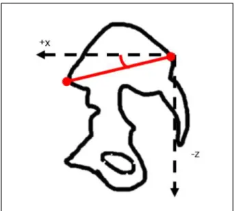
The angle of sagittal innominate rotation was defined as the angle between the A SIS-PSIS vector and the x-axis in the s agittal plane. The angle of transverse innominate rotation was defined as the angle between the A SIS-PSIS vector and a line perpendicular to the PSIS-PSIS vector in the transverse plane. Vis ual representations of these angles are presented in Figures 2 and 3, res pectively. Both angles were recorded for the right and left hips. The difference between right and left transverse and sagittal innominate rotation angles was calculated and recorded.
Passive Range of Motion and Adduction Drop Test
Passive ranges of motion were measured for hip internal rotation, hip external rotation, hip abduction, hip adduction, hip flexion, and hip extension. The testing procedures utilized for each range of motion measurement are described in Table 1. Each range of motion measurement was conducted three times; the mean of each measure was recorded. The Adduction Drop Test was conducted as described in the s creening protocol. A single examiner, trained and
experienced in analysis of lower extremity range of motion, measured all hip ranges of motion throughout the data collection protocol.
Experimental Protocol: Control Session
23
The participant then rested on the plinth for a period of twenty minutes, equal to the time of the intervention. The examiner then re-measured all outcome measures. The participant then walked on a treadmill at a s elf-selected speed for one half-mile. The participant returned to the plinth, and the examiner measured outcome measures for the final time. A ll s ensors were removed from the s ubject between assessments. The s ubject was instructed to avoid high-intensity physical activity or physical activity that deviates from their normal exercise s chedule in between data collection sessions.
Experimental Protocol: Intervention Session
The intervention session for each participant began with measurement of each outcome meas ure: innominate rotation angles, passive hip range of motion, and the adduction drop test. A s ingle examiner led all intervention exercises. This examiner was certified as completing the Myokinematic Restoration and Pelvis Restoration courses through the Postural Restoration Ins titute and had three years of experience in prescribing and directing PRI intervention
exercis es. Firs t, the examiner instructed the subject to take three deep breaths in a s upine, hook-lying position. The examiner viewed the subject’s breathing technique and provided cues to improve diaphragmatic breathing, such as “breathe in through your nose and out through your mouth,” “have your chest and stomach rise and fall together,” “exhale as if you we re blowing up a balloon,” and “think about rounding out your spine into the table.” The subject then completed three more diaphragmatic breaths to ensure retention of cuing. The examiner then led the
participant through an intervention of three exercises d esigned to correct left anterior innominate rotation.
24
The 90-90 Hip Lift was completed with the participants’ heels resting on a wooden block; the participant then performed isometric bilateral hamstring contraction and posterior pelvic tilting while completing diaphragmatic breathing. The Right Sidelying Left A dductor Pull Back was completed in a hook-lying position on the subject’s right side, with the participant completing is ometric left adductor contraction with diaphragmatic breathing. The Left Sidelying Right Glute Max was completed in a hook-lying position on the patient’s left s ide, with the participant completing is ometric right gluteus maximus contraction into a resistance band (Theraband, A kron, OH, USA) with diaphragmatic breathing. Four sets of four breaths (repetitions) of each exercis e were completed. The examiner instructed the s ubject directly from the Myokinematic Res toration Manual.27 During each exercise, the examiner provided feedback, s uch as cuing to expel all air from lungs, round out the back, and maintain is ometric contractions, during each exercis e to ensure proper completion.
A fter the intervention, the examiner re -measured all outcome measures. The participant then walked on a treadmill at a s elf-selected s peed for one half-mile, equal to the speed s elected during the control s ession. The participant returned to the plinth, and the examiner measured outcome measures for a final time. A ll s ensors were removed from the subject between as sessments.
Statistical Analysis
A n interclass correlation coefficient (ICC) was calculated for each measure within and
between days of measurement. Coefficients of 0.75 or higher considered to be good-to-excellent,
and coefficients of 0.75 or lower considered to be poor-to-moderate. The within-session ICC was
25
calculated between the pre-control and pre-intervention time-points. These ICC values were used in analysis of research question 1.
For res earch questions 2 through 5, two-way (time-point, session) repeated measures
A NOVA s were used to analyze the variance between time-points of collection for both the hip
range of motion measures and the innominate rotation angle measures. Bonferroni pairwise
comparisons were used to further determine differences in the presence of a s ignificant time x
intervention interaction. The alpha level for inferential s tatistical tests w as set at p = 0.05.
The s tandard error of measurement (SEM) and minimal detectable change (MDC) was
als o calculated to provide a measure of variability. SEM values we re calculated as follows: SEM
= SDpooled x √(1-ICC), with SDpooled representing the s tandard deviation of the measure. The
MDC reflects the magnitude of change necessary to provide confidence that any change is not
due to random variation or measurement error. MDC values were calculated as follo ws: MDC =
1.96 x SEM x √2.
For res earch questions 6 and 7, we ran Fis her's exact test of independence to determine
the interaction between intervention and Adduction drop test to compare the expected and
26
Table 1. Passive Range of Motion Measurement Procedures
Range of Motion Measurement Participant Body Position
Lower Extremity Position Passive Range of Motion Goniometer/ Inclinometer
Hip internal rotation
Prone Knee flexed to 90° angle
Femur internally rotated
Digital inclinometer perpendicular to medial tibia Hip external
rotation
Prone Knee flexed to 90° angle
Femur externally rotated
Digital inclinometer perpendicular to lateral tibia Hip extension Prone Leg straight Femur
extended Goniometer aligned across lateral midline of pelvis and femur Hip flexion Supine Knee maximally
flexed Femur flexed Goniometer aligned across lateral midline of pelvis and femur Hip adduction Supine Leg straight;
contralateral limb maximally abducted
Femur
adducted Goniometer aligned across ASIS and femur
Hip abduction Supine Leg straight Femur
abducted Goniometer aligned across ASIS and femur
27 Figure 2. Model of Sagittal Innominate Rotation Angle
28
CHAPTER 4: RESULTS
Data from all twenty-four participants (n=24) were used in the final analyses of hip range of motion and sagittal innominate rotation angles. Demographic data for the sample is lis ted in Table 2.
Data from four participants were not included in the final analyses of left and right transverse innominate rotation angles due to measurement error. Likely calibration error of the Flock of Birds s ystem caused inaccurate recording of Cartesian coordinates of the A SIS and PSIS four individuals. Calculation of the transverse innominate rotation angle from these data revealed impossible angle measures, such as transverse innominate rotation angles of above 180º, and these data were excluded in further s tatistical analysis. The corresponding sagittal innominate rotation angles for these s ubjects fell within the realm of anatomic possibility and were not excluded.
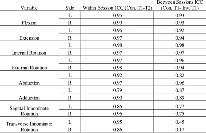
Reliability (RQ1)
29
W ithin- and between-session ICC values for all range of motion measures were considered good-to-excellent (ICC > 0.75). ICC values are lis ted in Table 3.
Range of Motion (RQ2 & RQ4)
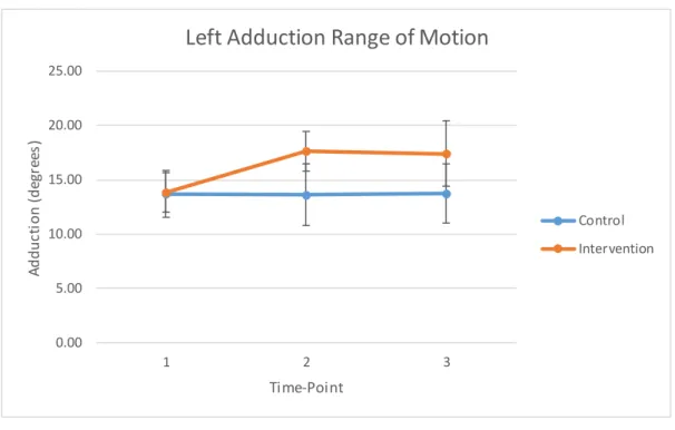
Completion of the PRI pelvic repositioning technique affected hip range of motion of the left limb in the frontal plane more than the control condition. A s ignificant interaction (p < 0.001) in left hip adduction range of motion was shown between the pre-intervention (µ = 13.85º ± 2.15º) and post-intervention (µ = 17.64º ± 2.84º) time-points, as compared to the difference between the pre-control (µ = 13.71º ± 1.82º) and post-control time points (µ = 13.64º ± 1.82º). This change (µ = 3.79º ± 2.45º) exceeded the minimal detectable change (MDC) of the measure (MDC = 2.41º). This finding was associated with a large effect size (1.85, 95% CI [1.17, 2.52]) as s een in figure 3. No difference in adduction range of motion was observed on the right limb after completion of the intervention.
Similarly, a s ignificant (p = 0.02) difference in left total arc frontal plane range of motion was seen between the pre-intervention (µ = 59.72º ± 6.59º) and post-intervention (µ = 64.35º ± 6.47º) time-points, as compared to the change between the pre- (µ = 60.64º ± 7.21º) and post-control time-points (µ = 60.00º ± 6.83º). This finding was associated with a large e ffect s ize (1.30) but the intervention change (µ = 4.63º ± 3.61º) did not exceed the calculated MDC s core (6.74º). This change is illus trated in Figure 4. No difference in total arc frontal plane range of motion was observed on the right limb after the completion of the intervention.
post-30
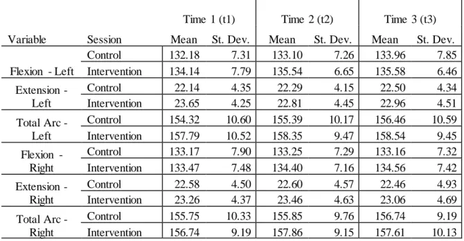
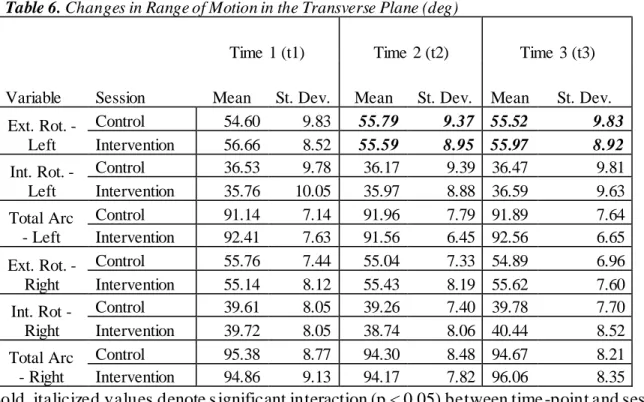
intervention (µ = 55.59º ± 8.95º) time-points. However, this difference between pre- and post-intervention (µ = -1.063º ± 3.07º) was well below the MDC (MDC = 4.93) of th is measure. Hip internal rotation, total arc transverse plane range of motion, hip flexion, hip extension, or total arc s agittal plane range of motion were not different between the control and intervention s essions at any time points. These data can be found in Tables 4, 5, and 6.
There was no change observed in any range of motion after completion of the walking tas k. Pairwise comparisons of the post-intervention time-point (Time 2) and the post-walking time-point (Time 3) in left hip adduction range of motion and left total arc frontal plane range of motion revealed no significant difference (p > 0.05).
Innominate Rotation Angles (RQ 3 & 5)
A paired samples t-test revealed a s ignificant difference (p = 0.002) between the mean left s agittal innominate rotation angle (µ = -11.2º ± 4.77º) and right s agittal innominate rotation angle (µ = -8.56º ± 8.70º) at the beginning of the control session. Omnibus repeated measures A NOVA analyses did not show significant Session x Time interactions in the sagittal or
transverse innominate rotation angles on the left or right limb , or in the difference between right and left s agittal and transverse angles. Further pairwise comparison between time -points 1, 2, and 3 s howed no significant differences in either s agittal or transverse innominate rotation angles before and after completion of the intervention and walking task, respectively. These data can be found in Table 7.
Adduction Drop Test (RQ 6 & 7)
31
participants (83.5%) s howed a change from a positive A DT to a negative ADT in the left leg. A fter completion of the walking task, one participant experienced a change of left A DT from negative to positive; nineteen of the twenty -four participants (79.2%) dis played a negative ADT on the left leg after completion of the walking task. All twenty-four participants presented with a negative A DT on the right leg after the intervention and after the walking task. Analysis via Fisher’s Exact Test revealed this change to be statistically significant at both Time 2 and Time 3 (p = 0.00).
Table 2. Patient Demographic Information
n Height (m) Weight (kg) Age (yrs)
Combined 24 1.69 ± .09 67.08 ± 12.38 21.46 ± 1.35
Males 11 1.76 ± .06 75.48 ± 12.32 21.82 ± 1.25
Females 13 1.62 ± .06 59.97 ± 6.91 21.15 ± 1.40
Table 3. Reliability of Range of Motion and Innominate Position Measures
Within Session ICC (Con. T1-T2)
Between Sessions ICC (Con. T1- Inv. T1)
Variable Side
Flexion
L 0.95 0.93
R 0.99 0.93
Extension
L 0.96 0.92
R 0.97 0.94
Internal Rotation
L 0.98 0.98
R 0.97 0.97
External Rotation
L 0.97 0.96
R 0.98 0.94
Abduction
L 0.92 0.82
R 0.97 0.96
Adduction
L 0.79 0.87
R 0.90 0.89
Sagittal Innominate Rotation
L 0.86 0.77
R 0.96 0.75
Transverse Innominate Rotation
L 0.95 0.45
32
Table 4. Changes in Range of Motion in the Sagittal Plane (deg)
Time 1 (t1) Time 2 (t2) Time 3 (t3)
Variable Session Mean St. Dev. Mean St. Dev. Mean St. Dev.
Flexion - Left
Control 132.18 7.31 133.10 7.26 133.96 7.85
Intervention 134.14 7.79 135.54 6.65 135.58 6.46
Extension - Left
Control 22.14 4.35 22.29 4.15 22.50 4.34
Intervention 23.65 4.25 22.81 4.45 22.96 4.51
Total Arc - Left
Control 154.32 10.60 155.39 10.17 156.46 10.59 Intervention 157.79 10.52 158.35 9.47 158.54 9.45
Flexion - Right
Control 133.17 7.90 133.25 7.29 133.16 7.32
Intervention 133.47 7.48 134.40 7.16 134.56 7.42
Extension - Right
Control 22.58 4.50 22.60 4.57 22.46 4.93
Intervention 23.26 4.37 23.46 4.63 23.06 4.69
Total Arc - Right
Control 155.75 10.33 155.85 9.76 156.74 9.19
Intervention 156.74 9.19 157.86 9.15 157.61 10.13
Table 5. Changes in Range of Motion in the Frontal Plane (deg)
Time 1 (t1) Time 2 (t2) Time 3 (t3)
Variable Session Mean St. Dev. Mean St. Dev. Mean St. Dev.
Abduction - Left
Control 46.93 6.97 46.36 6.35 46.64 5.84
Intervention 45.88 6.06 46.71 5.65 45.72 6.37
Adduction - Left
Control 13.71 1.82 13.64 1.82 13.75 3.01
Intervention 13.85 2.15 17.64 2.84 17.43 2.72
Total Arc - Left
Control 60.64 7.21 60.00 6.83 60.39 6.40
Intervention 59.72 6.59 64.35 6.47 63.15 6.92
Abduction - Right
Control 44.24 7.20 44.33 6.46 44.31 6.77
Intervention 44.83 7.27 45.57 6.33 45.44 6.27
Adduction - Right
Control 19.03 2.80 18.42 2.82 18.31 3.04
Intervention 19.06 3.55 18.93 2.65 19.07 3.28
Total Arc - Right
Control 63.27 8.44 62.75 7.29 62.61 7.82
Intervention 63.89 8.02 64.50 7.13 64.51 7.13
33
Table 6. Changes in Range of Motion in the Transverse Plane (deg)
Time 1 (t1) Time 2 (t2) Time 3 (t3)
Variable Session Mean St. Dev. Mean St. Dev. Mean St. Dev.
Ext. Rot. - Left
Control 54.60 9.83 55.79 9.37 55.52 9.83
Intervention 56.66 8.52 55.59 8.95 55.97 8.92
Int. Rot. - Left
Control 36.53 9.78 36.17 9.39 36.47 9.81
Intervention 35.76 10.05 35.97 8.88 36.59 9.63
Total Arc - Left
Control 91.14 7.14 91.96 7.79 91.89 7.64
Intervention 92.41 7.63 91.56 6.45 92.56 6.65
Ext. Rot. - Right
Control 55.76 7.44 55.04 7.33 54.89 6.96
Intervention 55.14 8.12 55.43 8.19 55.62 7.60
Int. Rot - Right
Control 39.61 8.05 39.26 7.40 39.78 7.70
Intervention 39.72 8.05 38.74 8.06 40.44 8.52
Total Arc - Right
Control 95.38 8.77 94.30 8.48 94.67 8.21
Intervention 94.86 9.13 94.17 7.82 96.06 8.35
Bold, italicized values denote s ignificant interaction (p < 0.05) between time -point and session. This interaction was observed at both time -point 2 and time-point 3.
Table 7. Changes in Innominate Position Angles (deg)
Time 1 Time 2 Time 3
Variable Session Mean St. Dev Mean St. Dev Mean St. Dev Left Sagittal Control -11.18 4.76 -10.94 5.57 -10.89 4.56 Inn. Angle Intervention -10.07 3.66 -9.99 5.09 -10.26 4.66
Right Sagittal Control -8.61 8.88 -8.12 9.45 -7.56 8.02 Inn. Angle Intervention -9.31 5.47 -9.21 5.77 -8.74 6.03
Sagittal Plane Control -2.57 7.12 -2.82 6.54 -3.33 7.50 Difference Intervention -0.77 3.80 -0.77 4.14 -1.52 4.29
Left Trans. Control 22.55 5.38 22.08 6.65 21.76 7.75
Inn. Angle Intervention 21.50 5.55 21.96 4.79 22.46 5.06
Right Trans. Control 32.53 7.91 31.08 9.80 29.39 9.70
Inn. Angle Intervention 32.94 8.15 30.01 6.21 29.79 7.90
Trans. Plane Control 9.98 8.21 9.01 11.56 7.63 10.53
34
Figure 4. Left Adduction Range of Motion (p < 0.001)
Figure 5. Left Frontal Plane Range of Motion (p = 0.02)
0.00 5.00 10.00 15.00 20.00 25.00
1 2 3
A d d u ct io n ( d eg re es ) Time-Point
Left Adduction Range of Motion
Control Intervention 50.00 55.00 60.00 65.00 70.00 75.00
1 2 3
Fr o n ta l P la n e M o ti o n ( d eg re es ) Time-Point
Total Arc Frontal Plane Range of Motion
Control
35
CHAPTER 5: DISCUSSION
The purpose of this investigation was to explore the effects of an intervention of rehabilitation exercis es, designed by the Postural Restoration Institute, on hip range of motion and innominate rotation. We found that completion of this intervention was associated with an increase in adduction range of motion and total arc frontal plane range of motion on the left hip. W e did not find any association between co mpletion of the intervention and innominate rotation angle in either the sagittal or transverse plane.
Reliability (RQ1)
Before we could assess any change in s agittal and transverse innominate rotation angles due to the intervention, it was vital to establish the reliability of the palpation-digitization technique at measuring these angles. Within-session reliability measures for both left and right s agittal and transverse innominate rotation were assessed as good-to-excellent. This provides confidence that any changes observed in innominate rotation angles during a single session were not due to the measurement technique. Our strong reliability measures also support our
hypothesis that palpation-digitization using an electromagnetic tracking system is a reliable technique for assessing innominate rotation in the s agittal and transverse planes, aligning with prior res earch that s upports the strong reliability of a palpation-digitization technique to assess changes in innominate rotation within a single data collection session.1-3,11-13
between-36
s ession reliability did not affect the s tatistical analysis of this study, as our analysis explored changes within each data collection session. However, this does weaken support for the use of an electromagnetic palpation-digitization technique as an accurate, non-invasive method of
as sessing innominate position.
There are a number of possible reasons for this poor between-session reliability in the transverse plane. As discussed earlier, there is sparse research related to the assessment of innominate position over time, and it is possible that the transverse innominate rotation angle is inherently variable between days. While the examiner instructed the participants to abstain from unusual physical activity between data collection s essions, there was no attempt to control for normal physical activity before each session. It is possible that the transverse innominate rotation angle would be different between days if the subject spent one morning s leeping in and the next on his or her feet. It is als o possibility that this poor reliability is due to the inconsistency of palpation in the examiner. The limitations of an examiner at accurately assessing innominate position through palpation of bony landmarks h ave been s hown repeatedly in prior research. 1-5,11-13,22
Range of Motion and Adduction Drop Test (RQ2, RQ4, RQ6, & RQ7)
The hypothesized effect of the repositioning exercise series on left hip adduction range of motion, as measured by both goniometry and the left Adduction Drop Test, was observed. This concurs with previous research and case s eries that have shown a change in left adduction ran ge of motion,8,42-43 and provides support for the theoretical claims of the Postural Res toration Ins titute.27-28
37
with the contralateral limb. This repositioning of the innominate bone also changes the
orientation of the acetabulum to the femoral head, allowing for an increase in adduction range of motion as an improved innominate position is achieved.
Des pite the present effect of the PRI intervention on left adduction range of motion, we did not observe any concomitant changes in s agittal or transverse plane range of motion that were hypothesized to accompany a change in the position of the acetabulum. Changes in acetabular position via anterior pelvic tilt have previously been associated with changes in
internal rotation range of motion,44 but no changes in internal rotation measures were observed in this study. We hypothesized that we would observe changes in the flexion, extension, abduction, internal rotation, and external rotation ranges of motion due to the change in acetabular position. W e also hypothesized that these changes of range of motion would not alter the total arc ra nge of motion in any one plane; an increase in adduction range of motion would be concomitant with a decrease in abduction range of motion. We did not observe any evidence to s upport these hypotheses. The significant increase shown in left adduction range of motion was not accompanied by a decrease in left abduction range of motion.
The changes observed in adduction range of motion, as measured by goniometry and by the A DT, remained present after walking one-half mile. Des pite the s hort duration and low intensity of this activity, this retention provides evidence that changes brought on by the PRI intervention last beyond the treatment table. Further research is required to assess how long these changes are retained, and if changes are retained after modera te- to high-intensity activity.
Innominate Position (RQ3 & RQ5)
38
date, this is the firs t data connecting biometric analysis of innominate position with the A dduction Drop Test, the PRI’s surrogate measure of innominate position. However, as
participants were required to have a positive ADT indicating left anterior inn ominate rotation to participate in this study, no comparison of these values to a population displaying s ymmetric innominate rotation was conducted.
A fter intervention, no changes in innominate rotation angle in the sagittal or transverse planes were observed. Prior exploration of innominate position using the palpation-digitization technique has only examined change in innominate position between varying femoro-acetabular positions. This is the firs t s tudy using this technique to quantify innominate position in a neutral s tance. As there has also been no prior research quantifying the effect of any intervention to correct for pelvic position, it is unclear how large a magnitude of a change in innominate position is to be expected. The MDC of our s ample was 4.53º in the s agittal plane and 6.58º in the transverse plane; it is possible that the hypothesized change in innominate position occurred, but we were unable to detect it using the palpation-digitization technique.
Alternate Mechanisms
It is als o possible that the observed increase in adduction range of motion is achieved by another mechanism entirely. Diaphragmatic breathing is a widely -used relaxation technique that has been shown to increase parasympathetic nervous system activity and decrease mus cle tone.9 However, effects from this systemic muscular relaxation would have been observed in all ranges of motion. During the Right Sidelying Adductor Pull Back exercis e, activation of the left
39
s erve as a form of proprioceptive neuromuscular facilitation (PNF), increasing the extensibility of the hip abductor muscles and allowing for greater adduction range of motion.24
A nother possible mechanism of increasing hip adduction range of motion is an alteration of the arthrokinematics at the femoro-acetabular joint. While the proposed mechanism of the PRI involves changing the position of the acetabulum via change of innominate position at the SIJ, it is possible that these exercises affect the position of the femoral head in the acetabulum.
Res earch has shown that exercises that correct anterior translation of the humeral head improve s houlder range of motion by restoring proper arthrokinematic motion to the glenohumeral joint.10 It is possible that the change observed in this study was due to the repositioning of the femoral head in the acetabulum, allowing for the proper inferior glide and superior roll required for adduction. Further studies are needed to explore these potential alternative mechanisms. Limitations
There was no attempt at blinding the investigator or participants in this study design. The principal investigator also implemented the intervention exercise series. Thus, bias could have been present in evaluation of range of motion measures after completion of the intervention s eries. Further s tudies should blind the investigator to the session (intervention v. control). As previously discussed, the use of the palpation-digitization system to evaluate innominate position in the neutral s tance has not been well s upported. While the observed reliability within each s ession was strong, further research is needed to compare this technique to computed tomography or radiography in order to validate the use of this technique. Comparison to
40
palpation-digitization technique to capture an accurate measure of transverse innominate rotation.
Prior res earch and the theoretical basis of the PRI have acknowledged the three-dimensional nature of movement of the innominate on the sacrum. Ou r s tudy only examined innominate position in the sagittal and transverse planes. Potentially, the change in adduction range of motion could have been due to repositioning of the innominate in the frontal plane. A nalysis of frontal plane motion would likely involve comparison of different bony landmarks of the pelvis, such as the is chial tuberosities or the pubic bones. Due to the depth of these
landmarks, palpation-digitization does not appear to be a feasible technique for measuring innominate position in the frontal plane.
Likewis e, any motion of the innominate occurring at the SI joint is inherently a tri-planar motion. To study this motion, we attempted to reduce this tri-planar motion to its planar
components. It is possible that a change could be observed in a combined, three-dimensional vector of innominate motion that was unable to be detected in the sagittal or transverse planes, individually. Assessing three-dimensional motion of the innominate would require advanced analysis in linear algebra and thus was beyond the scope of this study.
41 Clinical Implications
These results provide strong support for use of the PRI repositioning exercis e series to increase hip adduction range of motion, and the retention of these changes after the walking task s upport the claim that this intervention can provide short-term benefits beyond the treatment table. A change in left A dduction Drop Test result was observed in 83.5 percent of participants, providing a clear and easily observable change within one treatment. This intervention does not require expensive equipment and can be applied to a wide range of patient populations.
However, this study was done on a sample of healthy, active, college-aged s ubjects. Participants who reported back pain or hip pain that had limited athletic activity in the past s ix months were excluded from this study. Thus, this study cannot offer any recommendation for or against using the PRI intervention in the treatment of patients with low back or hip pain.
42 REFERENC ES
1. A dhia, D. B., Bus s ey, M. D., Mani, R., Jayakaran, P., A ldabe, D., & Milosavljevic, S. (2012). Inter-tester reliability of non-invasive technique for measurement of
innominate motion. Manual Therapy, 17(1), 71–76. https://doi.org/10.1016/j.math.2011.09.005
2. A dhia, D. B., Bus s ey, M. D., Ribeiro, D. C., Tumilty, S., & Milos avljevic, S. (2013). Validity and reliability of palpation-digitization for non-invasive kinematic
meas urement - A s ystematic review. Manual Therapy, 18(1), 26–34. https://doi.org/10.1016/j.math.2012.06.004
3. A dhia, D. B., Mani, R., Milos avljevic, S., Tumilty, S., & Bus sey, M. D. (2016). Do es repeated palpation-digitization of pelvic landmarks for measurement of innominate motion introduce a systematic error? - A ps ychometric investigation. Manual Therapy, 21, 282–286. https://doi.org/10.1016/j.math.2015.09.009
4. A dhia, D. B., Milos avljevic, S., Tumilty, S., & Bus sey, M. D. (2016). Innominate movement patterns, rotation trends and range of motion in individuals with low back pain of s acroiliac joint origin. Manual Therapy, 21, 100–108.
https://doi.org/10.1016/j.math.2015.06.004
5. A dhia, D. B., Tumilty, S., Mani, R., Milos avljevic, S., & Bus sey, M. D. (2016). Can hip abduction and external rotation discriminate sacroiliac joint pain? Manual Therapy, 21, 191–197. https://doi.org/10.1016/j.math.2015.08.002
6. Bagwell, J. J., Fukuda, T. Y., & Powers, C. M. (2016). Sagittal plane pelvis motion influences transverse plane motion of the femur: Kinematic coupling at the hip joint.
Gait and Posture, 43, 120–124. https://doi.org/10.1016/j.gaitpost.2015.09.010 7. Bedi, A., W arren, R. F., W ojtys, E. M., Oh, Y. K., A s hton-Miller, J. a., Oltean, H., &
Kelly, B. T. (2014). Res triction in hip internal rotation is associated with an increased ris k of ACL injury. Knee Surgery, Sports Traumatology, Arthroscopy, 1–8.
https://doi.org/10.1007/s00167-014-3299-4
8. Boyle, K. L. (2011). Managing a female patient with left low back pain and sacroiliac joint pain with therapeutic exercis e: A case report. Physiotherapy Canada, 63(2), 154–163. https://doi.org/10.3138/ptc.2009-37
9. Brannon Russell, M. E., Hoffman, B., Stromberg, S., & Carls on, C. R. (2014). Us e of Controlled Diaphragmatic Breathing for the Management of Motion Sickness in a Virtual Reality Environment, 269–277. https://doi.org/10.1007/s 10484-014-9265-6 10. Bullock, M. P., Foster, N. E., & W right, C. C. (2005). Shoulder impingement : the
43
11. Bus sey, M. D., Yanai, T., & Milburn, P. (2004). A non-invasive technique for as sessing innominate bone motion. Clinical Biomechanics, 19(1), 85–90. https://doi.org/10.1016/j.clinbiomech.2003.09.005
12. Bus sey, M. D., Bell, M. L., & Milos avljevic, S. (2009). The influence of hip abduction and external rotation on sacroiliac motion. Manual Therapy, 14(5), 520– 525. https://doi.org/10.1016/j.math.2008.08.009
13. Bus sey, M. D., Milosavljevic, S., & Bell, M. L. (2009). Sex differences in the pattern of innominate motion during passive hip abduction and external rotation. Manual Therapy, 14(5), 514–519. https://doi.org/10.1016/j.math.2008.09.004
14. Cibulka, M. T., Delitto, a, & Koldehoff, R. M. (1988). Changes in innominate tilt after manipulation of the sacroiliac joint in patients with low back pain. An experimental s tudy. Physical Therapy, 68(9), 1359–1363.
15. Cooperstein, R., & Lew, M. (2009). The relationship between pelvic torsion and anatomical leg length inequality: a review of the literature. Journal of Chiropractic Medicine, 8(3), 107–118. https://doi.org/10.1016/j.jcm.2009.06.001
16. Dagenais, S., Caro, J., & Haldeman, S. (2008). A s ystematic review of low back pain cost of illness studies in the United States and internationally. Spine Journal, 8(1), 8– 20. https://doi.org/10.1016/j.s pinee.2007.10.005
17. Dalton, S. L., Kerr, Z. Y., & Dompier, T. P. (2015). Epidemiology of Hamstring Strains in 25 NCA A Sports in the 2009-2010 to 2013-2014 A cademic Years . The American Journal of Sports Medicine, 43(11), 2671–9.
https://doi.org/10.1177/0363546515599631
18. Ellera Gomes , J. L., Palma, H. M., & Ruthner, R. (2014). Influence of hip restriction on noncontact A CL rerupture. Knee Surgery, Sports Traumatology, Arthroscopy,
22(1), 188–191. https://doi.org/10.1007/s00167-012-2348-0
19. Felix, T., Dilani, M., W arren, R., & Julie, A . (2015). The relationship between the piriformis mus cle, low back pain, lower limb injuries and motor control training among elite football players.
20. Franke, H., & Fryer, G. (2015). Muscle energy technique for non‐specific low‐back pain. The Cochrane …, (2).
https://doi.org/10.1002/14651858.CD009852.pub2.www.cochranelibrary.com 21. Gnat, R., Spoor, K., & Pool-Goudzwaard, A. (2015). The influence of s imulated
transversus abdominis muscle force on sacroiliac joint flexibility during asymmetric moment application to the pelvis. Clinical Biomechanics, 30(8), 827–831.