Article
Discovery of Human Signaling Systems: Pairing
Peptides to G Protein-Coupled Receptors
Graphical Abstract
Highlights
d
Universal characteristics enabled prediction of peptide
ligands and receptors
d
Multifaceted screening enabled detection of pathway- and
assay-dependent responses
d
Peptide ligands discovered for BB3, GPR1, GPR15, GPR55,
and GPR68
d
Each signaling system is a link to human physiology and is
associated with disease
Authors
Simon R. Foster, Alexander S. Hauser,
Line Vedel, ..., Bryan L. Roth,
Hans Bra¨uner-Osborne, David E. Gloriam
Correspondence
[email protected] (S.R.F.),
[email protected] (A.S.H.),
[email protected] (H.B.-O.),
[email protected] (D.E.G.)
In Brief
Features learned from comparative
sequence and structural analyses
enabled prediction of peptide ligands for
orphan GPCRs that, when coupled with
functional validation, expose
physiologically relevant signaling
systems.
Foster et al., 2019, Cell179, 895–908
Article
Discovery of Human Signaling Systems:
Pairing Peptides to G Protein-Coupled Receptors
Simon R. Foster,1,5,6,*Alexander S. Hauser,1,6,*Line Vedel,1Ryan T. Strachan,2Xi-Ping Huang,3Ariana C. Gavin,2
Sushrut D. Shah,4Ajay P. Nayak,4Linda M. Haugaard-Kedstro¨m,1Raymond B. Penn,4Bryan L. Roth,2,3
Hans Bra¨uner-Osborne,1,7,*and David E. Gloriam1,7,8,*
1Department of Drug Design and Pharmacology, University of Copenhagen, Universitetsparken 2, 2100 Copenhagen, Denmark
2Department of Pharmacology, University of North Carolina at Chapel Hill School of Medicine, Chapel Hill, NC 27599, USA
3Department of Pharmacology, School of Medicine, and the Division of Medicinal Chemistry and Chemical Biology, Eshelman School of
Pharmacy, and the NIMH Psychoactive Drug Screening Program, University of North Carolina at Chapel Hill, Chapel Hill, NC 27599, USA 4Department of Medicine, Center for Translational Medicine and Division of Pulmonary, Allergy and Critical Care Medicine; Jane and Leonard
Korman Respiratory Institute, Thomas Jefferson University, Philadelphia, PA 19107, USA
5Present address: Department of Biochemistry and Molecular Biology, Monash Biomedicine Discovery Institute, Monash University, Clayton,
VIC 3800, Australia
6These authors contributed equally
7Senior author
8Lead Contact
*Correspondence:[email protected](S.R.F.),[email protected](A.S.H.),[email protected](H.B.-O.),david.gloriam@ sund.ku.dk(D.E.G.)
https://doi.org/10.1016/j.cell.2019.10.010
SUMMARY
The peptidergic system is the most abundant
network of ligand-receptor-mediated signaling in
hu-mans. However, the physiological roles remain
elusive for numerous peptides and more than 100 G
protein-coupled receptors (GPCRs). Here we report
the pairing of cognate peptides and receptors.
Inte-grating comparative genomics across 313 species
and bioinformatics on all protein sequences and
structures of human class A GPCRs, we identify
universal characteristics that uncover additional
po-tential peptidergic signaling systems. Using three
orthogonal biochemical assays, we pair 17 proposed
endogenous ligands with five orphan GPCRs that
are associated with diseases, including genetic,
neoplastic, nervous and reproductive system
disor-ders. We also identify additional peptides for nine
re-ceptors with recognized ligands and
pathophysio-logical roles. This integrated computational and
multifaceted experimental approach expands the
peptide-GPCR network and opens the way for
studies to elucidate the roles of these signaling
sys-tems in human physiology and disease.
INTRODUCTION
Peptide hormones and neuropeptides are ubiquitous signaling molecules that predominantly stimulate cell surface receptors in numerous physiological processes. Over 85 endogenous pep-tide/protein-derived drugs target 51 proteins, half of which are G protein-coupled receptors (GPCRs) (Wishart et al., 2018). More-over, such biological agents and peptide-activated GPCRs are
gaining traction in current clinical trials (Hauser et al., 2017). However, despite their physiological importance and therapeutic potential, the cognate interactions for numerous peptides and over 100 GPCRs remain elusive; thus, they are referred to here as ‘‘orphan’’ receptors (Laschet et al., 2018) or, for simplicity, ‘‘oGPCRs.’’
Deorphanization, i.e., unambiguous pairing of cognate ligands and receptors, has consistently transformed the understanding of human biology (Civelli et al., 2013), and illumination of understudied drug targets is a key objective of modern drug discovery (Oprea et al., 2018; Roth and Kroeze, 2015). However, deorphanization has been slow in recent years (https:// www.guidetopharmacology.org/latestPairings.jsp). Furthermore, oGPCRs typically have uncharacterized signaling pathways (Roth et al., 2017), necessitating the use of promiscuous G proteins (Huang et al., 2015) and b-arrestin assays to report cellular re-sponses (Kroeze et al., 2015; Southern et al., 2013). Because not all GPCRs couple efficiently to promiscuous/chimeric G proteins and/or may not robustly induce b-arrestin recruitment, these assays may miss many bona fidereceptor-ligand interactions. Equally, the pluridimensional nature of GPCR signaling and the ability of some ligands to preferentially activate one signaling pathway at the expense of others (i.e., to bias their stimulus) requires the use of multiple complementary assays to effectively study oGPCRs.
some sequence motif-based modifications can be predicted, such as the introduction of C-terminal amidation and disulfide bridges. Mass spectrometry-based techniques have been used to discover endogenous ligands in the mouse (Fricker, 2010) and the human precursor proSAAS (Fricker et al., 2000), which contains bioactive peptides involved in circadian rhythms (Hatcher et al., 2008). How-ever, mass spectrometry can be limited in terms of detection of low-abundance peptides in complex samples.
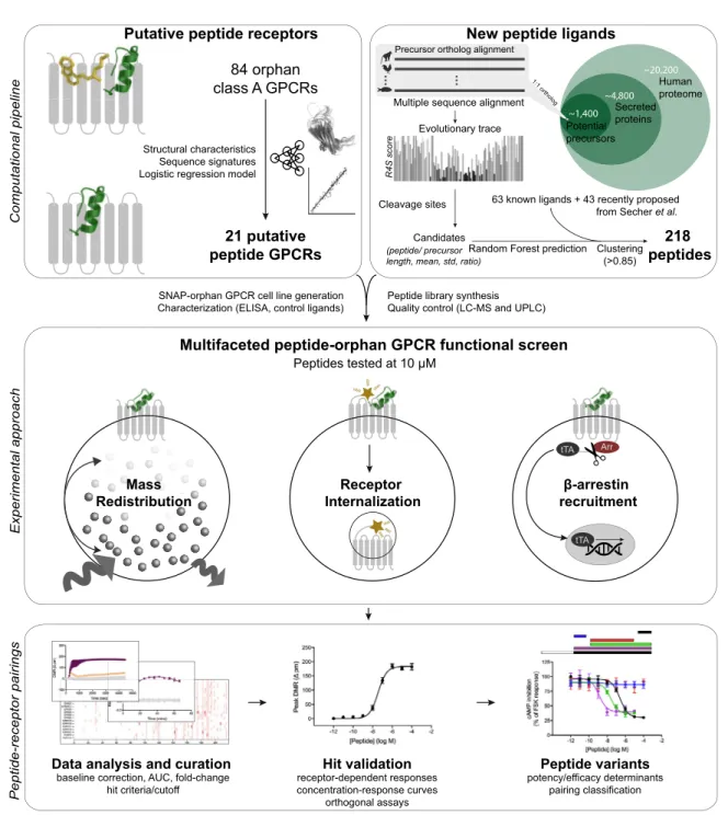
Here we provide an integrated computational and experi-mental approach for peptide-oGPCR pairing (Figure S1). We initially utilized comparative sequence and structural analyses to gain biological insights into the human peptide-receptor signaling landscape and leveraged these features to mine candi-date peptide ligands in the human genome. We then identified in-teractions via multiple orthogonal assay platforms to indepen-dently screen class A GPCRs against key signal transduction events. Ultimately, we discovered potential endogenous peptide ligands for five oGPCRs as well as secondary ligands for a num-ber of known peptide receptors.
RESULTS
Cognate Peptide Ligands and Receptors Co-evolved to Form the Largest Signal Transduction System in Humans
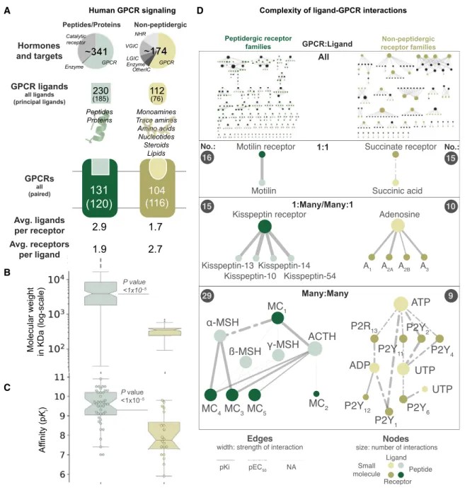
Initially, we explored the current knowledge regarding endoge-nous ligands and receptor systems by evaluating 341 peptide/ protein (encoded by 160 genes) and 174 non-peptide ligands (Harding et al., 2018). Both ligand classes mediate physiological functions predominantly through GPCRs (67% and 64%, respectively;Figure 1A;Table S1). The entire network of known interactions between GPCRs and cognate ligands spans 348 re-ported interactions between 120 receptors and 185 peptides. These interactions range from simple receptor-ligand systems with a one-to-one relationship to complex many-to-many sys-tems (Figure 1D). For instance, the peptide hormone motilin sig-nals through a single receptor, whereas the melanocortin and purinergic P2Y receptors are activated by multiple peptides and nucleotides, respectively. On average, each receptor is acti-vated by 2.9 peptide or 1.7 non-peptidergic ligands. Peptides are larger (average molecular weight, 7.7 kDa versus 0.4 kDa) and interact with their targets with higher affinity (average pKi:
9.4 versus 8.0) and potency (average pEC/IC50: 9.0 versus 6.9)
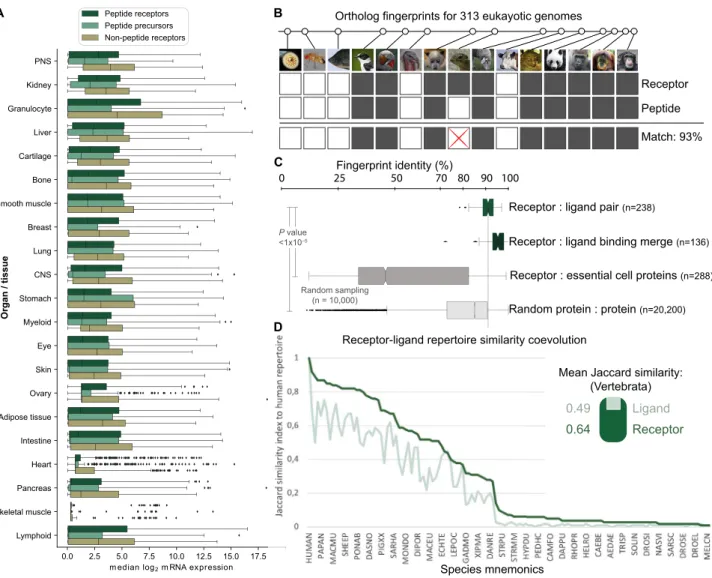
than non-peptides (Figures 1B and 1C;Table S1). The mRNA abundance is generally lower for peptide than non-peptide GPCRs (Figure S2A), although peptide ligand precursors and both types of receptors are expressed in all organs. Taken together, our most common and potent type of signaling mole-cules evolved through genetic encoding.
Next, we sought to investigate how the success of peptide ligands was shaped together with their cognate receptors ( Mira-beau and Joly, 2013) by analyzing 23,606,407 peptide-GPCR re-lationships across 313 eukaryotic genomes. When considering the minimal signaling system of one peptide and receptor, we found that 39 of 42 (93%) of the known human families arose dur-ing vertebrate evolution (Table S2), consistent with two early genome duplications (Holland et al., 1994). Notably, among all receptor families in all species, few have only the precursor
(4%) or GPCR (15%) gene, indicating nearly universal coevolu-tion. Moreover, by generating evolutionary fingerprints of conserved or lost gene orthologs (Figure S2B), we observed significantly higher coevolution of cognate ligand-receptor than random protein-protein pairs (average identities of 91% and 56%, respectively). This coevolution is higher (average identities of 95% and 89%, respectively) when merging the fingerprints of peptide precursors, but not of GPCRs, within the same receptor family (Figure S2C). These results suggest higher evolutionary pressure to conserve the distinct physiological function of recep-tors than ligands. Similarly, we found that the human receptor repertoire is more conserved than peptide ligands (average J = 0.64 versus 0.49; Figure S2D). Thus, cognate peptides and GPCRs have coevolved, and ligands have been more adaptive than receptors in shaping new signaling systems.
Precursors, Peptides, and Receptors Possess Universal Evolutionary, Sequence, and Structural Characteristics
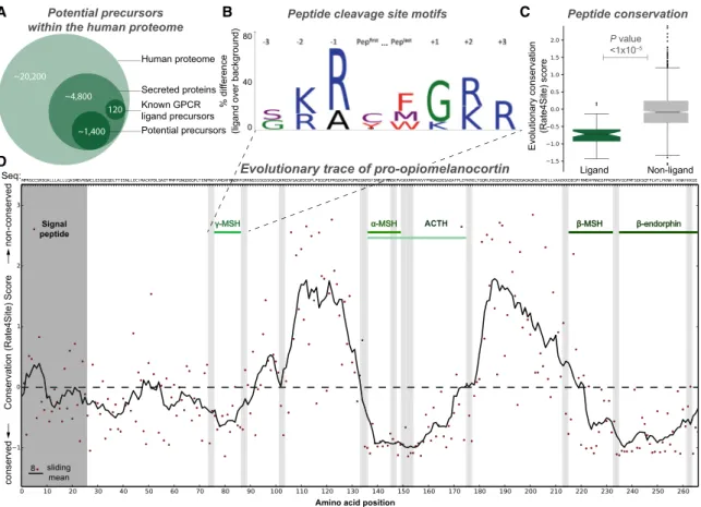
We next explored whether comparative genomics and biological processing paradigms could be predictively used to identify peptide precursors and peptides. We found that 99% of all pep-tide ligand precursors contain an N-terminal signal peppep-tide ( Fig-ure 2A), a requirement for extracellular secretion (Blobel and Dobberstein, 1975). Proteins are enzymatically cleaved at spe-cific sites (Ozawa et al., 2010), and we found that 80%/66% of the 184 human peptide ligand N/C termini are flanked by a dibasic motif (with a conserved glycine at C termini;Figure 2B). In addition, evolutionary trace analysis showed that known peptides make up the most conserved segments of precursor sequences (Wilcoxon rank-sum test, p < 13105;Figure 2C).
Strikingly, the peptide ligand subsequences can be recognized precisely within their precursors as highly conserved segments flanked by consensus cleavage sites, the signal peptide, or the C terminus (Figure 2D).
Universal Characteristics Reveal Plausible Additional Human Peptides and Receptors
We sought to investigate whether it was possible to mine poten-tial peptide ligands from the entire human proteome. First we identified putative precursors by sequentially filtering for proteins annotated in Swiss-Prot as secreted or with a signal peptide
(4,800) and those with unknown or precursor-compatible func-tional annotations (1,400) (Figure 2A). This yielded 1,227 ‘‘pep-tide cleavage variants,’’ representing candidate ligands that span the precursor signal peptide and C terminus or intermedi-ate consensus cleavage sites. We selected representative cleavage variants for pharmacological screening using a 5-fold
A
B
C
D
Figure 1. The Human G Protein-Coupled Receptor-Ligand System
(A) GPCRs represent the predominant targets for endogenous ligands. Peptides are more numerous, larger and bind with higher affinity than non-peptide ligands. From the top: (1) distinct endogenous ligands by target family; (2) endogenous GPCR ligands, of which ‘‘principal’’ ligands are considered most physiologically relevant; (3) peptide and small-molecule binding receptors, of which ‘‘paired’’ ones have a known principal endogenous ligand; and (4) ligands per receptor and vice versa (averages).
(B and C) Ligand molecular weight distribution (B) and cognate receptor affinity (C) (boxplots show a median and interquartile range of 1.5; Wilcoxon rank-sum test, p < 13105
). Data are from the Guide to Pharmacology database (Harding et al., 2018).
cross-validated random forest classifier based on length and several evolutionary conservation scores of known peptides and their precursors. This analysis resulted in 120 peptide se-quences representing the most plausible GPCR ligands. These were combined with 43 recently proposed unpaired rat peptides (Secher et al., 2016) to give a total of 163 putative peptide ligands, 112 of which came from precursors that have not been associated previously with GPCR activity. The final library containing 218 peptides was subsequently synthesized, including 55 known class A GPCR ligands (Table S5). To account for post-translational modification, we incorporated disulfide bridges and C-terminal amidation in 26 and 77 peptides, respec-tively. This synthetic peptide library contained the most GPCR ligand-like peptide cleavage variant, in effect ‘‘lead ligands’’ for primary screening.
In parallel, the conserved characteristics allowed us to predict peptide-activated receptors, of which we selected 21 class A GPCRs with rodent ortholog diverse disease associations (Table S5). Our cell lines for 15 (71%) oGPCRs displayed robust doxycycline-induced cell surface expression, and 12 (57%)
pro-moted constitutive G protein signaling, including couplings for three receptors not reported previously (Figure S3). Furthermore, 10 oGPCR cell lines were validated with commercially available compounds (Table S5).
A Multifaceted Screening Strategy Captures Pathway-and Assay-Dependent Receptor Responses
GPCRs can couple to multiple signaling pathways, with the com-bined signals constituting an overall response (Kenakin, 2017b). The measured response can appear different depending on the signal pathway, cell type, and time course investigated. Ligands can also intrinsically favor given receptor conformations that preferentially activate specific pathways (Kenakin, 2017b). These phenomena of assay-dependent observational bias and ligand-mediated signal bias are especially problematic for orphan and understudied receptors with poorly characterized signaling pathways (Huang et al., 2015; Roth et al., 2017).
For these reasons, we screened our 218 peptides and 21 pre-dicted peptide receptors in three complementary orthogonal assay platforms to cover multiple aspects of GPCR activation.
A
D
B C
Figure 2. Universal Precursor Processing and Peptide Ligand Gene Conservation Hotspots
(A) Potential precursors can be mined from the human proteome based on the presence of secretion signal peptides and an unknown or ligand-precursor-like function (Table S4).
(B) The vast majority of GPCR peptide ligands are cleaved from precursors at specific dibasic sites.
(C) GPCR peptide ligands are more evolutionarily conserved than random sequences of similar length (up to 45 residues) (Wilcoxon rank-sum test, p < 13105
).
A
B
C
E D
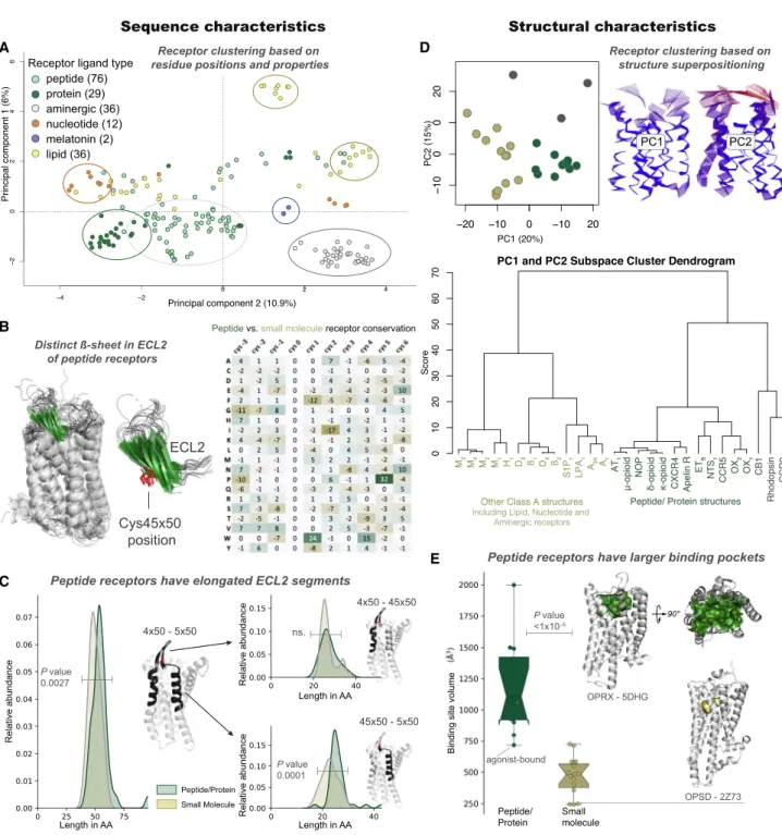
Figure 3. Peptide Receptors Share Distinct Sequence and Structural Characteristics
(A) The majority of class A GPCRs cluster by endogenous ligand type based on ligand-interacting residue analysis with multi-dimensional scaling.
(B) Peptide receptors with a structure (n = 21, left) share a characteristicbsheet (green) substructure (left) and sequence (right) in extracellular loop 2 (ECL2), which includes a conserved cysteine, Cys45350(red, center).
(C) A long ECL2 segment (>20 residues) after Cys45350is an overrepresented feature of peptide/protein receptors (Wilcoxon rank-sum test, p < 13105
). (D) Principal-component analysis of receptor structures in a 2D plot (top left) and dendrogram (bottom) demonstrate separation of peptide (green), non-peptide receptors (beige), and outliers (gray). Differences are predominantly found in the extracellularly facing ligand-binding domain, as shown by residue displacements from the mean (right).
A
B
The dynamic mass redistribution assay is ideal for investigating oGPCR activation because it captures most (including all G pro-tein) signaling pathways (Fang et al., 2008; Schro¨der et al., 2011). The second assay measures pathway-independent receptor internalization from the surface to the inside of cells (Foster and Bra¨uner-Osborne, 2018). These assays are both time resolved and provide valuable insights into receptor signaling kinetics. The third, theb-arrestin recruitment assay, is a highly amplified reporter gene-based readout of GPCR signaling (Kroeze et al., 2015) that increases the sensitivity of de-tecting positive pairings and allowed us to rapidly screen an additional 46 orphan/understudied and 27 known peptide-acti-vated class A GPCRs (Table S6). Collectively, these three assays provided an ideal platform to detect peptide ligands for under-characterized GPCRs and enabled physical interrogation of 21,446 potential peptide-receptor interactions.
We confirmed the activity of known agonists for 21 (78%) recognized peptide receptors using theb-arrestin recruitment assay. Our multifaceted screening identified peptide-mediated responses for all 21 predicted orphan peptide receptors ( Fig-ure 4A;Table S6). These results validate our computational pep-tide library design and provide good experimental coverage of receptor signaling. The three screening platforms demonstrated large variation in peptide pairings/GPCR targets (receptor inter-nalization, 24/6;b-arrestin recruitment, 57/21; and mass redistri-bution, 75/18;Table S6). We identified peptides that robustly activated bombesin receptor 3 (BB3), GPR1, GPR15, GPR55,
and GPR68 in multiple primary assays (Figure 4A;Table S6). Notably, all GPR55 and GPR68 peptides were inactive inb -ar-restin recruitment, and, conversely, GPR1 pairings were only observed in this assay, indicating potential ligand-mediated G protein and b-arrestin signal pathway bias, respectively. For the additional receptors only screened in theb-arrestin recruit-ment assay, we observed hits for 33 (72%) oGPCRs and nine known peptide receptors (Table S6). These findings underscore the importance of broadly covering receptor signaling using a multifaceted primary screening approach and additional func-tional assays for hit validation (Huang et al., 2015; Kroeze et al., 2015).
Discovery of Peptide-Receptor Pairs Expand the Human Signaling System
We extensively characterized our peptide-GPCR receptor inter-actions using additional orthogonal G protein and/orb-arrestin assays (Figure 4B;Figure S4;Table S7). Our peptide-receptor pairing criterion was activity in at least two assays. Furthermore, we characterized additional cleavage variants of these peptides to improve their potency (Figure 5;Figure S5). These
experi-ments also addressed the possibility that multiple peptide vari-ants can be endogenous agonists (Tatemoto et al., 1998) and provided insights into determinants of activity for the discovered peptide ligands.
BB3 is an orphan receptor, although it responds weakly to
physiologically relevant levels of the bombesin peptides neuro-medin B and gastrin-releasing peptide, the endogenous ago-nists of BB1and BB2, respectively (Alexander et al., 2017). We
observed considerably more potent neuromedin B responses in mass redistribution (pEC50, 7.43±0.08) and Gq/11signaling
as-says (IP1generation; pEC50, 6.39±0.42) (Figure 4B;Table S7)
than reported previously (Jensen et al., 2008). We observed no differences in BB3responses between the 10-amino acid
neuro-medin B peptide (designated NMB(47–56) based on its precur-sor residue number) and a longer, 32-residue cleavage variant, NMB(25–56), in any assay (Figure S5A;Table S7). We also found that the C terminus of gastrin-releasing peptide (GRP) activated BB3 with a potency comparable with the full-length peptide
tested in the screen, whereas a truncated N-terminal variant, GRP(24–40), was less potent/efficacious or inactive. In addition to these ligands, we identified less potent BB3-mediated
signaling for new cleavage variants derived from neuromedin-U and proenkephalin-A precursors (Figure 4B;Table S7). Taken together, these findings present multiple new peptide pairings for BB3, of which the neuromedin B peptides represent the
most likely endogenous ligands for BB3, albeit at lower potency
than at the BB1receptor.
GPR1 (recently renamed chemerin receptor 2) has been re-ported as a chemerin receptor, although its primary biological function is currently unknown (Kennedy and Davenport, 2018). No G protein has been unequivocally linked with GPR1 ( Fig-ure S3). Accordingly, we observed robust and selective GPR1-dependent responses for four different peptides in two different b-arrestin recruitment assays but not in other assays (Figures S3A andS4A). Peptide 141 (Osteocrin-2-19) is the most potent
(pEC50, 5.60±0.15 in Tango and 6.18 ±0.09 in PathHunter
b-arrestin recruitment assays, respectively) (Table S7). Alterna-tive osteocrin cleavage variants lacking two N-terminal amino acids had reduced potency (Figure S5B;Table S7). For another GPR1 hit, cholecystokinin (CCK-33), peptide cleavage variants were less active than the full-length peptide, except for a C-terminal 8-amino acid peptide, which was more potent in PathHunter b-arrestin recruitment assays than CCK-33 ( Fig-ure S5B;Table S7). We also found that gastrin-releasing peptide activated GPR1 inb-arrestin recruitment assays (Figure S5B; Table S7). Interestingly, we found that this activity was depen-dent on the peptide N terminus, whereas the C-terminal region GRP(41–50) was critical for BB3signaling.
Figure 4. General versus Assay-Specific Responses and Novel Peptide-Receptor Pairings
(A) The multifaceted screen of 218 peptides identified a variety of multiple and single-assay responses, including hits for all 21 predicted peptide receptors. Mass redistribution data revealed repeat hitters (denoted with asterisks) that reflect peptide-dependent responses from endogenous targets. Screening results for additional class A orphan and peptide GPCRs are provided inTable S6.
(B) Pairing of 17 peptides with five orphan receptors. Colored circles show pEC50values and concentration-response curves the most potent ligand for each
receptor. Other assays used were Gq/11(IP1), Gsand Gi/o(cAMP), andb-arrestin recruitment (PathHunter). An asterisk indicates a new cleavage variant of a known
GPCR peptide ligand with the amino acid range of the cleaved peptide shown in subscript; empty circles indicate inactivity.
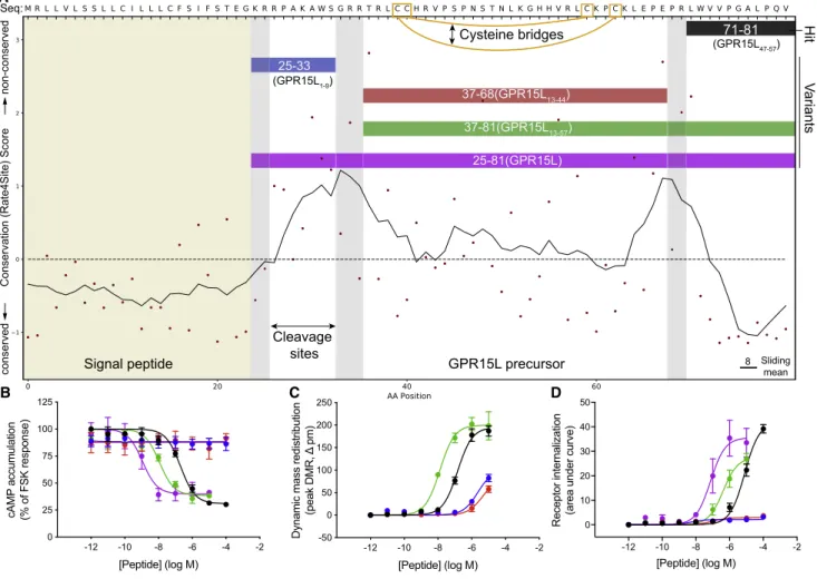
GPR15 was robustly activated by an 11-amino acid peptide derived from the C terminus of ‘‘Uniprot:C10orf99’’(Figure 4B; Table S7). We demonstrated that GPR15 is Gi/o-coupled
because this peptide reduced cyclic AMP (cAMP) production (pEC50, 6.72 ± 0.13). Notably, two cleavage variants of
this peptide (45 and 57 amino acids in length) had 10- and 100-fold improved potency, respectively (Figure 5; Table S7). In the course of our study, two other groups reported activation of GPR15 by the longest 57-residue cleavage variant, renamed GPR15L (Oco´n et al., 2017; Suply et al., 2017). GPR15L contains two intramolecular disulfide bridges characteristic of CC family chemokines; however, it differs because peptide activity is not dependent on the N terminus (Oco´n et al., 2017; Figure 5). Furthermore, we showed that the shortest, 11-residue C-terminal peptide, peptide 64 (GPR15L47-57) (lacking disulfide bridges) was sufficient to
acti-vate GPR15, although GPR15L represents the most potent and likely principal ligand.
We identified six peptides that promoted GPR55 internaliza-tion and mass redistribuinternaliza-tion, including five previously unde-scribed peptides and PACAP27, which exhibited a similar potency (pEC50, 9.51±0.07) as for its cognate receptor, PAC1
(Alexander et al., 2017; Figure 4B; Figure S5C; Table S7). Progressive truncations of the PACAP-27 peptide completely abrogated the GPR55 response (Figure S5C; Table S7). Conversely, we found that longer peptide cleavage variants (PACAP-38 and a 45-amino acid variant) elicited similar picomo-lar-potency internalization responses to PACAP-27. One of the six GPR55 ligands, peptide 143 originates from rat hypothala-mus (Secher et al., 2016), and testing of the human 143 confirmed activity, albeit with 10-fold lower potency (Table S7)
GPR68 is a proton-sensing receptor abundantly expressed in the hippocampus that is involved in learning and memory (Huang et al., 2015). We identified three peptides: 128 (Osteocrin33-55),
139 (CART(42-89)9-28), and rat 148 (Corticotropin17-40) that
led to GPR68-dependent mass redistribution responses with GPR15L precursor
25-81(GPR15L) 37-81(GPR15L13-57) 37-68(GPR15L13-44) 25-33
71-81
(GPR15L47-57)
(GPR15L1-9)
Signal peptide Sliding
mean 8
Hit
Cleavage sites
V
ariants
Cysteine bridges
A
B C D
Conservation (Rate4Site) Score
conserved
non-conserved
Seq:
z
Figure 5. Potential Peptide Cleavage Variants Elicit Increased GPR15 Signaling Responses
(A) Evolutionary trace and cleavage site (gray bars) analysis of theGPR15Lgene-encoded precursor presents potential alternative peptide cleavage variants. (B–D) GPR15-mediated responses forGPR15Lcleavage variants in (B) cAMP inhibition, (C) mass redistribution, and (D) receptor internalization assays. The most potent peptide is the longest, 57-residue form the recently named ‘‘GPR15L’’ (Suply et al., 2017) (excluded in C because of assay interference).
sub- or low-micromolar potencies (Figure 4B;Table S7). These responses were confirmed in the real-time internalization assay, although with lower potency compared with mass redistribution or G protein signaling assays (Roed et al., 2014). We further demonstrated that these peptides activated Gq/11 (calcium
mobilization) and Gs(cAMP signaling) (Figure 4B;Figure S4A;
Table S7), consistent with previously reported GPR68 signaling (Huang et al., 2015). We precluded a non-specific effect or direct proton-sensing mechanism of activity because none of these peptides induced responses in mock cells or elicited changes in extracellular pH (data not shown). Given the known allosteric modulation of proton-dependent GPR68 signaling by surrogate ligands, we performed further analyses of Gssignaling. These
studies revealed that the three peptides are positive allosteric modulators of the proton responses, with up to 2-fold improved allosteric activity (log(ab/KB)) over the small-molecule
GPR68 ligand ogerin (Huang et al., 2015;Figure 4B;Figure S4A; Table S7). Taken together, our pairings represent the first pep-tides and the very intriguing examples of putative endogenous allosteric modulators of proton-dependent agonism at GPR68.
In addition, we identified new peptide pairings for five other orphan and understudied receptors in theb-arrestin recruitment assays: GPR17, GPR161, GPR176, GPR183, and MAS1 ( Fig-ure S4B;Table S7). Because these receptors were not among the 21 oGPCRs selected for the mass redistribution and receptor internalization assays, we did not pursue these findings here. We also identified nine ‘‘repeat hitters’’ (in more than 5 recep-tor-expressing or untransfected cells) that represent ‘‘orphan peptide ligands’’ for receptors not assayed here (Table S6). Furthermore, our complete library of 1,227 ‘‘cleavage variants’’ (Table S4) comprises a resource of putative ligands for future deorphanization. Finally, all of our ‘‘lead peptides’’ may be alter-natively cleaved by carboxypeptidases or post-translationally modified into more potent biologically active receptor ligands, as for GPR15L (Figure 5;Table S7).
DISCUSSION
The discovery of ligand-GPCR signaling systems often trans-lates into clinical opportunities but first requires independent validation and characterization by the wider research commu-nity. Hence, we sought to explore the disease associations of our peptide-receptor pairs and to independently validate previously proposed pairings.
Therapeutic Potential of the Discovered Peptidergic Receptor Systems
We combined literature reports with mRNA expression data from ARCHS4 (Lachmann et al., 2018;Figure S6) and disease associa-tions fromhttps://www.opentargets.org(Figure 6;Table S4; Ko-scielny et al., 2017). BB3mouse knockout studies have
demon-strated an important role in energy homeostasis, making this receptor a target in obesity and metabolic disorders (Alexander et al., 2017). Interestingly, the Open Target data present several additional disease areas (the strongest being nervous system dis-ease) that strongly correlate with those of neuromedin-U and pro-enkephalin-A and moderately with neuromedin B (disease profile Pearson correlations of 0.82, 0.80, and 0.57, respectively;Figure 6).
GPR1 has been linked to cancer and cardiovascular and neurodegenerative disease (Kennedy and Davenport, 2018), which is reflected in a broad tissue expression profile. Osteocrin is a natriuretic peptide clearance (NPR3) receptor ligand impli-cated in bone and muscle function (Nishizawa et al., 2004; Thomas et al., 2003) and human brain development (Ataman et al., 2016). It has also been described as an endocrine hormone with potential therapeutic application to myocardial infarction (Chiba et al., 2017; Miyazaki et al., 2018).
GPR15 is expressed in immune cells, whereas GPR15L is ex-pressed in epithelial cells. GPR15L is secreted during inflamma-tion responses and therefore represents a promising target for inflammatory diseases, such as psoriasis and colitis (Suply et al., 2017). The Open Target database presents additional as-sociations of GPR15 with diseases of the endocrine, genetic, metabolic, and nervous systems (Figure 6).
GPR55 responds to lipids, but the direct receptor dependence of this signaling is somewhat controversial (Alhouayek et al., 2018). The agonist-dependent GPR55 trafficking shown here for PACAP27 and five new peptides open further avenues to investi-gate its function. GPR55 is widely expressed and has been pro-posed as a potential therapeutic target for a range of diseases, including cancer, metabolic disorders, pain, and inflammation ( Al-houayek et al., 2018). In Open Target, GPR55 and the precursors beta-microseminoprotein and clusterin-like protein 1 all have a strong link to neoplasm, supporting a potential link to cancer (Figure 6).
GPR68 acts as a proton sensor in bone, lung, and other tissue to regulate inflammatory responses, cell proliferation, and migra-tion (Huang et al., 2015). Accordingly, it is a potential target in inflammation and cancer (and a secondary target in anxiety; Weiß et al., 2017). Recently, GPR68 has been described as a flow sensor in arteriolar endothelium involved in cardiovascular pathophysiology (Xu et al., 2018). Our GPR68 peptides span multiple therapeutic areas (Figure 6). Most notably, peptide 139 (CART(42-89)9-28) is a shorter variant of cocaine- and
amphetamine-regulated portein (CART), which has been impli-cated in addiction (Kuhar, 2016). CART is an orphan peptide ligand of a GPCR; it has been shown to signal via protein kinase A, protein kinase C, and cAMP response element-binding protein (Chiu et al., 2009), as well as Gi/o. Furthermore, the
osteocrin-derived peptide 128 (Osteocrin33-50) shares disease
associations with GPR68, spanning cardiovascular, eye, ge-netic, immune system, metabolic, and nervous system diseases. Finally, the pro-opiomelanocortin-derived peptide 148 (Cortico-tropin17-40) and GPR68 both have strong associations with
genetic and neoplastic disease. These analyses suggest that GPR68 may hold (patho)physiological roles beyond proton and flow sensing. Moreover, the three peptides act as positive allo-steric modulators of the proton responses (Figure 4B; Fig-ure S4A;Table S7), suggesting that the two types of ligands may act in concert to regulate GPR68 activity.
Confirmation of Proposed Pairings and Identification of Secondary Ligands Expand the Peptide GPCR Network
Consensus regarding pairings and their physiological relevance is collated in the Guide to Pharmacology database (Harding et al., 2018). We sought to repeat literature pairings for our 21 predicted peptide receptors (Figure S7A). We confirmed previ-ously proposed pairings of chemerin/GPR1, the neuroendocrine peptide PEN/GPR83 (Gomes et al., 2016), the melanocortin re-ceptor ligandsa-MSH and ACTH/GPR139 (Nøhr et al., 2017), cortistatin and somatostatin/MRGPRX2, LPI/GPR55 (Henstridge et al., 2010), and LPS/P2RY10 (Inoue et al., 2012) by selective re-ceptor-dependent activation across multiple assays (Tables S5 andS7). In contrast, we found no activity for other proposed peptide/GPCR pairings such as adropin/GPR19 (Stein et al., 2016), head activator/GPR37 (Rezgaoui et al., 2006), prosap-tide/GPR37/GPR37L1 (Meyer et al., 2013; Rezgaoui et al., 2006), and galanin/GPR151 (Ignatov et al., 2004) or for the lipid/GPCR pairings resolvin D1/GPR32 (Krishnamoorthy et al., 2010) and lysophosphatidic acid/P2RY10 (Murakami et al., 2008) (Tables S5andS7). The majority of proposed pairings
eval-uated here were not assessed in previously published large orphan receptor screening studies, which measuredb-arrestin recruitment in PathHunter (Southern et al., 2013) or Tango assays (Kroeze et al., 2015).
Intriguingly, we also found that nine class A peptide recep-tors are activated by six previously published and 16 potential new peptide ligands (Figures S7B and S7C;Table S7). This included a truncated glucagon variant that stimulated the melanocortin MC4 receptor and a prolactin-releasing
peptide variant that activated NPY5. Although the potency of
these peptides is lower than for their principal agonists, the potential secondary/cross-pharmacology warrants further investigation.
In conclusion, our combined computational and pharmaco-logical approach has expanded the known human peptidergic signaling network from 348 to 407 interactions (an increase of 17%; Figure 7). 39 (74%) of the 53 peptides with validated receptor-dependent responses were first discovered here, Figure 6. Disease Associations for Novel Peptide-Receptor Pairs
demonstrating the predictive power of our approach, which could be transferred to many other peptide/protein systems. The discovery of peptide ligands for GPCRs has previously opened fields of research and is often closely followed by rapid translation into the clinic. Therefore, our findings are expected to fuel many future studies to establish their physiological roles and therapeutic potential.
STAR+METHODS
Detailed methods are provided in the online version of this paper and include the following:
d KEY RESOURCES TABLE
d LEAD CONTACT AND MATERIALS AVAILABILITY Figure 7. Expansion of the Human Peptidergic Receptor Signaling System
d EXPERIMENTAL MODEL AND SUBJECT DETAILS
B Mammalian cell culture conditions
d METHOD DETAILS
B Identification of human endogenous peptide and small molecule sets
B Evolutionary analysis of peptide receptor and peptide ligand repertoires
B Peptide receptor analysis
B Peptide ligand analysis and library design
B Selection of peptide variants
B Generation of expression datasets
B Peptide library preparation and quality control
B Generation of orphan GPCR cell lines
B Functional assays to measure orphan GPCR activation
d QUANTIFICATION AND STATISTICAL ANALYSIS
d DATA AND CODE AVAILABILITY
SUPPLEMENTAL INFORMATION
Supplemental Information can be found online athttps://doi.org/10.1016/j. cell.2019.10.010.
A video abstract is available at https://doi.org/10.1016/j.cell.2019.10. 010#mmc8.
ACKNOWLEDGMENTS
Helena Safavi and Elisabeth Kugelberg provided manuscript comments. Vignir Isberg, Andrius Senulis, and Christopher Southan contributed ideas regarding evolutionary fingerprinting. Karolina Sanicka contributed to characterization of receptor expression. This work was supported by the European Research Council (DE-ORPHAN 639125 to D.E.G. and A.S.H.), the Lundbeck Founda-tion (R163-2013-16327 to D.E.G. and R181-2014-2826 to S.R.F.), the Danish Council for Independent Research (4183-00243B to S.R.F.), the Carlsberg Foundation (to H.B.-O.), the A.P. Møller Foundation (to S.R.F.), the Augustinus Foundation (16-0313 to S.R.F.), the Toyota Foundation (to H.B.-O.), the NIH National Heart, Lung, and Blood Institute (P01 HL114471 to R.B.P.), NIH grant U24DK1116195 (to B.L.R.), the Michael Hooker Distinguished Professorship for Translational Proteomics (to B.L.R.), and the NIMH Psychoactive Drug Screening Program (to B.L.R. and X.-P.H.).
AUTHOR CONTRIBUTIONS
Conceptualization, D.E.G. and H.B.-O.; Project Administration and Writing – Original Draft, S.R.F., A.S.H., H.B.-O., and D.E.G.; Methodology, Formal Anal-ysis, and Visualization, S.R.F. and A.S.H.; Investigation, S.R.F., L.V., R.T.S., A.C.G., S.D.S., A.P.N., X.-P.H., and L.M.H.-K.; Software, A.S.H.; Supervision, D.E.G., H.B.-O., B.L.R., and R.B.P.; Funding Acquisition, D.E.G., H.B.-O., S.R.F., B.L.R., and R.B.P.; Writing – Review & Editing, all authors.
DECLARATION OF INTERESTS
L.M.H.-K. is an employee of PolyPeptide Group, Sweden.
Received: January 20, 2019 Revised: August 18, 2019 Accepted: October 8, 2019 Published: October 31, 2019
REFERENCES
Alexander, S.P., Christopoulos, A., Davenport, A.P., Kelly, E., Marrion, N.V., Peters, J.A., Faccenda, E., Harding, S.D., Pawson, A.J., Sharman, J.L., et al.; CGTP Collaborators (2017). THE CONCISE GUIDE TO
PHARMA-COLOGY 2017/18: G protein-coupled receptors. Br. J. Pharmacol.174(Suppl 1), S17–S129.
Alhouayek, M., Masquelier, J., and Muccioli, G.G. (2018). Lysophosphatidyli-nositols, from Cell Membrane Constituents to GPR55 Ligands. Trends Phar-macol. Sci.39, 586–604.
Altenhoff, A.M.,Skunca, N., Glover, N., Train, C.M., Sueki, A., Pilizota, I., Gori, K., Tomiczek, B., Mu¨ller, S., Redestig, H., et al. (2015). The OMA orthology database in 2015: function predictions, better plant support, synteny view and other improvements. Nucleic Acids Res.43, D240–D249.
Ataman, B., Boulting, G.L., Harmin, D.A., Yang, M.G., Baker-Salisbury, M., Yap, E.L., Malik, A.N., Mei, K., Rubin, A.A., Spiegel, I., et al. (2016). Evolution of Osteocrin as an activity-regulated factor in the primate brain. Nature539, 242–247.
Bercher, M., Hanson, B., van Staden, C., Wu, K., Ng, G.Y., and Lee, P.H. (2009). Agonists of the orphan human G2A receptor identified from inducible G2A expression and beta-lactamase reporter screen. Assay Drug Dev. Tech-nol.7, 133–142.
Blobel, G., and Dobberstein, B. (1975). Transfer of proteins across mem-branes. I. Presence of proteolytically processed and unprocessed nascent immunoglobulin light chains on membrane-bound ribosomes of murine myeloma. J. Cell Biol.67, 835–851.
Breiman, L. (2001). Random Forests. Mach. Learn.45, 5–32.
Chiba, A., Watanabe-Takano, H., Terai, K., Fukui, H., Miyazaki, T., Uemura, M., Hashimoto, H., Hibi, M., Fukuhara, S., and Mochizuki, N. (2017). Osteocrin, a peptide secreted from the heart and other tissues, contributes to cranial osteo-genesis and chondroosteo-genesis in zebrafish. Development144, 334–344.
Chiu, H.Y., Lin, H.H., and Lai, C.C. (2009). Potentiation of spinal NMDA-medi-ated nociception by cocaine- and amphetamine-regulNMDA-medi-ated transcript peptide via PKA and PKC signaling pathways in rats. Regul. Pept.158, 77–85.
Civelli, O., Reinscheid, R.K., Zhang, Y., Wang, Z., Fredriksson, R., and Schio¨th, H.B. (2013). G protein-coupled receptor deorphanizations. Annu. Rev. Phar-macol. Toxicol.53, 127–146.
Davenport, A.P., Alexander, S.P., Sharman, J.L., Pawson, A.J., Benson, H.E., Monaghan, A.E., Liew, W.C., Mpamhanga, C.P., Bonner, T.I., Neubig, R.R., et al. (2013). International Union of Basic and Clinical Pharmacology. LXXXVIII. G protein-coupled receptor list: recommendations for new pairings with cognate ligands. Pharmacol. Rev.65, 967–986.
Doi, M., Murai, I., Kunisue, S., Setsu, G., Uchio, N., Tanaka, R., Kobayashi, S., Shimatani, H., Hayashi, H., Chao, H.W., et al. (2016). Gpr176 is a Gz-linked orphan G-protein-coupled receptor that sets the pace of circadian behaviour. Nat. Commun.7, 10583.
Edgar, R.C. (2004). MUSCLE: multiple sequence alignment with high accuracy and high throughput. Nucleic Acids Res.32, 1792–1797.
Ehlert, F.J. (2005). Analysis of allosterism in functional assays. J. Pharmacol. Exp. Ther.315, 740–754.
Fang, Y., Frutos, A.G., and Verklereen, R. (2008). Label-free cell-based assays for GPCR screening. Comb. Chem. High Throughput Screen.11, 357–369.
Foster, S.R., and Bra¨uner-Osborne, H. (2018). Investigating Internalization and Intracellular Trafficking of GPCRs: New Techniques and Real-Time Experi-mental Approaches. Handb. Exp. Pharmacol.245, 41–61.
Fricker, L.D. (2010). Analysis of mouse brain peptides using mass spectrom-etry-based peptidomics: implications for novel functions ranging from non-classical neuropeptides to microproteins. Mol. Biosyst.6, 1355–1365.
Fricker, L.D., McKinzie, A.A., Sun, J., Curran, E., Qian, Y., Yan, L., Patterson, S.D., Courchesne, P.L., Richards, B., Levin, N., et al. (2000). Identification and characterization of proSAAS, a granin-like neuroendocrine peptide pre-cursor that inhibits prohormone processing. J. Neurosci.20, 639–648.
Fukusumi, S., Yoshida, H., Fujii, R., Maruyama, M., Komatsu, H., Habata, Y., Shintani, Y., Hinuma, S., and Fujino, M. (2003). A new peptidic ligand and its receptor regulating adrenal function in rats. J. Biol. Chem.278, 46387–46395.
GPR83 as the receptor for the neuroendocrine peptide PEN. Sci. Signal. 9, ra43.
Harding, S.D., Sharman, J.L., Faccenda, E., Southan, C., Pawson, A.J., Ireland, S., Gray, A.J.G., Bruce, L., Alexander, S.P.H., Anderton, S., et al.; NC-IUPHAR (2018). The IUPHAR/BPS Guide to PHARMACOLOGY in 2018: updates and expansion to encompass the new guide to IMMUNOPHARMA-COLOGY. Nucleic Acids Res.46(D1), D1091–D1106.
Hart, T., Brown, K.R., Sircoulomb, F., Rottapel, R., and Moffat, J. (2014). Measuring error rates in genomic perturbation screens: gold standards for hu-man functional genomics. Mol. Syst. Biol.10, 733.
Hatcher, N.G., Atkins, N., Jr., Annangudi, S.P., Forbes, A.J., Kelleher, N.L., Gil-lette, M.U., and Sweedler, J.V. (2008). Mass spectrometry-based discovery of circadian peptides. Proc. Natl. Acad. Sci. USA105, 12527–12532.
Hauser, A.S., Attwood, M.M., Rask-Andersen, M., Schio¨th, H.B., and Gloriam, D.E. (2017). Trends in GPCR drug discovery: new agents, targets and indica-tions. Nat. Rev. Drug Discov.16, 829–842.
Henstridge, C.M., Balenga, N.A., Schro¨der, R., Kargl, J.K., Platzer, W., Martini, L., Arthur, S., Penman, J., Whistler, J.L., Kostenis, E., et al. (2010). GPR55 li-gands promote receptor coupling to multiple signalling pathways. Br. J. Phar-macol.160, 604–614.
Holland, P.W., Garcia-Ferna`ndez, J., Williams, N.A., and Sidow, A. (1994). Gene duplications and the origins of vertebrate development. Dev. Suppl. 1994, 125–133.
Huang, Y., Niu, B., Gao, Y., Fu, L., and Li, W. (2010). CD-HIT Suite: a web server for clustering and comparing biological sequences. Bioinformatics26, 680–682.
Huang, X.P., Karpiak, J., Kroeze, W.K., Zhu, H., Chen, X., Moy, S.S., Saddoris, K.A., Nikolova, V.D., Farrell, M.S., Wang, S., et al. (2015). Allosteric ligands for the pharmacologically dark receptors GPR68 and GPR65. Nature 527, 477–483.
Ignatov, A., Hermans-Borgmeyer, I., and Schaller, H.C. (2004). Cloning and characterization of a novel G-protein-coupled receptor with homology to gal-anin receptors. Neuropharmacology46, 1114–1120.
Inoue, A., Ishiguro, J., Kitamura, H., Arima, N., Okutani, M., Shuto, A., Higa-shiyama, S., Ohwada, T., Arai, H., Makide, K., and Aoki, J. (2012). TGFa shed-ding assay: an accurate and versatile method for detecting GPCR activation. Nat. Methods9, 1021–1029.
Isberg, V., de Graaf, C., Bortolato, A., Cherezov, V., Katritch, V., Marshall, F.H., Mordalski, S., Pin, J.P., Stevens, R.C., Vriend, G., and Gloriam, D.E. (2015). Generic GPCR residue numbers - aligning topology maps while minding the gaps. Trends Pharmacol. Sci.36, 22–31.
Jacobsen, S.E., Nørskov-Lauritsen, L., Thomsen, A.R., Smajilovic, S., Wellen-dorph, P., Larsson, N.H., Lehmann, A., Bhatia, V.K., and Bra¨uner-Osborne, H. (2013). Delineation of the GPRC6A receptor signaling pathways using a mammalian cell line stably expressing the receptor. J. Pharmacol. Exp. Ther. 347, 298–309.
Jensen, R.T., Battey, J.F., Spindel, E.R., and Benya, R.V. (2008). International Union of Pharmacology. LXVIII. Mammalian bombesin receptors: nomencla-ture, distribution, pharmacology, signaling, and functions in normal and dis-ease states. Pharmacol. Rev.60, 1–42.
Jones, D.T., Taylor, W.R., and Thornton, J.M. (1994). A mutation data matrix for transmembrane proteins. FEBS Lett.339, 269–275.
Jordan, M., Schallhorn, A., and Wurm, F.M. (1996). Transfecting mammalian cells: optimization of critical parameters affecting calcium-phosphate precip-itate formation. Nucleic Acids Res.24, 596–601.
Kenakin, T. (2005). New concepts in drug discovery: collateral efficacy and permissive antagonism. Nat. Rev. Drug Discov.4, 919–927.
Kenakin, T. (2017a). A Scale of Agonism and Allosteric Modulation for Assess-ment of Selectivity, Bias, and Receptor Mutation. Mol. Pharmacol. 92, 414–424.
Kenakin, T. (2017b). Signaling bias in drug discovery. Expert Opin. Drug Dis-cov.12, 321–333.
Kennedy, A.J., and Davenport, A.P. (2018). International Union of Basic and Clinical Pharmacology CIII: Chemerin Receptors CMKLR1 (Chemerin1) and GPR1 (Chemerin2) Nomenclature, Pharmacology, and Function. Pharmacol. Rev.70, 174–196.
Koscielny, G., An, P., Carvalho-Silva, D., Cham, J.A., Fumis, L., Gasparyan, R., Hasan, S., Karamanis, N., Maguire, M., Papa, E., et al. (2017). Open Targets: a platform for therapeutic target identification and validation. Nucleic Acids Res. 45(D1), D985–D994.
Krishnamoorthy, S., Recchiuti, A., Chiang, N., Yacoubian, S., Lee, C.H., Yang, R., Petasis, N.A., and Serhan, C.N. (2010). Resolvin D1 binds human phago-cytes with evidence for proresolving receptors. Proc. Natl. Acad. Sci. USA 107, 1660–1665.
Kroeze, W.K., Sassano, M.F., Huang, X.P., Lansu, K., McCorvy, J.D., Gigue`re, P.M., Sciaky, N., and Roth, B.L. (2015). PRESTO-Tango as an open-source resource for interrogation of the druggable human GPCRome. Nat. Struct. Mol. Biol.22, 362–369.
Kuhar, M.J. (2016). CART Peptides and Drugs of Abuse: A Review of Recent Progress. J. Drug Alcohol. Res.5, 235984.
Lachmann, A., Torre, D., Keenan, A.B., Jagodnik, K.M., Lee, H.J., Wang, L., Silverstein, M.C., and Ma’ayan, A. (2018). Massive mining of publicly available RNA-seq data from human and mouse. Nat. Commun.9, 1366.
Laschet, C., Dupuis, N., and Hanson, J. (2018). The G protein-coupled recep-tors deorphanization landscape. Biochem. Pharmacol.153, 62–74.
Longo, P.A., Kavran, J.M., Kim, M.S., and Leahy, D.J. (2013). Transient mammalian cell transfection with polyethylenimine (PEI). Methods Enzymol. 529, 227–240.
Martin, A.L., Steurer, M.A., and Aronstam, R.S. (2015). Constitutive Activity among Orphan Class-A G Protein Coupled Receptors. PLoS ONE 10, e0138463.
Meyer, R.C., Giddens, M.M., Schaefer, S.A., and Hall, R.A. (2013). GPR37 and GPR37L1 are receptors for the neuroprotective and glioprotective factors pro-saptide and prosaposin. Proc. Natl. Acad. Sci. USA110, 9529–9534.
Mirabeau, O., and Joly, J.S. (2013). Molecular evolution of peptidergic signaling systems in bilaterians. Proc. Natl. Acad. Sci. USA110, E2028–E2037.
Mirabeau, O., Perlas, E., Severini, C., Audero, E., Gascuel, O., Possenti, R., Birney, E., Rosenthal, N., and Gross, C. (2007). Identification of novel peptide hormones in the human proteome by hidden Markov model screening. Genome Res.17, 320–327.
Miyazaki, T., Otani, K., Chiba, A., Nishimura, H., Tokudome, T., Takano-Wata-nabe, H., Matsuo, A., Ishikawa, H., Shimamoto, K., Fukui, H., et al. (2018). A New Secretory Peptide of Natriuretic Peptide Family, Osteocrin, Suppresses the Progression of Congestive Heart Failure After Myocardial Infarction. Circ. Res.122, 742–751.
Muppidi, J.R., Schmitz, R., Green, J.A., Xiao, W., Larsen, A.B., Braun, S.E., An, J., Xu, Y., Rosenwald, A., Ott, G., et al. (2014). Loss of signalling via Ga13 in germinal centre B-cell-derived lymphoma. Nature516, 254–258.
Murakami, M., Shiraishi, A., Tabata, K., and Fujita, N. (2008). Identification of the orphan GPCR, P2Y(10) receptor as the sphingosine-1-phosphate and ly-sophosphatidic acid receptor. Biochem. Biophys. Res. Commun. 371, 707–712.
Nishizawa, H., Matsuda, M., Yamada, Y., Kawai, K., Suzuki, E., Makishima, M., Kitamura, T., and Shimomura, I. (2004). Musclin, a novel skeletal muscle-derived secretory factor. J. Biol. Chem.279, 19391–19395.
Nøhr, A.C., Shehata, M.A., Hauser, A.S., Isberg, V., Mokrosinski, J., Andersen, K.B., Farooqi, I.S., Pedersen, D.S., Gloriam, D.E., and Bra¨uner-Osborne, H. (2017). The orphan G protein-coupled receptor GPR139 is activated by the peptides: Adrenocorticotropic hormone (ACTH),a-, andb-melanocyte stimu-lating hormone (a-MSH, andb-MSH), and the conserved core motif HFRW. Neurochem. Int.102, 105–113.
Oco´n, B., Pan, J., Dinh, T.T., Chen, W., Ballet, R., Bscheider, M., Habtezion, A., Tu, H., Zabel, B.A., and Butcher, E.C. (2017). A Mucosal and Cutaneous Che-mokine Ligand for the Lymphocyte Chemoattractant Receptor GPR15. Front. Immunol.8, 1111.
Oprea, T.I., Bologa, C.G., Brunak, S., Campbell, A., Gan, G.N., Gaulton, A., Gomez, S.M., Guha, R., Hersey, A., Holmes, J., et al. (2018). Unexplored ther-apeutic opportunities in the human genome. Nat. Rev. Drug Discov.17, 317–332.
Ozawa, A., Lindberg, I., Roth, B., and Kroeze, W.K. (2010). Deorphanization of novel peptides and their receptors. AAPS J.12, 378–384.
Pa´ndy-Szekeres, G., Munk, C., Tsonkov, T.M., Mordalski, S., Harpsøe, K., Hauser, A.S., Bojarski, A.J., and Gloriam, D.E. (2018). GPCRdb in 2018: adding GPCR structure models and ligands. Nucleic Acids Res.46(D1), D440–D446.
Pedersen, M.F., Wro´bel, T.M., Ma¨rcher-Rørsted, E., Pedersen, D.S., Møller, T.C., Gabriele, F., Pedersen, H., Matosiuk, D., Foster, S.R., Bouvier, M., and Bra¨uner-Osborne, H. (2019). Biased agonism of clinically approvedm-opioid receptor agonists and TRV130 is not controlled by binding and signaling ki-netics. Neuropharmacology, 107718.
Pele´, J., Be´cu, J.M., Abdi, H., and Chabbert, M. (2012). Bios2mds: an R pack-age for comparing orthologous protein families by metric multidimensional scaling. BMC Bioinformatics13, 133.
Pellegrini, M., Marcotte, E.M., Thompson, M.J., Eisenberg, D., and Yeates, T.O. (1999). Assigning protein functions by comparative genome analysis: pro-tein phylogenetic profiles. Proc. Natl. Acad. Sci. USA96, 4285–4288.
Pera, T., Deshpande, D.A., Ippolito, M., Wang, B., Gavrila, A., Michael, J.V., Nayak, A.P., Tompkins, E., Farrell, E., Kroeze, W.K., et al. (2018). Biased signaling of the proton-sensing receptor OGR1 by benzodiazepines. FASEB J.32, 862–874.
Petersen, T.N., Brunak, S., von Heijne, G., and Nielsen, H. (2011). SignalP 4.0: discriminating signal peptides from transmembrane regions. Nat. Methods8, 785–786.
Price, M.R., Baillie, G.L., Thomas, A., Stevenson, L.A., Easson, M., Goodwin, R., McLean, A., McIntosh, L., Goodwin, G., Walker, G., et al. (2005). Allosteric modulation of the cannabinoid CB1 receptor. Mol. Pharmacol.68, 1484–1495.
Pupko, T., Bell, R.E., Mayrose, I., Glaser, F., and Ben-Tal, N. (2002). Rate4Site: an algorithmic tool for the identification of functional regions in proteins by sur-face mapping of evolutionary determinants within their homologues. Bioinfor-matics18(Suppl 1), S71–S77.
Rezgaoui, M., Su¨sens, U., Ignatov, A., Gelderblom, M., Glassmeier, G., Franke, I., Urny, J., Imai, Y., Takahashi, R., and Schaller, H.C. (2006). The neu-ropeptide head activator is a high-affinity ligand for the orphan G-protein-coupled receptor GPR37. J. Cell Sci.119, 542–549.
Rivera, M.C., Jain, R., Moore, J.E., and Lake, J.A. (1998). Genomic evidence for two functionally distinct gene classes. Proc. Natl. Acad. Sci. USA95, 6239–6244.
Roed, S.N., Wismann, P., Underwood, C.R., Kulahin, N., Iversen, H., Cappe-len, K.A., Scha¨ffer, L., Lehtonen, J., Hecksher-Soerensen, J., Secher, A., et al. (2014). Real-time trafficking and signaling of the glucagon-like peptide-1 receptor. Mol. Cell. Endocrinol.382, 938–949.
Roth, B.L., and Kroeze, W.K. (2015). Integrated Approaches for Genome-wide Interrogation of the Druggable Non-olfactory G Protein-coupled Receptor Su-perfamily. J. Biol. Chem.290, 19471–19477.
Roth, B.L., Irwin, J.J., and Shoichet, B.K. (2017). Discovery of new GPCR li-gands to illuminate new biology. Nat. Chem. Biol.13, 1143–1151.
Saxena, H., Deshpande, D.A., Tiegs, B.C., Yan, H., Battafarano, R.J., Burrows, W.M., Damera, G., Panettieri, R.A., Dubose, T.D., Jr., An, S.S., and Penn, R.B. (2012). The GPCR OGR1 (GPR68) mediates diverse signalling and contraction of airway smooth muscle in response to small reductions in extracellular pH. Br. J. Pharmacol.166, 981–990.
Schro¨der, R., Schmidt, J., Bla¨ttermann, S., Peters, L., Janssen, N., Grund-mann, M., SeeGrund-mann, W., Kaufel, D., Merten, N., Drewke, C., et al. (2011).
Applying label-free dynamic mass redistribution technology to frame signaling of G protein-coupled receptors noninvasively in living cells. Nat. Protoc.6, 1748–1760.
Secher, A., Kelstrup, C.D., Conde-Frieboes, K.W., Pyke, C., Raun, K., Wulff, B.S., and Olsen, J.V. (2016). Analytic framework for peptidomics applied to large-scale neuropeptide identification. Nat. Commun.7, 11436.
Skjærven, L., Yao, X.Q., Scarabelli, G., and Grant, B.J. (2014). Integrating pro-tein structural dynamics and evolutionary analysis with Bio3D. BMC Bioinfor-matics15, 399.
Skjærven, L., Jariwala, S., Yao, X.Q., and Grant, B.J. (2016). Online interactive analysis of protein structure ensembles with Bio3D-web. Bioinformatics32, 3510–3512.
Sonmez, K., Zaveri, N.T., Kerman, I.A., Burke, S., Neal, C.R., Xie, X., Watson, S.J., and Toll, L. (2009). Evolutionary sequence modeling for discovery of pep-tide hormones. PLoS Comput. Biol.5, e1000258.
Southern, C., Cook, J.M., Neetoo-Isseljee, Z., Taylor, D.L., Kettleborough, C.A., Merritt, A., Bassoni, D.L., Raab, W.J., Quinn, E., Wehrman, T.S., et al. (2013). Screeningb-arrestin recruitment for the identification of natural ligands for orphan G-protein-coupled receptors. J. Biomol. Screen.18, 599–609.
Stein, L.M., Yosten, G.L., and Samson, W.K. (2016). Adropin acts in brain to inhibit water drinking: potential interaction with the orphan G protein-coupled receptor, GPR19. Am. J. Physiol. Regul. Integr. Comp. Physiol. 310, R476–R480.
Suply, T., Hannedouche, S., Carte, N., Li, J., Grosshans, B., Schaefer, M., Raad, L., Beck, V., Vidal, S., Hiou-Feige, A., et al. (2017). A natural ligand for the orphan receptor GPR15 modulates lymphocyte recruitment to epithelia. Sci. Signal.10, eaal0180.
Tatemoto, K., Hosoya, M., Habata, Y., Fujii, R., Kakegawa, T., Zou, M.X., Ka-wamata, Y., Fukusumi, S., Hinuma, S., Kitada, C., et al. (1998). Isolation and characterization of a novel endogenous peptide ligand for the human APJ re-ceptor. Biochem. Biophys. Res. Commun.251, 471–476.
Terskiy, A., Wannemacher, K.M., Yadav, P.N., Tsai, M., Tian, B., and Howells, R.D. (2007). Search of the human proteome for endomorphin-1 and endomor-phin-2 precursor proteins. Life Sci.81, 1593–1601.
Theobald, D.L., and Steindel, P.A. (2012). Optimal simultaneous superposi-tioning of multiple structures with missing data. Bioinformatics28, 1972–1979.
Thomas, G., Moffatt, P., Salois, P., Gaumond, M.H., Gingras, R., Godin, E., Miao, D., Goltzman, D., and Lanctoˆt, C. (2003). Osteocrin, a novel bone-spe-cific secreted protein that modulates the osteoblast phenotype. J. Biol. Chem. 278, 50563–50571.
Tian, W., Chen, C., Lei, X., Zhao, J., and Liang, J. (2018). CASTp 3.0: computed atlas of surface topography of proteins. Nucleic Acids Res. 46 (W1), W363–W367.
UniProt Consortium, T. (2018). UniProt: the universal protein knowledgebase. Nucleic Acids Res.46, 2699.
Weiß, K.T., Fante, M., Ko¨hl, G., Schreml, J., Haubner, F., Kreutz, M., Haver-kampf, S., Berneburg, M., and Schreml, S. (2017). Proton-sensing G protein-coupled receptors as regulators of cell proliferation and migration during tumor growth and wound healing. Exp. Dermatol.26, 127–132.
Wishart, D.S., Feunang, Y.D., Guo, A.C., Lo, E.J., Marcu, A., Grant, J.R., Sajed, T., Johnson, D., Li, C., Sayeeda, Z., et al. (2018). DrugBank 5.0: a major update to the DrugBank database for 2018. Nucleic Acids Res.46(D1), D1074–D1082.
Xu, J., Mathur, J., Vessie`res, E., Hammack, S., Nonomura, K., Favre, J., Gri-maud, L., Petrus, M., Francisco, A., Li, J., et al. (2018). GPR68 Senses Flow and Is Essential for Vascular Physiology. Cell173, 762–775.e16.
STAR
+
METHODS
KEY RESOURCES TABLE
REAGENT or RESOURCE SOURCE IDENTIFIER
Antibodies
Monoclonal anti-FLAG M2 Sigma Aldrich Cat # F1804; RRID: AB_262044
HRP labeled anti-Mouse IgG Vector Laboratories Cat # PI-2000; RRID: AB_2336177
Chemicals, Peptides, and Recombinant Proteins
Dulbecco’s modified Eagle’s medium (DMEM) without pyruvate
Thermo Fisher Scientific Cat # 61965026
Dialyzed FBS Thermo Fisher Scientific Cat # 26400036
Doxycycline hyclate Sigma Aldrich Cat # D9891
Zeocin Thermo Fisher Scientific Cat # R25001
Blasticidin Thermo Fisher Scientific Cat # A11139-03
Hygromycin B Thermo Fisher Scientific Cat # 10687010
Lipofectamine 2000 Transfection Reagent Thermo Fisher Scientific Cat # 11668027
Polyethylenimine (PEI), Linear, MW 25000, Transfection Grade
Polysciences Cat # 23966
Poly-L-lysine Sigma Aldrich Cat # P2636
SNAP-Lumi4-Tb (terbium) Cisbio Cat # SSNPTBD
Fluorescein-O0-acetic acid Sigma Aldrich Cat # 88596
Fluo-4 AM Invitrogen Cat # F14201
Probenecid Invitrogen Cat # P36400
MES, 2-(N-morpholino)ethanesulfonic acid Alfa Aesar Cat # H56472
HEPES, 4-(2-hydroxyethyl)-1-piperazineethanesulfonic acid
Fisher Scientific Cat # BP310
TAPS, (tris(hydroxymethyl)methylamino) propanesulfonic acid
VWR Cat # VWR J562
Ro 20-1724 Cayman Chemical Cat #18272
Luciferin Goldbio Cat # LUNCA
DMEM Corning Cat #10-013-CM
Dialyzed FBS Omega Scientific Cat # FB-03
Critical Commercial Assays
SuperSignal ELISA Femto Substrate Thermo Fisher Scientific Cat # 37075
Bright-Glo Luciferase assay system Promega Cat # E2620
IP-One - Gq kit Cisbio Cat # 62IPAPEC
cAMP - Gs Dynamic kit Cisbio Cat # 62AM4PEJ
Deposited Data
Crystal Structure of Bovine Rhodopsin at 2.2 Angstroms Resolution
Protein Data Bank PDB: 1U19
High resolution crystal structure of human B2-adrenergic G protein-coupled receptor.
Protein Data Bank PDB: 2RH1
The 2.5 A structure of the CXCR4 chemokine receptor in complex with small molecule antagonist IT1t
Protein Data Bank PDB: 3ODU
Structure of the human dopamine D3 receptor in complex with eticlopride
Protein Data Bank PDB: 3PBL
Structure of the human histamine H1 receptor in complex with doxepin
Protein Data Bank PDB: 3RZE
Continued
REAGENT or RESOURCE SOURCE IDENTIFIER
Structure of the human M2 muscarinic acetylcholine receptor bound to an antagonist
Protein Data Bank PDB: 3UON
Crystal Structure of a Lipid G protein-Coupled Receptor at 2.80A
Protein Data Bank PDB: 3V2Y
High Resolution Structure of Thermostable Agonist-bound Neurotensin Receptor 1 Mutant without Lysozyme Fusion
Protein Data Bank PDB: 4BUO
Ultra-thermostable beta1-adrenoceptor with cyanopindolol bound
Protein Data Bank PDB: 4BVN
Structure of the human kappa opioid receptor in complex with JDTic
Protein Data Bank PDB: 4DJH
Crystal structure of the mu-opioid receptor bound to a morphinan antagonist
Protein Data Bank PDB: 4DKL
1.8 A Structure of the human delta opioid 7TM receptor
Protein Data Bank PDB: 4N6H
M3-mT4L receptor bound to tiotropium Protein Data Bank PDB: 4U15
Crystal Structure of Human Lysophosphatidic Acid Receptor 1 in complex with ONO-3080573
Protein Data Bank PDB: 4Z36
Structures of the human OX1 orexin receptor bound to selective and dual antagonists
Protein Data Bank PDB: 4ZJ8
Crystal Structure of Human Angiotensin Receptor in Complex with Inverse Agonist Olmesartan at 2.8A resolution
Protein Data Bank PDB: 4ZUD
Structure of the human M1 muscarinic acetylcholine receptor bound to antagonist Tiotropium
Protein Data Bank PDB: 5CXV
The crystal structure of nociceptin/orphanin FQ peptide receptor (NOP) in complex with SB-612111
Protein Data Bank PDB: 5DHH
Structure of the M4 muscarinic acetylcholine receptor (M4-mT4L) bound to tiotropium
Protein Data Bank PDB: 5DSG
Crystal structure of the human CC chemokine receptor type 9 (CCR9) in complex with vercirnon
Protein Data Bank PDB: 5LWE
A2A Adenosine receptor room-temperature structure determined by serial femtosecond crystallography
Protein Data Bank PDB: 5NM4
High-resolution crystal structure of the human CB1 cannabinoid receptor
Protein Data Bank PDB: 5U09
Crystal structure of the human adenosine A1 receptor A1AR-bRIL in complex with the covalent antagonist DU172 at 3.2A resolution
Protein Data Bank PDB: 5UEN
Crystal Structure of CC Chemokine Receptor 5 (CCR5) in complex with high potency HIV entry inhibitor 5P7-CCL5
Protein Data Bank PDB: 5UIW
Structure of apelin receptor in complex with agonist peptide
Protein Data Bank PDB: 5VBL
Structure of the human D4 Dopamine receptor in complex with Nemonapride
Protein Data Bank PDB: 5WIU
Crystal structure of human orexin 2 receptor bound to the selective antagonist EMPA determined by the synchrotron light source at SPring-8.
Protein Data Bank PDB: 5WQC
Continued
REAGENT or RESOURCE SOURCE IDENTIFIER
Human endothelin receptor type-B in complex with antagonist K-8794
Protein Data Bank PDB: 5X93
Experimental Models: Cell Lines
Flp-In T-REx 293 cells Thermo Fisher Scientific Cat # R78007; RRID: CVCL_D585
Orphan GPCR T-REx 293 cell lines This study N/A
HTLA cells Gift from G. Barnea and R. Axel (Brown University and Columbia University)
N/A
HA-OGR1 (GPR68) HEK293 cells Saxena et al., 2012 N/A
pcDNA3 (mock) HEK293 cells Saxena et al., 2012 N/A
293T cells ATCC Cat # CRL-3216; RRID: CVCL_0063
Recombinant DNA
pcDNA5/FRT/TO Vector Kit Thermo Fisher Scientific Cat # V652020
pOG44 Flp-Recombinase Expression Vector Thermo Fisher Scientific Cat # V600520
pcDNA6/TR Thermo Fisher Scientific Cat # V102520
pcDNA5/FRT/TO FLAG SNAP Pedersen et al., 2019 N/A
Roth Lab PRESTO-Tango GPCR Kit Kroeze et al., 2015 Addgene Kit # 1000000068
GloSensor cAMP plasmid Promega Cat # E2301
Software and Algorithms
Graphpad Prism 7 Graphpad Software https://www.graphpad.com/
Masshunter Agilent
PyMOL Molecular Graphics System, Version 2.0
Schro¨dinger https://pymol.org/2/; RRID:SCR_000305
Bio3D v2.3 Skjærven et al., 2016 http://thegrantlab.org/bio3d/index.php
Arpeggio http://biosig.unimelb.edu.au/arpeggioweb/
Pandas v0.20.3 Wes McKinney https://pandas.pydata.org/
Scikit-learn v0.19.2 scikit-learn community https://scikit-learn.org/
CD-HIT Huang et al., 2010 http://weizhongli-lab.org/cd-hit/
GPCRdb Pa´ndy-Szekeres et al., 2018 https://github.com/protwis/protwis
Python v2.7.13 and v3.6.5 Python Software Foundation https://www.python.org/; RRID:SCR_008394
GetContacts Rasmus Fonseca and Anthony Ma https://getcontacts.github.io/
Flareplots Rasmus Fonseca https://gpcrviz.github.io/flareplot/
bio2mds Pele´ et al., 2012 https://cran.r-project.org/web/
packages/bios2mds/index.html
Rate4Site Pupko et al., 2002 https://www.tau.ac.il/itaymay/cp/
rate4site.html
Computed Atlas of Surface Topography of proteins (CASTp) 3.0
Tian et al., 2018 http://sts.bioe.uic.edu/castp/index.html?2r7g
Theseus Theobald and Steindel, 2012 https://theobald.brandeis.edu/theseus/
Maestro Schro¨dinger Release 2017-4 Schro¨dinger https://www.schrodinger.com/maestro
Custom scripts for analysis This study Available upon request
Other
Epic Benchtop (BT) System Corning Cat # 5053
Epic 384-well cell assay microplate, fibronectin-coated
Corning Cat # 5042
EnVision multimode plate reader PerkinElmer Cat # 2104
EnSpire multimode plate reader PerkinElmer Cat # 2300
LEAD CONTACT AND MATERIALS AVAILABILITY
Further information and requests for resources and reagents should be directed to and will be fulfilled by the Lead Contact, David E. Gloriam ([email protected]). The 21 orphan receptor cell lines generated in this study are available from the Lead Contact with a completed Materials Transfer Agreement.
EXPERIMENTAL MODEL AND SUBJECT DETAILS Mammalian cell culture conditions
Flp-In T-REx 293 Cells (Thermo Fisher Scientific) were used for the generation of all oGPCR stable cell lines that were used in dynamic mass redistribution (DMR) and receptor internalization screening, as well as subsequent cell signaling assays. This system is of particular utility for studies on orphan receptors, where heterologous overexpression may be toxic to the cells over prolonged periods (Bercher et al., 2009), or where the potential endogenous ligand may be present in the culture media. Cells were grown at 37C and 5% CO2 in Dulbecco’s modified Eagle’s medium (DMEM) with 4.5 g/L glucose and GlutaMAX Supplement, without pyruvate
(GIBCO), supplemented with 10% dialyzed FBS (Thermo Fisher Scientific), 100 U/mL penicillin-streptomycin and 15 mg/mL blasticidin (complete medium; Thermo Fisher Scientific). Prior to transfection with SNAP-tagged receptor constructs, parental Flp-In T-REx 293 cells were cultured in complete medium supplemented with 100mg/mL zeocin (Thermo Fisher Scientific). oGPCR transfections were performed using Lipofectamine 2000 (3mL of Lipofectamine per 1 mg of DNA). All stable cell lines were selected and maintained in complete medium supplemented with 200mg/mL hygromycin B (Thermo Fisher Scientific). HTLA cells were a gift from the laboratory of R. Axel and were maintained in DMEM (Corning) supplemented with 10% FBS, 100 U/mL penicillin and 100mg/ml streptomycin, 2 mg/mL puromycin and 100 mg/mL hygromycin B in a humidified atmosphere at 37C in 5% CO2.
HEK293T and HEK293 stable cell lines (HA-OGR1 (GPR68) and vector pcDNA) (Saxena et al., 2012) were cultured at 37C with 5% CO2to near confluence in DMEM with 10% FBS (Corning).
METHOD DETAILS
Identification of human endogenous peptide and small molecule sets
To identify all human genome encoded peptides, we extracted the human proteome annotation from UniProtKB/Swiss-Prot (version 2018.3; released 28/03/2018) and searched for allPEPTIDEannotations. This led to a unique set of 163 peptide precursor proteins and a total annotated set of 378 peptides, in many instances including multiple variants of similar peptides. For instance, apelin is represented in four versions including apelin-13, apelin-28, apelin-31 and apelin-36. In an orthogonal approach, we extracted all endogenous ligand entries including peptides and metabolites from the International Union of Basic and Clinical Pharmacology/ British Pharmacological Society (IUPHAR/BPS) Guide to Pharmacology database (version 2018.1; released 05/03/2018) (Harding et al., 2018) using custom Python scripts. Endogenous peptides were defined as ligands originating from within the studied organism shown to have activity at the receptor. Small molecules were defined by IUPHAR (‘‘Metabolites’’) as low molecular weight, non-pep-tide, biogenic compounds produced by life processes and their close analogs. Of the 341 endogenous human peptides, 230 target GPCRs (67.4%) and 185 are primary peptides annotated as ‘‘principal’’ endogenous ligands for the target (e.g., including angiotensin II and III for the angiotensin II type-1 receptor (AT1), but not angiotensin A and IV). Of the 174 endogenous human metabolites, 112 are
known to target GPCRs (64.4%) and 76 are primary metabolites that are annotated as main endogenous activators. For each endogenous ligand, molecular weight was calculated usingMolmass. Affinity and potency (pKi, pKd, pEC50and pIC50) values
were obtained from the Guide To Pharmacology database. When multiple potency/affinity values were provided, we used the maximum reported.
Evolutionary analysis of peptide receptor and peptide ligand repertoires
Determination of receptor and ligand repertoires