Potential Use of American College of
Radiology BI-RADS Mammography
Atlas for Reporting and Assessing
Lesions Detected on Dedicated
Breast CT Imaging:
Preliminary Study
Hae Kyoung Jung, MD, Cherie M. Kuzmiak, DO, Keum Won Kim, MD,
Na Mi Choi, MD, Hye Jeong Kim, MD, Eun Lee Langman, MD, Sora Yoon, MD,
Doreen Steen, RT, Donglin Zeng, PhD, Fei Gao
RationaleandObjectives: Dedicatedbreastcomputedtomography(DBCT)isanemergingandpromisingmodalityforbreastlesions. TheobjectiveofthisstudywastoevaluatethepotentialuseofapplyingtheBI-RADSMammographyAtlas5thEditionforreporting andassessingbreastlesionsonDBCT.Currently,noatlasexistsforDBCT.
MaterialsandMethods: Fourradiologiststrainedinbreastimagingwererecruitedinthisinstitutionalreviewboard-approved,Health InsurancePortabilityandAccountabilityAct-compliantstudy.Theenrolledradiologists,whowereblindedtomammographicand his-topathologicfindings,individuallyreviewed30randomizedDBCTcasesthatcontainedmarkedlesions.Thirty-fourlesionswereincluded inthisstudy:24(70.6%)masses,7(20.6%)calcifications,and3(8.8%)architecturaldistortions.Eight(23.5%)lesionsweremalignant and26(76.5%)werebenign.ThereaderwasaskedtospecifyaccordingtotheBI-RADSMammographyAtlasforeachmarkedDBCT lesion:primaryfindings,features,breastdensity,andfinalassessment.Wecalculatedreaders’diagnosticperformancesfor differenti-atingbetweenbenignandmalignantlesionsandinterobservervariabilityforreportingandassessinglesionsusingageneralizedestimating equationandtheFleisskappa(κ)statistic.
Results: Theestimatedoverallsensitivityofthereaderswas0.969,andthespecificitywas0.529.Therewerenosignificant differ-encesinthesensitivityandthespecificitybetweenlesiontypes.Forreportingthepresenceofaprimaryfinding,theoverallsubstantial agreement(κ =0.70)wasseen.Inassigningthebreastdensityandthefinalassessment,theoverallagreementwasmoderate(κ =0.53) andfair(κ =0.30).
Conclusion:TheuseoftheBI-RADSMammographyAtlas5thEditionforDBCTshowedhighperformanceandgoodagreementamong readers.
KeyWords: Breastneoplasm;breastCT;BI-RADS;mammography.
AcadRadiol2017;24:1395–1401
INTRODUCTION
B
reast Imaging Reporting and Data System (BI-RADS), established by the American College of Radiology, was begun in the late 1980s to address a lack of standard-ization and uniformity in mammography practice and reporting (1), and the BI-RADS lexicon has provided a valuable and reliable guide for reporting breast lesions on mammography, ultrasound, and magnetic resonance imaging (MRI), and has been familiar to most radiologists specializing in breast imaging. The descriptors in the BI-RADS lexicon have been selected on the basis of their ability to discriminate between benignity and malignancy as clear and standardized terms(2,3). BI-RADS has also recommended that a final impression be summarized by choos-ing only one among several standardized final assessment categories at the end of a report, each of which included a matched, stan-dardized management recommendation(4,5). The BI-RADS atlas is intended to be a “living” document that changes as new data are acquired and more sophisticated patterns of breast care emerge(4). With continued evolvement of lesion char-acterization and assessment for malignancy, the BI-RADS Mammography Atlas is now in its fifth edition(6).In addition to the updates in mammography, the fifth edition contains standardized breast lesion lexicons and assessment lan-guage for breast ultrasound and MRI. With advancements in breast imaging technologies, such as dedicated computed to-mography of the breast, the BI-RADS Mamto-mography Atlas can serve as the standard terminology upon which lexicons in other areas of radiology and research can be modeled.
Mammography is the current gold standard for detecting breast cancer in asymptomatic women and has been proven to decrease mortality(7–9). However, this technology does have some limi-tations because of the superimposition of anatomic structures. In women with dense breasts, mammography has not been proven as sensitive as in the population of women with nondense breasts(10,11). In reaction to this problem, dedicated breast computed tomography (DBCT), which provides three-dimensional data that can be reconstructed into multiple imaging planes, similar to breast MRI, has emerged as a new imaging modality in some researchers (12–31). DBCT is performed without breast compression and is not as limited as full-field digital mammography or digital breast tomosynthesis by breast density or breast implants(14,15,17). The radiation dose level is similar to the dose of a conventional two-view digital mam-mogram(23,24,26,28). Since the initial clinical experience of DBCT was begun by Lindfors in 2008(23), DBCT has showed promising results for the diagnostic evaluation of breast lesions, particularly for breast masses(12,20,25–29,31). Published ar-ticles have shown that DBCT has shown a significant improvement in the characterization or differentiation of breast lesions using BI-RADS descriptor and category terminology compared to digital mammography(20,31). However, to our knowledge, there is no published study about the reproduc-ibility of readers for reporting and assessing breast lesions on DBCT with the use of BI-RADS. Determining the repro-ducibility of BI-RADS is important because it can offer
standardized guidance in reporting and assessing breast lesions with DBCT. Currently, no atlas exists for DBCT.
The purpose of the present study was to evaluate the di-agnostic performance and the variability of multireaders for the use of the BI-RADS Mammography Atlas 5th Edition in reporting and assessing breast lesions on DBCT.
MATERIALS AND METHODS
Institutional review board approval was obtained for this ra-diologist reader study. Informed verbal and written consent was obtained from all of the readers involved in the present study. The deidentified DBCT cases that were used in the current study were from a DBCT image database of collect-ed cases from two other institutional review board-approvcollect-ed, Health Insurance Portability and Accountability Act-compliant DBCT clinical trials. A total of 34 lesions in 30 subjects were identified in the database and are included in our study. All the lesions in the database had been assessed as BI-RADS 4 or 5 lesions with standard of care imaging, which consisted of full-field digital mammography and their pathologic di-agnoses based on image-guided percutaneous core needle biopsy or surgical excision. All lesions were mammographically evident on standard of care diagnostic evaluation. Pathologic results for all lesions were evaluated with image-guided percutane-ous core biopsy. In cases referred for excisional biopsy after needle core biopsy because of the finding of atypia, malig-nancy, or radiology-pathology discordance, final surgical pathologic analysis was used for correlation with imaging find-ings. At our institution, our protocol for breast lesions that result in a diagnosis of atypia on needle core biopsy was to perform surgical excision to exclude histologic underestimation. Before the reader study, each lesion for each case was elec-tronically marked and numbered on the images by the principle investigator, who was familiar with the clinical, mammo-graphic, and pathologic information of each case in the study.
Readers
Eligible radiologists were identified by research staff review of their credentials from academic practice centers. A total of four fellowship trained breast imaging radiologists were re-cruited and enrolled in the present study. The readers had 1–13 years (mean of 7 years) of clinical experience and use of the BI-RADS Mammography Atlas. According to self-reports of the radiologists, they interpreted at least 140 mammography examinations (80–180) per week on average. The readers had no experience of DBCT imaging as part of their daily practice. To minimize reader bias, these breast imaging radiologists possessed no conflicts of interest in the research study or with the use of the device.
Data Description
comprised of 24 (71%) masses, 7 (21%) calcifications, and 3 (9%) architectural distortions. Pathologic diagnosis was avail-able for all lesions. Eight (24%) lesions were malignant and 26 (76%) were benign. Four subjects had bilateral lesions. Twelve of the 34 lesions (35.3%) were associated with symp-toms of palpable mass (n=10) and focal pain (n=2). The remaining 22 lesions in 22 subjects were asymptomatic. For the subsequent analysis, we combined the lesion types of mass and architectural distortion into one lesion group.
Image Interpretation
The readers underwent dedicated training with five pilot cases that were pathologically proven on a dedicated work-station in our Breast Imaging Research Lab before starting the reader study. For each case, the readers were allowed to roam and zoom, adjust the contrast, and display the images in coronal, sagittal, and axial planes. The readers then individually reviewed a total of 34 randomized, deidentified DBCT cases. The readers were blinded to the clinical, mammographic, and pathologic results. The readers were given a mandatory 15-minute break after each hour in the reader study.
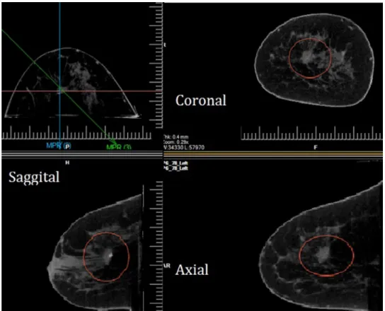
Each reader was asked for each case and for each lesion to evaluate the breast density, the lesion type, the lesion loca-tion, the lesion size in the longest dimension, the lesion characteristics, and the BI-RADS assessment score using the BI-RADS Mammography Atlas 5th Edition(6). All readers were given paper data recording sheets to mark their answers. Breast density was recorded as almost entirely fatty, scat-tered areas of fibroglandular density, heterogeneously dense, or extremely dense. Lesion type was classified as a mass, cal-cifications, architectural distortion, or asymmetry.Table 2lists the BI-RADS terminology used in the present study.Figures 1 and 2are an example of mass and calcification lesion cases, respectively. Final assessment was assigned based on lesion char-acterization according to the BI-RADS lexicon as previously mentioned, and was classified into five categories (1= neg-ative, 2=benign, 3=probably benign, 4=suspicious, and 5=highly suggestive of malignancy).
Data Analysis and Statistics
All data for each reader in both reading conditions were entered into a database and analyzed. We estimated the sensitivity and specificity of DBCT according to mammo-graphic lesion types from the four readers, where BI-RADS
categories 4 and 5 were diagnosed as malignancy. A gener-alized estimating equation was used to produce such estimates, and a compound symmetry working covariance was used. Normal distributions were used to construct 95% confi-dence intervals (CIs). The Fleisskstatistic was calculated to assess interobserver agreement for reporting a primary finding,
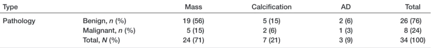
TABLE 1. Cross-Tabulation of Lesion Type and Pathology
Type Mass Calcification AD Total
Pathology Benign,n(%) 19 (56) 5 (15) 2 (6) 26 (76)
Malignant,n(%) 5 (15) 2 (6) 1 (3) 8 (24)
Total,N(%) 24 (71) 7 (21) 3 (9) 34 (100)
AD, architectural distortion.
TABLE 2. BI-RADS Atlas 5th Edition
Description Characteristic
Masses
Shape Oval
Round Irregular
Margin Circumscribed
Obscured Microlobulated Indistinct Spiculated
Density High density
Equal density Low density Fat-containing Calcifications
Typically benign Skin Vascular
Coarse or “popcorn-like” Large rodlike
Round Dystrophic Milk of calcium Suture Suspicious morphology Amorphous
Coarse heterogenous Fine pleomorphic
Fine linear or fine-linear branching
Distribution Diffuse
Regional Grouped Linear Segmental Architectural distortion
Yes No Asymmetry
lesion features, breast density, and final assessment among the four readers. The guidelines of Landis and Koch were followed in interpreting k values: a k value of equal to or less than 0.20 means slight agreement; 0.21–0.40, fair agreement;
0.41–0.60, moderate agreement; 0.61–0.80, substantial agree-ment; and 0.81–1.00, almost perfect agreement. The lesion types of mass and architectural distortion were combined in the statistical analysis.
Figure 1. Breast computed tomography images of a 44-year-old woman with a palpable mass in the upper central left breast diagnosed with invasive ductal cancer, grade I. Dedicated breast computed tomography displays the three-dimensional image data set (coronal, sag-ittal, and axial planes) and demonstrates a 1.5-cm irregular mass with a spiculated margin containing a calcification (circle).
Statistical analysis was performed using the SPSS version 20.0 software package (SPSS, Chicago, IL) and MedCalc version 14.10.2 (MedCalc Software, Mariakerke, Belgium).
RESULTS
Performances
The total reading time for all 34 cases was from 2 hours, 24 minutes, to 4 hours (mean of 3 hours, 27 minutes) in the four readers. We examined the sensitivities and specificities of DBCT according to mammographic lesion types from the four readers. The results are shown inTable 3. The estimated overall sen-sitivity was 0.969 (95% CI: 0.946–0.990), and the specificity was 0.529 (95% CI: 0.456–0.601). For lesion types, thePvalues comparing the sensitivity and specificity between the two types of groups were 0.5896 and 0.2916, respectively. There were no significant differences in the sensitivity and the specific-ity between the lesion types.
Interobserver Variability
Nineteen of 24 mass-type lesions (79.2%) were detected as mass by all of the four readers. The presence of all of the three architectural distortions and three of seven calcification-type lesions (42.9%) were noted by all four readers. For reporting the presence of a primary finding, the overall agreement was substantial (κ =0.70). The agreement for the presence of 24 mass-type and 3 architectural distortion-type lesions (κ =0.78) was higher than that for the 7 calcification-type lesions (κ =0.52). Meanwhile, in assigning the final assessment, the agreement was higher in calcification-type lesions (κ =0.56) than in mass- and architectural distortion-type lesions (κ =0.22). The results are shown inTable 3. In describing mass fea-tures, the overall agreement for shape was fair (κ =0.39); for margin, moderate (κ =0.455); and for density, slight (κ =0.17). Statistical analysis was not possible for agreement for assess-ing calcification features because only three cases were visualized by all four readers. The results are shown inTable 4.
DISCUSSION
We used the BI-RADS mammography atlas in reporting and assigning the assessment for DBCT breast lesions, and its di-agnostic performance and interobserver variability were
analyzed. We found that having radiologists use BI-RADS for DBCT lesions resulted in overall high performance and good agreement among readers.
According to Zhao et al. (31), in the receiver operating characteristic curve analysis, the area under the curve of DBCT was larger than that of two-view mammography (0.911 vs 0.827) for differentiating breast masses with BI-RADS. Our results were similar to those of Zhao et al. study, and the overall performance for assessing DBCT lesions with BI-RADS was relatively high with a sensitivity of 0.969 and a specificity of 0.529.
For lesion types, there were no significant differences in es-timated performances between mass- and architectural distortion-type lesions and calcification-distortion-type lesions (sensitivity, 0.958 vs 1.000; specificity 0.572 vs 0.350;P=0.5869 andP=0.2916, re-spectively). The performance of each radiologist can be affected by work experience duration in breast imaging. In our study, three of the four readers had 10 or more years of experience in breast imaging, and one reader only had 1 year of experience. However, we found that the number of years of the experi-ence in breast imaging had no influexperi-ence on the performance and the agreement of reporting a primary finding and assigning the BI-RADS final assessment and breast density. The reason might be that all the readers had no experience of DBCT imaging as part of their daily practice.
Lindfors et al. studied the clinical experience of DBCT and reported that DBCT was equal to mammography for the vi-sualization of breast masses, but mammography outperformed
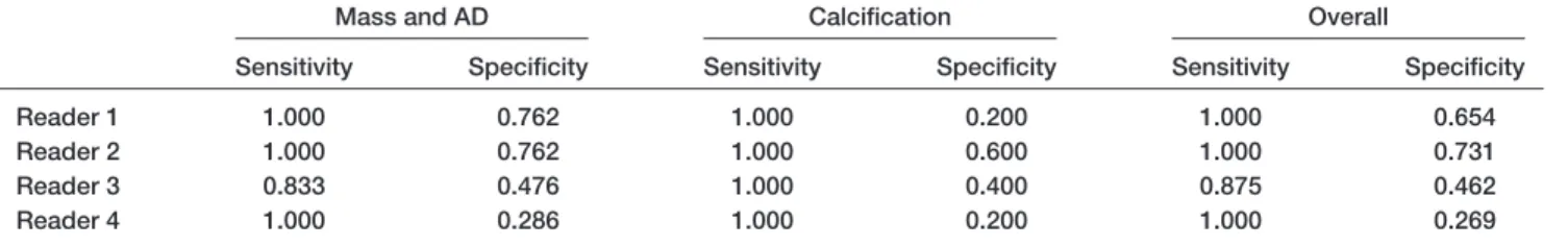
TABLE 3. Sensitivities and Specificities of Readers for Different Lesion Types
Mass and AD Calcification Overall
Sensitivity Specificity Sensitivity Specificity Sensitivity Specificity
Reader 1 1.000 0.762 1.000 0.200 1.000 0.654
Reader 2 1.000 0.762 1.000 0.600 1.000 0.731
Reader 3 0.833 0.476 1.000 0.400 0.875 0.462
Reader 4 1.000 0.286 1.000 0.200 1.000 0.269
AD, architectural distortion.
TABLE 4. Interobserver Variability in Description of Mammographic Lesions
BI-RADS Descriptor Kappa Value
Presence of primary finding
Overall 0.70
Mass and architectural distortion 0.78
Calcification 0.52
Breast density
Overall 0.53
Final assessment
Overall 0.30
Mass and architectural distortion 0.22
Calcification 0.56
CT for the visualization of microcalcifications(23). In the most recent study by Kuzmiak et al.(20), reader visualization con-fidence of mass characterization using BI-RADS mammography descriptors was significantly improved with DBCT com-pared to digital mammography, but reduced for calcifications. Currently, DBCT is regarded as inferior to mammography in the visualization of calcifications because of its lower spatial resolution, even though DCIS visualization on contrast-enhanced DBCT was equal to mammography in a recent study (32). The shape of a microcalcification group and the spatial distribution of an individual microcalcification within it are important indicators of malignancy; however, the resolution of DBCT was too low to resolve the three-dimensional shape of calcifications in our study. Among the seven calcification cases in the present study, only three cases were visualized by all of the four readers. In two of the calcification cases, only two of the four readers visualized them. The remain-ing two calcification cases were interpreted as mass or architectural distortion by the readers. Therefore, our study also suggested that the agreement for reporting the presence of calcification-type lesions among the readers was lower than that for mass- and architectural distortion-type lesions (κ =0.52 vs κ =0.78), but moderate agreement was obtained for calcification-type lesions. Meanwhile, in assigning the final assessment, the agreement was higher for calcification-type lesions (κ =0.56) than for mass- and architectural distortion-type lesions (κ =0.22). We noted that the overall agreement on the final assessment was not high (κ =0.30) in the present study but was similar to the result (κ =0.28) of a prior study on interobserver variability for mammography using the BI-RADS 4th Edition(33). Moreover, the agreement for mass descriptors was also comparable to the prior results(33).
In this preliminary study, these results are encouraging in lesion evaluation, especially in women with dense breasts where the phenomenon of masking of lesions by normal breast tissue is encountered.
Our study has several limitations. The first limitation is the small cohort size, especially with regard to patients with ma-lignant lesions. The small number of these cases may have statistical bias. The second was that each lesion was marked on the images for each study for the reader. This may result in bias for the lesion perception of the readers. Third, we com-bined the lesion type of mass and architectural distortion in our data analysis. Consequently, this may result in data anal-ysis error in the mass lesion type. Future studies with larger numbers of lesions and different lesions types are needed.
In conclusion, the use of the BI-RADS Mammography Atlas 5th Edition for DBCT showed high performance and good agreement among readers. As with our current breast imaging modalities, when new breast imaging tools emerge, a standard terminology needs to be developed and updated for radiologists to practice wisely and to provide outstand-ing patient care. The BI-RADS Mammography Atlas 5th Edition can be a potential starting point in breast lesion characterization and assessment category with breast lesions detected on DBCT.
REFERENCES
1. McLelland R, Hendrick R, Zinninger MD, et al. The American College of Radiology mammography accreditation program. AJR Am J Roentgenol 1991; 157:473–479.
2. Getty DJ, Pickett RM, D’Orsi CJ, et al. Enhanced interpretation of di-agnostic images. Invest Radiol 1988; 23:240–252.
3. Swets JA, Getty DJ, Pickett RM, et al. Enhancing and evaluating diag-nostic accuracy. Med Decis Making 1991; 11:9–17.
4. Burnside ES, Sickles EA, Bassett LW, et al. The ACR BI-RADS® expe-rience: learning from history. J Am Coll Radiol 2009; 6:851–860.
5. Committee ACoRB-R, Radiology ACo. Breast Imaging Reporting and Data System. Reston, VA: American College of Radiology, 1998.
6. D’Orsi CJ. ACR BI-RADS Atlas: Breast Imaging Reporting and Data System. Reston, VA: American College of Radiology, 2013.
7. Coburn NG, Chung MA, Fulton J, et al. Decreased breast cancer tumor size, stage, and mortality in Rhode Island: an example of a well-screened population. Cancer Control 2004; 11:222–230.
8. Jatoi I, Chen BE, Anderson WF, et al. Breast cancer mortality trends in the United States according to estrogen receptor status and age at di-agnosis. J Clin Oncol 2007; 25:1683–1690.
9. Otto S, Fracheboud J, Looman C, et al. National evaluation team for breast cancer screening initiation of population-based mammography screen-ing in Dutch municipalities and effect on breast-cancer mortality: a systematic review. Lancet 2003; 361:411–417.
10. Gordon PB, Goldenberg SL. Malignant breast masses detected only by ultrasound. A retrospective review. Cancer 1995; 76:626–630.
11. Pisano ED, Gatsonis C, Hendrick E, et al. Diagnostic performance of digital versus film mammography for breast-cancer screening. NEJM 2005; 353:1773–1783.
12. Bach AG, Abbas J, Jasaabuu C, et al. Comparison between incidental malignant and benign breast lesions detected by computed tomogra-phy: a systematic review. J Med Imaging Radiat Oncol 2013; 57:529– 533.
13. Boone JM, Nelson TR, Lindfors KK, et al. Dedicated breast CT: radiation dose and image quality evaluation 1. Radiology 2001; 221:657– 667.
14. Chang C, Sibala JL, Fritz SL, et al. Computed tomographic evaluation of the breast. Am J Roentgenol 1978; 131:459–464.
15. Chang CJ, Sibala JL, Gallagher JH, et al. Computed tomography of the breast: a preliminary report 1. Radiology 1977; 124:827–829.
16. Chen B, Ning R. Cone-beam volume CT breast imaging: feasibility study. Med Phys 2002; 29:755–770.
17. Gisvold J, Karsell P, Reese E. Clinical evaluation of computerized to-mographic mammography. Mayo Clin Proc 1977; 181–185.
18. Gong X, Vedula AA, Glick SJ. Microcalcification detection using cone-beam CT mammography with a flat-panel imager. Phys Med Biol 2004; 49:2183.
19. Inoue K, Liu F, Hoppin J, et al. High-resolution computed tomography of single breast cancer microcalcifications in vivo. Mol Imaging 2012; 11:1.
20. Kuzmiak CM, Cole EB, Zeng D, et al. Dedicated three-dimensional breast computed tomography: lesion characteristic perception by radiolo-gists. J Clin Imaging Sci 2016; 6.
21. Kwan AL, Boone JM, Yang K, et al. Evaluation of the spatial resolution characteristics of a cone-beam breast CT scanner. Med Phys 2007; 34:275–281.
22. Lai C-J, Shaw CC, Chen L, et al. Visibility of microcalcification in cone beam breast CT: effects of x-ray tube voltage and radiation dose. Med Phys 2007; 34:2995–3004.
23. Lindfors KK, Boone JM, Nelson TR, et al. Dedicated breast CT: initial clinical experience 1. Radiology 2008; 246:725–733.
24. McKinley RL, Tornai MP, Tuttle LA, et al. Development and initial dem-onstration of a low-dose dedicated fully 3D breast CT system. International Workshop on Digital Mammography. Heidelberg, Germany: Springer, 2012; 442–449.
25. Mettivier G, Russo P, Lanconelli N, et al. Cone-beam breast computed tomography with a displaced flat panel detector array. Med Phys 2012; 39:2805–2819.
26. O’Connell A, Conover DL, Zhang Y, et al. Cone-beam CT for breast imaging: radiation dose, breast coverage, and image quality. Am J Roentgenol 2010; 195:496–509.
28. O’Connell AM, Kawakyu-O’Connor D. Dedicated cone-beam breast com-puted tomography and diagnostic mammography: comparison of radiation dose, patient comfort, and qualitative review of imaging findings in BI-RADS 4 and 5 lesions. J Clin Imaging Sci 2012; 2:7.
29. Prionas ND, Lindfors KK, Ray S, et al. Contrast-enhanced dedicated breast CT: initial clinical experience 1. Radiology 2010; 256:714–723.
30. Shen Y, Zhong Y, Lai C-J, et al. Cone beam breast CT with a high pitch (75µm), thick (500µm) scintillator CMOS flat panel detector: visibility of simulated microcalcifications. Med Phys 2013; 40:101915.
31. Zhao B, Zhang X, Cai W, et al. Cone beam breast CT with multiplanar and three dimensional visualization in differentiating breast masses com-pared with mammography. Eur J Radiol 2015; 84:48–53.
32. Aminololama-Shakeri S, Abbey CK, Gazi P, et al. Differentiation of ductal carcinoma in-situ from benign micro-calcifications by dedicated breast computed tomography. Eur J Radiol 2016; 85:297–303.