2017 ESC Guidelines on the Diagnosis and Treatment of Peripheral Arterial
Diseases, in collaboration with the European Society for Vascular Surgery
(ESVS)
Document covering atherosclerotic disease of extracranial carotid and vertebral, mesenteric, renal, upper and lower extremity arteries
Endorsed by: the European Stroke Organization (ESO)
The Task Force for the Diagnosis and Treatment of Peripheral Arterial Diseases of the European Society of Cardiology (ESC) and of the European Society for Vascular Surgery (ESVS)
Authors/Task Force Membersa, Victor Aboyans*, Jean-Baptiste Ricco*, Marie-Louise E.L. Bartelink, Martin Björck, Marianne Brodmann, Tina Cohnert, Jean-Philippe Collet, Martin Czerny, Marco De Carlo, Sebastian Debus,
Christine Espinola-Klein, Thomas Kahan, Serge Kownator, Lucia Mazzolai, A. Ross Naylor, Marco Roffi, Joachim Röther, Muriel Sprynger, Michal Tendera, Gunnar Tepe, Maarit Venermo, Charalambos Vlachopoulos, Ileana Desormais
The disclosure forms of all experts involved in the development of these guidelines are available on the ESC websitehttp://www.escardio.org/guidelines The Addenda andQuestions and Answerscompanion documents of these guidelines are available at: www.escardio.org/Guidelines/Clinical-Practice-Guidelines/Peripheral-Artery-Diseases-Diagnosis-and-Treatment-of
ESC Committee for Practice Guidelines (CPG) and National Cardiac Societies (NCS) document reviewers:listed in theAppendix. ESC entities having participated in the development of this document:
Associations: European Association of Preventive Cardiology (EAPC), European Association of Cardiovascular Imaging (EACVI), European Association of Percutaneous Cardiovascular Interventions (EAPCI).
Councils: Council for Cardiology Practice (CCP), Council on Cardiovascular Primary Care (CCPC), Council on Hypertension (CHT).
Working Groups: Atherosclerosis and Vascular Biology, Cardiovascular Pharmacotherapy, Cardiovascular Surgery, Peripheral Circulation, Thrombosis. The content of these European Society of Cardiology (ESC) Guidelines has been published for personal and educational use only. No commercial use is authorized. No part of the ESC Guidelines may be translated or reproduced in any form without written permission from the ESC. Permission can be obtained upon submission of a written request to Oxford University Press, the publisher of the European Heart Journal and the party authorized to handle such permissions on behalf of the ESC ([email protected]).
Disclaimer. The ESC Guidelines represent the views of the ESC and were produced after careful consideration of the scientific and medical knowledge and the evidence available at the time of their publication. The ESC is not responsible in the event of any contradiction, discrepancy and/or ambiguity between the ESC Guidelines and any other official recommendations or guidelines issued by the relevant public health authorities, in particular in relation to good use of healthcare or therapeutic strategies. Health professionals are encouraged to take the ESC Guidelines fully into account when exercising their clinical judgment, as well as in the determination and the implementation of preventive, diagnostic or therapeutic medical strategies; however, the ESC Guidelines do not override, in any way whatsoever, the individual responsibility of health professionals to make appropriate and accurate decisions in consideration of each patient’s health condition and in consultation with that patient and, where appropriate and/or necessary, the patient’s caregiver. Nor do the ESC Guidelines exempt health professionals from taking into full and careful consideration the relevant official updated recommendations or guidelines issued by the competent public health authorities, in order to manage each patient’s case in light of the scientifically accepted data pursuant to their respective ethical and professional obligations. It is also the health professional’s responsibility to verify the applicable rules and regulations relating to drugs and medical devices at the time of prescription.
aAuthors/Task Force Members:Victor Aboyans*(ESC Chairperson) (France), Jean-Baptiste Ricco*,c(Co-Chairperson) (France), Marie-Louise E. L. Bartelink (The Netherlands), Martin Björckc(Sweden), Marianne Brodmann (Austria), Tina Cohnertc(Austria), Jean-Philippe Collet (France), Martin Czerny (Germany), Marco De Carlo (Italy), Sebastian Debusc(Germany), Christine Espinola-Klein (Germany), Thomas Kahan (Sweden), Serge Kownator (France), Lucia Mazzolai (Switzerland), A. Ross Naylorc(UK), Marco Roffi(Switzerland), Joachim Rötherd(Germany), Muriel Sprynger (Belgium), Michal Tendera (Poland), Gunnar Tepe (Germany), Maarit Venermoc(Finland), Charalambos Vlachopoulos (Greece), Ileana Desormais (France)
b
Document Reviewers:Petr Widimsky (ESC Review Coordinator) (Czech Republic), Philippe Kolh (ESVS Review Coordinator) (Belgium), Stefan Agewall (Norway), Héctor Bueno (Spain), Antonio Coca (Spain), Gert J. De Borstc(The Netherlands), Victoria Delgado (The Netherlands), Florian Dickc(Switzerland), Cetin Erol (Turkey), Marc Ferrini (France), Stavros Kakkosc(Greece/UK), Hugo A. Katus (Germany), Juhani Knuuti (Finland), Jes Lindholtc(Denmark), Heinrich Mattled(Switzerland), Piotr Pieniazek (Poland), Massimo Francesco Piepoli (Italy), Dierk Scheinert (Germany), Horst Sievert (Germany), Iain Simpson (UK), Jakub Sulzenko (Czech Republic), Juan Tamargo (Spain), Lale Tokgozoglu (Turkey), Adam Torbicki (Poland), Nikolaos Tsakountakis (Greece), José Tuñón (Spain), Melina Vega de Cenigac(Spain), Stephan Windecker (Switzerland), Jose Luis Zamorano (Spain)
* Corresponding authors:Victor Aboyans, Department of Cardiology CHRU Dupuytren Limoges, 2 Avenue Martin Luther King, 87042 Limoges, France. Tel:
þ33 5 55 05 63 10, Fax:þ33 5 55 05 63 34, Email:[email protected] Ricco, Department of Vascular Surgery, University Hospital, rue de la Miletrie, 86021 Poitiers, France. Tel:þ33 549443846, Fax:þ33 5 49 50 05 50, Email:[email protected]
c
Representing the European Society for Vascular Surgery (ESVS)
d
Representing the European Stroke Organisation (ESO)
1078-5884/Ó2017 European Society of Cardiology. Published by Oxford University Press and Elsevier Limited on behalf of European Society for Vascular Surgery. All rights reserved.
The article has been co-published with permission in theEuropean Heart Journal[DOI: 10.1093/eurheartj/ehx095] on behalf on the European Society of Cardiology andEuropean Journal of Vascular and Endovascular Surgery[DOI: 10.1016/j.ejvs.2017.07.018] on behalf of the European Society for Vascular Surgery.All rights reserved in respect ofEuropean Heart Journal,ÓEuropean Society of Cardiology 2017. The articles are identical except for minor stylistic and spelling differences in keeping with each journal’s style. Either citations can be used when citing this article.
Document Reviewersb, Petr Widimsky, Philippe Kolh, Stefan Agewall, Héctor Bueno, Antonio Coca, Gert J. De Borst, Victoria Delgado, Florian Dick, Cetin Erol, Marc Ferrini, Stavros Kakkos, Hugo A. Katus, Juhani Knuuti, Jes Lindholt, Heinrich Mattle, Piotr Pieniazek, Massimo Francesco Piepoli, Dierk Scheinert, Horst Sievert, Iain Simpson, Jakub Sulzenko, Juan Tamargo, Lale Tokgozoglu, Adam Torbicki, Nikolaos Tsakountakis, José Tuñón, Melina Vega de Ceniga, Stephan Windecker, Jose Luis Zamorano
Keywords:Guidelines, Peripheral arterial diseases, Carotid artery disease, Vertebral artery disease, Upper
extremity artery disease, Mesenteric artery disease, Renal artery disease, Lower extremity artery disease, Multisite artery disease
TABLE OF CONTENTS
Abbreviations and acronyms . . . .3
1. Preamble . . . .5
2. Introduction . . . .6
3. Epidemiology and risk factors . . . .8
3.1. Epidemiology . . . 8
3.2. Risk factors . . . 8
3.3. Prognosis . . . 8
4. General aspects . . . .8
4.1. Diagnostic approach . . . 8
4.1.1. Clinical history . . . 8
4.1.2. Clinical examination . . . 8
4.1.3. Laboratory testing . . . 8
4.1.4. Diagnostic methods for PADs . . . 9
4.2. Treatment approach . . . 10
4.2.1. Smoking cessation . . . 10
4.2.2. Lipid-lowering drugs . . . 10
4.2.3. Antithrombotic drugs . . . 10
4.2.4. Antihypertensive drugs . . . 10
5. Antithrombotic drugs in PADs . . . 11
5.1. Antithrombotic treatment in carotid artery disease . . . 11
5.1.1. Single antiplatelet therapy . . . 11
5.1.2. Dual antiplatelet therapy . . . 11
5.2. Antithrombotic therapy in lower extremity artery disease . . . 12
5.2.1. Single antiplatelet therapy . . . 12
5.2.2. Dual and triple antiplatelet therapy . . . .. . . 13
5.2.3. Antithrombotic therapy after lower-extremity bypass grafting . . . 13
5.2.4. Antithrombotic drugs after endovascular therapy for lower extremity artery disease . . . 13
5.2.5. Patients with lower extremity artery disease and concomitant coronary artery disease . . . 13
5.3. Antithrombotic therapy in lower extremity artery disease patients requiring long-term oral anticoagulant . . . 14
5.4. Antithrombotic therapy after endovascular therapy in other territories . . . 15
6. Extracranial carotid and vertebral artery disease . . . 16
6.1. Carotid artery disease . . . 16
6.1.1. Definition . . . 16
6.1.2. Diagnosis . . . 17
6.1.3. Treatment . . . 17
6.1.4. Management of carotid artery disease . . . 18
6.2. Vertebral artery disease . . . 22
6.2.1. Definition and natural history . . . 22
6.2.2. Imaging . . . 22
6.2.3. Management of vertebral artery disease . . . 22
7. Upper extremity artery disease . . . 22
7.4. Diagnostic methods . . . 22
7.4.1. Duplex ultrasound . . . 22
7.4.2. Computed tomography angiography . . . 22
7.4.3. Magnetic resonance angiography . . . 23
7.4.4. Digital subtraction angiography . . . 23
7.4.5. Positron emission tomography . . . 23
7.5. Treatment . . . .. . . 23
7.5.1. Endovascular treatment . . . 23
7.5.2. Open surgery . . . 23
7.5.3. Medical therapy . . . 23
8. Mesenteric artery disease . . . 24
8.1. Acute mesenteric ischaemia . . . 24
8.1.1. Diagnosis . . . 24
8.1.2. Treatment . . . 24
8.2. Chronic mesenteric artery disease . . . 25
8.2.1. Diagnosis . . . 25
8.2.2. Treatment . . . 25
8.3. Secondary prevention . . . 25
9.1. Introduction . . . 26
9.2. Clinical presentation . . . 26
9.3. Natural history . . . 26
9.4. Diagnostic strategy . . . 26
9.5. Prognosis . . . 27
9.6. Treatment . . . 27
9.6.1. Medical therapy . . . 27
9.6.2. Revascularization . . . .. . . 28
10. Lower extremity artery disease . . . 29
10.1. Clinical presentation and natural history . . . 29
10.2. Diagnostic tests . . . 30
10.2.1. Ankle-brachial index . . . 30
10.2.2. Treadmill test . . . 30
10.2.3. Imaging methods . . . 30
10.2.4. Other tests . . . 30
10.3. Medical treatment . . . 31
10.4. Revascularization options: general aspects . . . 31
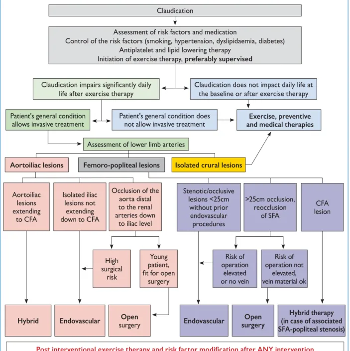
10.5. Management of intermittent claudication . . . 31
10.5.1. Exercise therapy . . . 31
10.5.2. Pharmacotherapy to decrease walking impairment . . . 31
10.5.3. Revascularization for intermittent claudication . . . 32
10.5.4. Management strategy for intermittent claudication . . . 32
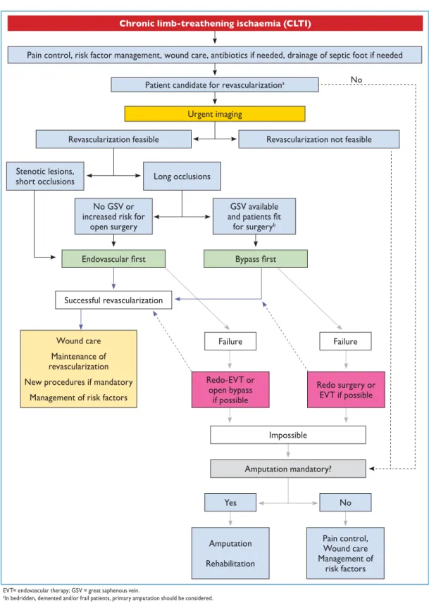
10.6. Chronic limb-threatening ischaemia . . . 33
10.6.1. Chronic limb-threatening ischaemia severity and risk stratification: the WIfI classification . . . 33
10.6.2. Management of patients with chronic limb-threatening ischaemia . . . 35
10.6.3. Spinal cord stimulation . . . 37
10.6.4. Stem cell and gene therapy . . . 37
10.6.5. Amputation . . . 37
10.7. Acute limb ischaemia . . . 37
10.8. Blue toe syndrome . . . 38
11. Multisite artery disease . . . 39
11.1. Multisite artery disease: epidemiology and impact prognosis . . . 39
11.2. Screening for and management of multisite artery disease . . . 40
11.2.1. Peripheral arterial diseases in patients presenting with coronary artery disease . . . 40
11.2.2. Coronary artery disease in patients presenting with peripheral arterial diseases . . . 42
11.2.3. Other peripheral localizations in patients with peripheral arterial diseases . . . 43
12. Cardiac conditions in peripheral arterial diseases . . . 44
12.1. Introduction . . . 44
12.2. Heart failure and peripheral arterial diseases . . . 44
12.2.1. Epidemiology . . . 44
12.2.2. Heart failure in patients with peripheral arterial diseases . . . 44
12.2.3. Peripheral arterial diseases in patients with heart failure . . . 45
12.3. Peripheral arterial diseases and atrial fibrillation . . . 45
12.3.1. General considerations . . . 45
12.3.2. Antithrombotic treatment in patients with atrial fibrillation . . . 45
12.4. Peripheral arterial diseases and valvular heart disease . . . 45
12.5. Peripheral arterial diseases and vascular access site for cardiac interventions . . . 45
13. Gaps in evidence . . . 46
14. To do and not to do messages from the Guidelines . . . 47
Web addenda and companion document . . . 50
Appendix . . . 50
References . . . 50
ABBREVIATIONS AND ACRONYMS
AAA abdominal aorta aneurysm ABI ankle-brachial index
ACAS Asymptomatic Carotid Atherosclerosis Study
ACEIs angiotensin-converting enzyme inhibitors
ACS acute coronary syndrome
ACSRS asymptomatic carotid atherosclerosis risk of stroke
ACST Asymptomatic Carotid Surgery Trial ACT Asymptomatic Carotid Trial
AF atrialfibrillation
AMERICA Aggressive detection and Management of the Extension of atherothrombosis in high Risk coronary patients In compari-son with standard of Care for coronary Atherosclerosis
ARBs angiotensin-receptor blockers ARR absolute risk reduction
ASTRAL angioplasty and stenting for renal artery lesions
BEST-CLI Best Endovascular vs. Best Surgical Ther-apy in Patients with Critical Limb Ischaemia BMT best medical therapy
BP blood pressure
CABG coronary artery bypass grafting CAD coronary artery disease
CAPRIE Clopidogrel versus Aspirin in Patients at Risk of Ischaemic Events
CAPTURE Carotid ACCULINK/ACCUNET Post-Approval Trial to Uncover Rare Events CARESS Clopidogrel and Aspirin for the Reduc-tion of Emboli in Symptomatic carotid Stenosis
CASPAR Clopidogrel and Acetylsalicylic Acid in Bypass Surgery for Peripheral Arterial disease
CAS carotid artery stenting CCA common carotid artery CEA carotid endarterectomy CFA common femoral artery
CHA2DS2-VASc Congestive heart failure, Hypertension, Age75 (2 points), Diabetes mellitus, Stroke or TIA (2 points), Vascular dis-ease, Age 65e74 years, Sex category CHARISMA Clopidogrel for High Atherothrombotic
Risk and Ischemic Stabilization, Man-agement and Avoidance
CI confidence interval CKD chronic kidney disease
CLEVER Claudication: exercise versus endolumi-nal revascularization
CLTI Chronic limb-threatening ischaemia CMI chronic mesenteric ischaemia
CONFIRM Coronary CT Angiography Evaluation for Clinical Outcomes: an International Multicenter
CORAL Cardiovascular Outcomes in Renal Atherosclerotic Lesions
CPB cardiopulmonary bypass
CPG Committee for Practice Guidelines CREST Carotid Revascularization
Endarterec-tomy versus Stenting Trial
CTA computed tomography angiography
CV cardiovascular
DAPT dual antiplatelet therapy DES drug eluting stent
DSA digital subtraction angiography DUS duplex ultrasound
ECG electrocardiogram
ECST European Carotid Surgery Trial EPD embolus protection device ESC European Society of Cardiology ESO European Stroke Organisation ESVS European Society of Vascular Surgery EUCLID Effects of Ticagrelor and Clopidogrel in
Patients with Peripheral Artery Disease
EVA-3S Endarterectomy vs Stenting in Patients with Symptomatic Severe Carotid Stenosis
EVT endovascular therapy
ExT exercise therapy
FMD fibromuscular dysplasia GSV great saphenous vein
HDL-C high-density lipoprotein cholesterol HF-ACTION Heart Failure: A Controlled Trial
Investi-gating Outcomes of Exercise Training HITS high-intensity transient signal HOPE Heart Outcomes Prevention Trial
HR hazard ratio
IC intermittent claudication ICA internal carotid artery
ICD implantable cardioverter defibrillator ICSS International Carotid Stenting Study INR international normalized ratio
INVEST INternational VErapamil-SR/Trandolapril Study
LDL-C low-density lipoprotein cholesterol LEAD lower extremity artery disease LV left ventricular
MACE major adverse cardiovascular event MI myocardial infarction
MRA magnetic resonance angiography MR CLEAN MultiCenter Randomized Clinical Trial of
Ischemic Stroke in the Netherlands MRI magnetic resonance imaging MSAD multisite artery disease MWD maximal walking distance
NASCET North American Symptomatic Carotid Endarterectomy Trial
NNH number needed to harm NNT number needed to treat
NOAC non-vitamin K oral anticoagulant OAC oral anticoagulation
ONTARGET Ongoing Telmisartan Alone and in Combination With Ramipril Global Endpoint Trial
OR odds ratio
PADs peripheral arterial diseases
PCI percutaneous coronary intervention PEGASUS-TIMI 54 Prevention of Cardiovascular Events in
Patients with Prior Heart Attack Using Ticagrelor Compared to Placebo on a Background of AspirineThrombolysis in
Myocardial Infarction 54
PRODIGY PROlonging Dual antiplatelet treatment after Grading stent-induced intimal hY-perplasia study
PTA percutaneous transluminal angioplasty QOL quality of life
RAAS renineangiotensinealdosterone system RAD renal artery disease
1. PREAMBLE
Guidelines summarize and evaluate available evidence with the aim of assisting health professionals in selecting the best management strategies for an individual patient with a given condition. Guidelines and their recommendations should facilitate decision making of health professionals in their daily practice. However, thefinal decisions concerning an individual patient must be made by the responsible health professional(s) in consultation with the patient and caregiver as appropriate.
A great number of guidelines have been issued in recent years by the European Society of Cardiology (ESC), by the European Society of Vascular Surgery (ESVS) and by the European Stroke Organization (ESO), as well as by other societies and organisations. Because of the impact on clin-ical practice, quality criteria for the development of guide-lines have been established in order to make all decisions transparent to the user. The recommendations for formu-lating and issuing ESC Guidelines can be found on the ESC Website ( https://www.escardio.org/Guidelines/Clinical- Practice-Guidelines/Guidelines-development/Writing-ESC-Guidelines). ESC Guidelines represent the official position of the ESC on a given topic and are regularly updated.
Members of this Task Force were selected by the ESC, including representation from the ESVS and ESO to repre-sent professionals involved with the medical care of patients with this pathology. Selected experts in thefield undertook a comprehensive review of the published evidence for man-agement of a given condition according to ESC Committee for Practice Guidelines (CPG) policy and approved by the ESVS and ESO. A critical evaluation of diagnostic and ther-apeutic procedures was performed, including assessment of the riskebenefit ratio. The level of evidence and the strength of the recommendation of particular management options were weighed and graded according to predefined scales, as outlined inTables 1and2.
The experts of the writing and reviewing panels provided declaration of interest forms for all relationships that might be perceived as real or potential sources of conflicts of in-terest. These forms were compiled into onefile and can be found on the ESC Website (http://www.escardio.org/ guidelines). Any changes in declarations of interest that arise during the writing period were notified to the ESC and updated. The Task Force received its entirefinancial support from the ESC and ESVS without any involvement from the healthcare industry.
RCT randomized clinical trial
REACH Reduction of Atherothrombosis for Continued Health
ROCKET-AF Rivaroxaban Once Daily Oral Direct Factor Xa Inhibition Compared with Vitamin K Antagonism for Prevention of Stroke and Embolism Trial in Atrial Fibrillation
RR relative risk
RRI renal resistive index
SAPPHIRE Stenting and Angioplasty with Protection in Patients at High Risk for
Endarterectomy
SAPT single antiplatelet therapy SBP systolic blood pressure SFA superficial femoral artery
SPACE Stent Protected Angioplasty versus Ca-rotid Endarterectomy
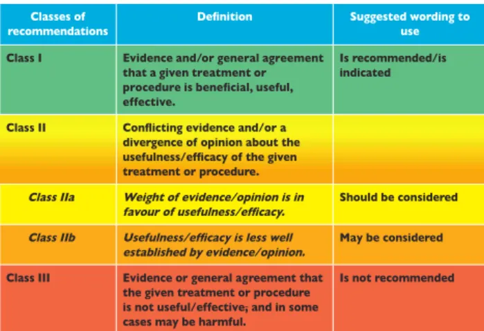
Table 1.Classes of recommendations.
Table 2.Levels of evidence. Level of
evidence A
Data derived from multiple randomized clinical trials or meta-analyses. Level of
evidence B
Data derived from a single randomized clinical trial or large non-randomized studies. Level of
evidence C
Consensus of opinion of the experts and/or small studies, retrospective studies, registries.
STAR Stent Placement in Patients With Atherosclerotic Renal Artery Stenosis and Impaired Renal Function
TAMARIS Efficacy and Safety of XRP0038/NV1FGF in Critical Limb Ischaemia Patients With Skin Lesions
TAVI transcatheter aortic valve implantation TBI toe-brachial index
TcPO2 transcutaneous oxygen pressure TIA transient ischaemic attack TTE transthoracic echocardiography UEAD upper extremity artery disease
VA vertebral artery
VAST Vertebral Artery Stenting Trial VHD valvular heart disease VKA vitamin K antagonist
WD walking distance
The ESC CPG supervises and coordinates the prepara-tion of new Guidelines. The Committee is also responsible for the endorsement process of these Guidelines. The ESC Guidelines undergo extensive review by the CPG and external experts, and in this case by ESVS- and ESO-appointed experts. After appropriate revisions the Guidelines are approved by all the experts involved in the Task Force. The finalized document is approved by the CPG and ESVS for publication in the European Heart Journal and in the European Journal of Vascular and Endovascular Surgery. The Guidelines were developed af-ter careful consideration of the scientific and medical knowledge and the evidence available at the time of their dating.
The task of developing ESC Guidelines in collaboration with ESVS also includes the creation of educational tools and implementation programmes for the recom-mendations including condensed pocket guideline ver-sions, summary slides, booklets with essential messages, summary cards for non-specialists and an electronic version for digital applications (smartphones, etc.). These versions are abridged and thus, if needed, one should always refer to the full text version, which is freely available via the ESC Website and hosted on the EHJ Website. The National Societies of the ESC are encour-aged to endorse, translate and implement all ESC Guidelines. Implementation programmes are needed because it has been shown that the outcome of disease may be favourably influenced by the thorough application of clinical recommendations.
Surveys and registries are needed to verify that real-life daily practice is in keeping with what is recommended in the guidelines, thus completing the loop between clinical research, writing of guidelines, disseminating them and implementing them into clinical practice.
Health professionals are encouraged to take the ESC Guidelines developed in collaboration with ESVS fully into account when exercising their clinical judgment, as well as in the determination and the implementation of preven-tive, diagnostic or therapeutic medical strategies. How-ever, the ESC Guidelines do not override in any way whatsoever the individual responsibility of health pro-fessionals to make appropriate and accurate decisions in consideration of each patient’s health condition and in consultation with that patient or the patient’s caregiver where appropriate and/or necessary. It is also the health professional’s responsibility to verify the rules and regu-lations applicable to drugs and devices at the time of prescription.
2. INTRODUCTION
In 2011, the ESC published itsfirst Guidelines on the
Diag-nosis and Management of Peripheral Arterial Diseases.1
This publication filled an important gap within the ESC Guidelines documents compendium. Meanwhile, the ESVS
released on a regular basis several guidelines documents on the management of specific localizations of arterial diseases. Both societies emphasized the need for multidisciplinary management of these patients. When the decision was made to update these guidelines, it appeared obvious that a combination of efforts from both societies would provide the most comprehensive single document, providing updated guidelines on peripheral arterial diseases (PADs) for clinicians.
It is of the outmost importance that every cardiologist should be sensitive in regard to the diagnosis and man-agement of patients with PADs, as many of them are seen and managed for concomitant cardiac conditions. In the ESC 2011 Guidelines, a specific chapter was dedicated to patients with combined coronary and peripheral artery diseases, as they mostly share the same aetiology and risk factors. In these guidelines, the Task Force made a step forward and proposed a new chapter on other cardiac conditions frequently encountered among patients with PADs. Also, as the options for the use and combination of antithrombotic drugs have increased, a specific chapter has been dedicated to their use in the management of PADs.
In this document, the term ‘peripheral arterial diseases’ encompasses all arterial diseases other than coronary ar-teries and the aorta. This should be clearly distinguished from the term ‘peripheral artery disease’, which is often used for lower extremity artery disease (LEAD). Indeed, other peripheral localizations, including the carotid and vertebral, upper extremities, mesenteric and renal arteries, are also frequently affected, mainly by atherosclerosis, and complete the family of PADs. Regarding the carotid and vertebral arteries, this document covers only their extra-cranial segments, as specialists other than cardiologists and vascular surgeons often manage intracranial arterial diseases.
The Task Force has decided to address only PADs sec-ondary to atherosclerosis, with a few exceptions in spe-cific areas where non-atherosclerotic diseases are a frequent differential diagnosis (e.g. fibromuscular dysplasia in renal arteries). For other cases, readers should always bear in mind the possibility for non-atherosclerotic conditions and refer to specific docu-ments. Readers are also invited to refer to the Web addendafor further information.
3. EPIDEMIOLOGY AND RISK FACTORS
3.1. Epidemiology
The epidemiology of different patterns of PADs is presented in theWeb addenda 3.1.
3.2. Risk factors
Although different localizations of PADs share common major risk factors for atherosclerosis, the impact of those and/or available evidence differ per arterial site. SeeWeb addenda 3.2.
3.3. Prognosis
Atherosclerosis is often generalized. Patients affected at one site are overall at risk for fatal and non-fatal CV events. Beyond the risk of cerebrovascular events, patients with CAD are also at risk for myocardial infarction (MI) and cardiac death.3 In a systematic review of 17 studies including 11 391 patients with >50% asymptomatic ca-rotid stenosis, 63% of late deaths were related to cardiac events, with a mean cardiac-related mortality rate of 2.9%/ year.4
Many studies have shown an increased risk of mortality, CV mortality and morbidity (MI, stroke) in patients with symptomatic or asymptomatic LEAD, even after adjust-ment for conventional risk factors.5 An ankle-brachial in-dex (ABI)0.90 is associated with more than doubling of
the 10-year rates of coronary events, CV mortality and total mortality.6 After 5 years, 20% of patients with intermittent claudication (IC) present an MI or stroke and mortality is 10e15%.7
All these data emphasize the importance of general CV prevention beyond the management of the disease related to a specific site of atherosclerosis.
4. GENERAL ASPECTS
4.1. Diagnostic approach
4.1.1. Clinical history. Personal and family clinical history
should always be assessed. Family history includes CAD, cerebrovascular disease, aortic aneurysm as well as LEAD.8e10Clinical history includes the evaluation of CV risk
factors and comorbidities as well as a review of the symptoms related to different vascular territories (seeWeb Table 1). Lifestyle habits, dietary patterns, walking perfor-mances and physical activity need to be systematically interrogated. Physical activity should be assessed.11 Questionnaires and functional status provide reasonably accurate outcome measures. They may be useful for determining the impairment level and selection of appro-priate care.12,13
4.1.2. Clinical examination. Although physical examination
alone is of relatively poor sensitivity and reproducibility, a systematic approach is mandatory (see Web Table 2). Beyond their diagnostic importance, clinical signs have a Key messages
Overall, the risk of different localizations of PADs increases sharply with age and with exposure to major cardiovascular (CV) risk factors, including smoking, hypertension, dyslipidaemia and diabetes. Other risk factors are still under investigation.
The strength of association between each risk factor and each vascular territory is variable, but all the major risk factors should be screened and considered.
When a vascular territory is affected by athero-sclerosis, not only is the corresponding organ
endangered [e.g. the brain for carotid artery disease (CAD)], but also the total risk of any CV event is increased (e.g. coronary events). Each vascular territory affected by atherosclerosis can be considered as marker of CV risk.
General recommendations on the management of patients with peripheral arterial diseases
Recommendations Classa Levelb
In healthcare centres, it is recommended to set up a multidisciplinary Vascular Team to make decisions for the management of patients with PADs.
I C
It is recommended to implement and support initiatives to improve medical and public awareness of PADs, especially cerebrovascular and lower extremity artery diseases.
I C
PADs¼peripheral arterial diseases. a
Class of recommendation. b
Level of evidence.
Key messages
Thorough clinical history and physical examination are key steps in PADs management.
Beyond the diagnosis of LEAD, ABI is also a strong marker for CV events.
The management of PADs includes all interventions to address specific arterial symptoms as well as general CV risk prevention.
prognostic value. Individuals with carotid bruits have twice the risk of MI and CV death as compared with those without.14 Interarm blood pressure (BP) asymmetry (15 mmHg) is a marker of vascular disease risk and death.15 A femoral bruit is an independent marker for ischaemic cardiac events.16
4.1.3. Laboratory testing. Investigations should progress
from the ‘minimal’ biological assessment17 to comple-mentary laboratory tests if necessary (outlined in Web Table 3).
4.1.4. Diagnostic methods for PADs
4.1.4.1. Ankle-brachial index.The ABI is a non-invasive tool useful for the diagnosis and surveillance of LEAD. It is also a strong marker of generalized atherosclerosis and CV risk (see Table 3). An ABI0.90 is associated on average with a 2- to 3-fold increased risk of total and CV death. An ABI >1.40 represents arterial stiffening (medial arterial calcification) and is also associated with a higher risk of CV events and mortality.6,18It is more prevalent in elderly patients, mostly in those with diabetes or chronic kidney disease (CKD). When added to a risk score, ABI enables the risk estimation to be upgraded in one-third and one-fifth of ‘low-risk’ women and men, respectively.6It is a valid method of CV risk assessment in diverse ethnic groups, independent of risk factors.18In contrast to coronary calcium score and carotid intima-media thickness, ABI is inexpensive and minimally time consuming. Good training is mandatory.
In addition to the general CV risk, ABI measurement can identify a patient’s risk for lower-extremities events, requiring close attention and education for foot wound prevention.
4.1.4.2. Duplex ultrasound. Duplex ultrasound (DUS) is often afirst step in the vascular workup both for screening and diagnosis. DUS includes B-mode echography, pulsed-wave, continuous, colour and power Doppler modalities to detect and localize vascular lesions and quantify their extent and severity through velocity criteria. More recent tech-niques, such asflow imaging or live three-dimensional (3D) echography, as well as the use of ultrasound contrast agents, further improve DUS performances, although their use is still limited. DUS can detect subclinical artery disease (e.g. carotid plaque), which is important for CV risk assessment.17
4.1.4.3. Digital subtraction angiography.Digital subtraction angiography (DSA) was considered the standard reference in vascular imaging. Given its invasive character and risk of complications, it has been mostly replaced by other less invasive methods except for below-the-knee arterial dis-ease. It may be used in the case of discrepancy between non-invasive imaging tools.
4.1.4.4. Computed tomography angiography.Multidetector computed tomography angiography (CTA) has a short exam-ination time with reduced motion and respiration artefacts while imaging vessels and organs. Advantages of CTA include rapid non-invasive acquisition, wide availability, high
Table 3.The Ankle-Brachial Index.
1. Who should have an ABI measurement in clinical practice? Patients with clinical suspicion for LEAD:
Lower extremities pulse abolition and/or arterial bruit
Typical intermittent claudication or symptoms suggestive for LEAD
Non-healing lower extremity wound
Patients at risk for LEAD because of the following clinical conditions:
Atherosclerotic diseases: CAD, any PADs
Other conditions: AAA, CKD, heart failure
Asymptomatic individuals clinically-free but at-risk for LEAD:
Men and women aged>65 years
Men and women aged<65 years classified at high CV risk according the ESC Guidelinesa
Men and women aged>50 years with family history for LEAD
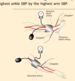
2. How to measure the ABI?
In supine position, with cuff placed just above the ankle, avoiding wounded zones. After a 5e10 minute rest, the SBP is measured by a Doppler probe (5e10 MHz) on the posterior and the anterior tibial (or dorsal pedis) arteries of each foot and on the brachial artery of each arm. Automated BP cuffs are mostly not valid for ankle pressure and may display overestimated results in case of low ankle pressure. The ABI of each leg is calculated by dividing the highest ankle SBP by the highest arm SBP.
3. How to interpret the ABI?
For diagnosis of LEAD interpret each leg separately (one ABI per leg).
For the CV risk stratification: take the lowest ABI between the two legs.
Interpretation:
AAA ¼ abdominal aorta aneurysm; ABI ¼ ankle-brachial index; BP ¼ blood pressure; CAD ¼ coronary artery disease; CKD ¼ chronic kidney disease; CV ¼ cardiovascular; ESC¼ European Society of Cardiology; LEAD¼ lower extremity artery disease; PADs ¼ peripheral arterial diseases; SBP¼systolic blood pressure.
a
resolution and 3D reformatting. Similar to DSA and magnetic resonance angiography (MRA), CTA displays a‘roadmap’of the vascularization, essential for determining interventional strategies (lesion localization and severity, upstream/down-stream status). The drawbacks of CTA include the lack of functional and haemodynamic data, exposure to radiation and the use of iodinated contrast agents, which should be limited in the case of CKD, with precautions in case of al-lergies. Nephrotoxicity can be limited by minimizing contrast agent volume and ensuring adequate hydration before and after imaging. The benefit of acetyl-cysteine to limit nephro-toxicity is uncertain.19,20Recent studies have suggested that statins or sodium bicarbonate could prevent contrast agent nephrotoxicity.21,22Further research is required.
4.1.4.5. Magnetic resonance angiography.MRA is used for peripheral artery imaging using contrast (i.e. gadolinium) and non-contrast techniques (i.e. phase contrast and time-of-flight sequences). These latter techniques have inferior resolution and are susceptible to artefacts, limiting their interpretation. They are a valuable alternative for use in patients with mild to moderate CKD. Compared with CTA, MRA does not need iodine contrast and has higher soft tissue resolution; however, motion artefacts are more frequent and contraindications include pacemakers and implantable cardioverter defibrillators (ICDs) [except magnetic resonance imaging (MRI)-conditional and compatible pacemakers, ICDs and leads], claustrophobia and severe CKD. In the latter case, the risk of nephrogenic systemic fibrosis following gadolinium administration should not be underestimated.23 Vascular calcifications, potentially affecting revascularization procedures, can be underestimated. Endovascular stents are not evaluable by MRI.
4.2. Treatment approach
The therapeutic approach to patients with PADs includes two aspects. Thefirst is to address specific symptoms of any localization and the risk related to a specific lesion. This is addressed in the next sections.
The second aspect of management in these patients is related to their increased risk of any CV event (see section 3.2). General CV prevention is of the utmost importance and management should be multidisciplinary. Best medical therapy (BMT) includes CV risk factor management, including best pharmacological therapy, as well as non-pharmacological measures such as smoking cessation, healthy diet, weight loss and regular physical exercise.24,25 The pharmacological component of BMT includes antihy-pertensive, lipid-lowering and antithrombotic drugs. In diabetic patients, optimal glucose level control should be obtained as recommended.26
4.2.1. Smoking cessation. A body of evidence supports
the benefits of smoking cessation in reducing CV events and mortality, especially in patients with cerebrovascular disease and LEAD.27,28 Management and support for smoking cessation was extensively addressed in the 2016
ESC guidelines on CV disease prevention.25Passive smoking should be assessed and prevented.29
4.2.2. Lipid-lowering drugs. All patients with PADs should
have their serum low-density lipoprotein cholesterol (LDL-C) reduced to <1.8 mmol/L (<70 mg/dL) or decreased by 50% if the initial LDL-C level is between 1.8 and 3.5 mmol/ L (70 and 135 mg/dL).25In observational studies and limited randomized clinical trials (RCTs) in patients with LEAD (from asymptomatic to severe cases), statin therapy has been shown to cause reductions in all-cause mortality and CV events.30e32 In the Reduction of Atherothrombosis for
Continued Health (REACH) registry, among patients with LEAD, statin use was associated with a 17% decrease in adverse CV events rates.33 Even in the most advanced stages of disease, statin therapy is associated with lower 1-year rates of mortality and major CV adverse events.34 Combination treatment with ezetimibe in selected patients is also beneficial.35 In a randomized trial, bezafibrate showed no benefit over placebo to reduce coronary and cerebrovascular events in patients with LEAD.36 In those with CAD, statins reduce the stroke risk.37,38 Recently the Fourier trial demonstrated the additional benefits of evo-locumab, a monoclonal antibody inhibiting the proprotein convertase subtilisin/kexin type 9 to reduce CV events in patients with atherosclerotic disease over statins alone.39 The results were consistent in the subgroup of 1505 pa-tients with LEAD alone. Further results are awaited.
4.2.3. Antithrombotic drugs. Antiplatelet agents are used
for secondary prevention of CV events in patients with symptomatic PADs. The evidence is mostly available in patients with LEAD and cerebrovascular disease (see chapter 5).
4.2.4. Antihypertensive drugs. Lowering systolic blood
pressure (SBP) reduces CV events.40 According to the current ESC/European Society of Hypertension guide-lines,41 a target BP <140/90 mmHg is recommended except in patients with diabetes, for whom a diastolic blood pressure85 mmHg is considered safe. In patients with LEAD, this is mainly based on data from the INter-national VErapamil-SR/Trandolapril (INVEST) study.42 Caution should be exercised to avoid an SBP decrease below 110e120 mmHg, since a J-shape relationship
be-tween SBP and CV events has been reported in that trial in LEAD patients.42 In old and frail patients, these levels should be achieved only if well tolerated, without ortho-static hypotension.43,44 In patients with PADs, an appro-priate lifestyle and salt intake (<5e6 g/day) are
The Heart Outcomes Prevention Trial (HOPE) and the Ongoing Telmisartan Alone and in Combination With Ramipril Global Endpoint Trial (ONTARGET) have shown that ACEIs and ARBs significantly reduce CV events in patients with PADs.46,47According to these trials, ACEIs or ARBs are recommended for secondary prevention, even in patients with chronic limb-threatening ischaemia (CLTI). In this subgroup of patients, the use of ACEIs or ARBs is associated with decreased major adverse cardiovascular events (MACEs) and mortality without any effect on limb outcomes.48
Importantly, beta-blockers are not contraindicated in patients with LEAD, as they do not alter walking capacity in patients with mild to moderate LEAD.49In an observational study, patients with LEAD and prior MI and taking beta-blockers had a significant 53% coronary events risk decrease at 32 months.50 Nevertheless, they should be carefully prescribed to patients with CLTI.
5. ANTITHROMBOTIC DRUGS IN PADS
Antiplatelet therapy is part of BMT for symptomatic PADs (seechapter 4). The specific issues about CAD and LEAD are addressed here. The question of DAPT after endovascular
therapy in other territories as well as the sensitive issue of PADs patients requiring anticoagulation [e.g. with concomitant atrialfibrillation (AF)] are also addressed.
5.1. Antithrombotic treatment in carotid artery disease
5.1.1. Single antiplatelet therapy.While the benefit of SAPT
for preventing stroke in asymptomatic patients with carotid artery stenosis>50% is not evidenced through an RCT, lifelong low-dose aspirin should be part of BMT to reduce the risk of stroke and other CV events,54as these patients are also at twice the risk of MI.14 In symptomatic extracranial carotid stenosis, antiplatelet monotherapy is recommended.54,55 Clopidogrel (75 mg/day) is an alternative in patients with aspirin intolerance.51
5.1.2. Dual antiplatelet therapy.In the randomized
Clopi-dogrel for High Atherothrombotic Risk and Ischemic Stabi-lization, Management and Avoidance (CHARISMA) trial, asymptomatic CAD was an inclusion criteria in 7% of
pa-tients enrolled. No benefit was observed between DAPT vs. SAPT.56 The Clopidogrel and Aspirin for the Reduction of Emboli in Symptomatic carotid Stenosis (CARESS) study, conducted in 108 patients, demonstrated that DAPT vs. aspirin reduced silent cerebral micro-emboli by 37% after 7 days.57 No life-threatening intracranial or major bleeding was observed, but the sample size was small. For these reasons, DAPT may be considered within 24 h of a minor ischaemic stroke or transient ischaemic attack (TIA) and may be continued for 1 month in patients treated conservatively.58
DAPT is recommended in patients undergoing CAS. Two small RCTs comparing aspirin alone with DAPT for CAS were terminated prematurely due to high rates of stent throm-bosis and neurological events in the aspirin-alone group.59,60 These data were obtained at 30 days. Most events were procedure related. The optimal duration of DAPT following CAS is unknown. Recent studies showing
Recommendations in patients with peripheral arterial diseases: best medical therapy
Recommendations Classa Levelb
Smoking cessation is recommended in all patients with PADs.27,28 I B
Healthy diet and physical activity are recommended for all patients with PADs. I C
Statins are recommended in all patients with PADs.31,32 I A In patients with PADs, it is recommended to reduce LDL-C to<1.8 mmol/L (70 mg/dL) or decrease it by
50% if baseline values are 1.8e3.5 mmol/L (70e135 mg/dL).25
I C
In diabetic patients with PADs, strict glycaemic control is recommended. I C
Antiplatelet therapy is recommended in patients with symptomatic PADs.51 I Cd
In patients with PADs and hypertension, it is recommended to control blood pressure at<140/ 90 mmHg.41,42,52
I A
ACEIs or ARBs should be considered asfirst-line therapycin patients with PADs and hypertension.47,53 IIa B
ACEIs¼angiotensin-converting enzyme inhibitors; ARBs¼angiotensin-receptor blockers; LDL-C¼ low-density lipoprotein cholesterol; PADs¼peripheral arterial diseases.
a
Class of recommendation. b
Level of evidence. c
Calcium channel blockers should be proposed in black individuals. d
Evidence is not available for all sites. When evidence is available, recommendations specific for the vascular site are presented in corresponding sections.
Key messages
Antiplatelet therapy is indicated in all patients with carotid artery stenosis irrespective of clinical symptoms and revascularization. Dual antiplatelet therapy (DAPT) should be given for at least 1 month after CAS.
Single antiplatelet therapy (SAPT) is indicated only if LEAD patients are symptomatic or have undergone revascularization. Clopidogrel is the preferred antiplatelet drug in LEAD patients.
late brain lesions on diffusion-weighted MRI after CAS question whether DAPT beyond the first month may be required.61 However, potential risks include haemorrhagic transformation in patients with recent stroke and intracra-nial bleeding in patients at risk of reperfusion injury following revascularization. DAPT may be prolonged beyond 1 month after CAS in the presence of recent (<12 months) MI and low bleeding risk (Figure 1).62
5.2. Antithrombotic therapy in lower extremity artery disease
Antiplatelet agents are used in patients with LEAD to prevent limb-related and general CV events. A number of antiplatelet strategies are available, but their specific indications remain unclear.63One study compared clopidogrel with aspirin51and two studies compared clopidogrel plus aspirin to aspirin alone.64,65No specific trial addressed the role of antiplatelet agents in the full spectrum of LEAD (asymptomatic, IC and CLTI). Also, the Task Force is aware of the premature halting of the COMPASS trial for ‘overwhelming’ efficacy. The trial compared rivaroxaban monotherapy (5 mg twice a day) with dual therapy (aspirin plus rivaroxaban 2.5 mg twice a day) and with aspirin monotherapy in 27 402 patients with CAD or LEAD. As the data were neither presented nor published at the time of guideline printing, the Task Force was unable to address these results and their potential clinical
consequences. Hence the Task Force will consider the results when they become available, as well as the option for an update if necessary.
5.2.1. Single antiplatelet therapy.Two trials, one in a
gen-eral population (with ABI<0.95)66 and another in diabetic patients (with ABI<1.0),67found no benefit from aspirin in subclinical LEAD.
In symptomatic LEAD, the strongest evidence in favour of aspirin to protect against MACE (combining non-fatal MI and stroke with CV death) comes from the Antithrombotic Trialists Collaboration.54 In 6200 patients with IC, aspirin significantly reduced MACE vs. controls (6.4 vs. 7.9%). Another meta-analysis of RCTs comparing aspirin to placebo in patients with LEAD (symptomatic or asymptomatic) showed a non-significant reduction in MACE {relative risk [RR] 0.75 [95% confidence interval (CI) 0.48e1.18]}.68 No
significant benefit was found within the individual compo-nents except for a reduction in non-fatal stroke [RR 0.64 (95% CI 0.42e0.99)].68In a post hoc analysis of the
Clopi-dogrel versus Aspirin in Patients at Risk of Ischaemic Events (CAPRIE) trial, at 3 years, clopidogrel was superior to aspirin in the subgroup of patients with clinical LEAD (n¼6452), with significant reductions in CV mortality [hazard ratio (HR) 0.76 (95% CI 0.64e0.91)] and MACE [HR 0.78 (95% CI 0.65e
0.93)], with similar benefit in the subgroup of LEAD patients with diabetes.51In the randomized Effects of Ticagrelor and Clopidogrel in Patients with Peripheral Artery Disease (EUCLID) trial, ticagrelor was compared to clopidogrel in 13 885 patients50 years of age with symptomatic LEAD.69 The trial failed to show any difference regarding MACE [HR 1.02 (95% CI 0.92e1.13)] or major bleeding [HR 1.10 (95%
CI 0.84e1.43)].
5.2.2. Dual and triple antiplatelet therapy. So far, data
proving the superiority of DAPT (with clopidogrel) over aspirin alone to reduce CV events in patients with LEAD are lacking.63In the subgroup of patients with LEAD enrolled in the CHARISMA trial (n¼3906), DAPT led to a reduction in MI [HR 0.63 (95% CI 0.42e0.95)], with a neutral effect on all
the other vascular events, at the cost of increased severe, fatal or moderate bleeding [HR 1.99 (95% CI 1.69e2.34)].65
Because of the post hoc nature of this analysis and the negative results of the overall trial, these findings need confirmation.
Vorapaxar, a protease-activated receptor-1 inhibitor, was tested vs. placebo on top of standard antiplatelet therapy in secondary prevention in patients with clinical LEAD (n¼ 3787).70 Vorapaxar did not reduce the risk of MACE [HR 0.94 (95% CI 0.78e1.14)] but significantly reduced the risk of acute limb ischaemia [HR 0.58 (95% CI 0.39e0.86)] and peripheral revascularization [HR 0.84
(95% CI 0.73e0.97)].70 This benefit was observed
irre-spective of the underlying mechanism of acute limb ischaemia, including surgical graft thrombosis and native vessel thrombosis.71 These beneficial effects were coun-terbalanced by an increased risk of bleeding [HR 1.62 (95% CI 1.21e2.18)].
5.2.3. Antithrombotic therapy after lower-extremity
bypass grafting. Antiplatelet agents are mostly used
after peripheral percutaneous revascularization, while warfarin has a small role (Figure 2). No conclusive data are yet available for direct oral thrombin and factor Xa inhibitors.72
5.2.3.1. Aspirin vs. placebo.In a meta-analysis of 952 pa-tients, graft patency was significantly improved with aspirin (with or without dipyridamole) vs. placebo (HR 0.42, P¼0.01).72Notably, at any of the time points, this effect was not observed for venous grafts alone but for prosthetic grafts (at 12 months: OR 0.19,P<0.00001). Amputation, survival and bleeding rates were similar.
5.2.3.2. Aspirin vs. oral anticoagulation.In the Dutch Bypass Oral Anticoagulants or Aspirin Study, no difference in graft patency was found between aspirin (or aspirin/dipyridamole) and vitamin K antagonist (VKA) over 2 years of follow-up [HR 0.64 (95% CI 0.25e1.63)].73There was no difference in
mor-tality [OR 1.02 (95% CI 0.83e1.26)] or amputation [OR 0.99 (95% CI 0.75e1.30)]. Major bleeding risk doubled with VKA [with high target international normalized ratios (INRs)>3].73 There were significantly fewer venous bypass occlusions under
VKA vs. aspirin [HR 0.69 (95% CI 0.51e0.94)]. In another study,
the addition of warfarin to aspirin failed to show any improvement in graft patency vs. aspirin alone, with a 2-fold increased risk of major bleeding.74DAPT has been compared with VKA plus clopidogrel (n¼341) in femoro-popliteal bypass, with marginal benefit on graft failure, more bleeding and no effect on MACE.75
5.2.3.3. Aspirin vs. dual antiplatelet therapy. Among the 851 patients with below-the-knee bypass grafting enrolled in the Clopidogrel and Acetylsalicylic Acid in Bypass Surgery for Peripheral Arterial disease (CASPAR) randomized controlled trial, no difference between aspirin plus placebo vs. aspirin plus clopidogrel was found regarding the occurrence of index graft occlusion or revascularization, above-ankle amputation of the affected limb or death [HR 0.98 (95% CI 0.78e1.23)].64
In the pre-specified subgroup of patients with a prosthetic graft, the primary efficacy endpoint was reduced in DAPT patients vs. aspirin alone [HR 0.65 (95% CI 0.45e0.95)] with a significant interaction according to the type of graft (venous vs. prosthetic). There was no statistically significant difference in the incidence of primary events when a venous graft was used [HR 1.25 (95% CI 0.94e1.67)]. Although total
bleeding was more frequent on DAPT [HR 2.65 (95% CI 1.69e4.15)], there was no significant difference regarding
severe or fatal bleeding (2.1 vs. 1.2%).
5.2.4. Antithrombotic drugs after endovascular therapy for
lower extremity artery disease.DAPT is currently
recom-mended for at least 1 month after intervention, irrespective of the stent type (bare metal vs. drug eluting). In the Zilver PTX randomized trial comparing provisional drug-eluting stents to bare-metal stents, DAPT was mandated for 2 months.76In the IN.PACT SFA trial, half of the patients were on DAPT at 1 year.77 Stenting below-the-knee arteries is often followed by a longer period of DAPT, but no specific evidence is available. Anticoagulation has been prospec-tively tested after percutaneous infra-inguinal revasculari-zation. Vascular patency was not improved, while bleeding was significantly increased.78
5.2.5. Patients with lower extremity artery disease and
concomitant coronary artery disease.In patients with CAD,
(95% CI 0.92e1.77)]. A significant interaction (P ¼ 0.01)
suggests specific benefits only in patients with concomitant LEAD.79 In the Prevention of Cardiovascular Events in Pa-tients with Prior Heart Attack Using Ticagrelor Compared to Placebo on a Background of AspirineThrombolysis in
Myocardial Infarction 54 (PEGASUS-TIMI 54) trial, the addition of ticagrelor 90 mg twice a day or 60 mg twice a day on top of low-dose aspirin in stable patients with prior MI (1e3 years) was investigated.80 Among patients with known LEAD (5% of the entire population), ticagrelor
(pooled doses) reduced significantly the risk of major adverse limb outcomes (acute limb ischaemia and periph-eral revascularization) [HR 0.65 (95% CI 0.44e0.95)]. In
addition, in patients with LEAD, ticagrelor showed the greatest benefit, with an absolute risk reduction (ARR) of 4.1% [number needed to treat (NNT)¼25] for MACE and an absolute excess of major bleeding of 0.12% [number
needed to harm (NNH) ¼ 834].81 Therefore, long-term ticagrelor on top of low-dose aspirin may be considered in LEAD patients with prior MI (<3 years).
DAPT duration in these settings should follow the current guidelines.82In LEAD patients who underwent infra-inguinal percutaneous revascularization, DAPT may be prolonged beyond 1 month when there is a prior history (<1 year) of ACS and/or percutaneous coronary intervention (PCI) (Figure 2). Yearly reassessment of DAPT should be consid-ered according to the patient’s clinical status.
5.3. Antithrombotic therapy in lower extremity artery disease patients requiring long-term oral anticoagulant
AF is frequent in patients with LEAD, with a worse outcome as compared to those without AF (see section 12.3).83,84 Although evidence is scarce to support a spe-cific antithrombotic regimen in patients with LEAD and an indication for oral anticoagulation (OAC), the first step is
to reassess the indication for OAC. OAC should be continued only if a compelling indication exists (e.g. paroxysmal, persistent or permanent AF with a Conges-tive heart failure, Hypertension, Age 75 (2 points), Diabetes mellitus, Stroke or TIA (2 points), Vascular dis-ease, Age 65e74 years, Sex category (CHA2DS2-VASc)
score 2; mechanical heart valve; recent or a history of recurrent deep venous thrombosis or pulmonary embo-lism). Importantly, LEAD accounts for 1 point in the CHA2DS2-VASC score and can shift the indication for OAC. A post hoc analysis of the Rivaroxaban Once Daily Oral Direct Factor Xa Inhibition Compared with Vitamin K Antagonism for Prevention of Stroke and Embolism Trial in Atrial Fibrillation (ROCKET-AF) trial reported a signifi -cant interaction for major and non-major clinically rele-vant bleeding in patients with LEAD (n ¼ 839) treated with rivaroxaban vs. warfarin [HR 1.40 (95% CI 1.06e
1.86)] compared to patients without LEAD [HR 1.03 (95% CI 0.95e1.11); interaction P ¼ 0.037].85 Additional
studies are needed.
The duration of combined therapy should be as limited as possible (1 month), depending on the clinical indication
and bleeding risk.82,83 The addition of an antiplatelet treatment may depend on concomitant CAD and the need for LEAD endovascular revascularization. With the excep-tion of below-the-knee stenting or complex lesions at very high risk of thrombosis, triple therapy (i.e. aspirin, clopi-dogrel and an anticoagulant) is discouraged in this setting. The proposed treatment algorithm taking into account the management strategy and bleeding risk is shown in Figure 3. Gastric protection with a proton pump inhibitor is recommended and the dose intensity of OAC should be carefully monitored with a target INR of 2.0e2.5 in pa-tients treated with VKA, with the exception of individuals with mechanical prosthetic valves in the mitral position. In patients treated with non-vitamin K oral anticoagulants (NOACs), the lowest dose in approval studies for stroke prevention should be applied when combined with anti-platelet therapy.83,86
5.4. Antithrombotic therapy after endovascular therapy in other territories
SeeWeb addenda 5.4.
6. EXTRACRANIAL CAROTID AND VERTEBRAL ARTERY DISEASE
6.1. Carotid artery disease
6.1.1. Definition.The different presentation modes of
ce-rebrovascular events are detailed in Web Table 4.92 This chapter primarily deals with stroke secondary to carotid and vertebral artery disease but not cardioembolism. Ca-rotid artery stenosis refers to a 50% stenosis of the extracranial internal carotid artery (ICA), with stenosis severity estimated using the North American Symptomatic Carotid Endarterectomy Trial (NASCET) method (Web
Recommendations on antithrombotic therapy in patients with peripheral arterial diseases
Recommendations Classa Levelb
Carotid artery disease
In patients with symptomatic carotid stenosis, long-term SAPT is recommended.87 I A
DAPT with aspirin and clopidogrel is recommended for at least 1 month after CAS.60 I B
In patients with asymptomatic>50% carotid artery stenosis, long-term antiplatelet therapy (commonly low-dose aspirin) should be considered when the bleeding risk is low.c
IIa C
Lower extremities artery disease
Long-term SAPT is recommended in symptomatic patients.51,54,68 I A
Long-term SAPT is recommended in all patients who have undergone revascularization.72 I C
SAPT is recommended after infra-inguinal bypass surgery.72,88,89 I A
In patients requiring antiplatelet therapy, clopidogrel may be preferred over aspirin.51,69 IIb B
Vitamin K antagonists may be considered after autologous vein infra-inguinal bypass.73 IIb B
DAPT with aspirin and clopidogrel for at least 1 month should be considered after infra-inguinal stent implantation.
IIa C
DAPT with aspirin and clopidogrel may be considered in below-the-knee bypass with a prosthetic graft.64 IIb B
Because of a lack of proven benefit, antiplatelet therapy is not routinely indicated in patients with isolateddasymptomatic LEAD.66,67
III A
Antithrombotic therapy for PADs patients requiring oral anticoagulant
In patients with PADs and AF, OAC:83,90
is recommended when the CHA2DS2-VASc score is2 I A
should be considered in all other patients. IIa B
In patients with PADs who have an indication for OAC (e.g. AF or mechanical prosthetic valve), oral anticoagulants alone should be considered.91
IIa B
After endovascular revascularization, aspirin or clopidogrel should be considered in addition to OAC for at least 1 month if the bleeding risk is low compared with the risk of stent/graft occlusion.
IIa C
After endovascular revascularization, OAC alone should be considered if the bleeding risk is high compared with the risk of stent/graft occlusion.
IIa C
OAC and SAPT may be considered beyond 1 month in high ischaemic risk patients or when there is anotherfirm indication for long-term SAPT.
IIb C
AF¼atrialfibrillation; CAS¼carotid artery stenosis; CHA2DS2-VASc¼Congestive heart failure, Hypertension, Age75 (2 points), Diabetes mellitus, Stroke or TIA (2 points), Vascular disease, Age 65e74 years, Sex category; DAPT ¼ dual antiplatelet therapy; LEAD ¼ lower extremity artery disease; OAC¼oral anticoagulation; PADs¼peripheral arterial diseases; SAPT¼single antiplatelet therapy.
CHA2DS2-VASc score is calculated as follows: congestive heart failure history (1 point), hypertension (1 point), age>75 years (2 points), diabetes mellitus (1 point), stroke or TIA or arterial thromboembolic history (1 point), vascular disease history (1 point), age 65e74 years (1 point), sex category (1 point if female).
a
Class of recommendation. b
Level of evidence. c
With the exception of patients with an indication for long-term OAC. d
Without any other clinical cardiovascular condition requiring antiplatelet therapy (e.g. coronary artery disease or other multisite artery diseases).
Key messages
Of all strokes, 10e15% follow thromboembolism from a 50e99% internal carotid artery stenosis.
The majority of recently symptomatic patients will gain maximum benefit when carotid interventions are performed within 14 days of symptom onset.
Given the improved prognosis with BMT, the management of asymptomatic carotid disease remains controversial. However, some subgroups of patients may benefit from revascularization.
Predicting the magnitude of the perioperative risk of stroke can determine whether carotid endarterectomy or CAS is safer in individual patients, especially in the early time period after the onset of symptoms and in
patients>70 years of age. After the perioperative period, late stroke rates after carotid endarterectomy and CAS are similar.
Figure 1).93 According to the definitions in major trials, carotid stenosis is defined as ‘symptomatic’if associated with symptoms in the preceding 6 months and ‘ asymp-tomatic’ if no prior symptoms can be identified or when symptoms occurred>6 months ago.
6.1.2. Diagnosis
6.1.2.1. Clinical evaluation. The different presentation modes of cerebrovascular events are presented in theWeb addenda 6.1.2.1.
6.1.2.2. Imaging.In patients with TIA/stroke, urgent imag-ing of the brain and supra-aortic vessels is mandatory. DUS is usually the first-line carotid imaging modality to assess extracranial ICA stenoses. It includes Doppler velocity measurements and ratios for accurate evaluation of stenosis severity. Multiple criteria should be used for reliable esti-mation of stenosis. Further details are presented in a recent consensus document.94
Plaque morphological evaluation using MRI or DUS (echolucency, intraplaque haemorrhage, surface irregular-ity) may identify patients with asymptomatic stenoses at higher risk of ipsilateral ischaemic stroke. Other markers are silent infarction on CT/MRI and the detection of sponta-neous embolization using transcranial Doppler moni-toring.95e97Combining DUS with transcranial Doppler and/
or transcranial colour-coded DUS enables a more thorough assessment of intracranial stenoses and an evaluation of impaired cerebrovascular reserve.98
The main advantage of CTA/MRA over DUS is their ability to image simultaneously from the aortic arch up to the intracranial circulation as well as brain parenchyma. While CT is more widely available and differentiates between ischaemic and haemorrhagic stroke, MRI is more sensitive in detecting brain ischaemia, especially in the early post-stroke period. CTA offers excellent sensitivity and specificity for detecting carotid stenosis.99 Severe calcification may overestimate stenosis severity. MRA does not visualize vascular calcification, an important issue should CAS be considered. In a meta-analysis, DUS, MRA and CTA were equivalent for detecting significant carotid stenosis.99 Intra-arterial DSA, necessary for guiding CAS but not carotid endarterectomy (CEA), is rarely required for diagnostic purposes and is used only in highly selected situations with discordant non-invasive imaging results or additional intracranial vascular disease. In a patient with recent TIA or
stroke with 50e99% ICA stenosis, echocardiography and
24e72-h rhythm monitoring remains suitable to detect the
potential source of cardioembolism, but this should not delay any carotid intervention.
6.1.3. Treatment
6.1.3.1. Medical therapy.The medical management of pa-tients with carotid disease is detailed inchapters 4 and 5.
6.1.3.2. Open surgery 6.1.3.2.1. Technical aspects
Details about the technical performance of CEA (type of anaesthesia, patching, shunting and other details) are summarized in theWeb addenda 6.1.3.2.1.
6.1.3.2.2. Postoperative outcomes
Several studies have identified prognostic factors and markers for an increased risk of stroke after CEA. SeeWeb addenda 6.1.3.2.2.
6.1.3.3. Endovascular techniques.CAS is a potentially less invasive alternative to CEA, with a low risk of cranial nerve injury, wound complications and/or neck haematoma, but it is vulnerable to access complications. CAS offers advantages over CEA in the presence of a‘hostile neck’(previous radi-ation, recurrent stenosis), contralateral recurrent laryngeal nerve palsy or in the case of challenging surgical access [very high ICA lesions, proximal common carotid artery (CCA) le-sions], though not necessarily with a lower risk of periop-erative stroke. Patients at higher risk for suffering perioperative cardiac complications may benefit from CAS in order to reduce perioperative MI (more common after CEA).100 In a subgroup analysis from the Carotid Revascu-larization Endarterectomy versus Stenting Trial (CREST), the 4-year mortality was significantly higher [HR 3.40 (95% CI 1.67e6.92)] in patients suffering a perioperative MI.100
6.1.3.3.1. Carotid stenting: technical aspects
6.1.3.3.1.1. Criteria associated with increased difficulty for carotid artery stenting
SeeWeb addenda 6.1.3.3.1.1.
6.1.3.3.1.2. Embolic protection devices
The rationale for cerebral protection devices is supported by the presence of embolic material in distalfilters,101but their use remains controversial. Using diffusion-weighted MRI, studies have reported lower rates of cerebral embolization
Recommendations for imaging of extracranial carotid arteries
Recommendations Classa Levelb
DUS (asfirst-line imaging), CTA and/or MRA are recommended for evaluating the extent and severity of extracranial carotid stenoses.99
I B
When CAS is being considered, it is recommended that any DUS study be followed by either MRA or CTA to evaluate the aortic arch as well as the extra- and intracranial circulation.99
I B
When CEA is considered, it is recommended that the DUS stenosis estimation be corroborated by either MRA or CTA (or by a repeat DUS study performed in an expert vascular laboratory).99
I B
CAS¼ carotid artery stenting; CEA¼ carotid endarterectomy; CTA¼ computed tomography angiography; DUS¼ duplex ultrasound; MRA¼magnetic resonance angiography.
a
Class of recommendation. b
with a proximal embolus protection device (EPD), but none was powered to address clinical outcomes.102e106A
meta-analysis of 24 studies observed that EPD use was associated with a lower risk of perioperative stroke (RR 0.59; P < 0.001).107 A pooled analysis of RCTs also reported significantly lower rates of perioperative stroke/death (RR 0.57), favouring EPD.108 The benefit of EPDs was also evident in a prospective registry of 1455 patients: in those treated with EPD, in-hospital death/stroke rates were at 2.1% vs. 4.9% in patients treated without EPD (P¼0.004).109The best results within RCTs were seen in the CREST and the Asymptomatic Carotid Trial (ACT-1), where cerebral protection was mandatory and CAS practitioners were trained in its use.110 In contrast, the Stent-Protected Angioplasty versus Carotid Endarterectomy (SPACE) trial observed lower ipsilateral stroke rates in CAS patients without EPD (6.2%) vs. with EPD (8.3%).111Given the lack of high-quality data, the revised recommendation in these guidelines is based on a broad consensus that protection devices should be considered when performing CAS.
6.1.3.3.2. Carotid artery stenting: operator experience and outcome
Evidence suggests that experience plays a role in CAS out-comes.112,113 SeeWeb addenda 6.1.3.3.2.
6.1.4. Management of carotid artery disease
6.1.4.1. Asymptomatic carotid artery disease. 6.1.4.1.1. Open surgery vs. medical therapy
The Asymptomatic Carotid Atherosclerosis Study (ACAS) and the Asymptomatic Carotid Surgery Trial (ACST-1) compared CEA with medical therapy in asymptomatic patients with 60e 99% carotid stenosis.114e116In ACAS, 5-year rates of ipsilateral
stroke/death under CEA vs. medical therapy were 5.1% vs. 11.0%, respectively (P¼0.0001, NNT¼18). The 10-year risk of ‘any’ stroke rates were 13.4% vs. 17.9%, respectively (P¼0.009, NNT¼22). ACST-1 reported 5-year rates of any stroke of 6.4% vs. 11.8%, respectively (P<0.0001, NNT¼19). Fatal/disabling stroke rates were 3.5% vs. 6.1%, respectively (P¼0.004, NNT¼38). In a combined analysis of both trials, CEA conferred less benefit in women at 5 years.117At 10 years, however, ACST-1115reported that females gained a small but significant benefit following CEA (ARR 5.8%,P¼0.05). How-ever, both trials are now rather dated. In a meta-analysis of 41 studies, the rate of ipsilateral stroke was 2.3/100 person-years in studies completing recruitment before 2000, compared with 1.0/100 person-years during the 2000e2010 period (P<0.001).118A 60e70% decline in annual stroke rates was
also observed in medically treated patients in both trials over the recruitment period from 1995 to 2010.114e116,119
Despite the small but significant benefit favouring CEA over medical therapy, the ARR in stroke was only 4.6% at 10 years, indicating that 95% of asymptomatic patients ultimately un-derwent unnecessary interventions.97,115There is a need to target revascularization in a subgroup of patients with clinical and/or imaging features that may make them higher risk for stroke on BMT97(Table 4). Pending the development of better algorithms for patient selection, the presence of one or more of these clinical or imaging features might be useful for selecting patients for revascularization.
Importantly, ACST found no evidence that age>75 years at baseline was associated with any ipsilateral stroke reduction at 5 or 10 years. Additionally, the stenosis severity cannot be a criterion for stratifying late stroke risk. In a meta-analysis of 41 studies, ipsilateral stroke in patients with 50e69% and
70e99% stenosis were at 1.9 and 2.1/100 person-years,
respectively (Pvalue).118Neither the ACAS nor ACST found any evidence that stenosis severity or contralateral occlusion increased late stroke risk.114,115,120
6.1.4.1.2. Carotid revascularization: surgery vs. stenting Five RCTs compared CEA with CAS in‘average risk for CEA’ asymptomatic patients (Web Table 6), while SPACE-2 also included a third limb for BMT. The two biggest RCTs (CREST
Table 4. Features associated with increased risk of stroke in patients with asymptomatic carotid stenosis treated medically (for details seeWeb Table 5).
Clinicala Contralateral TIA/stroke121
Cerebral imaging Ipsilateral silent infarction122 Ultrasound imaging Stenosis progression
(>20%)123
Spontaneous embolization on transcranial Doppler (HITS)124
Impaired cerebral vascular reserve125
Large plaquesb 126
Echolucent plaques96
Increased juxta-luminal black (hypoechogenic) area127 MRA Intraplaque haemorrhage128
Lipid-rich necrotic core
HITS¼high intensity transient signal; MRA¼magnetic resonance angiography; TIA¼transient ischaemic attack.
a
Age is not a predictor of poorer outcome. b
More than 40 mm2on digital analysis.
Recommendation on the use of embolic protection device during carotid stenting
Recommendation Classa Levelb
The use of embolic protection devices should be considered in patients undergoing carotid artery stenting.
IIa C
a
Class of recommendation. b