Address for correspondence Dr. Yousuf Abd Mallick Consultant Dermatologist,
R-386, Sector 16 A, Buffer Zone (Gulshan-e-Waseem), North Karachi, Karachi, Pakistan. Ph: 03432687716
Email: [email protected]
Original Article
Frequency of thyroid autoimmunity and thyroid
dysfunction in patients of chronic spontaneous
urticaria in our population
Introduction Chronic spontaneous urticaria (CSU) is a common skin disorder and it is characterized by recurrent development of transient, itchy, erythematous weals (hives) daily or almost daily for at least 6 weeks duration.1 It affects almost 0.5-5% of the world’s population in different studies.2 CSU is divided into two main types;
Yousuf Abd Mallick, Sidra Akhlaq Hussain*, Maria Mansoor**, Sana Gul†, Madiha Izhar††
Dermatology Unit, The Indus Hospital, Karachi. * Dermatology Unit, Abbasi Shaheed Hospital, Karachi.
** Dermatology Unit, Dow University of Health Sciences & Civil Hospital, Karachi. †
Dermatology Unit, Jinnah Medical and Dental College & Hospital, Karachi. †† Dermatology Unit, Al-Tibri Medical College and Hospital Karachi.
Abstract
Objective To calculate the frequency of thyroid autoimmunity (TA) and thyroid dysfunction in patients of chronic spontaneous urticaria (CSU) in our population.Methods This cross-sectional study was conducted in Dermatology outpatient department of Saifee Hospital Trust, Karachi for a period of twelve months. Clinically diagnosed, 55 cases of CSU of either gender, age ≥12 years, having urticaria for 6 months or more were included via non-probability consecutive sampling technique. Thyroid functions tests (TFT), thyroid autoantibodies (TAAbs) i.e. anti-thyroid peroxidase and anti-thyroglobulin antibodies, and antinuclear antibodies (ANA) were performed and results were recorded.
Results In our study; the mean age was 32.7±12.8 years and mean duration of symptoms was 24.3±25.1 months. Out of 55 patients; 39 (70.9%) were female, 16 (29.1%) were male, and male to female ratio was 1:2.4. TA was present in 30 (54.5%) patients. 9 (16.4%) patients were ANA positive. TFT showed that 40 (72.7%), 10 (18.2%) and 5 (9.1%) patients were euthyroid, hypothyroid and hyperthyroid respectively. As compared to men, high percentage of women were found to be anti-thyroid peroxidase (P=0.005), anti-thyroglobulin (P=0.011), and ANA (P=0.046) positive. Patients having urticaria for longer duration were found to be TAAbs positive (P=0.014). Higher proportion of patients with TA diagnosed with hyperthyroidism and hypothyroidism. Anti-thyroid peroxidase antibody was more common but anti-thyroglobulin antibody was more associated with hypothyroidism (P=0.008).
Conclusion The authors conclude that association between CSU and TA is statistically significant and a notable percentage of CSU patients have thyroid dysfunction. We recommend full TFT and TAAbs screening in all patients with CSU.
Key words
chronic idiopathic urticaria (CIU) and chronic
autoimmune urticaria (CAU).1 Idiopathic
indicates that no cause, relation or aetiology is identified for development of weals while the relation of CAU with autoimmunity is supported by detection of various autoantibodies in blood and positive association with HLA subtypes
DRB*04 and DQB1*0302.3 Furthermore,
resolution of symptoms with plasmapheresis and intravenous immunoglobulins also potentiate the concept of autoimmune aetiopathogenesis.4,5
Chronic autoimmune urticaria is associated with many autoimmune disorders. The most common among these is thyroid autoimmunity.6 The prevalence of thyroid autoimmunity (TA) in patients of CSU has been reported from 4.3-57% in the literature.3 Upon further evaluation of patients with positive TA, another 5-10% patients had been detected with different thyroid disorders.7
Aim
The objectives of this study were: 1) to
determine the frequency of thyroid
autoimmunity in patients of CSU in our population by measuring anti-thyroid peroxidase and anti-thyroglobulin antibodies, and 2) to ascertain frequency of thyroid dysfunction in antibody positive and negative patients of CSU.
Methods
This cross sectional study was conducted in OPD of Dermatology department, Saifee Hospital Trust, Karachi, Pakistan from 1st January 2018 to 31st December 2018. The cases were selected via non probability consecutive sampling technique. 55 subjects meeting the selection criteria were enrolled in this study after verbal informed consent. Consent was taken from parents in cases under 18 years of age.
Cases of CSU were diagnosed clinically, age 12 years and above, of either gender with history of chronic urticaria for 6 months or more, not received any treatment or off from all types of treatment (oral and topical) for at least 1 month were included. Patients with active infections, worm infestations, anemia, inducible urticarias, urticarial vasculitis, drug-induced urticaria, patients on steroids or immunosuppressive therapies, known case of thyroid, liver or kidney diseases, pregnant and lactating mothers were excluded.
After enrollment of cases; complete blood picture, thyroid function tests (TFT), antinuclear antibodies (ANA), and thyroid autoantibodies (TAAbs) were performed in all cases. Anti-double stranded DNA (anti-ds-DNA) was performed only in ANA positive cases. TFT include free levels of tri-iodothyronine (FT3) &
thyroxine (FT4), and serum thyroid stimulating
hormone (TSH) levels. While in TAAbs profile, only serum anti-thyroid peroxidase (anti-TPO) and anti-thyroglobulin (anti-Tg) antibodies were performed because of limited availability of thyroid immunoassays.
cases. P-value <0.05 was considered as statistically significant.
Results
A total of 55 patients were enrolled in the study, out of which 39 (70.9%) were females and 16 (29.1%) were males. The male to female ratio in our study was 1:2.4. Mean age of patients was 32.7±12.8 years and mean duration of symptoms was 24.3±25.1 months. Thyroid autoimmunity was detected in a total of 30 (54.5%) patients.
In our study; 25 (45.5%) patients were positive
for both anti-TPO and anti-Tg antibodies while 5 (9%) patients were only anti-TPO antibody positive. Overall anti-TPO positivity was present in 30 (54.5%) patients. None of the patients showed anti-Tg antibody positivity alone. 25 (45.5%) patients did not show any positive antibody titer in the blood. Only 9 (16.4%) patients were ANA positive and 2 out of these were anti-ds-DNA positive (Figure 1). Results of TFT showed that 40 (72.7%), 10 (18.2%) and 5 (9.1%) patients were euthyroid, hypothyroid and hyperthyroid respectively in our study
(Figure 1).
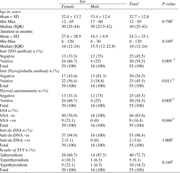
Table 1 Comparative analysis between genders
Sex
Total P-value
Female Male Age in years
Mean ± SD 32.4 ± 13.2 33.4 ± 12.4 32.7 ± 12.8
0.798ɫ
Min-Max 12 - 65 17 - 60 12 - 65
Median (IQR) 30 (25-44) 30 (23.5-42) 30 (25-42)
Duration in months
Mean ± SD 27.6 ± 28.9 16.1 ± 6.9 24.3 ± 25.1
0.349ƪ
Min-Max 6 - 120 6 - 30 6 - 120
Median (IQR) 16 (12-24) 15.5 (12-22.8) 16 (12-24)
Anti-TPO antibody n (%)
Negative 13 (33.3) 12 (75) 25 (45.5)
0.005 *†
Positive 26 (66.7) 4 (25) 30 (54.5)
Total 39 (100) 16 (100) 55 (100)
Anti-Thyroglobulin antibody n (%)
Negative 17 (43.6) 13 (81.3) 30 (54.5)
0.011*†
Positive 22 (56.4) 3 (18.8) 25 (45.5)
Total 39 (100) 16 (100) 55 (100)
Thyroid autoimmunity n (%)
Negative 13 (33.3) 12 (75) 25 (45.5)
0.005*†
Positive 26 (66.7) 4 (25) 30 (54.5)
Total 39 (100) 16 (100) 55 (100)
ANA n (%)
ANA -ve 30 (76.9) 16 (100) 46 (83.6)
0.046*ⱡ
ANA +ve 9 (23.1) 0 (0) 9 (16.4)
Total 39 (100) 16 (100) 55 (100)
Anti-ds-DNA n (%)
Anti-ds-DNA -ve 37 (94.9) 16 (100) 53 (96.4)
1.000ⱡ
Anti-ds-DNA +ve 2 (5.1) 0 (0) 2 (3.6)
Total 39 (100) 16 (100) 55 (100)
Results of TFT n (%)
Euthyroidism 26 (66.7) 14 (87.5) 40 (72.7)
0.349ⱡ
Hyperthyroidism 4 (10.3) 1 (6.3) 5 (9.1)
Hypothyroidism 9 (23.1) 1 (6.3) 10 (18.2)
Total 39 (100) 16 (100) 55 (100)
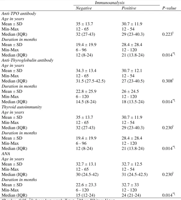
Table 2 Immunoanalysis of thyroid autoantibodies
Immunoanalysis
P-value
Negative Positive
Anti-TPO antibody Age in years
Mean ± SD 35 ± 13.7 30.7 ± 11.9
0.223ɫ
Min-Max 12 - 65 12 - 54
Median (IQR) 32 (27-43) 29 (23-40.3)
Duration in months
Mean ± SD 19.4 ± 19.9 28.4 ± 28.4
0.014*ƪ
Min-Max 6 - 96 12 - 120
Median (IQR) 12 (8-24) 21 (13.8-24)
Anti-Thyroglobulin antibody Age in years
Mean ± SD 34.3 ± 13.4 30.7 ± 12.1
0.308ɫ
Min-Max 12 - 65 12 - 54
Median (IQR) 31.5 (27.5-42.5) 27 (23-40.5)
Duration in months
Mean ± SD 22.8 ± 25.9 26 ± 24.5
0.014*ƪ
Min-Max 6 - 120 12 - 120
Median (IQR) 14.5 (8-24) 18 (13.5-24)
Thyroid autoimmunity Age in years
Mean ± SD 35 ± 13.7 30.7 ± 11.9
0.230ɫ
Min-Max 12 - 65 12 - 54
Median (IQR) 32 (27-43) 29 (23-40.3)
Duration in months
Mean ± SD 19.4 ± 19.9 28.4 ± 28.4
0.014*ƪ
Min-Max 6 - 96 12 - 120
Median (IQR) 12 (8-24) 21 (13.8-24)
ANA Age in years
Mean ± SD 32.7 ± 13.1 32.7 ± 12.5
0.230ɫ
Min-Max 12 - 65 12 - 54
Median (IQR) 30 (24.5-42) 31 (24.5-42.5)
Duration in months
Mean ± SD 22.6 ± 23.3 32.7 ± 33
0.014*ƪ
Min-Max 6 - 120 12 - 120
Median (IQR) 15 (12-24) 24 (21-24)
*P-value <0.05, ɫ Independent sample T-test, ƪ Mann-Whitney U test
Figure 1 Overview of our results
On comparative analysis; significantly higher proportion of women were found to be anti-TPO, anti-thyroglobulin, and ANA positive as compared to men. P values were 0.005, 0.011 and 0.046 respectively (Table 1). While differences in age, duration of urticaria and results of TFT were not statistically significant between the genders. P values were 0.798, 0.349 and 0.349 respectively (Table 1).
P values were statistically significant with duration of urticaria but not with age of the two groups (Table 2). Patients who had urticaria for longer duration were found to be thyroid autoantibodies positive. Furthermore, it was found that significantly higher proportion of
patients with positive TAAbs had
hyperthyroidism and hypothyroidism as
compared to those who are antibody negative
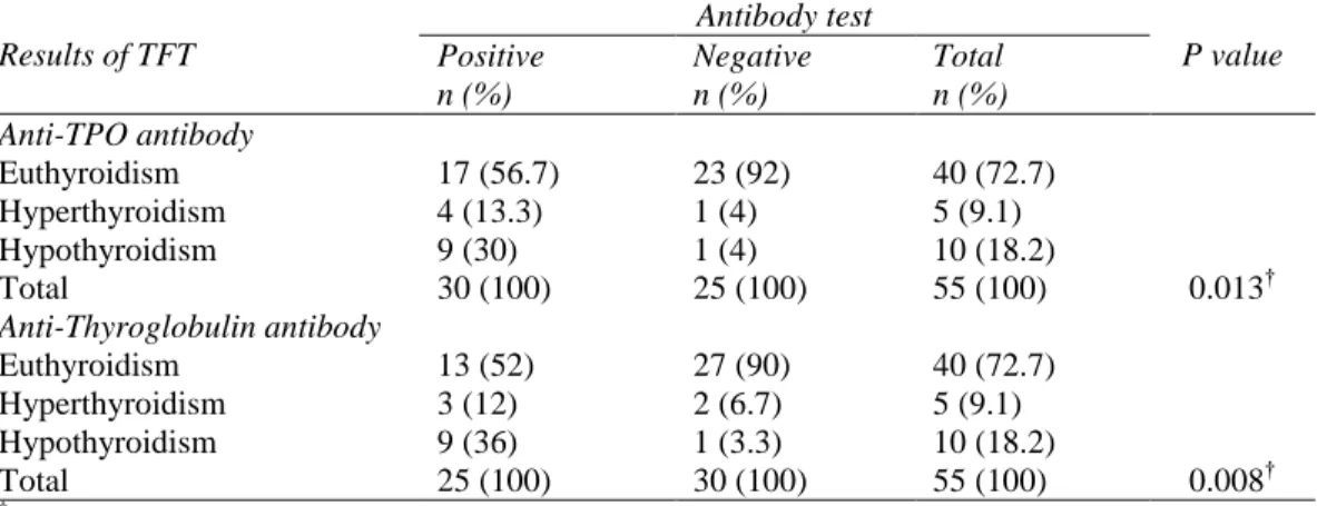
(Table 3). Overall, 27.3% patients in our study were diagnosed with thyroid dysfunction; 18.2% were having hypothyroidism while 9.1% had hyperthyroidism. Upon comparison of both autoantibodies; we found that patients who were
anti-Tg positive were more commonly
associated with hypothyroidism. P value was 0.008 (Table 3).
Discussion
The cause of chronic spontaneous urticaria (CSU) remains a complex mystery for centuries. However, in last few decades, detection of different functional autoantibodies in the sera of patients suffering from CSU provided a clue towards autoimmune aetiopathogenesis of this disease, although not in all sufferers but at least in half of them.8 The most important in this regard is thyroid autoimmunity. TA is a classical
autoimmune, organ-specific disorder. The
distinctive feature of TA is presence of multiple
thyroid autoantibodies (TAAbs). The only way to confirm it is by measuring serum TAAbs.9 First time urticaria was linked with thyroiditis by Midelfart et al.10 in 1983 and first ever thyroid autoantibody was detected in blood and reported by Leznoff et al.11 in the same year.
In our study, the mean±SD for age of CSU patients was 32.7±12.8 years. Similar age of patients was also reported by Aamir et al. from Pakistan7, Siddalingappa et al.12, Jindal et al. 13, and Mariyath et al.1 from India, and Kasumagic-Halilovic from Bosnia and Herzegovina.3
In our study, 70.9% patients were females while 29.1% were males with a male to female ratio of 1:2.4. Nearly similar ratios were also reported by
many others from Asia. Mariyath,1
Siddalingappa,12 and Kim14 reported ratios of 1:3, 1:2.1, and 1:4 respectively in their studies which is consistent with our study.
In our study, 54.5% patients were showing thyroid autoimmunity. Among these 45.5% patients were positive for both anti-TPO and anti-Tg antibodies while 9% patients were only anti-TPO antibody positive. Overall, 27.3%
patients were diagnosed with thyroid
dysfunction as well; 18.2% with hypothyroidism and 9.1% with hyperthyroidism.
Table 3 Comparative analysis of thyroid autoantibodies positive and negative cases
Results of TFT
Antibody test
P value Positive Negative Total
n (%) n (%) n (%)
Anti-TPO antibody
Euthyroidism 17 (56.7) 23 (92) 40 (72.7)
0.013†
Hyperthyroidism 4 (13.3) 1 (4) 5 (9.1)
Hypothyroidism 9 (30) 1 (4) 10 (18.2)
Total 30 (100) 25 (100) 55 (100)
Anti-Thyroglobulin antibody
Euthyroidism 13 (52) 27 (90) 40 (72.7)
0.008†
Hyperthyroidism 3 (12) 2 (6.7) 5 (9.1)
Hypothyroidism 9 (36) 1 (3.3) 10 (18.2)
Total 25 (100) 30 (100) 55 (100)
Kasumagic-Halilovic et al. from Bosnia and Herzegovina reported TA in 52.85% patients; anti-TPO positivity in 30% and anti-Tg positivity in 22.85% of patients and a further 18.57% patients were both antibodies positive. P value was <0.001. Thyroid function abnormalities was found in 11.43% patients.3
Aamir et al. from Pakistan15 compared frequencies of anti-Tg and anti-TPO antibodies in patients of chronic urticaria with healthy controls and reported anti-Tg positivity in 42% and anti-TPO positivity in 57%. P value was <0.001. Very similar results were published in another study from Pakistan, i.e. 42.6% and 57.4% CSU cases were positive for anti-Tg and anti-TPO antibodies, but they lacked the controls and all patients were females. P value was <0.05. Furthermore, 42% patients were diagnosed with hypothyroidism while none was having hyperthyroidism.7
Sibgatulina et al. from Russia noticed anti-TPO and anti-Tg antibodies were positive in 35.4% and 30.7% respectively in patients of chronic relapsing urticaria. They also proposed that these TAAbs are involved in mechanism of formation of chronic relapsing urticaria. TFT were not checked.16
Palma-Carlos et al. from Portugal notified TA in 28.6% of patients. Anti-TPO positivity in 26.8% and anti-Tg positivity in 23.2% of patients, while 21.4% patients were both antibodies positive. Only 7% patients were having abnormal TFT.17
Diaz-Angulo et al. from Spain performed a case-control study and detected anti-TPO and anti-Tg positivity in 20.4% and 15.2% of CSU patients respectively, while 8.7% were positive for both antibodies. P value was <0.001. They also reported that 14.9% patients were detected with some form of thyroid dysfunction; most
common were subclinical hypothyroidism
(5.8%) and subclinical hyperthyroidism
(5.5%).18
Bakos and Hillander strongly believed that there is a significant relation between CAU and TA. In their comparative study which involved CAU and CIU patients; they reported anti-TPO positivity in 42.3% of CAU and 13.6% of CIU patients (P=0.03) and presence of Helicobacter pylori infection in 90.9% of CAU and 46.7% of CIU patients (P=0.02). They also proposed that Helicobacter pylori infection might have a triggering role in aetiopathogenesis of CAU as well as in cases of CIU. TFT were not mentioned.19
Cebeci et al. in a case-control study from Turkey delineated presence of TAAbs in 29.28% of CSU patients and 5.52% of controls. Anti-TPO, anti-Tg and both antibodies were positive in 19.3%, 16.4% and 6.4% respectively. P value was <0.001. Among TAAbs positive cases; 24.4% were detected with thyroid dysfunction.20
Zauli et al. from Italy documented presence of TAAbs in 29% of chronic urticaria patients while 40% of these found to have concomitant thyroid dysfunction upon subsequent work up.6
Asero et al. also from Italy reported detection of circulating TAAbs in 26% of CSU patients, and one-fourth of these antibodies positive patients were diagnosed with hypothyroidism on further investigation.21
Results of all above mentioned studies are consistent with our research. We are describing a high frequency of TAAbs (54.5%) as compared to most of the studies, high percentage
of CSU patients (45.5%) with both
autoantibodies positive, anti-TPO is more common (54.5%) in our CSU cohort (Figure 1)
hypothyroidism (36%) (Table 3). These findings have not been documented in literature more often by others. Reasons could be multifactorial and explanations are also not unidirectional.
First; the frequencies of TAAbs in different populations is different because of some genetic
predisposition to autoimmune diseases.
Especially studies from Pakistan7,15 reported high frequencies of TA in patients of CSU, as by our study as well.
Second; our patients were mostly adolescents and young (mean age: 32.7 years) and Chanprapaph and colleagues from Thailand documented that chances of having CAU is more among CSU patients when they are younger than 35 years of age.22
Third; many of our patients were also ANA positive (16.4%). Lazurova and colleagues23 from Slovakia proposed that the presence of different non-organ-specific antibodies (like Rheumatoid factor and ANA etc.) may induce production of multiple other autoantibodies (organ-specific and non-organ-specific) by immune cells and also by thyroid epithelial cells which can result in production of high amount of TAAbs, and subsequently lead to development of autoimmune thyroid disease and CAU.
Recently, Bagnasco and colleagues24 from Italy have demonstrated high levels of circulating TAAbs in blood of chronic urticaria patients. They showed that when these autoantibodies bound to the surface of mast cells and basophils, can directly increase their sensitivity to many circulating agents leading to easy susceptibility of mast cells and basophils for degradation and release of chemokines and cytokines which then result in production of urticarial weals.
Conclusion
The existence of thyroid autoimmunity with chronic urticaria is an established fact. Early
detection of autoantibodies and thyroid
dysfunction may help in better control of urticaria. Proper management of thyroid disorder may also prevent patients from going in to disease related complications. We recommend to perform thyroid autoantibody profile and thyroid function tests in all patients of CSU, especially in females and all patients under 35 years of age.
References
1. Mariyath OKR, Binitha MP, Anilakumari VP, Biju G, Nikhila PK, Pradeep M. Association between chronic spontaneous urticaria and thyroid autoimmunity: a case control study from a tertiary care centre. Int J Res Dermatol 2017; 3(1): 64-8.
2. Kim DH, Sung NH, Lee AY. Effect of levothyroxine treatment on clinical symptoms in hypothyroid patients with chronic urticaria and thyroid autoimmunity. Ann Dermatol 2016; 28(2): 199-204. 3. Kasumagic-Halilovic E, Beslic N,
Ovcina-Kurtovic N. Thyroid autoimmunity in patients with chronic urticaria. Med Arch 2017; 71(1): 29-31.
4. Grattan CE, Francis DM, Slater NG, Barlow RJ, M. Graves MW. Plasmapheresis for severe, unremitting, chronic urticaria. Lancet. 1992; 339(8801): 1078-80.
5. O’Donnell BF, Barr RM, Black AK, Fransis DM, Kermani F, Niimi N. et al. Intravenous immunoglobulin in autoimmune chronic urticaria, Br J Dermatol 1998;138(1):101-6. 6. Zauli D, Deleonardi G, Foderaso S, Grassi
A, Bortolotti R, Ballardini G, et al. Thyroid autoimmunity in chronic urticaria. Allergy Asthma Proc 2001; 22: 93-5.
7. Aamir IS, Tauheed S, Majid F, Atif A. Frequency of autoimmune thyroid disease in chronic urticaria. J Coll Physicians Surg Pak 2010; 20(3): 158-61.
9. Kawashima A, Tanigawa K, Akama T, Yoshihara A, Ishii N, Suzuki K. Innate immune activation and thyroid autoimmunity. J Clin Endocrinol Metab. 2011; 96(12): 3661-71.
10. Midelfart K, Moseng D, Kavli G, Stenvold SE, Volden G. A case of chronic urticaria and vitiligo, associated with thyroiditis, treated with PUVA. Dermatologica 1983; 167(1): 39-41.
11. Leznoff A, Josse RG, Denburg J, Dolovich J. Association of chronic urticaria and angioedema with thyroid autoimmunity. Arch Dermatol 1983; 119(8): 636-40. 12. Siddalingappa K, Murthy SC, Herakal K,
Deepika M. Thyroid autoantibodies in chronic urticaria: a case-control study in a South Indian population. Iran J Dermatol 2017; 20: 50-3.
13. Jindal R, Roy S, Nagrani P. Chronic idiopathic urticaria and autoimmunity: frequency and association in patients with positive versus negative autologous serum skin test. Int J Res Med Sci. 2017; 5(3): 1103-6.
14. Kim YS, Han K, Lee JH, Kim NI, Roh JY, Seo SJ, et al. Increased risk of chronic spontaneous urticaria in patients with autoimmune thyroid diseases: a nationwide, population-based study. Allergy Asthma Immunol Res 2017; 9(4): 373-7.
15. Aamir IS, Tauheed S, Majid F, Atif A. Serum antithyroid antibodies in female patients with chronic urticaria. J Coll Phys Surg Pak 2008; 18(8): 498-501.
16. Sibgatulina NA, Kuzmina NS, Rakhmatullina NM, Gevarzieva VB. Autoantibodies to thyroid gland antigens in chronic relapsing urticaria. Zh Mikrobiol Epidemiol Immunobiol 2002; (5): 69-71. 17. Palma-Carlos AG, Palma-Carlos ML.
Chronic urticaria and thyroid autoimmunity.
Eur Ann Allergy Clin Immunol. 2005; 37(4): 143-6.
18. Dıaz-Angulo S, Lopez-Hoyos M, Munoz Cacho P, Fernandez M, Lopez-Escobar M, Rodrıguez F, et al. Prevalence of thyroid autoimmunity in Spanish patients with chronic idiopathic urticaria: a case–control study involving 343 subjects. J Eur Acad Dermatol Venereol 2016; 30(4): 692-3. 19. Bakos N, Hillander M. Comparison of
chronic autoimmune urticaria with chronic idiopathic urticaria. Int J Dermatol 2003; 42(8): 613-5.
20. Cebeci F, Tanrikut A, Topcu E, Onsun N, Kurtulmus N, Uras AR. Association between chronic urticaria and thyroid autoimmunity. Eur J Dermatol 2006; 16(4): 402-5.
21. Asero R, Lorini M, Tedeschi A. Association of chronic urticaria with thyroid autoimmunity and raynaud phenomenon with anticentromere antibodies. J Allergy Clin Immunol 2003; 111: 1129-30.
22. Chanprapaph K, Iamsumang W, Wattanakrai P, Vachiramon V. Thyroid autoimmunity and autoimmunity in chronic spontaneous urticaria linked to disease severity, therapeutic response, and time to remission in patients with chronic spontaneous urticaria. Biomed Res Int. 2018; 2018: 9856843.
23. Lazúrová I, Benhatchi K, Rovenský J, Kozáková D, Wagnerová H, Tajtáková M, et al. Autoimmune thyroid disease and autoimmune rheumatic disorders: a two-sided analysis. Ann N Y Acad Sci. 2009; 1173: 211-6.