The Effect of Estrogen on Cardiac-Skeletal Muscle Biomarkers Following Prolonged Aerobic Exercise in Eumenorrheic Women
Timmons Williams
An honors thesis submitted to the faculty of the University of North Carolina at Chapel Hill in partial fulfillment of the requirement for the honors thesis in the Department of
Exercise and Sports Science.
Approved by:
Anthony C. Hackney Ph.D., D.Sc.
Rebecca Battaglini, M.A.
ABSTRACT
TIMMONS E. WILLIAMS: The Effect of Estrogen on Cardiac Biomarkers Following Prolonged Aerobic Exercise in Eumenorrheic Women
(Under the direction of Anthony C. Hackney, Ph.D., D.Sc.)
Purpose: This study assessed the influence of estrogen (E2) on skeletal muscle creatine kinase (CK) and cardiac muscle CK (CK-MB) responses to prolonged aerobic exercise. Elevations in these biomarkers are indicative of muscle trauma-damage. Methods: Eumenorrheic women (n=10) who were physically active completed two 60-minute treadmill running sessions at ~60-65% maximal intensityduring low E2
(midfollicular menstrual phase) and high E2 (midluteal menstrual phase) hormonal conditions. Blood samples were collected prior to exercise (following supine rest), immediately post-, 30 min post-, and 24 hr post-exercise to determine changes in muscle biomarkers. Resting blood samples confirmed appropriate E2 hormonal levels Results:
CK concentrations increased following exercise and at 24 hr post-exercise were higher in the midfollicular phase (p<0.001). However, CK-MB concentrations were unaffected by E2 level or exercise (p=0.442). Conclusions: E2 levels influence the CK biomarkers
response for skeletal but not cardiac muscle following prolonged aerobic exercise. These findings imply elevated E2 is protective of skeletal muscle from exercise-induced
ACKNOWLEDGEMENTS
I am eternally grateful to Dr. Anthony C. Hackney for allowing me to work with him
on my senior honors thesis. The enthusiasm and passion he puts into all of his work
is contagious. Thank you to my committee members, Dr. Barbara Osborne and Mrs.
Rebecca Battaglini, for graciously agreeing to review my work and for giving me
valuable feedback. To my study subjects, I am so thankful for your willingness to
participate in my study and to run for 60-minutes on a treadmill on two separate
occasions. Lastly, thank you to all of the Endocrine Group: Liz, Michelle, Colin, and
TABLE OF CONTENTS
PAGE
LIST OF TABLES ……….. 7
LIST OF FIGURES ……… 8
CHAPTER I ……….. 9
INTRODUCTION……….….. 9
Basis For Study……….….11
Purpose………..11
Research Hypothesis………...11
Definition of terms………..11
Estrogen………...….11
Creatine Kinase………...11
Delimitations ……….…11
Limitations ……….…12
Significance of study ……….…13
CHAPTER II………..… 14
REVIEW OF LITERATURE………... 14
CHAPTER III……….………... 20
METHODOLOGY……….………... 20
Participants……….... 20
Instrumentation………...………...21
Protocol……….………..… 22
Orientation Session I……….………. 22
Experimental Sessions……….………..………… 25
Follow Up Blood Draws Session III and V……….……...… 26
Blood Procedures……….……….……. 26
Hematocrit……….……….………… 26
Hemoglobin……….………... 27
Plasma Volume Shift……….……….……… 27
Creatine Kinase (CK), CK-MB, Estradiol, and Lactate Assays………...……….. 27
Data Analysis……….……….……….. 28
CHAPTER IV……….……….………29
RESULTS……….……….………29
Subject Characteristics……….29
Hormonal Condition Determination…………..………..30
Experimental Treadmill Running Protocol (60 min)………..31
Plasma Volume Shift………..32
Blood Responses to Experimental Treadmill Running Protocol…………...……33
CHAPTER V……….……….………..36
DISCUSSION……….……….………36
Hormonal Condition Determination……….…….………36
Experimental Treadmill Running Session……….………....……37
Creatine Kinase (Total CK) ………..…….………....…….39
Creatine Kinase- MB…………..………...………....……….39
Limitations………..41
Future Studies………42
APPENDIX A: SAMPLE SIZE CALCULATIONS……….……43
APPENDIX B: INFORMED CONSENT……….……..44
APPENDIX C: MEDICAL SCREENING FORMS……….….……49
Medical History Questionnaire……….………50
Physical Examination Screening……….…55
APPENDIX D: ACSM METABOLIC EQUATION FOR DETERMINING EXERCISE INTENSITY………..56
APPENDIX E: MENSTRUAL CYCLE QUESTIONNAIRE………58
APPENDIX F: FOOD LOG……….61
APPENDIX G: DATA COLLECTION FORMS………..…………63
Orientation Session………64
Experimental Session………65
Hematocrit and Hemoglobin……….………66
Subject Compliance………67
LIST OF TABLES
1. Bruce protocol treadmill test
2. Participant physical and menstrual characteristics
3. Descriptive data (mean ± SD) for VO2, HR and RPE for each experimental
treadmill running protocol in each hormonal condition (MF and MF)
4. Mean (± SD) plasma volume shifts
5. Mean ± SD for CK at rest, immediately post exercise (IP), 30 minutes post
exercise (30P), and 24 hours post exercise (24P)
6. Mean ± SD for CK-MB at rest, immediately post exercise (IP), 30 minutes post
LIST OF FIGURES
1. An overview of the menstrual cycle
CHAPTER I
INTRODUCTION
Basis For Study:
The protective benefits of estrogen on the cardiovascular system are well
established. There is still debate, however, on the influence of estrogen on cardiac
muscle damage during and following prolonged aerobic exercise, and if estrogen
acts as a protectant against cardiac damage, and the mechanism by which estrogen
protects the cardiac muscle (Squadrito et al., 1997). The intent of this study is to
examine the influence of estrogen on cardiac muscle in women following exercise.
Estrogen is a sex and steroid hormone produced in the ovaries that regulates
the menstrual and reproductive cycles in women. There are three types of estrogen:
estrone, estradiol, and estriol. Estradiol is the strongest and most potent of the three
types and is considered the most important type of estrogen in females between the
menarche and menopause stages of life (National Institute on Aging, 2013).
Estrogen acts by diffusing through a cell membrane into the cytoplasm, binds to an
estrogen receptor and then moves into the nucleus in order to modulate the
expression of many genes (Babiker et al., 2001). There are two dominant types of
estrogen receptors: ER and ER . α β
Estrogen receptors have been found to be present in many different types of
tissues including skeletal and cardiac muscle (Babiker et al., 2001). Skeletal and
cardiac muscle cells are structurally and functionally very similar as they both
consist of the structural proteins actin and myosin myofilaments that form
creating a striated appearance. This structural similarity allows both cells to
generate muscle contractions through actin and myosin cross-bridge cycling.
It is widely known that unaccustomed exercise results in temporary muscle
damage. Estrogen has shown to protect skeletal muscle from inflammation in
various studies through its attenuation of circulating cytokines and as a result
attenuating inflammation as a result of the inflammation. (Tiidus, P.,2003;
Pfeilschifter et al., 2002; Pottratz et al., 1994; Puder et al., 2001; Schwarz et al., 2000). However, a clear relationship between estrogen and inflammation after exercise induced cardiac muscle damage in women is yet to be determined. As cardiac and
skeletal muscles are very similar structurally and functionally, it is logic to assume
the protective effect of estrogen observed for skeletal muscle would exist for cardiac
too.
Creatine kinase is a leakage enzyme used to make ATP via phosphorylation of
ADP from creatine phosphate by catalyzing the reversible phosphorylation of
creatine by ATP to form phosphocreatine + ADP (Cornell University, 2013). Creatine
kinase is composed of two types of monomers: the M and B subunits. Together,
these monomers form the isoenzyme creatine kinase-MB (CK-MB), which is present
in highest concentrations in cardiac muscle. Creatine kinase is assayed in blood tests
as a marker of myocardial damage as elevated levels of CK indicate muscular
damage. As prolonged exercise causes increased muscular damage, CK-MB levels
Purpose
The purpose of this study will be to determine if differing levels of estrogen
during a menstrual cycle influences the cardiac muscle damage markers of CK and
CK-MB following moderate intensity prolonged exercise in eumenorrheic women.
Research Hypothesis
If estrogen is related to the cardiac biomarker CK and CK-MB then higher
estrogen levels in eumenorrheic women will attenuate CK and CK-MB
concentrations following moderate intensity exercise.
Definition of Terms
Estrogen
Estrogen is a 18-carbon corticosteroid molecule secreted by the ovary in
females. Estrogen is primarily involved in the development and reproductive
functions in women, but also plays a role in many cardiovascular, musculoskeletal,
immune and central nervous system functions (Enns & Tiidus 2010). Creatine Kinase
Creatine Kinase is an enzyme in the muscles that functions by making ATP through the phosphorylation of ADP from creatine phosphate. As creatine kinase is found mostly in muscle tissue, it is the most common blood marker of muscle damage
(Ebbeling & Clarkson, 1989). High CK values are an indicator of muscle damage. Eumenorrhea
Eumenorrhea is normal or regular menstruation by a woman that occurs at least nine times in the preceding year and lasts between 21 to 35 days.
Amenorrhea is an absence of menstruation. An absence of three or more periods in a row classifies women as amenorrheic. Strenuous physical activity, excessive weight loss, use of some antidepressants, and stress can cause a woman to have amenorrhea. Delimitations
1. Participants will be healthy females between the ages of 18-30.
2. Must be eumenorrheic and not currently taking or have taken oral contraceptives or other hormone therapy six months prior to participation in this study.
3. Have not sustained an injury within the last six months that has limited ability to exercise.
4. Must not be currently taking anti-inflammatory medicines, such as ibuprofen, naproxen, or aspirin.
5. Are not pregnant or become pregnant during the study.
6. Become ill with an immune responding condition, such as a cold or respiratory infection during the study.
7. Must have a current minimum training volume of 3-5 days a week, 45-120 minutes per session of aerobic activity, and have a VO2 max of at least 45ml/kg/min.
8. Abstain from strenuous physical activity and maintain a diet similar in calories and carbohydrate content 24 hours prior to experimental protocol.
9. Women must be premenopausal.
10. Estrogen’s role in cardiac illnesses or pathologies will be not considered within this study.
Limitations
1. Results may not be applicable to men and some women (amenorrheic or post-menopausal).
2. Subjects may not comply with specific pre-test instructions.
3. CK and CK-MB concentrations will be the only cardiac biomarkers measured in the blood. Other cardiac biomarkers such as troponin T (cTnT), troponin I (cTnI), and lactate dehydrogenase (LDH) will not be measured.
4. Technician error Significance of study
Understanding estrogen’s role on cardiac muscle damage following high intensity exercise is important for both women and researchers. There has been an increased amount of research on post-menopausal women and the possibility of a correlation between decreased estrogen levels and cardiac illness over the past few decades.
Chapter II
REVIEW OF LITERATURE
In this review, the influence of estrogen on exercise-induced cardiac muscle
damage will be examined. There appears to be little research in the influence of
estrogen on cardiac muscle in humans. However, skeletal and cardiac muscle is
structurally and functionally very similar and since there is research supporting
estrogen’s role in skeletal muscle, it can logically be hypothesized that estrogen
could play a role in cardiac muscle also. This review will discuss how estrogen acts a
protectant against muscle damage, research supporting estrogen’s protective role in
skeletal muscle, and research on estrogen’s influence on cardiac muscle.
Estrogen is a steroid hormone that regulates and maintains sexual and
reproductive function in females. While estrogen is present in both males and
females, it is significantly higher in women in their reproductive years compared to
males. There are three types of estrogen: estrone, estradiol, and estriol. Estrogen
affects many physiological systems including the cardiovascular, musculoskeletal,
immune and central nervous systems. The most potent and abundant form of
estrogen that has the largest effect on these systems is 17 -estradiol (Enns & Tiidus,β
Creatine kinase (CK) and the isoenzyme creatine kinase-MB (CK-MB) are two
cardiac biomarkers that are examined during blood assays as markers of cardiac
damage. CK-MB is very useful indicator of myocardial damage as only myocardium
tissue has substantial amounts of it present. During unaccustomed exercise the
muscles of the body such as the heart are damaged which causes a release of CK and
CK-MB. This was demonstrated in a study of fifteen young men who performed 90
minutes of cycling for 3 consecutive days. Blood assays showed that CK levels were
significantly elevated after the first training protocol and steadily increased after
each consecutive training protocol (Totsuka et al., 2002). This increase in CK levels
was also shown to correlate with increased CK-MB levels. This was seen in a study
on mongrel dogs that analyzed ventricular myocardium for total CK and CK-MB
content and found that CK-MB levels were slightly although insignificantly higher in
trained versus caged dogs (Miller et al., 1989). Another study had rats swim for 3.5
hours straight and then sacrificed the rats immediately, 2 hours, 24 hours, and 48
hours post exercise. The researchers found that CK-MB was released into circulation
in the highest concentrations immediately post exercise and returned to normal 24
hours post exercise (Chen, Serfass & Apple, 2000). This indicates that intense
endurance exercise induces myocardial damage and is highest immediately post
exercise.
Estrogen is an antioxidant and membrane stabilizer making it unique as a
steroid hormone. During physical activity, oxygen consumption increases to meet
metabolic demand. This increase in oxygen consumption corresponds with an
molecules, which cause inflammation. Estrogen is structurally similar to cholesterol,
which allows it to insert within membrane phospholipids (Ennus & Tiidus &; 2010).
This allows estrogen to act as a membrane stabilizer by preventing disruptions of
the muscle membrane, which leads to muscle damage and inflammation. (Tiidus,
2005). These properties have allowed researchers to deduce that estrogen may help
mitigate the effects on post-exercise muscle damage (Tiidus, 2005). However, the
direct effect of estrogen on muscle structural damage after exercise is still debated.
The potential influence of estrogen on post-damage inflammation in various
tissues has increased with research in the field over the past two decades. In the few
human studies conducted, estrogen has shown to prevent inflammation related
leucocyte infiltration into both skeletal and cardiac muscle after unaccustomed
exercise or damaging insult (Tiidus, 2005; Tiidus & Bombardier et al., 2001). Bar
and his associates (1998) proposed that that the levels of creatine kinase after
exercise-induced stress differed between males and females as a result of estrogen’s
protective role on the female muscle membrane. Research conducted in animals has
found that estrogen influences skeletal muscle damage and leukocyte infiltration
after exercise (Komulainen et al., 1999). The infiltration of leukocytes such as
neutrophils and macrophages remove damaged muscle tissue and stimulate the
repair processes. A study conducted by Tiidus and Bombardier reported that male
rats had significantly attenuated neutrophil infiltration into skeletal muscles 24
hours after running exercise compared to female rats. When the same male rats
were supplemented with estrogen, however, they exhibited a similar response of
Estrogen is also thought to effect the activation and proliferation of satellite
cells. Satellite cells proliferate in response to injury in order to provide the
necessary materials needed to initiate muscle growth and repair. In a study
conducted in male rats, Tiidus et al. (2005) observed that the skeletal muscles of
estrogen-supplemented male rats had increased numbers of satellite cells 72 hours
after downhill running. Further studies performed by Tiidus et al. (1999; 2005;
2008) found that sex-mediated differences in muscle fiber regeneration and satellite
cell numbers may be directly influenced by estrogen.
Estrogen’s influence on cardiovascular health is well established. In relation
to cardiac disease, pre-menopausal women have a lower incidence of cardiac illness
compared to age-matched men (Milne & Noble; 2008). It has also been shown that
estrogen reduces the degree of myocardial injury following ischaemia-reperfusion
injury (Booth et al., 2008). Oestradiol-17 , a form of estrogen, was also shown to β
positively affect the heart in a study on the acute effects of oestradiol-17β on
myocardial ischaemia. Researchers found that oestradiol-17β had a beneficial effect
on myocardial ischaemia in women with coronary artery disease due to a direct
coronary-relaxing effect and to peripheral vasodilation (Rosano et al., 1993).
Research, however, is limited in respect to the influence of estrogen on healthy,
pre-menopausal women’s cardiac muscle tissue. With this said, research has shown to
support that the levels of creatine kinase (CK), a prominent blood biomarker of
cardiac muscle damage, is higher in male rats compared with female rats following
muscle injury (Bar et al.; 1988, Amelink et al.; 1990). These findings, however, can
from the blood due to sex differences. Rose et al and Smith et al. found that
postmenospausal women and premenarchial girls both display high levels of serum
CK activity that correspond with their low levels of estrogen (Lane and Roses, 1981;
Smith et al., 1979). This is thought to be due to estrogens stabilizing effect on the
muscle membrane, which in turn reduce the level of CK activity caused by muscle
damage. A sex difference in serum CK levels was also shown to be present at rest
and after acute eccentric exercise training suggesting that estrogen might be
responsible for this difference (Amelink & Bar, 1986).
In summary, research on the role of estrogen on cardiac muscle damage in
humans after prolonged endurance exercise is still unclear. The knowledge gained
through this research will be beneficial towards expanding the area of research
literature pertaining to estrogens role in protecting the heart against self-induced
muscle damage from exercise. As demonstrated in this review, estrogen is an
important hormone in many physiological systems as well as sexual and
reproductive functions. After looking at the research pertaining to estrogens effect
on skeletal muscle, it has been suggested in this review that estrogen may attenuate
cardiac muscle damage in a similar fashion. The results of this study will contribute
to this body of knowledge and provide insight into what role estrogen has on the
Gilbert, S. F. (2013). Hormones and Mammalian Egg Maturation. Retrieved from
Chapter III
METHODOLOGY
Participants made five visits to the Applied Physiology Laboratory (APL) at
the University of North Carolina (UNC-CH). The first visit consisted of an orientation
where consent will be obtained. The visit also determined if a participant was
eligible for the study and allowed for the acquirement of descriptive characteristics,
record of menstrual history, and measurement of maximal oxygen consumption
(VO2max). The second session was an experimental protocol requiring subjects to
run 60 minutes on a treadmill at ~60-65% of their predetermined VO2max. The
variables CK and CK-MB were measured at baseline, immediately post-exercise, and
30 minutes post-exercise. The third session was the day after the exercise protocol
and was a 24-hour post-exercise blood draw. The fourth and fifth sessions repeated
the second and third sessions.
Participants
Fifteen healthy, highly trained, pre-menopausal women between the ages of
18-30 were recruited. Samples size was estimated from previous research in the
literature to ensure adequate statistical power (Appendix A). Participants eligible
for the study needed to be eumenorrheic for the past six months, not taken oral
contraceptives or other hormone therapy prior to participation in the study, and not
be currently using anti-inflammatory medications, such as ibuprofen, naproxen, or
aspirin. Participants were also required to report a current minimum training
volume of 3-5 days of aerobic activity lasting between 45-120 minutes per sessions
exercise. If a participant reports an injury in the past 6 months, a physician’s signed
clearance must be obtained before beginning participation in the study. Participants
were dropped from the study if they become pregnant or ill with an immune
responding condition, such as a cold or respiratory infection. Consent was obtained
from the participants after being cleared of all eligibility criteria and thoroughly
informing them of the experimental protocol and any risks or rewards related to the
study. Participants were asked to abstain from strenuous physical activity and
maintain a normal diet in calories and carbohydrate content 24 hours prior to the
two experimental trials.
Instrumentation
Height was measured with a portable stadiometer (Perspectives Enterprises,
Portage, MI USA) and body mass was measured using a mechanical scale (Detecto,
Webb City, MO USA). Skinfolds was measured with a Lange skin caliper (Beta
Technology, Inc., Santa Cruz, CA USA). Maximal oxygen consumption was measured
during a continuous, incremental treadmill run on a Quinton Q65 treadmill (Bothell,
WA USA). Respiratory gases were obtained for the orientation and two
experimental sessions using the Parvo Medics TrueMax 2400 Metabolic System
(Parvo Medics, Salt Lake City, UT USA). A continuous measurement of heart rate
was taken using the Polar telemetry system (Polar Electro, Inc., Lake Success, NY
USA). Ratings of perceived exertion (RPE) were determined using Borg’s 20-point
scale. Hematocrit was measured using an Adams MHCT II microhematocrit
centrifuge (Becton Dickinson, Franklin Lakes, NJ USA) and an International
USA). Hemoglobin will be determined from a Stanbio Lab Hemopoint H2 analyzer
(Boerne, TX USA). Whole blood samples were kept in an IEC Centra-8R refrigerated
centrifuge (International Equipment Company, Needham Heights, MA USA) and the
resultant separated plasma were stored and frozen at -80˚C. Serum CK and CK-MB
were measured using a Ortho-Clinical Diagnostics Vitros DT60II analyzer
(Rochester, NY). Plasma E2 was measured using the enzyme-linked immunoassay
(ELISA) technique, (Abnova Corporation, Taiwan, China).
Protocol Pre-screening
Interested participants in the study emailed the investigator directly in order
to determine eligibility. Eligible participants then were scheduled for an orientation
and experimental session.
Orientation Session I
The orientation session took place in the Applied Physiology Laboratory at
The University of North Carolina, Chapel Hill. Participants arrived and were
informed of the experimental protocol, possible risks and rewards associated with
the study, and sign an informed consent form. The Office of Human Research Ethics,
Institutional Review Board, of The University of North Carolina, Chapel Hill,
approved the informed consent form before beginning the experimental protocol.
After giving informed consent, participants filled out the Exercise and Sports Science
medical history questionnaire, and underwent and passed a medical screening.
Next, a VO2max test was performed using a continuous, incremental
treadmill protocol. Participants performed a five-minute warm up before beginning
the test consisting of biking at their preferred pace and light stretching. After the
warm up, resting oxygen consumption VO2 were measured for three minutes.
Participants then began the exercise test using the Bruce treadmill protocol (ACSM
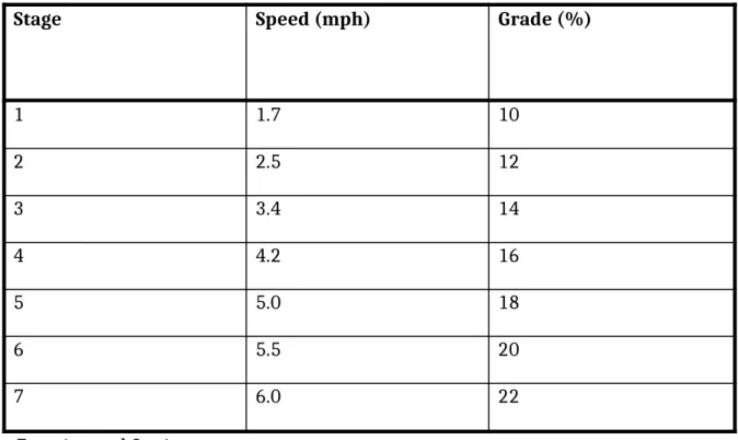
Guidelines, 2010). Table 1 lists the Bruce protocol treadmill test. Heart rate and
respiratory gas exchange were measured continuously throughout the test as well
as the 20-point scale ratings of perceived exertion (RPE). At the conclusion of the
test, participants were allowed to recover either actively or passively and were
permitted to leave the laboratory when heart rate drops below 100 beats per
minute (bpm). The VO2 test was determined as a maximal test rather than a peak
test through the participants displaying three of the four criteria: a plateau or
decrease in VO2 with increases in workload, respiratory exchange ratio (RER)
greater than or equal to 1.1, RPE ≥ 18, and a HR within 10% of predicted heart rate
max (220-age of participant). ACSM guidelines were used to estimate exercise
intensity corresponding to ~60-65% of VO2 (American College of Sports Medicine,
Table 1: Speed in miles per hour (mph) and grade for each three-minute stage of the
Bruce protocol incremental exercise test.
Stage Speed (mph) Grade (%)
1 1.7 10
2 2.5 12
3 3.4 14
4 4.2 16
5 5.0 18
6 5.5 20
7 6.0 22
Experimental Sessions
Experimental Sessions II and IV
Participants visited the Applied Physiology Laboratory once during their
mid-follicular (MF, low estrogen) and mid-luteal (ML, high estrogen) phases
respectively. Appointment dates were determined using the forward counting
method (Chavanne & Gallup, 1998). The two experimental sessions were
counterbalanced to prevent order effects. The testing occurred during the MF and
ML phases of the menstrual cycle in order to find the largest differences between
naturally fluctuating estradiol levels and maximize effect size. The MF phase
typically occurs between days 3-7 of the menstrual cycle when estradiol is lower,
whereas, the ML phase occurs roughly between days 20-24 of the menstrual cycle
when estradiol is much higher. These date ranges can vary among participants
depending upon the length of a participant’s menstrual cycle and are only used as
approximations. These will be calculated and are reported exactly once the study is
complete.
Participants were asked to refrain from intense physical activity and
maintain a similar diet 24 hours prior to the experimental sessions. Participants
were asked if they followed all guidelines prior to the testing sessions, and a food
diary will be used to ensure similar nutrient intake between trials, as well as
adequate caloric and carbohydrate intake. If participants complied to these terms,
the experimental trial continued. Participants rested supine in a quiet environment
for 10 minutes. After this resting period, blood was obtained via individual blood
draws or catheter, based on participant preference. The blood sample was split in
Vacutainer ™ tubes and then immediately put on ice. Participants then completed a
five minute warm-up consisting of cycling and light stretching, followed by 60
minutes of treadmill running at their previously determined workload of 65% of
VO2 as determined by the ACSM formula (ACSM, 2010). Blood samples were taken
immediately post exercise and 30 minutes post exercise and promptly put on ice.
Plasma was separated from blood samples and stored until later analysis for CK and
CK-MB. Blood samples will be stored for three years.
The 60-minute time length for the treadmill protocol was chosen to mimic a
normal exercise training session. Previous research from the Applied Physiology
Laboratory at the University of North Carolina (UNC-CH) has shown that this length
of time as sufficient in inducing muscle creatine kinase responses (Kallman, 2011).
Follow Up Blood Draws Session III and V
Participants returned to the laboratory 24 hours post exercise for additional blood
draws. Participants were asked to lay supine for 10 minutes in a quiet environment.
Blood samples were obtained to measure for CK and CK-MB using the same
procedures specified in sessions I and II. Participants were asked to refrain from
any exercise that is more than what is necessary for activities of daily living for 24
hours after the blood draw.
Blood Procedures Hematocrit
Immediately post each exercise test, resting and post-exercise Hematocrit
(Hct) values were determined in triplicate from whole blood samples. Whole blood
Pittsburgh, PA) and sealed using Critoseal (Krackeler Scientific, Albany, NY).
Capillary tubes were spun in a microhematocrit centrifuge for three minutes at
10,000 RPM and then placed on a hematocrit wheel to determine the hematocrit
values of each sample. A mean was calculated from three samples and used in data
analysis.
Hemoglobin
Resting, immediately post-exercise, and recovery hemoglobin (Hb) values
from each experimental session were measured in duplicate from the whole blood
samples using the Stanbiolab Hemopoint H2 analyzer (Boerne, TX). These values
were determined immediately after completion of the exercise tests. A mean was
calculated from three samples and used in data analysis.
Plasma Volume Shift
Hb and Ht values were used to calculate exercise induced plasma volume
shifts according to the Dill and Costill equation (Dill & Costill 1974).
Creatine Kinase (CK and CK-MB), and Estradiol Assays
To separate plasma and serum from whole blood, the blood samples were
centrifuged at 3,000 x g for 10 minutes. The separated plasma and serum were
transferred to storage tubes and stored until analyses are conducted.
Enzyme-linked immunoassay (ELISA) techniques (Abnova Corporation, Taiwan, China) were
used to measure plasma estradiol concentrations. The assay manufacturer reports a
minimum detectable concentration of 2.0 pg/mL. The Vitros DT60II analyzer uses a
All blood assays (unknown samples) were performed in duplicate while standards
were done in triplicate.
Data Analysis
SPSS statistical software was used to analyze data (version 18.0, Chicago, IL
USA). Significance for all data was set at < 0.05. Descriptive statistics was shownα
as means ± standard deviations (SD). Effect size was calculated for all significant
measures to determine if significance has practical meaning.
A 2x3 (estradiol level x time) totally within, repeated measures ANOVA and
where appropriate, Bonferoni post hoc test, was used to assess the effects of
estradiol on CK and CK-MB concentrations. A 2x2 (estradiol level vs. time) ANOVA
was used to evaluate the percent change (% ) in CK values between pre and postΔ
exercise, as well as pre- exercise and recovery during each menstrual cycle phase
(estradiol level). These two same procedures were used to assess the effects of
estradiol on CK-MB. The % will be calculated using this equation:Δ
% = [(Post-exercise – Baseline)/Baseline] x 100Δ
At dependent t-test analysis was used to evaluate statistical significance
CHAPTER IV
RESULTS
Originally, the goal of the study protocol was to have a sample size of fifteen
subjects in order to ensure adequate statistical power based off of previous research
literature. However, due to difficulties of finding women who met the inclusion
criteria, only ten women completed the study protocol.
Additionally, one participant was unable to complete the second
experimental session treadmill running (ML phase) due to medical reasons.
Therefore, the subject’s missing data were approximated using mean substitution
through the SPSS (version 18.0) missing variables function in order to maintain the
integrity of both experimental session results within the statistical analysis.
Subject Characteristics
Ten highly trained, eumenorrheic women participated in the study. The
subjects met the inclusion criteria: healthy females between the ages of 18-30 years,
eumenorrheic, not currently taking or have taken oral contraceptives or other
hormone therapy 6 months prior to participation, have not had an injury in the
previous six months, not currently taking anti-inflammatory medication, and have a
current minimum training volume of 3-5 days per week, 45-120 minutes per session
of aerobic activity and a VO2 max of at least 45 mL/kg/min. The physical and
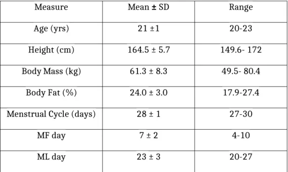
menstrual characteristics of the 10 subjects who completed the study are reported
Table 2: Participant physical and menstrual characteristics (n=10)
Measure Mean ± SD Range
Age (yrs) 21 ±1 20-23
Height (cm) 164.5 ± 5.7 149.6- 172
Body Mass (kg) 61.3 ± 8.3 49.5- 80.4
Body Fat (%) 24.0 ± 3.0 17.9-27.4
Menstrual Cycle (days) 28 ± 1 27-30
MF day 7 ± 2 4-10
ML day 23 ± 3 20-27
MF= Midfollicular; ML=Midluteal phases of menstrual cycle
VO2Peak Testing
The criteria for a maximal oxygen consumption test was not achieved by all
subjects, thus all maximal oxygen consumption tests (VO2max) are referred to as
VO2 peak tests. The average VO2peak (mean ± SD) was 53.5 ± 4.7 ml/kg/min (range
45.1- 60.0 ml/kg/min). The average peak RPE obtained was 18 ± 1Borg Units (range
16-20 Borg Units). The average peak heart rate (HR) was 191 ± 1 bpm (range
180-208 bpm). The time to reach VO2peak ranged between 12:35 to 16:10 mins:sec.
Based upon the results of this maximal testing, the average calculated 65% of VO2
peak to use in the 60-minute exercise session was 34.8 ± 3.0 ml/kg/min.
Hormonal Condition Determination
Menstrual cycle length was calculated for each subject. The average
menstrual cycle length (mean ± SD) was 28 ± 1 days (range 27- 30 days). Individual
mentioned in the methodology chapter. The onset of menses was denoted as day 1.
Subjects were tested at 7 ± 2 days (range 4-10 days) after the onset of menses
during the midfollicular phase (MF). Subjects were tested at 23 ± 3 days (range
20-27) after the onset of menses during the midluteal phase (ML) of the cycle.
Hormonal analysis of resting blood samples at the MF and ML testing
sessions for 17β-estradiol (E2) confirmed the appropriate menstrual phase and
hormonal status of the subjects. The MF E2 concentration was 39.8 ± 18.3 pg/ml,
while the ML E2 concentration was 148.1 ± 35.2 pg/ml. These concentrations were
significantly different (p< 0.01) which confirms the desired hormonal treatment
effect were achieved. The biochemical within assay coefficient of variation for E2
was 5.20%.
Experimental Treadmill Running Protocol (60 min)
The subjects followed and complied with protocol guidelines as stated in the
methodology chapter. Specifically, they reporting to the laboratory well hydrated
(urine specific gravity < 1.030 cc3) and having replicated a standardized diet, and
abstained from strenuous physical activity for the 24 hours prior. Three subjects
participated in light exercise prior to the first testing session, and this exercise was
replicated exactly prior to the second prolonged running session. The order of the
subject testing (MF ML vs. ML MF) was counter-balanced. Mean body mass
prior to running for MF was 61.4 ± 8.6 kg and for ML was 61.1 ± 8.3 kg.
All subjects completed the 60-minute sessions at the calculated running
speed to elicit 65% of the individual’s VO2 peak . Actual running speed was
with a corresponding actual VO2 of 61.7 ± 5.0 % during MF and 59.7 ± 2.8 % during
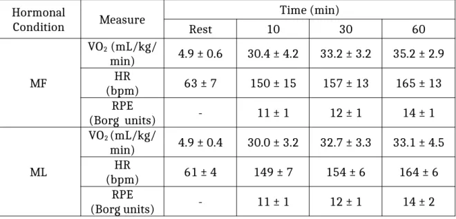
ML. Measurements obtained for VO2, HR and RPE were nearly identical for each
running session. Descriptive data at each measurement time for these variables is
shown in Table 3 below.
Table 3: Descriptive data (mean ± SD) for VO2, HR and RPE for each experimental
treadmill running protocol in each hormonal condition (MF and MF)
Hormonal
Condition Measure
Time (min)
Rest 10 30 60
MF
VO2 (mL/kg/
min) 4.9 ± 0.6 30.4 ± 4.2 33.2 ± 3.2 35.2 ± 2.9
HR
(bpm) 63 ± 7 150 ± 15 157 ± 13 165 ± 13
RPE
(Borg units) - 11 ± 1 12 ± 1 14 ± 1
ML
VO2 (mL/kg/
min) 4.9 ± 0.4 30.0 ± 3.2 32.7 ± 3.3 33.1 ± 4.5
HR
(bpm) 61 ± 4 149 ± 7 154 ± 6 164 ± 6
RPE
(Borg units) - 11 ± 1 12 ± 1 14 ± 2
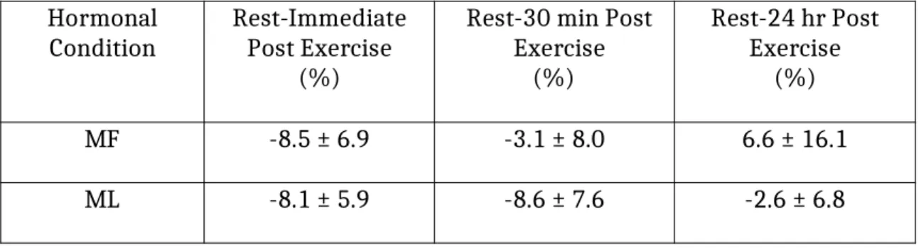
Plasma Volume Shift
The Dill and Costill method of determining plasma volume shifts was used in
this study. Plasma volume decreased over the prolonged treadmill running protocol.
Table 4 reports the average percentage plasma volume shifts for fluid moving from
the vascular spaces during the exercise and the recovery periods from exercise. The
responses for plasma volumes shifts were very comparable in each of the 60-minute
Table 4: Mean (± SD) plasma volume shifts
Hormonal
Condition Rest-ImmediatePost Exercise (%)
Rest-30 min Post Exercise
(%)
Rest-24 hr Post Exercise
(%)
MF -8.5 ± 6.9 -3.1 ± 8.0 6.6 ± 16.1
ML -8.1 ± 5.9 -8.6 ± 7.6 -2.6 ± 6.8
Blood Responses to Experimental Treadmill Running Protocol Creatine Kinase
Resting, immediate exercise, 30 min post exercise, and 24 hr
post-exercise CK activity is reported in Table 5. The main effect for hormonal status was significant (p < 0.05; MF>ML). The main effect for time was also significant (p < 0.01), with hoc revealing levels were significantly increased from 30 min
post-exercise to 24 hr post-post-exercise from rest. Most importantly, the interaction of
hormonal status and time was significant (p < 0.01). Post hoc tests indicated the
increase observed at 24 hr post-exercise was significantly greater in MF than ML.
Examination of the statistical analysis using the percent change data (see Table 5)
did not change the interpretation of the responses.
Creatine Kinase - MB
Creatine kinase - MB (CK-MB) response to the experimental treadmill
not significant (p= 0.72). The main effect for time was not significant (p= 0.47). Furthermore, there was not a significant interaction effect for hormonal condition
and time (p= 0.44). Examination of the statistical analysis using the percent change
data (see Table 6) did not change the interpretation of the responses.
The within assay coefficient of variation for the CK biochemical analysis was
9.50%. The within assay coefficient of variation for the CK-MB biochemical analysis
was 9.50%
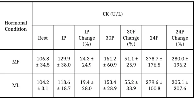
Table 5: Mean ± SD for CK at rest, immediately post exercise (IP), 30 minutes post
exercise (30P), and 24 hours post exercise (24P)
Hormonal Condition
CK (U/L)
Rest IP ChangeIP
(%) 30P
30P Change
(%) 24P
24P Change
(%)
MF ± 34.5106.8 ± 38.0129.9 24.3 ±24.9 ± 60.9161.2 51.1 ±25.9 378.7 ±176.5 280.0 ±196.2
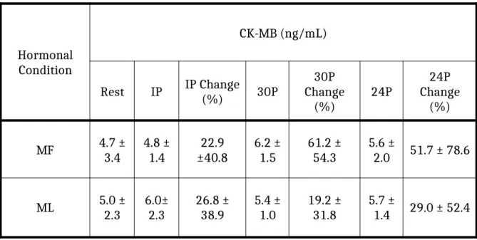
Table 6: Mean ± SD for CK-MB at rest, immediately post exercise (IP), 30 minutes
post exercise (30P), and 24 hours post exercise (24P)
Hormonal Condition
CK-MB (ng/mL)
Rest IP IP Change(%) 30P Change30P
(%) 24P
24P Change
(%)
MF 4.7 ±3.4 4.8 ±1.4 ±40.822.9 6.2 ±1.5 61.2 ±54.3 5.6 ±2.0 51.7 ± 78.6
CHAPTER V
DISCUSSION
The purpose of this study was to determine if fluctuations in E2,
corresponding to menstrual cycle phase, influenced blood CK and CK-MB response
to prolonged aerobic exercise. It was hypothesized that if E2 influences CK and
CK-MB response then higher E2 levels (ML phase) would attenuate CK and CK-MB
concentrations following prolonged aerobic activity. This hypothesis was tested by
examining the response of 10 highly trained females who completed two 60-minute
moderate intensity treadmill running sessions during the MF phase and ML phase o
the menstrual cycle. Subjects returned 24-hours after each exercise session for follow up
blood samples to assess CK and CK-MB responses. Hormonal Condition Determination
In the present study, there was a significant treatment condition between the
MF (low E2) and ML (high E2) menstrual cycle phases (p=0.003). E2 concentration
was approximately four times greater in the ML than MF (39.3 ± 18.3pg/ml to 148.1
± 35.2 pg/ml, respectively). These results fell within expected values determined by
the ELISA biochemical procedures used in analysis in which MF levels typically
range from 30-100pg/ml and ML levels typically range from 60-400pg/ml (Human
E2, Abnova, Walnut, CA & Taiwan, China). Also, the difference in E2 concentration
between hormonal conditions measured in this study agrees with other studies
investigating the effect of estrogen on inflammatory markers in eumenorrheic
Experimental Treadmill Running Session
The running speed of the participants within each of the 60-minute treadmill
runs corresponded to an average intensity of 61.7 ± 5.0 % VO2peak during the MF
phase and 59.7 ± 2.8 % VO2peak during the ML phase, slightly lower than the
desired intensity of 65% VO2peak. However, the mean VO2 responses between
hormonal conditions were similar at each time measurement in both of the
60-minute exercise sessions and did not differ. The VO2 measurements slightly
increased from the start to end of the treadmill run due to the manual adjustments
in running speed to meet target VO2 levels, and as the result of the cardiac drift
phenomenon [see below] (Dawson et al., 2005).
Heart rate responses between hormonal conditions were very similar too,
but did show a gradual increase over the 60-minute treadmill running sessions. This
gradual increase in heart rate during prolonged aerobic activity is not unusual.
During such prolonged aerobic exercise, body temperature gradually increases. To
maintain homeostasis in this situation, blood is redirected towards the skin to
release water within blood plasma through the form of sweat so that evaporative
cooling can occur. This action reduces the plasma volume making the blood more
viscous and decreases venous return to the heart. Therefore, the heart contractility
must increase (increasing heart rate) to maintain cardiac output and exercise
intensity. This is known as the cardiac drift phenomenon. This concept is supported
by the data obtained in this study by the reduced plasma volume observed following
Ratings of perceived exertion (RPE) in each of the 60-minute running
sessions were nearly identical between hormonal conditions too; although, during
both experimental running sessions the RPE did gradually increase which is a
typical response. Prolonged aerobic activity requires adequate motivation,
hydration levels, and nutritional status. Thus, RPE could have increased from a lack
of any of these factors; lack of motivation, monotony of treadmill running,
dehydration, or prior nutritional status, towards the end of the running session.
Although, it should be pointed out the subjects were allowed to drink water as often
as desired.
The similar findings of RPE between the two experimental sessions are
somewhat atypical. Previous research has shown an increase in RPE during the ML
phase. This is speculated to be due to the increase in body temperature during the
luteal phase, which negatively affects a woman’s capability of maintaining thermal
balance which can increase the perception of the difficulty of the exercise (Pivarnik
et al., 1992). That is, the body must work harder and sweat more to prevent
overheating making exercise seem more difficult. Why the present findings are at
odds with the work of Pivarnik et al. is unclear
Overall, the VO2, HR, and RPE measurements obtained in each of the
60-minute exercise sessions were in line with expected physiological outcomes caused
by prolonged aerobic exercise and were very similar between the hormonal
conditions (ACSM, 2011; Kallman, 2011). This suggests that any differences in blood
levels of CK or CK-MB response observed in this study were the result of differences
Creatine Kinase (Total CK)
A significant interaction effect of hormonal status and time was observed for
CK (i.e., exercise induced CK changes but the magnitude of response varied with E2
levels). Further statistical analyses indicated that the increase in CK observed at 24
hr post-exercise was significantly greater in MF than ML. These results are similar
and relate to a study by Carter et al. who found naturally elevated estrogen levels
during the ML had a protective effect on muscle following eccentric exercise (Carter
et. al, 2001). Specifically, Carter et al. found significantly less CK response 72 hours
after exercise in subjects during high estrogen (ML) versus low estrogen menstrual
phases (MF) when doing downhill running to introduce muscular trauma. These and
the current results also are in line with previous research supporting the notion that
high levels of estrogen attenuate inflammation that causes delayed onset muscle
soreness (DOMS) after exercise (Tiidus et al., 2001).
In skeletal muscle, studies on animals have shown that estrogen may
potentially prevent the release of CK into the bloodstream (Amelink et al., 1990;
Enns & Tiidus, 2010). The mechanism by which estrogen reduces muscle damage
and CK release is not fully understood, but it is thought that estrogens exhibit
antioxidant properties, act as a membrane stabilizer, and has an ability to bind to
estrogen receptors and regulate downstream genes inducing decreased
inflammation (Tiidus et al, 2010).
Creatine Kinase - MB
There was not a significant interaction effect for hormonal condition and
response vary with E2 levels). CK-MB is very useful indicator of myocardial damage
as essentially only myocardium tissue has substantial amounts of this biomarker
present. Therefore, this lack of a significant interaction effect (i.e., increase in
response to the exercise) for CK-MB suggests that the increase seen in CK levels 24
hrs post-exercise was a result of skeletal inflammation and not cardiac muscle
damage (i.e., a component of the CK response is made up on the CK-MB
sub-fraction). These results are not in line with some previous research on long-distance
runners that found elevated CK-MB levels 1, 24, 48, and 72 hours after a marathon
race in men (Apple et al, 1984, 1985). Another study in rats found that after 3.5
hours of swimming with an 8% bodyweight workload attached to their tails, CK-MB
concentrations from the heart significantly increased (Chen et al., 2000). These
studies supported the notion that prolonged aerobic exercise elicited cardiac muscle
damage; but the duration of the exercise used in these studies was substantially
greater than that in the current study.
The lack of an interaction effect for CK-MB values may be a result of the
limitations in the current study design. It is possible that the duration and intensity
of the treadmill running protocol were not great enough to elicit a significant
difference in CK-MB levels between hormonal conditions. Therefore, it is possible
that the heart was not put under enough stress to elicit the release of a significant
amount of CK-MB between hormonal conditions and thus may have masked the
influence of E2. A study conducted by Hutcheson et al. found that strenuous exercise
(i.e., marathon running) elicited elevated CK-MB levels in men and women
required 15 subjects to be part of the study. Therefore the sample size of 10 subjects
may have been too small to detect changes, which limits the statistical strength of
the results and could have contributed to the lack of a relationship found between
CK-MB and E2.
Conclusion
In conclusion, the inflammatory marker CK significantly increases 24 hrs
post-exercise during periods of low E2 (MF phase). However, the lack of a significant
increase in CK-MB levels following exercise suggests that this increase in CK is from
skeletal inflammation not from cardiac muscle damage. Therefore, it is unclear if
estrogen hormonal status influences the potential cardiac muscle damage from
prolonged aerobic exercise. Due to the lack of significance for changes in CK-MB
levels, suggestions for women to alter their training programs in relation to their
menstrual phase to possibly prevent cardiac muscle damage cannot yet be made.
However, the significant interaction effect for hormonal condition and time for the
skeletal inflammatory marker CK suggest that women should consider ingesting an
NSAID while training during their MF phase (low E2) to alleviate exercise induced
inflammation. Given the limited research present on the influence of E2 on cardiac
biomarkers following prolonged aerobic exercise, additional research is
recommended.
Limitations
Several limitations in the current study do exist. The sample size relative to
CK-MB may have been inadequate to reach statistical significance. Therefore,
of the subjects did not reach the criteria for a maximal oxygen consumption test
(VO2max) during their initial exercise testing session. Also, the intensity of the
exercise was not as difficult as originally intended in the study design, which could
have affected the results obtained.
Future Studies
Future studies should consider increasing the intensity or duration of the
running protocol to elicit a greater physiological stress response. Future studies
should also obtain a greater number of subjects to reach optimal statistical power.
Also, additional cardiac biomarkers such as troponin should be examined in order to
have a greater understanding of the relationship between E2 and cardiac damage.
Overall, more future research is needed to further understand E2 effect on exercise
APPENDIX A
APPENDIX B
INFORMED CONSENT
University of North Carolina at Chapel Hill Consent to Participate in a Research Study Adult Participants
Consent Form Version Date: _______2013-2014_______
IRB Study # 10-2109
Title of Study: Influence of Estrogen on Cytokine Response to Prolonged Treadmill Running
Principal Investigator: Anthony Hackney
Principal Investigator Department: Exercise And Sport Science
Principal Investigator Phone number: (919) 962-0334
Principal Investigator Email Address: [email protected] _________________________________________________________________
What are some general things you should know about research studies?
You are being asked to take part in a research study. To join the study is voluntary.
You may refuse to join, or you may withdraw your consent to be in the study, for any reason, without penalty.
Research studies are designed to obtain new knowledge. This new information may help people in the future. You may not receive any direct benefit from being in the research study. There also may be risks to being in research studies. Deciding not to be in the study or leaving the study before it is done will not affect your relationship with the researcher, your health care provider, or the
University of North Carolina-Chapel Hill. If you are a patient with an illness, you do not have to be in the research study in order to receive health care.
Details about this study are discussed below. It is important that you understand this information so that you can make an informed choice about being in this research study.
You will be given a copy of this consent form. You should ask the researchers named above, or staff members who may assist them, any questions you have about this study at any time.
What is the purpose of this study?
Recent work has shown a negative relationship between concentration of estrogens and the inflammatory response, specifically cytokines, at rest. Females in the midluteal phase of the
menstrual cycle, high estrogen concentration, exhibited significantly lower circulating cytokines (and creatine kinase) compared to females in the midfollicular phase, low estrogen concentration. These demonstrated fluctuations at rest begs the question - Is there an altered cytokine response during exercise at different points within the menstrual cycle as estrogen changes?
To date, few exercise studies on human female subjects with respect to estrogen concentration and cytokines exist. The studies that do exist present divergent results. The studies performed have limitations; primarily small sample sizes, potential inaccurate menstrual phase determination, or dosage of exercise was not enough to provoke a response.
The main aim of the study is to determine if there is a significant difference in cytokine response at rest, immediately post exercise, 30 minutes post exercise, and 24 hours post exercise between two phases of the menstrual cycle, midluteal and midfollicular. On separate days, you will perform an exercise test on a treadmill to determine your maximal aerobic capacity (VO2max), two 60-minute
running bouts at approximately 65-70% VO2max, and have blood drawn before, immediately post, 30
minutes post, and 24-hours post the 60-minute running bout. Blood samples will be assessed for estrogen and cytokine concentration and creating kinase levels.
You are being asked to be in the study because you are a healthy, highly trained woman between the ages of 18 and 30 with a normal menstrual cycle for at least 6 months. You have not used oral contraceptives for at least six months prior. Have no major injuries within the last six months that limit ability to engage in exercise or if have sustained an injury are completely recovered and cleared by a physician to partake in exercise. Your current VO2max is at least 45 ml/kg/min. Your current
minimum training volume is 3-5 days a week, 45-120 minutes per session of aerobic activity.
Are there any reasons you should not be in this study?
You should not be in this study if you are knowingly pregnant or become pregnant during the study, if you have an irregular or absent menstrual cycle, are currently taking or have taken within the six months prior oral contraceptives, you have sustained an injury within the last six months that has limited your ability to exercise, use substances known to alter immune response (e.g. NSAIDS) the week before each 60-minute exercise session, or you become ill with immune responding conditions (i.e., colds, respiratory infections…etc.).
How many people will take part in this study?
There will be approximately 25 people in this research study.
How long will your part in this study last?
You will be enrolled in the study for approximately 6 weeks. Within the 6 weeks, 3 visits are made to the Applied Physiology Laboratory at the University of North Carolina at Chapel Hill.
Visit 1: Orientation Session, duration is approximately 90 minutes
Visit 2 (approximately 1-6 weeks after visit 1): Prolonged treadmill running bout (60 min) with a before and immediately, and 30 minutes after exercise blood draws performed by a certified phlebotomist (NCPT 56147) visit, duration is approximately 2 hours
Visit 3: 24-hours after prolonged running bout blood draw, duration is approximately 30 minutes
Visits 4-5 will be a repeat of visits 2-3 approximately 2-6 weeks after visit 2. Blood specimens will be stored for 3 years following the completion of the study.
What will happen if you take part in the study?
Orientation/Familiarization Session, duration approximately 90 minutes (visit 1):
The study protocol, schedule, inherent benefits, and potential risks will be explained to you, followed by signing the informed consent.
Height and mass will be obtained and you will be fitted for a heart rate monitor and then asked to rest lying down for 10 minutes. After obtaining your resting heart rate, you will be fitted for a mouthpiece that will be used to collect expired air.
You will then perform a modified Bruce Protocol to volitional fatigue to determine VO2max.
The protocol consists of 3-minute stages with progressive speed at 0% inclined to near max, then grade will be introduced to maximal exertion (e.g. 3 minutes: 6.0, 0%, 3 minutes: 7.5, 0%, 3 minutes: 9.0, 0%, 3 minutes, 9.0, 2.5%). Heart rate (HR), rating of perceived exertion (RPE), and expired air will be monitored throughout the test. VO2max attained will be used
to determine running speed for the specific menstrual phase prolonged running bout for each individual subject.
Menstrual Phase Determination:
You will need to inform the principal investigator at the start of menses (the first menses following visit 1 to the lab), which will be denoted as day one. Scheduling of the prolonged treadmill running bout will correspond to a specific menstrual phase.
Prolonged treadmill running bout 1, duration approximately 2 hours (Visit 2 and 4):
The time lapse between visit 1 and visit 2 is not determined by investigators, rather it is determined based upon your menstrual cycle. The start of your menstrual cycle (e.g. menses, day 1) and the approximate length of your menstrual cycle (day 1 of menses to the start of the next menses) will determine when you are scheduled for the prolonged running bouts so as to ensure you are in the appropriate menstrual cycle phase. The approximate time between visit 1 and visit 2 can be 1-6 weeks. Between visit 1 and 24-hours before visit 2 you can partake in normal daily activities and exercise training with no restrictions. Twenty-four hours prior to the prolonged running bout you will be asked to refrain from exercise, drink plenty of fluids, and eat a diet rich in carbohydrates.
Upon arrival, you will be asked to urinate into a sterile specimen container. The urine will be assessed for hydration status. If the urine analysis comes back as dehydrated you will not participate in the prolonged running bout, you will be encouraged to consume plenty of fluids, and you will be rescheduled.
If the urine analysis comes back normal you will be weighed, fitted with a HR monitor, and asked to rest lying down for 10 minutes. After, a resting HR will be recorded and a 1-teaspoon blood sample will be drawn from your arm by a certified phlebotomist (NCPT 56147), placed into a K3-EDTA blood collection tube and immediately put on ice. The blood
sample will be used to confirm menstrual cycle status and resting IL-6, and estrogen levels.
You will then be transferred to the treadmill and fitted for a mouthpiece. You will be asked to sit quietly for 4 minutes as your expired air is collected to determine resting VO2.
You will then have 10 minutes to warm-up: 5 minutes will be dedicated to easy walking on the treadmill followed by 5 minutes of stretching appropriate muscles used in the upcoming prolonged running bout (e.g. calf stretch, hamstring stretch, quadriceps stretch, and hip flexor stretch). During the walking, you will practice going on and off the mouthpiece as you are moving.
Following warm-up, you will run on a treadmill at a 0% incline and a speed to elicit 65-70% VO2max. At 6 minutes, 26 minutes, and 56 minutes you will be asked to return to the
mouthpiece and expired air will be recorded for four minutes. This is to ensure appropriate intensity and make adjustments in running speed if necessary. Heart rate and RPE will be recorded during the last 10 seconds of minutes 9, 29, and 59 of the running bout.
Throughout the running bout you will have a fan to keep you cool, can drink water at your convenience, and listen to music.
At the completion of exercise, another 1-teaspoon blood sample will be drawn following the same procedure, placed in K3-EDTA blood collection tube, and immediately placed on ice.
easy pace for 5 minutes, stretching muscles used during the prolonged running bout (e.g. calf stretch, hamstring stretch, quadriceps stretch, and hip flexor stretch), and sitting quietly in a chair.
30 minute post blood draw
Once your heart rate has returned to 100 bpm you are free to leave the laboratory.
Follow-up Blood Draws, duration approximately 30 minutes:
At 24 hours after the running bout you will report to the laboratory for additional blood draws. You will be asked to rest lying down for 10 minutes. Blood samples will be obtained following the same blood draw procedures as explained above. These blood samples will be analyzed for IL-6 and estrogen concentrations. During the 24 hours of recovery from the exercise you are asked to refrain from performing any physical activity other than that of daily routine living.
Prolonged treadmill running bout, duration approximately 2 hours:
You will be asked to repeat the aforementioned protocol during two different phases of your menstrual cycle. The time frame between the prolonged running bouts is approximately 2 to 6 weeks. Between visit 4 and 24-hours before visit 5 you can partake in normal daily activities and exercise training with no restrictions.
Blood Analysis:
The blood samples will be separated by centrifuging and frozen until later analysis. The blood plasma will be analyzed for estrogen levels, creatine kinase and immune markers.
What are the possible benefits from being in this study?
Research is designed to benefit society by gaining new knowledge. There is little chance you will benefit from being in this research study. The benefits to you from being in this study may be the obtaining of your Vo2max (aerobic capacity) which you can use in setting up a specific exercise training program.
What are the possible risks or discomforts involved from being in this study?
The potential risks to you from participating in this study may be related to exercise or the blood draw process.
Potential risks associated with exercise are outlined by American College of Sports Medicine as: sudden cardiac death, musculoskeletal injury, and falling.
The risk of sudden cardiac death is low in healthy individuals; however, to minimize risk a health history questionnaire and physical examination will occur prior to testing.
To minimize risk of musculoskeletal injury a proper warm-up will be completed prior to all testing.
Given the prolonged nature of the exercise bout dehydration is a potential risk. To minimize this risk your hydration status will be determined before testing begins ensuring you are in normal hydrated state; if dehydrated the testing session will be cancelled and rescheduled. You will be asked to consume plenty of fluids 24 hours before testing and encouraged to drink water throughout and after the running bout.
Furthermore, the potential risk of exercise for you will be minimal because you have performed similar exercise intensities and durations within previous training programs.
Risks associated with blood draws include infection, bruising of the area around the needle insertion, and dizziness/fainting.
To minimize infection, cleaning of the puncture area and sterile equipment will be used.
Proper needle gauge and firm pressure applied to the puncture following the blood draw will help minimize risk of bruising.
Following the blood draw, to minimize the risk of syncope you will be asked to move from a supine position to sitting and eventually standing slowly. Research technicians will monitor complexion and skin temperature for adverse signs.
A certified phlebotomist will perform all blood draws. First aid procedures and universal precautions will be followed during blood draws and handling of blood samples.
There may be uncommon or previously unknown risks. You should report any problems to the researcher.
What if we learn about new findings or information during the study?
You will be given any new information gained during the course of the study that might affect your willingness to continue your participation.
How will information about you be protected?
Following initial screening, an identification number will be assigned to you for future identification. A hard copy of records will be stored in a locked cabinet in the Applied Physiology Laboratory. Electronic records will be maintained on a secured, password-protected computer. All identifiable hard-copy files will be shredded and disposed of using UNC-CH mechanisms and procedures. Blood samples will be stored in a secured ultra-freezer behind a access code protected door within a laboratory involving only electric ID card access. These specimens will be encoded and labeled so that no personal identifying information will be revealed. The identification number will consist of a unique number along with phase and the sample time (e.g. 00913, 009 is the subject ID, 1 is
indicative of menstrual phase, 3 is time sample). Study data and specimens will only contain the identification number. These numbers will be indiscernible unless access to the master list which will be locked in a file cabinet in the Applied Physiology Laboratory. Only the principal investigator will have access to the records.
Participants will not be identified in any report or publication about this study. Although every effort will be made to keep research records private, there may be times when federal or state law requires the disclosure of such records, including personal information. This is very unlikely, but if disclosure is ever required, UNC-Chapel Hill will take steps allowable by law to protect the privacy of personal information. In some cases, your information in this research study could be reviewed by
representatives of the University, research sponsors, or government agencies (for example, the FDA) for purposes such as quality control or safety.
What will happen if you are injured by this research?