Review Article
Theme: Integrating Microdialysis and Imaging Tools in Systems Pharmacology Guest Editors: Robert E. Stratford, Nimita Dave, and Richard F. BergstromMicrodialysis: the Key to Physiologically Based Model Prediction of Human CNS
Target Site Concentrations
Yumi Yamamoto,
1Meindert Danhof,
1and Elizabeth C. M. de Lange
1,2Received 22 November 2016; accepted 25 January 2017
Abstract.
Despite the enormous research efforts that have been put into the development
of central nervous system (CNS) drugs, the success rate in this area is still disappointing. To
increase the successful rate in the clinical trials,
fi
rst the problem of predicting human CNS
drug distribution should be solved. As it is the unbound drug that equilibrates over
membranes and is able to interact with targets, especially knowledge on unbound
extracellular drug concentration-time pro
fi
les in different CNS compartments is important.
The only technique able to provide such information
in vivo
is microdialysis. Also, obtaining
CNS drug distribution data from human subjects is highly limited, and therefore, we have to
rely on preclinical approaches combined with physiologically based pharmacokinetic (PBPK)
modeling, taking unbound drug CNS concentrations into account. The next step is then to
link local CNS pharmacokinetics to target interaction kinetics and CNS drug effects. In this
review, system properties and small-molecule drug properties that together govern CNS drug
distribution are summarized. Furthermore, the currently available approaches on prediction
of CNS pharmacokinetics are discussed, including
in vitro,
in vivo,
ex vivo, and
in silico
approaches, with special focus on the powerful combination of
in vivo
microdialysis and
PBPK modeling. Also, sources of variability on drug kinetics in the CNS are discussed.
Finally, remaining gaps and challenges are highlighted and future directions are suggested.
KEY WORDS:brain extracellularfluid (brainECF); central nervous system (CNS); cerebrospinalfluid
(CSF); mastermind research approach; physiologically based pharmacokinetic (PBPK) model.
INTRODUCTION
There is a huge unmet medical need for central nervous
system (CNS) disease therapies because of the growing of
chronic and complex diseases associated with aging. However,
development of CNS drugs is one of the most challenging tasks
for the pharmaceutical industry (1). Actually, drug development
for CNS drugs has suffered a higher attrition rate compared to
that of other therapeutic areas drugs; it has been reported that
only around 8
–
9% of CNS drugs that entered phase 1 were
approved to launch (2). And around 50% of the attrition of
potential CNS drugs has resulted due to a lack of ef
fi
cacy and
safety issues in phase 2 (2,
3). Knowledge of human CNS drug
concentrations forms the basis for understanding
exposure-response relationships; therefore, the lack of appropriate
consideration of these target concentrations is one of the factors
contributing to this high degree of attrition.
Obtaining the target site concentrations of CNS drugs is
not straightforward because plasma concentrations do not
adequately re
fl
ect CNS exposure, primarily due to the
presence of the brain barrier (BBB) and the
blood-cerebrospinal
fl
uid barriers (BCSFB), and additional speci
fi
c
physiological characteristics of the CNS. Furthermore,
signif-icant variation in the rate and extent of mechanisms that
govern target site pharmacokinetics (PK), target engagement,
and signal transduction is known to exist, due to differences
in system conditions such as species, gender, genetic
back-ground, age, diet, disease, and drug treatment (4). Moreover,
with regard to CNS drug action, there is a lack of suf
fi
ciently
established clinical biomarkers and proof-of-concept (5).
Thus, it is clear that there is a need for more predictive
approaches. These predictive approaches have to be
inter-connected to the system conditions and must be performed
using adequate (including bound and unbound drug)
The original version of this article was revised: Figure 3 in the PDF and electronic versions of the published article contains formatting errors caused by the typesetter.
1Division of Pharmacology, Cluster Systems Pharmacology, Leiden
Academic Centre for Drug Research, Leiden University Gorlaeus Laboratories, Einsteinweg 55, 2333 CC, Leiden, The Netherlands. 2To whom correspondence should be addressed. (e-mail:
The AAPS Journal (#2017)
DOI: 10.1208/s12248-017-0050-3
concentrations. Also processes should preferably not be studied
in isolation and then combined, but instead studied in
conjunc-tion with each other as this will provide insight about the
interdependencies of these processes (4). Since measurements
on CNS target site concentration in the clinical setting are highly
restricted, we have to develop an approach based on integrated
preclinical data that is translatable to human.
Even though drug properties have been investigated
well, information of CNS system properties (CNS physiology
and biochemistry) is sparse and has a large variability. CNS
pharmacokinetics of drugs is determined by their interaction.
System properties depend on the condition of the system,
which means that we have to use approaches to distinguish
between system and drug properties, as this would allow us to
translate the model to other species and also other disease
conditions, by using physiologically based pharmacokinetic
(PBPK) modeling.
Currently, many more or less complex semi-PBPK
models have been published for CNS drug distribution. At
present, four preclinical translational models have been
validated with human CNS concentration pro
fi
les (6
–
9). In
these models, however, the parameters were estimated using
in vivo
data to describe CNS distribution of individual drug in
animals. Ultimate goal of the PBPK modeling is to build a
generic PBPK model in which the parameters are derived
from
in vitro
and/or
in silico
data. To achieve this,
in vivo
data
is needed to validate the generic PBPK model. Furthermore,
an investigation is needed on the relationship between drug
physicochemical properties and CNS distribution.
In this review, system properties and small-molecule
drug properties that together govern CNS drug distribution
are summarized, followed by currently available approaches
on prediction of CNS pharmacokinetics, including
in vitro,
in vivo,
ex vivo, and
in silico
approaches, with special focus
on the powerful combination of
in vivo
microdialysis and
PBPK modeling. Also, sources of variability on drug
kinetics in the CNS are discussed. Finally, remaining gaps
and challenges will be discussed and future directions will
be provided.
INTERACTION BETWEEN CNS SYSTEM AND DRUG
PROPERTIES
Many CNS system properties and drug speci
fi
c
proper-ties are known to in
fl
uence drug kinetics in the brain, as
shown in Fig.
1. Here, we focus on the relevant factors from
each that contribute to the drug kinetics and summarize their
function.
CNS SYSTEM PROPERTIES
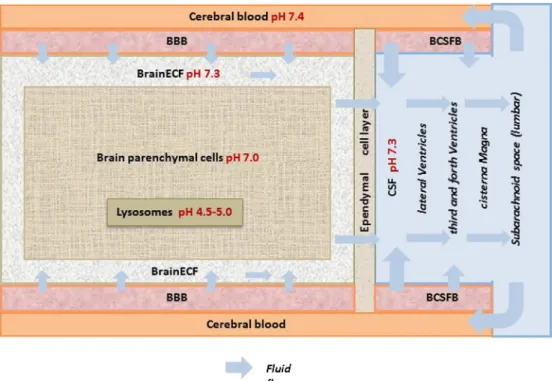
Physiological Compartments, Flows, and pH
The CNS is a complex system composed of many
physiological components and
fl
ows (Fig.
2): Physiological
compartments are the BBB, the BCSFB, brain extracellular
fl
uid (brain
ECF), cerebral blood, brain parenchymal cells, and
the cerebrospinal
fl
uid (CSF) in the ventricles, the cisterna
magna, and the subarachnoid space (4). There are pH
differences among the compartments (10
–
16). Then, there
are the CNS
fl
uid
fl
ows that include the cerebral blood
fl
ow
(CBF), brain
ECFbulk
fl
ow, and CSF
fl
ow. All relevant
physiological parameter values are summarized in Table
I.
Active Transporters
The localization of transporters and their expression
level are also important factors to determine drug
distribu-tion in the brain. Transporters are present at the BBB and
at the BCSFB, also on the membrane of brain parenchyma.
Active transporters on the BBB and BCSFB consist of
facilitated transport and ATP-dependent transport. The
solute carrier (SLC) family, such as organic
anion-transporting polypeptide (OATP) and organic anion
transporters (OATs), is categorized as a facilitated transport,
while ABC transporters, such as P-glycoprotein (P-gp),
multidrug resistance protein (MRPs), and breast
cancer-resistant protein (BCRP) are categorized as an
ATP-Fig. 2. Brain physiological components andflow. Figure is modified from de Lange (4)
Table I. Values of CNS system properties for rat and human
Parameter Human Refs Rat Refs
Volumes BBB volume 8.25 mL (calculated using thickness endothelial cell of 550 nm)
(17) 5.02μL (18)
BCSFB volume 107.25 mL (calculated using thickness 14.3μm of endothelial cell)
(19) 37.5μL (18)
Brain volume 1400 g (20) 1.8 g, 1880μL (21,22)
BrainECFvolume 240–280 mL (23,24) 290μL (25)
BrainICFvolume 960 mL (24) 1440μL (24)
CSF volume 130-150 mL (26,27) 250μL (21)
CSFLVvolume 20–25 mL (26,28) 50μL (29,30)
CSFTFVvolume 20–25 mL (26,28) 50μL (29,30)
CSFCMvolume 7.5 mL (31,32) 17μL (31,32)
CSFSASvolume 90–125 mL (26,28) 180μL (33,34)
Flows cerebral bloodflow 610–860 mL/min (35–37) 1.1–1.3 mL/min (38,39)
brainECFflow 0.15–0.2 mL/min, (50% of CSF production) (27) 0.00018–0.00054 mL/min (40)
CSFflow 0.3–0.4 mL/min (27) 0.0022 mL/min (25,41)
Surfaces BBB SA 12–18 m2 (17) 155–263 cm2 (42,43)
BCSFB SA 6–9 m2 (17) 25–75 cm2 (42,44)
(assumed 50% of BBB SA) (assumed 50% of BBB SA)
brain ECF/ICF SA 228 m2 Calculateda) 3000 cm2 (18)
brain ICF/lysosome SA 12 m2 Calculateda) 162 cm2 Calculateda)
pH Plasma 7.4 (13) 7.4 (10)
BrainECF NA 7.3 (11)
BrainICF 7.0 (14) 7.0 (11)
lysosome 4.5-5.0 (15) 5.0 (11)
CSF 7.3 (13) 7.3 (12)
brainECF, brain extracellularfluid compartment,brainICFbrain intracellularfluid compartment,CSFLVcompartment of cerebrospinalfluid in
lateral ventricle,CSFTFVcompartment of cerebrospinalfluid in the third and fourth ventricle,CSFCMa compartment of cerebrospinalfluid in
the cisterna magna,CSFSAScompartment of cerebrospinalfluid in the subarachnoid space,SAsurface area a
dependent transport (45). Table
II
summarizes an overview
of transporters with their localization and their endogenous
and exogenous substrates.
Metabolic Enzymes
Presence and localization of enzymes in the brain are
also important factors to determine drug kinetics in the brain.
In the brain, the following enzymes are found:
oxidoreduc-tases such as cytochrome P450 (CYPs) and monoamine
oxidase (MAO), membrane-bound and soluble
catechol-O-methyltransferase (COMT), and transferases such as uridine
5-diphospho (UDP) -glucuronosyltransferases (UGTs) and
phenol sulfotransferase (PST) (68). In Table
III, an overview
is provided of the different enzymes with their localization
and examples of their endogenous and exogenous substrates.
SMALL-MOLECULE DRUG PROPERTIES AND
INTERACTION WITH THE CNS SYSTEM
A combination of CNS system properties and drug
properties determines the pharmacokinetics of a drug in the
CNS, including the CNS target site. Important
physicochem-ical properties for determination of drug CNS
pharmacoki-netics are summarized in Fig.
1.
Physicochemical properties of a drug, such as
lipophilic-ity, size, charge, hydrogen binding potential and polar surface
area (PSA), are important determinants for pharmacokinetics
of a drug. Many studies have investigated the in
fl
uence of
individual physicochemical properties on the BBB
penetra-tion in isolapenetra-tion. However, as physicochemical properties are
highly inter-correlated, it is more appropriate to consider
these properties in combination.
First of all, it should be noted that it is the unbound and
neutral form of a drug molecules that is able to diffuse across
barriers like the BBB and BCSFB, depending on the
concentration gradient of the unbound and neutral form of
the drug on either side of a membrane. Lipophilicity relates
to the BBB permeability, as transcellular diffusion rate
(93,94). Furthermore, as a rule of thumb, higher lipophilicity
increases CNS tissue binding. Molecular size is an important
factor for paracellular drug diffusion rate and also has an
impact on transcellular diffusion rate at the BBB (93,
95,
96).
The degree of ionization depends on the pKa of the drug and
actual pH in a body compartment. Thus, the BBB
perme-ability rate is in
fl
uenced by lipophilicity, size, and pKa of a
drug (93,
97). Using quantitative structure-activity
relation-ship (QSAR) modeling, it has been shown that the
descrip-tors for the prediction of BBB penetration are different for
different charge classes (98). As there are pH differences
between plasma, brain
ECFand CSF (Fig.
2), charge is an
important factor for CNS drug disposition (99).
The hydrogen bonding potential re
fl
ects the necessary
energy for a molecule to move out of the aqueous phase into
the lipid phase of a membrane. Recent studies have shown
that the relationship between chemical structure and
Kp,uu,brain (the ratio of the unbound concentration in the
brain over that in plasma at equilibrium which measures the
extent of CNS distribution) was dominated by hydrogen
bonding (100).
PSA is generally de
fi
ned as the sum of the van der Waals
surface areas of oxygen and nitrogen atoms. Therefore, PSA
of a compound can be related to its hydrogen bonding
potential. Some studies have shown that PSA is highly
correlated with the permeability coef
fi
cient (Pc) of
mem-branes (94,101,102). A recent study for Kp,uu,brain has been
shown that PSA is one of the important factors to predict the
Kp,uu,brain for each compound (103).
BBB and BCSFB Transport
Protein Binding.
It is generally accepted that unbound
drug in plasma is able to cross the BBB and BCSFB. Two major
proteins in plasma are albumin and
α1-acid glycoprotein (104).
For passive diffusion, the free concentration gradient between
plasma and brain determines the rate of transport. The extent of
BBB and BCSFB transport are investigated using Kp,uu,brain:
If there is only diffusion, Kp,uu,brain is 1. If there is active
transport processes, then Kp,uu,brain is larger than 1 (active in)
or Kp,uu,brain is smaller than 1 (active out).
Ionization of the Drug in Plasma and in the Brain.
There
are similar pH differences among the CNS physiological
compartments in human and in rat (Table
I). Because of the
pH differences, the ratio of neutral form of a compound
among the compartments is different. It is generally accepted
that neutral form can pass barriers; therefore, ionization that
is determined by the pKa of a compounds and pH in the
physiological compartments will have an impact on drug
disposition in the brain.
Cerebral Blood Flow
—
Flow Versus Permeability-Limited
Transport Rate.
Lipophilic compounds usually have a large
permeability coef
fi
cient; therefore, a permeability surface
area product (PA), which is determined by the permeability
coef
fi
cient and surface area of tissue, becomes large. If the PA
is larger than the physiological cerebral blood
fl
ow, then the
physiological cerebral blood
fl
ow determines the transport
rate of the compound.
Modes of BBB Transport
—
Different Modes.
The
combi-nation of transport modes at the BBB, BSCFB, and
membrane of brain parenchyma determines the rate and
extent of drug exchange at the BBB, BCSFB and membrane
of brain parenchyma (105,106). Therefore, the operative
transport mechanism(s) may differ for each drug. Each
transport mode is summarized in Table
IV
.
Active Transporter Function.
Active transporters
medi-ate in
fl
ux and ef
fl
ux of drug transport. The magnitude of
interaction of active transport is drug- and species-dependent
(107). The functions of individual transporters are
summa-rized in Table
II.
Brain Distribution and Elimination
CSF compartments. At the same time, the drug in brain
ECFis
transported to brain parenchymal cell intracellular
fl
uid
(brain
ICF). It should be noted that also on the brain
parenchyma cell membranes active transport may occur
(106).
Tissue Binding.
Tissue binding can occur as being
speci
fi
c at the target or non-speci
fi
c to tissue components.
Lysosomal Trapping.
In the brain parenchyma cells,
there is a physiological pH gradient between the intracellular
compartment (cytoplasm) and the lysosome compartment
(Fig.
2). Especially basic compounds are known to be trapped
in the lysosomes (11).
Drug Dispersion Within CSF.
Some studies have shown
that intrathecally administered drugs distribute faster than
what can be accounted only by molecular diffusion (108,
109). Thus, it is thought that molecular diffusion makes only
a small contribution to the total drug dispersion within CSF.
This leads to the need to take into account also the
convection due to oscillatory CSF
fl
ow to adequately
explain this dispersion (110). Recently, the drug dispersion
has been considered to be enhanced by the CSF pulsatility
(heart rate and CSF stroke volume), and it leads to high
inter- and intra-patient variability in drug distribution in the
brain (110,
111).
Elimination from the Brain.
Apart from transport across
the BBB and BCSFB as discussed earlier, drug may leave the
brain
via
the BBB, but also
via
CSF re
fl
ux into the blood
stream at the level of the arachnoid villi.
Metabolism.
In the brain, several metabolic enzymes are
present. Enzyme interaction with drugs is important
informa-tion not only on the drug PK pro
fi
le but also the drug
pharmacological effect in the brain since it may create active
metabolites. Presence and localization of several enzymes
have been reported in the brain (Table
III), although their
activity is reported to be relatively small compared to the
liver (68,
87).
CURRENT APPROACHES TO INVESTIGATE CNS
DRUG DISTRIBUTION
Since obtaining a human drug target site concentration in
the brain is not feasible in most of the clinical studies,
quantitative prediction of target site concentration is
impor-tant. To achieve this, we need information from
in vitro,
ex vivo,
in vivo, and
in silico
approaches. Here, we summarize
the current approaches to obtain the necessary information to
predict human drug target site concentration.
IN SILICO
APPROACHES
For decades, QSAR studies have been performed using
Kp,brain (total concentration ratio of the brain to plasma) or log
BB, either of which may not re
fl
ect the relevant drug exposure in
the brain to assess the ef
fi
cacy of the drug since this ef
fi
cacy is
in
fl
uenced by binding of compounds to plasma proteins and brain
tissue. Eventually, log BB was replaced by the PA, as an estimate
of the net BBB in
fl
ux clearance (112). However, it has been
argued that the PA cannot predict the unbound drug
concentra-tion in the CNS by itself. Recently, the most relevant parameter
Kp,uu,brain has been used, with QSAR being conducted to
model this parameter (100,103,113,114). Other than Kp,uu,brain,
physiological meaningful parameter, Vu,brain (the volume of
distribution of the unbound drug in the brain) or Kp,uu,cell
(unbound concentration ration between brain
ECFand brain
ICF)
are also reported using molecular descriptors (103).
IN VITRO
APPROACHES
In vitro
approaches to investigate the BBB permeability
have been conducted using BBB models (115). BBB models can
be classi
fi
ed into non-cell based surrogate models, such as
parallel arti
fi
cial membrane permeability assay (PAMPA), and
cell-based models such as primary cultures cells, immortalized
brain endothelial cells, or human-derived stem cells (116).
Although primary cultured cells from human tissue have been
reported, acquiring human brain tissue is dif
fi
cult as it can be
Table IV. Blood-brain barrier main modes of transport and their characteristics
BBB/BCSFB transport mode
Characteristics
Concentration-dependent transport kinetics?
Drug concentration-gradient dependent?
Consumes energy?
Paracellular Passive; No Yes No
Between tight junctions of the BCEC and the CPEC
Transcellular Passive; No Yes No
Across the membranes of the BCEC and the CPEC
Facilitated Passive; Yes Yes No
Active influx Active; Yes No Yes
Active efflux Active; Yes No Yes
Transcytosis Receptor (specific, low capacity) or absorptive mediated (non-specific, high capacity)
No No Yes
obtained postmortem and should be fresh enough (117).
Therefore, alternative models based on immortalized brain
endothelial cells or human-derived stem cells are also used
(118,119). Even though some models have been developed for
measuring the BBB permeability, an ideal cell culture model of
the BBB is yet to be developed. Furthermore, reliable
in vitro-in vivo
correlation data is needed to enable the use of
in vitro
results for the prediction of
in vivo
permeability.
However,
in vitro
results have not been consistent in their
ability to predict
in vivo
permeability, probably because of
Plasma
Periphery 1
CSF
SASCSF
CMCSF
TFVCSF
LVBrain
ECFQDIFF
QDIFF
Brain
ICFPeriphery 2
QDIFF
QDIFF
QDIFF
CLPL_ECF
QPL_PER2
CLPL
QECF_ICF
CLLV_PL
QPL_PER1
a
b
Neutr
al
non P-gp
subs
tr
a
te
Basic
non P-gp
subs
tr
a
te
Acid
non P-gp
subs
tr
a
te
acetaminophen
methotrexate
Concen
tr
a
on (ng
/mL)
Time (min)
Concen
tr
a
on (ng
/mL)
Time (min)
Concen
tr
a
on (ng
/mL)
atenolol
remoxipride
Time (min)
+pr
obenecid
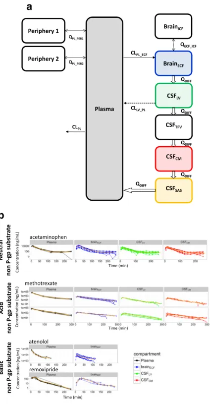
Fig. 3. A generic multi-compartmental CNS distribution model. (a)
different
in vitro
models and different sets of compounds used in
the
in vitro
studies (120).
Currently, the biopharmaceutics classi
fi
cation system
(BCS) and biopharmaceutics drug distribution classi
fi
cation
system (BDDCS) are used for CNS drugs. The BDDCS is a
modi
fi
cation of BCS that utilizes drug metabolism to predict
drug disposition and potential drug-drug interactions in the
brain (121). However, this classi
fi
cation approach needs to be
further investigated because of inconsistencies. For example,
it was proposed that 98% of BDDCS class 1 drugs would be
able to get into the brain even though the drugs were P-gp
substrates based on
in vitro
studies (122), while it has also
been reported that the
in vitro
ef
fl
ux ratio re
fl
ects the
in vivo
brain penetration regardless of the class in BDDCS (123).
EX VIVO
APPROACHES
As mentioned before, it is the unbound drug molecules
that are able to pass membranes and to interact with the
target (21). Thus, measuring unbound drug concentrations is
very important. Vu,brain or Fu,brain (the unbound fraction in
the brain) is used to investigate unbound fraction of drugs in
the brain. Fu,brain can be derived from brain homogenate
(124), and Vu,brain can be obtained from the brain slice
technique (125
). The brain slice method is more
physiologically relevant because the cell-cell interactions, pH
gradients, and active transport systems are all conserved
(114).
IN VIVO
APPROACHES
Microdialysis can be considered as a key technique to
time-dependent information regarding unbound drug concentrations.
With microdialysis, both the rate and extent of drug transport and
distribution processes can be determined (126,127). Thus, it can
be used to obtain Kp,uu,brain in conjunction with the rate of
transport processes. Moreover, this can be done at multiple
locations, and this feature has shown that even for a drug like
acetaminophen that is not subjected to any active transport,
substantial differences in pharmacokinetic pro
fi
les exist in
different brain compartments (6). While there is some limit to
use this water-based technique for the highly lipophilic drugs, lots
of microdialysis experiments have contributed to a boost in the
understanding on drug exchange across the BBB (126,128,129).
Especially the use of microdialysis at multiple brain locations has
provided insight into the relative contribution of CNS distribution
and elimination processes to the local (differences in)
pharmaco-kinetics of a compound (6,
7,
130). It has paved the way to the
development of a generic multi-compartmental CNS distribution
c
Basic
P-gp
sub
str
a
te
Neutr
al
P-gp
sub
str
a
te
phenytoin
morphine
Concen
tr
a
on
(ng/mL)
Concen
tr
a
on (ng
/mL)
+t
ariquid
a
r
+t
ariquid
ar
+pr
obenecid
Time (min)
paliperidone
quinidine
risperidone
Time (min)
+t
ariquid
a
r
+t
ariquid
a
r
+t
ariquid
a
r
+t
ariquid
a
r
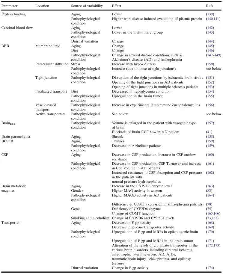
Table V. Sources of variability in CNS pharmacokinetics
Parameter Location Source of variability Effect Refs
Protein binding Aging Lower (139)
Pathophysiological condition
Higher with disease induced evaluation of plasma protein (140,141)
Cerebral bloodflow Aging Lower (142)
Pathophysiological condition
Lower in the multi-infarct group (143)
Diurnal variation Change (144)
BBB Membrane lipid Aging Change (145)
Diet Change (146)
Pathophysiological condition
Change in several disease conditions, such as Alzheimer’s disease (AD) and schizophrenia
(147–149)
Paracellular diffusion Stress Increase with hypoxic stress (150)
Pathophysiological condition
Increase (due to loose of tight junctions) see below
Tight junction Pathophysiological condition
Disruption of the tight junctions by ischaemic brain stroke (151) Opening of the tight junctions in AD patients (152) Opening of tight junctions in multiple sclerosis patients (153)
Facilitated transport Diet Decreased in hypoglycemia condition (154)
Pathophysiological condition
Upregulation in the brain tumor (155)
Vesicle-based transport
Pathophysiological condition
Increase in experimental autoimmune encephalomyelitis (156)
Active transporters Pathophysiological condition
See below see below
BrainECF Pathophysiological
condition
Volume is enlarged in the patient with vasogenic type of brain
(157)
Blockade of brain ECFflow in AD patient (41)
Brain parenchyma Aging Shrunk (158)
BCSFB Aging Thinner (159)
Pathophysiological condition
Decrease in Alzheimer patients (159)
CSF Aging Decrease in CSF production, increase in CSF outflow
resistance
(160)
Pathophysiological condition
Decrease in CSF production, CSF Turnover and increase in CSF volume in AD patients
(161)
Increased resistance to CSF absorption and CSF pressure in the patients with
normal‐pressure hydrocephalus
(162)
Brain metabolic enzymes
Aging Increase in the CYP2D6 enzyme level (163)
Gender Higher MAO activity in women (83)
Pathophysiological condition
Higher MAOB activity in AD patients (164)
Difference of COMT expression in schizophrenia patients (78)
Gene Deficiency of CYP2D6 enzyme (75)
Change of COMT function (165,166)
Smoking and alcoholism Change of CYP2B6 and CYP2E1 levels (73,167)
Transporter Aging Decrease in P-gp activity (168)
Decrease in glucose transporter activity (169) Pathophysiological
condition
Upregulation of P-gp and MRPs in epileptogenic brain (170)
Upregulation of P-gp and MRP1 in the brain tumor (171) Alteration of the levels of glutamate transporter in the
various brain disorders, including cerebral ischemia, amyotrophic lateral sclerosis, AD, AIDs,
traumatic brain injury, schizophrenia, and epilepsy (seizure)
(172,173)
model (Fig.
3), with some validated human CNS predictions that
will be discussed later in this review.
Then, positron emission tomography (PET) is a valuable
non-invasive
in vivo
monitoring technique that can be used to
visualize drug CNS distribution in living animals and human.
However, the PET technique cannot distinguish parent
com-pounds from their metabolites or bound and unbound drug.
Furthermore, it may also encounter dif
fi
culties in obtaining
useful data when a very high non-speci
fi
c binding (NSB) to
non-target proteins and phospholipid membranes occurs (131).
Recently, a novel lipid membrane binding assay (LIMBA) was
established as a fast and reliable tool for identifying compounds
with unfavorably high NSB in the brain tissue (132).
COMBINATORY MAPPING APPROACH
Combinatory mapping is an approach that combines three
compound-speci
fi
c parameters obtained from
in vitro,
ex vivo,
and
in vivo
data: Kp,brain, Vu,brain, and Fu,plasma, for
calculation of Kp,uu,brain (133). This approach also can be
used to obtain not only Kp,uu,brain but also to understand
unbound drug disposition in the cell cytosol and the lysosomes.
Recently, this approach has been extended to predict drug
exposure in different brain regions such as frontal cortex,
striatum, hippocampus, brainstem, cerebellum, and
hypothalamus, in which also the impact of transporters and
receptors in each region was taken into account (134). Although
this approach is useful to support the selection of potential CNS
drugs in drug discovery, it has two limitations. The
fi
rst limitation
is that it can only predict the parameters at steady-state. The
second limitation is that the approach cannot be translated to
predict the parameters, for instance, species or
inter-disease conditions because the processes to obtain the
param-eters in this approach are not connected with system properties
which will be changed in these conditions.
CONDITION DEPENDENCY AND PBPK MODELING
Condition Dependency
Drug distribution into and within the brain depends on the
interaction between system and drug properties. Drug
proper-ties remain the same, whatever the species and conditions are in
which the drug has been administered. This indicates that
interspecies variability in drug distribution into and within the
brain is the result of differences in physiological and biochemical
parameters. Factors which cause variation in drug
pharmacoki-netics include genetic background, species differences, gender,
age, diet, disease states, and drug treatment (4). Factors which
cause variation in drug pharmacodynamics include seasonal
Table VI. Currently published (semi-) PBPK model for CNS drugs
effect (135), age (136), gender (137), and species (138).
In
fl
uences of these conditions on CNS system properties are
summarized in Table
V.
(Semi-) PBPK Modeling
PBPK models need to be informed on system and on
drug properties to model the interaction and predict the
PK in different compartments. Especially as obtaining
pharmacokinetic data from the human brain is highly
restricted, working in the PBPK model framework is
valuable as it can be translated to predict the target site
concentrations in inter-species and inter-disease situations
(4). Some translational studied have been reported by
using an animal (semi-) PBPK model for CNS drugs but
they are relatively sparse and range from simple to more
advanced (Table
VI).
Recently, a generic multi-compartmental CNS
distribu-tion model structure has been proposed, that could
success-fully describe the pharmacokinetics in plasma and different
CNS compartments (brain
ECF, CSF in the lateral ventricle
(CSF
LV) and CSF in the cisterna magna (CSF
CM)), using
microdialysis data for 9 paradigm compounds with substantial
differences in physicochemical properties (9) (Table
VI,
Fig.
3). These compounds are acetaminophen, atenolol,
methotrexate, morphine, paliperidone, phenytoin, quinidine,
remoxipride, and risperidone. This is the
fi
rst model that can
nicely predict human brain
ECFand CSF time concentration
pro
fi
les which were obtained from physiologically
B
close to
normal
^
brain for morphine and acetaminophen (9).
For remoxipride, Stevens
et al.
have shown that brain
ECFpharmacokinetics, as measured with microdialysis,
repre-sented the target site concentrations, because these
concen-trations could be directly linked to the effect of remoxipride
Concen
tr
a
tion (
µ
g
/mL)
Time (min)
Fig. 4. Simulation of time concentration profiles of brainECF, CSFLV, CSFCM
on plasma prolactin levels in an advanced mechanism-based
model (185). After scaling to human, this indeed could also
be concluded for human CNS remoxipride effects on human
plasma prolactin levels. This underscores the importance of
having information on pharmacokinetics at the CNS target
region.
Using our generic multi-compartmental CNS
distribu-tion model, we can provide predicdistribu-tions of human CNS
pharmacokinetics for all the nine compounds. For a direct
comparison of rat and human pharmacokinetics in the
different CNS compartments in response to plasma
pharmacokinetics, the same plasma exposure was used
for individual compound. In Fig.
4, it can be seen that, in
general, human CNS pharmacokinetics, especially that in
the CSF in the subarachnoid space (CSF
SAS), which is
including the lumbar CSF is typically slower than that in
the rat. This provides important information on the
relationship between brain
ECF(which often is the target
site) pharmacokinetics and the lumbar CSF concentrations
that are often used as biomarker of brain target site
concentrations. Also, it can be seen that the differences in
the pharmacokinetics at the more early time points of the
different CNS compartments is larger in human than in
the rat. With time, these differences fade out. The
consequences for drug-target interaction kinetics (186)
and further processes towards CNS drug effects remain
to be determined.
Remaining Gaps and Challenges on PBPK Modeling,
Towards a Generic PBPK Model
The ultimate aim is to have a CNS PBPK model that
can predict human brain compartment concentrations on
the basis of the physicochemical properties of a
com-pound, which can be determined by
in vitro
measure-ments, or
in silico
prediction. Thus, in the overview in
Table
VI, it can be seen that we still have a number of
gaps in the currently available (semi-) PBPK models of
CNS drugs. Most of the models require
in vivo
data on
the compound(s), and most of the predictions have not
been validated on human data. Even the most
compre-hensive model (9), with validated prediction of human
CNS drug distribution (for acetaminophen and morphine),
still requires
in vivo
data for individual compound
predictions. Thus, it can be seen that there is a need for
further development of a generic, fully PBPK model for
CNS drug distribution (187
–
189).
To have a PBPK model that would predict CNS drug
distribution, based the physicochemical properties of an
individual drug, for different species and in different
condi-tions, a number of challenges remain:
&
Having a PBPK model structure with all relevant
compartment/parameters, as physiological parameter
values reported are sparse and variable (see Table
I).
&
Having drug physicochemical parameter values
from
in vitro, and/or
in silico, or even some
in vivo
measurements, which may not necessarily be correct.
For example,
in vitro
or
in vivo
data may depend on
the experimental setting, while
in silico
information
really depends on the data availability, used to obtain
the equation.
&
Having human data sets for validation of
predic-tion by the model, with typically limited availability.
&
Having information on pathophysiological
changes in human CNS properties in (the many)
disease conditions. For example, BBB characteristics
may change in Alzheimer
’
s disease, multiple sclerosis,
and pharmacoresistant epilepsies (190).
DISCUSSION AND CONCLUSION
Pharmacokinetics of drugs in the CNS is governed by a
combination of CNS system physiology and drug properties.
This means that variability in CNS system physiological
parameters (condition dependency) may lead to variability
of CNS pharmacokinetics. Therefore, it is important to
explicitly distinguish between system physiology and drug
properties, either by changing conditions and investigating
the pharmacokinetics of one drug, or investigating the
pharmacokinetics of different drugs in the same condition.
PBPK models make this distinction; however, being
based on total drug plasma and total tissue concentrations
at equilibrium (SS), while more recent PBPK models
include, at best, unbound plasma SS concentrations.
However, as body processes are based on the interaction
with the unbound drug and are time-dependent, it is crucial
to include measuring the unbound drug in each
compart-ment as a function of time (Mastermind Research Approach
(MRA)) (4), for which microdialysis has been proven the
key technique. Using the MRA, microdialysis has provided
lots of valuable data that pave the way towards a
semi-physiological generic CNS drug distribution model, yet
applicable for nine compounds with highly different
phys-icochemical properties with excellent description of the rat
data for all these compounds, and adequate prediction of
human CNS data that were available for acetaminophen
and morphine (9).
One microdialysis experiment in a single freely moving
animal can provide a lot of data points, obtained under the
same experimental condition of the animal, and thereby
revealing the interrelationships of processes. With this
microdialysis has already contributed to reduction and
re
fi
nement in the use of animals. Furthermore, all this
information can further be
B
condensed
^
into a generic
PBPK model and will thereby help in the reduction in the
future use of animals (replacement) (191).
So, in order to be able to predict CNS drug effects in
human, next steps would be a development of a full PBPK
CNS drug distribution model, and combine it with target
binding kinetics, receptor occupancy, and signal transduction
(186,192), and include system changes by human disease
condition.
ACKNOWLEDGEMENTS
COMPLIANCE WITH ETHICAL STANDARDS
Con
fl
ict of Interest
The authors declare that they have no con
fl
ict
of interest.
Open Access
This article is distributed under the terms
of the Creative Commons Attribution 4.0 International
License (http://creativecommons.org/licenses/by/4.0/), which
permits unrestricted use, distribution, and reproduction in
any medium, provided you give appropriate credit to the
original author(s) and the source, provide a link to the
Creative Commons license, and indicate if changes were
made.
REFERENCES
1. Kola I, Landis J. Can the pharmaceutical industry reduce attrition rates? Nat Rev Drug Discov. 2004;3:1–5.
2. Hurko O, Ryan JL. Translational research in central nervous system drug discovery. J Am Soc Exp Neurother. 2005;2(4):671– 82.
3. Cook D, Brown D, Alexander R, March R, Morgan P, Satterthwaite G, et al. Lessons learned from the fate of AstraZeneca’s drug pipeline: a five-dimensional framework. Nat Rev Drug Discov. 2014;13(6):419–31.
4. De Lange ECM. The mastermind approach to CNS drug therapy: translational prediction of human brain distribution, target site kinetics, and therapeutic effects. Fluids Barriers CNS. 2013;10(1):1–16.
5. Palmer AM, Stephenson FA. CNS drug discovery: challenges and solutions. Drug News Perspect. 2005;18(1):51–7. 6. Westerhout J, Ploeger B, Smeets J, Danhof M, de Lange ECM.
Physiologically based pharmacokinetic modeling to investigate regional brain distribution kinetics in rats. AAPS J. 2012;14(3):543–53.
7. Westerhout J, Van Den Berg D-J, Hartman R, Danhof M, De Lange ECM. Prediction of methotrexate CNS distribution in different species—influence of disease conditions. Eur J Pharm Sci. 2014;57:11–24.
8. Gaohua L, Neuhoff S, Johnson TN, Rostami-hodjegan A, Jamei M. Development of a permeability-limited model of the human brain and cerebrospinalfluid (CSF) to integrate known physiological and biological knowledge: estimating time vary-ing CSF drug concentrations and their variability usvary-ing in vitro data. Drug Metab Pharmacokinet. 2016;31(3):224–33. 9. Yamamoto Y, Välitalo PA, van den Berg D-J, Hartman R, van
den Brink W, Wong YC,et al. A generic multi-compartmental CNS distribution model structure for 9 drugs allows prediction of human brain target site concentrations. Pharm Res. 2017;34(2):333–51.
10. Mutch W. a, Hansen a J. Extracellular pH changes during spreading depression and cerebral ischemia: mechanisms of brain pH regulation. J Cereb Blood Flow Metab. 1984;4(1):17–27. 11. Fridén M, Bergström F, Wan H, Rehngren M, Ahlin G,
Hammarlund-Udenaes M, et al. Measurement of unbound drug exposure in brain: modeling of pH partitioning explains diverging results between the brain slice and brain homogenate methods. Drug Metab Dispos. 2011;39(3):353–62.
12. Shu C, Shen H, Teuscher NS, Lorenzi PJ, Keep RF, Smith DE. Role of PEPT2 in peptide/mimetic trafficking at the blood-cerebrospinal fluid barrier: studies in rat choroid plexus epithelial cells in primary culture. J Pharmacol Exp Ther. 2002;301(3):820–9.
13. Shen DD, Artru AA, Adkison KK. Principles and applicability of CSF sampling for the assessment of CNS drug delivery and pharmacodynamics. Adv Drug Deliv Rev. 2004;56(12):1825–57.
14. Jwnsen KE, Thomsen C, Henriksen O. In vivo measurement of intracellular pH in human brain during different tensions of carbon dioxide in arterial blood. A31P-NMR study. Acta Physiol Scand. 1988;134(2):295–8.
15. Mindell JA. Lysosomal acidification mechanisms. Annu Rev Physiol. 2012;74:69–86.
16. Touitou E, Barry BW. Enhancement in Drug Delivery. 2006. 575–591 p.
17. Wong AD, Ye M, Levy AF, Rothstein JD, Bergles DE, Searson PC. The blood-brain barrier: an engineering perspec-tive. Front Neuroeng. 2013;6:1–22.
18. Trapa PE, Belova E, Liras JL, Scott DO, Steyn SJ. Insights from an integrated physiologically based pharmacokinetic model for brain penetration. J Pharm Sci. 2016;105(2):965–71. 19. Serot JM, Béné MC, Foliguet B, Faure GC. Morphological alterations of the choroid plexus in late-onset Alzheimer’s disease. Acta Neuropathol. 2000;99(2):105–8.
20. Dekaban AS. Changes in brain weights during the span of human life: relation of brain weights to body heights and body weights. Ann Neurol. 1978;4(4):345–56.
21. Hammarlund-Udenaes M, Fridén M, Syvänen S, Gupta A. On the rate and extent of drug delivery to the brain. Pharm Res. 2008;25(8):1737–50.
22. Kawakami J, Yamamoto K, Sawada Y, Iga T. Prediction of brain delivery of ofloxacin, a new quinolone, in the human from animal data. J Pharmacokinet Biopharm. 1994;22(3):207–27.
23. Begley DJ, Bradbury MW, Kreuter J. The blood–brain barrier and drug delivery to the CNS. New York: Marcel Dekker, Inc.; 2000.
24. Thorne RG, Hrabe S, Nicholson C, Robert G. Diffusion of epidermal growth factor in rat brain extracellular space measured by integrative optical imaging. J Neurophysiol. 2004;92(6):3471–81.
25. Cserr H, Cooper D, Suri P, Patlak C. Efflux of radiolabeled polyethylene glycols and albumin from rat brain. Am J Physiol. 1981;240(4):319–28.
26. Sakka L, Coll G, Chazal J. Anatomy and physiology of cerebrospinal fluid. Eur Ann Otorhinolaryngol Head Neck Dis. 2011;128(6):309–16.
27. Kimelberg HK. Water homeostasis in the brain: basic concepts. Neuroscience. 2004;129(4):851–60.
28. Pardridge WM. Drug transport in brain via the cerebrospinal
fluid. Fluids Barriers CNS. 2011;8(7):1–7.
29. Condon P, Wyper D, Grant R, Patterson J, Hadley D, Teasdale G,
et al. Use of magnetic resonance imaging to measure intracranial cerebrospinalfluid volume. Lancet. 1986;327(8494):1355–7. 30. Kohn MI, Tanna NK, Herman GT, Resnick SM, Mozley PD,
Gur RE, et al. Analysis of brain and cerebrospinal fluid volumes with MR imaging. part I. methods, reliability, and validation. Radiology. 1991;178(1):115–22.
31. Robertson EG. Developmental defects of the cisterna magna and dura mater. J Neurol Neurosurg Psychiatry. 1949;12(1):39–51. 32. Adam R, Greenberg JO. The mega cisterna magna. J
Neurosurg. 1978;48(2):190–2.
33. Bass NH, Lundborg P. Postnatal development of bulkflow in the cerebrospinalfluid system of the albino rat: clearance of carboxyl-(14 C)inulin after intrathecal infusion. Brain Res. 1973;52:323–32. 34. Levinger IM. The cerebral ventricles of the rat. J Anat.
1971;108(3):447–51.
35. Stange K, Greitz M, Ingvar T, Hindmarsh T, Sollevi A. Global cerebral bloodflow during infusion of adenosine in humans: assessment by magnetic resonance imaging and positron emission tomography. Acta Physiol Scand. 1997;160(2):117–22. 36. Ito H, Inoue K, Goto R, Kinomura S, Taki Y, Okada K,et al. Database of normal human cerebral bloodflow measured by SPECT: I. comparison between I-123-IMP, Tc-99m-HMPAO, and Tc-99m-ECD as referred with O-15 labeled water PET and voxel-based morphometry. Ann Nucl Med. 2006;20(2):131–8. 37. Fagerholm U. The highly permeable blood–brain barrier: an
evaluation of current opinions about brain uptake capacity. Drug Discov Today. 2007;12(23–24):1076–82.
39. Peng B, Andrews J, Nestorov I, Brennan B, Nicklin P, Rowland M. Tissue distribution and physiologically based pharmacokinetics of antisense phosphorothioate oligonucleo-tide ISIS 1082 in rat. Antisense Nucleic Acid Drug Dev. 2001;11(1):15–27.
40. Abbott NJ. Prediction of blood–brain barrier permeation in drug discovery from in vivo, in vitro and in silico models. Drug Discov Today Technol. 2004;1(4):407–16.
41. Abbott NJ. Evidence for bulk flow of brain interstitialfluid: significance for physiology and pathology. Neurochem Int. 2004;45(4):545–52.
42. Skipor J, Thiery JC. The choroid plexus—cerebrospinalfluid system: undervaluated pathway of neuroendocrine signaling into the brain. Acta Neurobiol Exp. 2008;68(3):414–28. 43. Kumar G, Smith QR, Hokari M, Parepally J, Duncan MW.
Brain uptake, pharmacokinetics, and tissue distribution in the rat of neurotoxic N-butylbenzenesulfonamide. Toxicol Sci. 2007;97(2):253–64.
44. Strazielle N, Ghersi-Egea JF. Choroid plexus in the central nervous system: biology and physiopathology. J Neuropathol Exp Neurol. 2000;59(7):561–74.
45. Kusuhara H, Sugiyama Y. Active efflux across the blood–brain barrier : role of the solute carrier family. J Am Soc Exp Neurother. 2005;2:73–85.
46. Schinkel AH. P-Glycoprotein, a gatekeeper in the blood–brain barrier. Adv Drug Deliv Rev. 1999;36(2–3):179–94.
47. Tsuji A, Tamai I. Blood-brain barrier function of P-glycopro-tein. Adv Drug Deliv Rev. 1997;25(2–3):287–98.
48. Bendayan R. In situ localization of P-glycoprotein (ABCB1) in h u m a n a n d r a t b r a i n . J H i s t o c h e m C y t o c h e m . 2006;54(10):1159–67.
49. Löscher W, Potschka H. Role of drug efflux transporters in the brain for drug disposition and treatment of brain diseases. Prog Neurobiol. 2005;76(1):22–76.
50. Schinkel AH, Jonker JW. Mammalian drug efflux transporters of the ATP binding cassette (ABC) family: an overview. Adv Drug Deliv Rev. 2003;55:3–29.
51. Shapiro AB, Ling V. Stoichiometry of coupling of rhodamine 123 transport to ATP hydrolysis by P - glycoprotein. Eur J Biochem. 1998;254:189–93.
52. Rao VV, Dahlheimer JL, Bardgett ME, Snyder AZ, Finch RA, Sartorelli AC, et al. Choroid plexus epithelial expression of MDR1 P glycoprotein and multidrug resistance-associated protein contribute to the blood–cerebrospinal-fluid drug-permeability barrier. Med Sci. 1999;96(7):3900–5.
53. Cordon-Cardo C, O’Brien JP, Casals D, Rittman-Grauer L, Biedler JL, Melamed MR,et al. Multidrug-resistance gene (P-glycoprotein) is expressed by endothelial cells at blood-brain barrier sites. Proc Natl Acad Sci U S A. 1989;86(2):695–8. 54. Kassem NA, Deane R, Segal MB, Chen R, Preston JE.
Thyroxine (T4) transfer from CSF to choroid plexus and ventricular brain regions in rabbit: contributory role of P-glycoprotein and organic anion transporting polypeptides. Brain Res. 2007;1181:44–50.
55. Ronaldson PT, Bendayan M, Gingras D, Piquette-Miller M, Bendayan R. Cellular localization and functional expression of P-glycoprotein in rat astrocyte cultures. J Neurochem. 2004;89(3):788–800.
56. Miller DS. Regulation of ABC transporters at the blood-brain barrier. Clin Pharmacol Ther. 2015;97(4):395–403.
57. Löscher W, Potschka H. Blood-brain barrier active efflux transporters: ATP-binding cassette gene family. NeuroRx. 2005;2(1):86–98.
58. Nies AT, Jedlitschky G, König J, Herold-Mende C, Steiner HH, Schmitt H-P,et al. Expression and immunolocalization of the multidrug resistance proteins, MRP1–MRP6 (ABCC1–ABCC6), in human brain. Neuroscience. 2004;129(2):349–60.
59. Hipfner DR, Deeley RG, Cole SPC. Structural, mechanistic and clinical aspects of MRP1. Biochim Biophys Acta. 1999;1461:359–76.
60. Hirrlinger J, König J, Dringen R. Expression of mRNAs of multidrug resistance proteins (Mrps) in cultured rat astrocytes, oligodendrocytes, microglial cells and neurones. J Neurochem. 2002;82:716–9.
61. Westerhout J, Danhof M, Lange ECMDE. Preclinical predic-tion of human brain target site concentrapredic-tions: considerapredic-tions in extrapolating to the clinical setting. J Pharm Sci. 2011;100(9):3577–93.
62. Begley DJ. ABC transporters and the blood-brain barrier. Curr Pharm Des. 2004;10(12):1295–312.
63. Wijnholds J, De Lange ECM, Scheffer GL, Van Den Berg DD, Mol CAAM, Van Der Valk M. Multidrug resistance protein 1 protects the choroid plexus epithelium and contributes to the b l o o d - c e r e b r o s p i n a l flu i d b a r r i e r. J C l i n I n v e s t . 2000;105(3):279–85.
64. Gao B, Stieger B, Noé B, Fritschy JM, Meier PJ. Localization of the organic anion transporting polypeptide 2 (Oatp2) in capillary endothelium and choroid plexus epithelium of rat brain. J Histochem Cytochem. 1999;47(10):1255–64.
65. Hogue Angeletti R, Novikoff PM, Rao Juvvadi S, Fritschy J-M, Meier PJ, Wolkoff AW. The choroid plexus epithelium is the site of the organic anion transport protein in the brain. Neurobiology. 1997;94:283–6.
66. Gao B, Hagenbuch B, Kullak-Ublick GA, Benke D, Aguzzi A, Meier PJ. Organic anion-transporting polypeptides mediate transport of opioid peptides across blood-brain barrier. J Pharmacol Exp Ther. 2000;294(1):73–39.
67. Sekine T, Cha SH, Endou H. The multispecific organic anion transporter (OAT) family. Pflugers Arch. 2000;440(3):337–50. 68. Minn A, Ghersi-Egea J-F, Perrin R, Leininger B, Siest G. Drug
metabolizing enzymes in the brain and cerebral microvessels. Brain Res Rev. 1991;16(1):65–82.
69. Miksys SL, Tyndale RF. Drug-metabolizing cytochrome P450s in the brain. J Psychiatry Neurosci. 2002;27(6):406–15. 70. Miksys S, Tyndale RF. Cytochrome P450-mediated drug
metab-olism in the brain. J Psychiatry Neurosci. 2013;38(3):152–63. 71. Decleves X, Jacob A, Yousif S, Shawahna R, Potin S,
Scherrmann J. Interplay of drug metabolizing CYP450 en-zymes and ABC transporters in the blood-brain barrier. Curr Drug Metab. 2011;12(8):732–41.
72. Favetta P, Degoute CS, Perdrix JP, Dufresne C, Boulieu R, Guitton J. Propofol metabolites in man following propofol induction and maintenance. Br J Anaesth. 2002;88(5):653–8. 73. Miksys S, Lerman C, Shields PG, Mash DC, Tyndale RF.
Smoking, alcoholism and genetic polymorphisms alter CYP2B6 levels in human brain. Neuropharmacology. 2003;45(1):122–32.
74. Miksys S, Rao Y, Sellers EM, Kwan M, Mendis D, Tyndale RF. Regional and cellular distribution of CYP2D subfamily mem-bers in rat brain. Xenobiotica. 2000;30(6):547–64.
75. Zanger UM, Raimundo S, Eichelbaum M. Cytochrome P450 2D6: overview and update on pharmacology, genetics, bio-chemistry. Naunyn Schmiedebergs Arch Pharmacol. 2004;369(1):23–37.
76. D’empaire I, Guico-Pabia CJ, Preskorn SH. Antidepressant treatment and altered CYP2D6 activity. J Psychiatr Pract. 2011;17(5):330–9.
77. Leysen J, Janssen P, Megens A, Schotte A. Risperidone: a novel antipsychotic with balanced serotonin-dopamine antag-onism, receptor occupancy profile, and pharmacologic activity. J Clin Psychiatry. 1994;55:5–12.
78. Matsumoto M, Weickert CS, Beltaifa S, Kolachana B, Chen J, Hyde TM, et al. Catechol O-methyltransferase (COMT) mRNA expression in the dorsolateral prefrontal cortex of patients with schizophrenia. Neuropsychopharmacology. 2003;28(8):1521–30.
79. Huotari M, Gogos JA, Karayiorgou M, Koponen O, Forsberg M, Raasmaja A. Brain catecholamine metabolism in catechol-O-methyltransferase (COMT)-de ® cient mice. Neuroscience. 2002;15:246–56.
80. Pintar JE, Breakefield XO. Monoamine oxidase (MAO) activity as a determinant in human neurophysiology. Behav Genet. 1982;12(1):53–68.
81. Bogdanski DF, Weissbach H, Udenfriend S. THe distribution of serotonin, 5-hydroxytryptophan decarboxylase, and mono-amine oxjdase in brain. J Neurochem. 1957;1:272–8.
83. Robinson DS, Davis JM, Nies A, Ravaris CL, Sylwester D, Burlington V. Relation of sex and aging to monoamine oxidase activity of human brain, plasma, and platelets. Arch Gen Psychiat. 1971;24:536–9.
84. Nagatsu T. Progress in Monoamine Oxidase (MAO) research in relation to genetic engineering. Neurotoxicology. 2004;25(1– 2):11–20.
85. Levitt P, Pintart JE, Breakefieldt XO. Immunocytochemical demonstration of monoamine oxidase B in brain astrocytes and serotonergic neurons (central nervous system/rat/mono-amine). Neurobiology. 1982;79:6385–9.
86. Ohno S, Kawana K, Nakajin S. Contribution of UDP-glucuronosyltransferase 1A1 and 1A8 to morphine-6-glucuronidation and its kinetic properties. Drug Metab Dispos. 2008;36(4):688–94.
87. Ghersi-Egea JF, Leninger-Muller B, Suleman G, Siest G, Minn A. Localization of drug-metabolizing enzyme activities to blood-brain interfaces and circumventricular organs. J Neurochem. 1994;62:1089–96.
88. King CD, Rios GR, Assouline JA, Tephly TR. Expression of UDP-Glucuronosyltransferases (UGTs) 2B7 and 1A6 in the human brain and identification of 5-hydroxytryptamine as a substrate. Arch Biochem Biophys. 1999;365(1):156–62. 89. Suleman FG, Abid A, Gradinaru D, Daval J-L, Magdalou J,
Minn A. Identification of the uridine diphosphate glucurono-syltransferase isoform UGT1A6 in rat brain and in primary cultures of neurons and astrocytes. Arch Biochem Biophys. 1998;358(1):63–7.
90. Foldes A, Meek JL. Rat brain phenolsulfotransferase-partial purification and some properties. Biochim Biophys Acta 1973. 1973;327:365–74.
91. Meek JL, Neff NH. Biogenic amines and their metabolites as substrates for phenol sulphotransferase (EC 2.8.2.1) of brain and liver. J Neurochem. 1973;21(9):1–9.
92. Renskers KJ, Feor KD, Roth JA. Sulfation of dopamine and other biogenic amines by human brain phenol sulfotransferase. J Neurochem. 1980;34(6):1362–8.
93. Levin VA. Relationship of octanol/water partition coefficient and molecular weight to rat brain capillary permeability. J Med Chem. 1980;23:682–4.
94. Van de Waterbeernd er al H, van de Waterbeemd H, Camenisch G, Folkers G, Raevsky OA. Estimation of Caco-2 cell permeability using calculated molecular descriptors. Quant Struct-Act Relat. 1996;15:480–90.
95. Lipinski CA, Lombardo F, Dominy BW, Feeney PJ. Experimental and computational approaches to estimate solubility and permeability in drug discovery and development settings. Adv Drug Deliv Rev. 2012;64:4–17.
96. Chan OH, Stewart BH. Physicochemical and drug- delivery considerations for oral drug bioavailability. Drug Discov Today. 1996;1(11):461–73.
97. Upton RN. Regional pharmacokinetics I. Physiological and physicochemical basis. Biopharm Drug Dispos. 1990;11(8):647–62. 98. Shayanfar A, Soltani S, Jouyban A. Prediction of blood-brain distribution: effect of ionization. Biol Pharm Bull. 2011;34(2):266– 71.
99. Macintyre AC, Cutler DJ. The potential role of lysosomes in tissue distribution of weak bases. Biopharm Drug Dispos. 1988;9:513–26.
100. Fridén M, Winiwarter S, Jerndal G, Bengtsson O, Wan H, Bredberg U,et al. Structure-brain exposure relationships in rat and human using a novel data set of unbound drug concentra-tions in brain interstitial and cerebrospinalfluids. J Med Chem. 2009;52(20):6233–43.
101. Palm K, Luthman K, Ungell AL, Strandlund G, Beigi F, Lundahl P. Evaluation of dynamic polar molecular surface area as predictor of drug absorption: comparison with other computational and experimental predictors. J Med Chem. 1998;41(27):5382–92.
102. Krarup LH, Christensen thager I, Hovgaad L, Frokjaer S. Predicting drug absorption from molecular surface properties based on molecular dynamics simulations. Pharmacetical Res. 1998;15(7):972–8.
103. Loryan I, Sinha V, Mackie C, Van Peer A, Drinkenburg WH, Vermeulen A,et al. Molecular properties determining unbound
intracellular and extracellular brain exposure of CNS drug candidates. Mol Pharm. 2015;12(2):520–32.
104. Peletier LA, Benson N, van der Graaf PH. Impact of plasma-protein binding on receptor occupancy: an analytical descrip-tion. J Theor Biol. 2009;256(2):253–62.
105. Abbott NJ, Rönnbäck L, Hansson E. Astrocyte–endothelial interactions at the blood–brain barrier. Nat Rev Neurosci. 2006; 7(41–53).
106. Lee G, Dallas S, Hong M, Bendayan R. Drug transporters in the central nervous system: brain barriers and brain parenchyma considerations. Pharmacol Rev. 2001;53(4):569– 96.
107. Ohtsuki S, Terasaki T. Contribution of carrier-mediated transport systems to the blood-brain barrier as a supporting and protecting interface for the brain; importance for CNS drug discovery and development. Pharm Res. 2007;24(9):1745–58.
108. Plassat R, Verbe BP, Menei P, Menegalli D, Mathe JIR. Treatment of spasticity with intrathecal Baclofen administra-tion long-term follow-up, review of 40 patients. Spinal Cord. 2004;42:686–93.
109. Kroin JS. The distribution of medication along the spinal canal after chronic intrathecal administration. Neurosurgery. 1993;33:226–30.
110. Hettiarachchi HDM, Hsu Y, Harris TJ, Penn R, Linninger A. a. The effect of pulsatileflow on intrathecal drug delivery in the spinal canal. Ann Biomed Eng. 2011;39(10):2592–602. 111. Hsu Y, Hettiarachchi HDM, Zhu DC, Linninger AA. The
frequency and magnitude of cerebrospinal fluid pulsations influence intrathecal drug distribution. Anesth Analg. 2012;115(2):386–94.
112. Pardridge WM. Log(BB), PS products and in silico models of drug brain penetration. Drug Discov Today. 2004;9(9):392–3. 113. Chen H, Winiwarter S, Fridén M, Antonsson M, Engkvist O.
In silico prediction of unbound brain-to-plasma concentration ratio using machine learning algorithms. J Mol Graph Model. 2011;29(8):985–95.
114. Loryan I, Fridén M, Hammarlund-Udenaes M. The brain slice method for studying drug distribution in the CNS. Fluids Barriers CNS. 2013;10(6):1–9.
115. Helms HC, Abbott NJ, Burek M, Cecchelli R, Couraud P-O, Deli MA,et al. In vitro models of the blood-brain barrier: an overview of commonly used brain endothelial cell culture models and guidelines for their use. J Cereb Blood Flow Metab. 2016;36(5):862–90.
116. Bicker J, Alves G, Fortuna A, Falcão A. Blood-brain barrier models and their relevance for a successful development of CNS drug delivery systems: a review. Eur J Pharm Biopharm. 2014;87(3):409–32.
117. Bernas MJ, Cardoso FL, Daley SK, Weinand ME, Campos AR, Ferreira AJG,et al. Establishment of primary cultures of human brain microvascular endothelial cells to provide an in vitro cellular model of the blood-brain barrier. Nat Protoc. 2010;5(7):1265–72.
118. Weksler BB, Subileau EA, Perrière N, Charneau P, Holloway K, Leveque M. Blood-brain barrier-specific properties of a human adult brain endothelial cell line. FASEB J. 2005;19(13):1872–4.
119. Cecchelli R, Aday S, Sevin E, Almeida C, Culot M, Dehouck L,et al. A stable and reproducible human blood-brain barrier model derived from hematopoietic stem cells. PLoS One. 2014;9(6):1–11.
120. Stanimirovic DB, Bani-Yaghoub M, Perkins M, Haqqani AS. Blood-brain barrier models: in vitro to in vivo translation in preclinical development of CNS-targeting biotherapeutics. Expert Opin Drug Discov. 2015;10(2):141–55.
121. Larregieu CA, Benet LZ. Distinguishing between the perme-ability relationships with absorption and metabolism to im-prove BCS and BDDCS predictions in early drug discovery. Mol Pharm. 2014;11(4):1335–44.
122. Broccatelli F, Larregieu CA, Cruciani G, Oprea TI, Benet LZ. Improving the prediction of the brain disposition for orally administered drugs using BDDCS. Adv Drug Deliv Rev. 2012;64(1):95–109.